Embed Size (px)
Citation preview

Università degli studi Roma Tre
Scuola Dottorale in Biologia
Sezione Biologia applicata alla salute dell’Uomo
XXIV CICLO
(A.A. 2010/2011)
"Role of the MUTYH protein in the response to oxidative damage to DNA"
“Ruolo della proteina MUTYH nella risposta al danno ossidativo al DNA”
Dottoranda
Vitalba Ruggieri
Docente guida: Dr.ssa Margherita Bignami
Tutor: Prof. Antonio Antoccia Coordinatore: Prof. Paolo Visca

Pag 2

Pag. 3
INDEX
ABBREVIATIONS 5
RIASSUNTO 7
SUMMARY 13
I.INTRODUCTION 17
1. Damage to DNA and human disease 17
1.1. The response to DNA damage: the genome-maintenance network 19
1.2. DNA-damage repair and checkpoint pathways 22
2. The oxidative damage to DNA 24
2.1. Response to oxidative damage: the special problem of 8-oxo-dG 25
2.2. Sources of cellular 8-oxo-dG: potassium bromate and
combination of 6-thioguanine and UVA 26
2.3. Base excision repair: structural and functional aspects 28
2.4. The problem of 8-oxo-dG: the special function of MUTYH 30
2.5. Defective MUTYH: MUTYH-associated polyposis 32
II. RESULTS 35
III. DISCUSSION 53
IV. CONCLUSIONS 59
V. REFERENCES 61
ACKNOWLEDGEMENTS 75

Pag 4

Pag. 5
ABBREVIATIONS
AP sites: Apurinic/Apyrimidinic sites
APE1: AP endonuclease
Aza: Azathioprine
BER: Base Excision Repair
DDR: DNA-damage response
dNTPs: deoxynucleoside triphosphates
DSBs: double strand breaks
FAP: Familial adenomatous polyposis
HR: homologous recombination
IR: ionizing radiation
LCLs: lymphoblastoid cell lines
LP-BER: Long-Patch BER
MAP: MUTYH-associated polyposis
MEFs: Mouse embryo fibroblasts
me6-TG: methyl-6-TG
MMR: Mismatch Repair
NER: Nucleotide Excision Repair
NHEJ: nonhomologous end-joining
Pol: polymerase
RNS: reactive nitrogen species
ROS: reactive oxygen species

Pag 6
SSBs: single-strand breaks
SP-BER: Short-Patch BER
ssDNA: single-strand DNA
UV: ultraviolet radiation
5’dRp: 5’-deoxyribose-5’-phosphate
6-TG: 6-thioguanine
8-oxo-dG: 7,8-dihydro-8-hydroxyguanine
8-oxo-dGTP: 8-oxo-2’-deoxyguanosine triphosphate
8-oxo-dGMP: 8-oxo-2’-deoxyguanosine monophosphate
9-1-1 complex: Rad1, Rad9, and Hus1 complex

Pag. 7
RIASSUNTO
Da un numero sempre più crescente di evidenze sperimentali emerge che il
danno al DNA è uno dei principali fattori causali nell’insorgenza del cancro e di
molte patologie legate all’invecchiamento. Il DNA, al pari di tutte le molecole
biologiche, rappresenta un target preferenziale di potenziali agenti di danno, sia
esogeni che endogeni, che ne mettono a repentaglio l’integrità.
Le radiazioni ultraviolette, i raggi X, gli agenti chimici genotossici come anche i
prodotti del metabolismo endogeno o di reazioni chimiche spontanee possono
indurre lesioni permanenti nel genoma con gravi conseguenze a livello cellulare.
Le rotture a singolo (SSBs) o doppio filamento (DSBs) come anche gli
appaiamenti errati di basi azotate, i foto-prodotti indotti dai raggi ultravioletti e
le modificazioni chimiche a livello delle basi sono solo alcune delle potenziali modificazioni indotte da tali agenti di danno (Hoeijmakers, 2009), (figura 1,
pagina 18). L’importanza del rischio associato a queste lesioni si riflette
nell’esistenza di numerosi sistemi di riparazione del DNA (tabella 1, pagina 21),
veri e propri meccanismi di salvaguardia del genoma dai quali dipende il destino
della cellula in termini di sopravvivenza, senescenza o morte, in caso di danno.
Nei mammiferi, alcuni fra questi sono rappresentati dal sistema di riparazione
per escissione di basi (BER), dal sistema di riparazione per escissione di
nucleotidi (NER), la riparazione degli appaiamenti errati (MMR) e la
ricombinazione omologa (HR) e non omologa (NHEJ), (Maynard et al., 2009).
Il processo di riparazione del danno, così come la replicazione del DNA, è
coordinato in maniera fine e strettamente regolata con la progressione del ciclo
cellulare attraverso dei complessi meccanismi di sorveglianza, noti come checkpoint del ciclo cellulare. Questi sono composti da una rete di sensori,
trasduttori ed effettori (Bartek et al., 2004; Sancar et al., 2004), e vengono
considerati essenziali per la sopravvivenza cellulare e dell’intero organismo
(Brown et al., 2000; de Klein et al., 2000), (figura 2, pagina 23).
Alcuni fra i principali fattori endogeni di danno al DNA includono le specie
reattive dell’ossigeno (ROS) generate dal normale metabolismo cellulare che,
pur esercitando importanti funzioni in numerosi processi fisiologici, svolgono un
ruolo causale in fenomeni quali la mutagenesi, la carcinogenesi e
l’invecchiamento (Valko et al., 2006).
Le basi azotate sono fra i bersagli più sensibili all’ossidazione da parte dei ROS,
e tra di esse la guanina risulta essere particolarmente suscettibile a causa del suo basso potenziale redox. Non sorprende dunque che la 8-oxo-7,8-diidroguanina
(8-oxo-dG) sia una delle lesioni più abbondanti e meglio caratterizzate, spesso
usata come bio-marcatore cellulare di stress ossidativo a carico del DNA. A
causa della sua capacità di mimare funzionalmente la timina nella sua
conformazione syn, essa si appaia stabilmente con l’adenina, introducendo nel
genoma trasversioni di tipo G>T (David et al., 2007), identificate come
mutazioni somatiche predominanti in diverse tipologie di cancro. Alla rimozione
di tale lesione sono preposti molteplici sistemi di riparazione del DNA e tra

Pag 8
questi il BER è in prima linea. Il processo di eliminazione della 8-oxo-dG
catalizzato dalle proteine del BER consiste in una sequenza di reazioni finemente coordinate in cui l’evento chiave è l’idrolisi del legame N-glicosidico
tra la base modificata e il deossiribosio, catalizzata da DNA glicosilasi aventi
ciascuna una diversa specificità di substrato (Sharma and Dianov 2007), (figura
3, pagina 29). Tra queste, la proteina MUTYH (mutY homolog (E.Coli))
rimuove l’adenina erroneamente appaiata con la 8-oxo-dG e contribuisce in tal
modo, in cooperazione con la DNA glicosilasi OGG1 (8-Oxoguanine
glycosylase), ad eliminare tale lesione dal genoma (figura 4, pagina 32).
Diversi domini funzionali sono stati identificati nella struttura proteica di
MUTYH, deputati a processi quali il legame al DNA, la escissione della 8-oxo-
dG e l’interazione con fattori della replicazione (PCNA, RPA), della
segnalazione del danno al DNA (il complesso 9-1-1), o proteine di altri sistemi di riparazione come il MMR (MSH6) (Oka and Nakabeppu, 2011).
La capacità di MUTYH di interagire con proteine appartenenti a sistemi diversi
dalla semplice riparazione del DNA suggerisce il suo possibile coinvolgimento
in un network complesso di intercomunicazione che lascia supporre un suo ruolo
più ampio e rilevante nel mantenimento dell’integrità del genoma.
L’importanza funzionale di questa proteina è inoltre sottolineata
dall’associazione fra la presenza di mutazioni germinali nel gene MUTYH ed
una malattia autosomica recessiva, la poliposi associata a MUTYH (MAP),
caratterizzata da poliposi colorettale e da predisposizione al cancro. In tale
sindrome ereditaria le mutazioni bialleliche di MUTYH sono tipicamente
associate alla presenza di trasversioni G>T nel gene oncosoppressore APC
(Adenomatous Polyposis Coli), oltre che, in un’alta percentuale di tumori derivati da pazienti MAP, nell’oncogene K-RAS.
Partendo da tali osservazioni, nel presente studio sono stati analizzati gli effetti
prodotti dalla mutazione o dalla inattivazione del gene che codifica per questa
proteina nella risposta biologica al danno ossidativo al DNA. In particolare,
nella prima parte della tesi si è esaminato l’impatto prodotto a livello cellulare
dalla presenza di mutazioni diverse di MUTYH in linee linfoblastoidi di pazienti
affetti da MAP, in termini di ossidazione basale del DNA, riparazione del danno
ossidativo, mutagenesi spontanea ed indotta.
Diverse varianti geniche di MUTYH sono state finora caratterizzate mediante
l’uso di proteine purificate (Bai et al., 2005; Bai et al., 2007; Ali et al., 2008;
Yanaru-Fujisawa et al., 2008; D’Agostino et al., 2010) sebbene gli studi biochimici siano stati limitati ad un numero relativamente ridotto di esse. Inoltre,
mancano informazioni riguardanti il fenotipo mutatore associato
all’inattivazione di MUTYH nell’uomo. Da uno studio condotto in precedenza
nel nostro laboratorio era emerso che tutte le varianti di MUTYH associate a
MAP erano accomunate dall’accumulo di 8-oxo-dG nel DNA e
dall’ipersensibilità agli effetti citotossici del bromato di potassio (KBrO3), un
noto agente ossidante. Tale studio era stato condotto utilizzando un saggio in cui
le singole proteine mutanti venivano espresse in fibroblasti embrionali di topo

Pag. 9
(MEFs) derivate da topi Mutyh difettivi (Molatore et al., 2010). Tale tipo di
approccio, tuttavia, non può essere usato per analizzare mutazioni eterozigoti composte di MUTYH, che si riscontrano piuttosto comunemente tra i pazienti
italiani affetti da MAP. Allo scopo di studiare tali varianti complesse abbiamo
utilizzato linee linfoblastoidi derivate da pazienti MAP con mutazioni missenso
e troncanti in condizioni di omozigosi o di eterozigosi composta (Tabella 2,
pagina 35 e figura 5, pagina 36).
La quantificazione dei livelli di espressione di MUTYH mediante RT-PCR e
Western blotting ha rivelato che, mentre in alcune linee cellulari esiste una
buona correlazione fra i livelli di trascritto e quelli di proteina, in altri casi
invece, per lo più in linee caratterizzate da mutazioni di tipo frameshift, la
presenza del trascritto non corrisponde a quella della proteina (Figura 6, pagina
37), lasciando ipotizzare che alcune mutazioni siano associate ad instabilità dei relativi trascritti.
In seguito alla messa a punto di un saggio in cui viene valutata l’attività
glicosilasica sia di MUTYH che di OGG1 (figura 7, pagina 38) è stato possibile
dimostrare che tutte le linee mutanti sono difettive nella rimozione dell’adenina
da un substrato 8-oxoG:A, pur presentando una normale attività della glicosilasi
OGG1 (Figura 8, pagina 39). In accordo con quanto dimostrato da altri autori
(Wooden et al., 2004; Parker et al., 2005; Ali et al., 2008; Kundu et al., 2009) si
è identificata una residua benché minima attività glicosilasica nelle due varianti
p.Tyr179Cys/Tyr179Cys e p.Tyr179Cys/Gly396Asp caratterizzate dalla
presenza di livelli di proteina simili alle linee wild-type di donatori sani.
E’stato inoltre interessante osservare che mentre la quasi totalità delle linee
esaminate mostra livelli basali di 8-oxo-dG maggiori rispetto alle linee wild-type, le linee caratterizzate dalle mutazioni frameshift p.Gly264TrpfsX7 e
p.Ala385ProfsX23, che non esprimono livelli rilevabili di proteina, fanno
eccezione. Tale osservazione potrebbe suggerire la presenza di un effetto
dominante negativo associato alla proteina mutante, probabilmente connesso
all’interferenza con altri meccanismi di riparazione della 8-oxo-dG (Figura 9A,
pagina 40).
Con l’obiettivo di verificare se l’incremento nei livelli basali della base ossidata
fosse dovuto ad una sua difettiva rimozione nelle linee mutanti, si è eseguito un
saggio di cinetica di riparazione dopo trattamento con KBrO3, un agente
ossidante che introduce un alto livello di 8-oxo-dG nel DNA. Tutte le linee con
mutazioni di MUTYH hanno mostrato in effetti una rimozione più lenta della lesione quando confrontate con una linea di donatore sano (Figura 9B, pagina
40). La presenza di specifiche trasversioni G>T nei geni APC o K-RAS nei
tumori di pazienti MAP può essere plausibilmente ritenuta un’evidenza indiretta
della esistenza di un fenotipo mutatore associato alla presenza di una proteina
MUTYH non funzionale (Al-Tassan et al., 2002; Jones et al., 2002, Lipton et al.,
2003; van Pujienbroek et al., 2008). Partendo da tale osservazione, si è misurata,
per la prima volta in linee di pazienti MAP, la frequenza di mutazioni spontanea,
utilizzando una metodologia innovativa messa a punto dal gruppo di L. Luzzatto

Pag 10
(Araten et al., 2005) basata sulla determinazione per via citofluorimetrica della frequenza di mutazione di un gene sentinella, PIG-A, già applicata allo studio
del fenotipo mutatore associato ad altri difetti della riparazione del DNA. Dai
risultati ottenuti si è identificato un aumento medio di quattro volte nel valore
della frequenza di mutazione di PIG-A nelle linee di pazienti rispetto al valore
medio di tre linee wild-type (Figura 10, pagina 42). Inoltre, la ricomparsa di
cellule mutanti dopo previa eliminazione tramite “sorting” dalla popolazione
cellulare iniziale, in una delle linee con più alta frequenza di mutazione, ha
dimostrato che il fenotipo mutatore identificato è una caratteristica intrinseca
della linea cellulare. Nella stessa linea mutante trattata con KBrO3, si è per di
più osservata sia ipersensibilità agli effetti citotossici dell’agente ossidante che un fenotipo di iper-mutabilità rispetto ad una linea di donatore sano (Figura 11,
pagina 43), confermando la presenza di un forte impatto biologico associato alla
presenza di mutazioni di MUTYH nella risposta al danno ossidativo.
Nella seconda parte della tesi sono invece riportati e discussi i risultati ottenuti
in un progetto condotto in parallelo in cui si sono studiati gli effetti dovuti
all’assenza di MUTYH nella risposta ad un interessante modello di danno
ossidativo prodotto dalla interazione del farmaco immunosoppressore
Azatioprina (Aza) con i raggi ultravioletti di tipo A (UVA).
Come suggerito da diversi studi, il danno al DNA indotto dalla combinazione di
tali agenti è stato riconosciuto come uno dei possibili fattori responsabili
dell’aumentata insorgenza di cancro della pelle in pazienti immunosoppressi con
Aza dopo trapianto d’organo (Brem et al., 2009). Il trattamento sistemico con Aza causa l’incorporazione nel DNA di 6-tioguanina (6-TG) (Relling and
Dervieux, 2001) che, a differenza delle basi azotate canoniche assorbe le
radiazioni UVA, agendo da cromoforo e generando ROS.
La stessa 6-TG è fortemente esposta al processo di ossidazione. In particolare, la
sua interazione con i raggi UVA genera una lesione nota come guanina 6-
sulfonato che costituisce un blocco alla replicazione del DNA ed è
potenzialmente mutagena.
Studi realizzati su cellule umane hanno dimostrato che la 6-TG e i raggi UVA
sono sinergicamente citotossici e mutageni inducendo pericolose lesioni a livello
del DNA e delle proteine (O' Donovan et al., 2005).
Sulla base di tali osservazioni, e considerando l’importante funzione di MUTYH nella risposta al danno ossidativo, abbiamo esaminato il ruolo di questa proteina
nella risposta al danno cellulare indotto dalla combinazione di 6-TG ed UVA.
Dato il suo coinvolgimento nella regolazione della risposta cellulare alla 6-TG
come anche nella rimozione dell’8-oxo-dG abbiamo esteso parte degli studi
anche alla proteina MSH2 facente parte del sistema MMR.
Dai nostri saggi di sopravvivenza in vitro sulle MEFs è risultato che la
combinazione di 6-TG e raggi UVA produce effetti tossici in cellule wild-type
(WT), mentre il trattamento singolo con 6-TG o con UVA non ha effetti
rilevanti sulla sopravvivenza cellulare. Sorprendentemente, sia l’assenza di
Mutyh che di Msh2 conferisce resistenza all’effetto tossico prodotto dal

Pag. 11
trattamento combinato, (Figura 12, pagina 44) sebbene tutte le linee,
indipendentemente dal genotipo, mostrano aumenti simili dei livelli di 8-oxo-dG nel DNA (Figura 13, pagina 45).
Poichè numerosi dati sperimentali indicano che, in cellule trattate con 6-TG,
tale analogo di base risulta essere presente nel “pool” cellulare di
deossinucleosidi trifosfato (dNTPs) dove può agire da fonte di ROS per
esposizione ai raggi UVA (Cooke et al., 2008), si è pensato di determinare il
potenziale contributo di questa “riserva” di 6-TG agli effetti biologici osservati.
Esperimenti svolti a tale scopo (Figura 14A, pagina 46) hanno rivelato che anche
nel sistema da noi analizzato la 6-TG del “pool” contribuisce notevolmente sia
agli effetti di citotossicità (Figura 14B, pagina 47) che di ossidazione (Figura 15
pagina 47) osservati dopo il trattamento combinato. Con l’obiettivo di studiare il meccanismo alla base della tossicità riportata nelle
cellule WT ed il ruolo esercitato da MUTYH in questo processo abbiamo
analizzato la progressione del ciclo cellulare mediante citometria a flusso. Dai
profili ottenuti è emerso che l’assenza MUTYH è associata al mancato arresto
delle cellule in fase S dopo esposizione a 6-TG ed UVA, un arresto che si
osserva invece in maniera prominente nelle cellule WT (Figura 16, pagina 48).
Tale fenomeno si riflette in una diversa cinetica di attivazione del checkpoint:
l’attivazione per fosforilazione della proteina Chk1, coinvolta nell’attivazione
del checkpoint di fase S e G2/M, si osserva a tempi brevi dal trattamento
combinato nelle cellule WT, mentre nelle cellule Mutyh difettive il segnale della
proteina fosforilata compare in tempi molto più lunghi e con un’intensità notevolmente ridotta (Figura 17, pagina 49). Tali osservazioni sembrano
supportare il coinvolgimento di MUTYH nell’attivazione precoce come anche
nella regolazione del checkpoint di fase S in risposta a tale tipo di danno. E’
noto che la fosforilazione di Chk1 da parte della chinasi ATR è coinvolta nel
controllo della riparazione dei DSBs mediante il sistema di riparazione HR
(Sørensen et al.; 2005). La quantificazione dei DSBs misurati come numero di
foci dell’istone fosforilato, H2AX, ha rivelato che, anche nel nostro modello murino, come nelle cellule umane (Brem et al.; 2010), il trattamento combinato
con 6-TG e raggi UVA produce questo tipo di danno al DNA, e che i livelli sono
paragonabili fra i due genotipi. Sorprende tuttavia che le due linee si distinguano
nella modalità di processamento di tale danno. La determinazione numerica dei
foci della proteina RAD51, facente parte del sistema HR, ha infatti rivelato che
se da un lato nelle cellule WT vi è un’induzione più elevata di tale proteina nonché una sua persistenza a tempi lunghi dopo il trattamento con 6-TG ed
UVA, dall’altro invece l’assenza di MUTYH è associata a livelli decisamente
più bassi di foci e ad un graduale decremento degli stessi nel tempo (Figura 18,
pagina 50). Se si considera che RAD51 esercita una funzione chiave nella
sopravvivenza cellulare contro i potenziali effetti letali associati ai DSBs,
l’ipersensibilità delle cellule WT alle conseguenze del trattamento combinato è
in apparente contraddizione con l’induzione di una risposta chiaramente
protettiva. Una possibile spiegazione di questo fenomeno consiste nell’ipotizzare

Pag 12
che il tentativo di riparazione del danno prodotto dall’interazione della 6-TG con
i raggi UVA mediante attivazione del sistema HR induce la formazione di una quantità considerevole di intermedi riparativi estremamente tossici incompatibili
con la sopravvivenza cellulare. In quest’ottica, l’assenza MUTYH e quindi di
una modalità efficiente e/o canonica di riparazione del danno comporterebbe un
vantaggio selettivo per la cellula stessa in termini di vitalità, associato tuttavia
ad un possibile aumento dell’instabilità genomica.
Lo dimostrano infatti i dati derivati da uno studio parallelo condotto in vivo su
animali wild-type e Mutyh-difettivi, sottoposti a trattamenti singoli o combinati
(Aza somministrata per via intraperitoneale seguita o meno da esposizione ad
UVA) per 12 mesi consecutivi. Come nel modello in vitro, il trattamento con
entrambi gli agenti ha determinato una notevole differenza nella tossicità: l’80%
dei topi difettivi è sopravvissuto al trattamento contro il 20% dei topi wild-type. Il trattamento singolo con i raggi UVA non ha invece prodotto effetti sulla
sopravvivenza in nessuno dei due genotipi, mentre l’immunosoppressione
prodotta dall’Aza ha determinato livelli di mortalità simile nei due genotipi.
E’ stato inoltre osservato un incremento notevole nei livelli di 8-oxo-dG nella
pelle di entrambi i gruppi di animali, sebbene non siano state riscontrate
differenze significative nei livelli di ossidazione del DNA tra i due genotipi
(Figura 19A, pagina 52). E’ degno di nota, invece, che l’analisi istologica della
pelle ha identificato la presenza di due carcinomi a cellule squamose in due topi
Mutyh-/- (Figura 19B, pagina 52) dopo trattamento combinato suggerendo che
l’assenza di una proteina fondamentale nella salvaguardia del genoma è
associata ad una possibile predisposizione all’insorgenza di tumori della pelle.
Considerati nel loro complesso i dati ottenuti da tali studi mettono in luce l’importanza dell’effetto protettivo esercitato dalla proteina MUTYH nella
risposta al danno ossidativo al DNA. Le osservazioni condotte sulle linee di
pazienti affetti da MAP, oltre a sottolineare l’aspetto di patogenicità associato
alla presenza della proteina mutante, consentono anche di individuare
nell’accumulo di danno ossidativo nel DNA e nella presenza di un fenotipo
mutatore sia spontaneo che indotto da stress ossidativo, fattori di estrema
rilevanza per una migliore valutazione clinica della patogenesi associata alle
varianti di MUTYH. Infine, i risultati ottenuti dallo studio condotto nel modello
murino di inattivazione di MUTYH confermano la rilevanza biologica di tale
proteina nella salvaguarda della stabilità genomica, indicando tuttavia un suo
ruolo più complesso nella risposta al danno al DNA che va oltre il semplice meccanismo di riparazione del danno ossidativo.

Pag. 13
SUMMARY
The oxidized base 7,8-dihydro-8-hydroxyguanine (8-oxo-dG) is one of the most
deleterious injuries induced by oxidative stress. Multiple DNA repair proteins
have evolved to protect the genome against the detrimental effects of this
promutagenic lesion. One of the major ones is the Base Excision Repair (BER)
MUTYH DNA glycosylase that removes adenine from 8:oxoG-containing
mispairs originated by DNA polymerases and via the OGG1 DNA glycosylase
contributes to 8-oxo-dG repair. Germline mutations in the MUTYH gene lead to
MUTYH-associated polyposis (MAP), an autosomal recessive syndrome
characterized by colorectal polyposis and cancer predisposition. Although
several reports characterized MUTYH variants using purified proteins, relatively
few mutations have been investigated from the biochemical point of view. In addition no information is available on the mutator phenotype associated with
MUTYH inactivation in humans. In our previous study accumulation of 8-oxo-
dG and hypersensitivity to killing by the oxidizing agent KBrO3 were identified
as a common phenotype among the investigated MAP-associated variants. These
results were based on an assay in which single mutant MUTYH proteins were
expressed in mouse embryonic fibroblasts (MEFs) derived from Mutyh-null
mice (Molatore et al., 2010). This approach however cannot be exploited to
analyze compound heterozygous MUTYH mutations, a very common situation
among Italian MAP patients. With the aim of studying these particularly
complex variants, we derived human lymphoblastoid cell lines (LCLs) from
MAP patients harbouring missense and truncating mutations. RT-PCR and
Western blotting analyses revealed that while in some cell lines there is a good correlation between transcripts and protein levels, in other instances no MUTYH
protein is detectable.
When basal levels of 8-oxo-dG were measured in these cell lines, increases were
detected in DNA of six LCLs expressing MUTYH variants when compared to
two wild-type cell lines. Interestingly the only two exceptions were cells in
which no detectable expression of the MUTYH protein could be identified.
To determine whether this increase in steady-state levels was due to a defective
repair of 8-oxo-dG in these mutants, repair kinetics of this oxidized base were
determined following exposure to KBrO3. All the LCLs harbouring MUTYH
mutations showed a defective repair of 8-oxo-dG when compared to a cell line from a healthy donor.
Results of a novel assay where both MUTYH and OGG1 activity could be
evaluated indicate that all these variants were defective in removing adenine
from an 8-oxoG:A DNA substrate, but retained wild-type OGG1 activity.
Mutation frequency measurements at the PIG-A gene identified a four-fold
increase in spontaneous mutagenesis in six LCLs from MAP patients when
compared to three LCLs from healthy donors. Finally KBrO3 hypersensitivity
was accompanied by a hyper-mutable phenotype in a MUTYH mutant cell line.
These observations support the pathogenic role of these MUTYH mutations and

Pag 14
identify accumulation of 8-oxo-dG and a mutator phenotype as relevant factors
for a better clinical assessment of MUTYH variant pathogenesis. In the second part of this thesis the results of a parallel study on the effects of
MUTYH loss in response to a different type of oxidative damage are reported.
This study addresses the toxicity and carcinogenicity of the anti-cancer
immunosuppressant drug Azathioprine (Aza) combined with UVA radiation.
Systemic treatment with Aza causes the incorporation of 6-thioguanine (6-TG)
into DNA. 6-TG is a chromophore which generates reactive oxygen species
(ROS) on exposure to UVA and is itself highly susceptible to oxidation. DNA
damage induced by the Aza/UVA combination is thought to contribute to the
huge incidence of skin cancer in immunosuppressed organ transplant patients.
Several studies have shown indeed that Aza combined with low doses of UVA
may cause mutagenic damage in human cells. In particular, 6-TG/UVA generates a novel DNA lesion, the guanine-6-sulfonate that blocks DNA
replication and is potentially mutagenic. Here we examined the role of the
MUTYH DNA glycosylase and the mismatch repair (MMR) MSH2 protein in
the cellular response to 6-TG/UVA-induced DNA damage.
6-TG and UVA were synergistically toxic to wild-type mouse embryo
fibroblasts (MEFs) while neither 6-TG or UVA alone detectably affected
survival. Mutyh- or Msh2-defective cells were more resistant than wild-type
MEFs to killing by 6-TG/UVA. Nevertheless, the combined treatment
significantly increased the levels of DNA 8-oxo-dG irrespectively of the
genotype. Interestingly, we also found in wild-type cells that the
deoxynucleoside triphosphates (dNTP) pool contributed to both the increased
levels of DNA 8-oxo-dG and the enhanced toxicity of a combined 6-TG/UVA treatment.
To better understand the mechanism of 6-TG/UVA toxicity and the relative role
of the MUTYH protein we also analysed cell cycle progression by flow
cytometry. The data suggest that the MUTYH protein is involved in the S phase
arrest induced by the combined 6-TG/UVA treatment
We also reported a difference between the two cell lines in the timing of the
checkpoint activation: phosphorylation of the Chk1 protein occurred
immediately after 6-TG/UVA treatment in wild-type cells, while a later and
weaker appearance of phosphorylated Chk1 occurred in Mutyh-/- cells. These
data suggest that MUTYH might be involved in the activation of an S phase
checkpoint following this type of oxidative damage. We also confirmed that in this mouse model, as in human cells, treatment with 6-TG/UVA produces an
increase in double strand breaks (DSBs). These DSBs, as measured by γH2AX
foci, were formed immediately and maintained up to 48 hr from the end of the
treatment. The comparable increase in DSBs observed in Mutyh-defective cells
indicates that the initial level of DNA damage is similar in the two genotypes.
However analysis of the homologous recombination (HR) protein RAD51 foci
following the combined treatment indicates that the DSBs resolution differs
between the two genotypes. Mutyh-/- MEFs showed a significantly lower

Pag. 15
induction of RAD51 foci in comparison to wild-type cells with a gradual
decrease in the levels of foci at late post-treatment times. This contrasts with the persistence in wild-type cells of high levels of these foci indicating the presence
of unresolved DSBs.
Finally we report the results of in vivo experiments in which the long-term
toxicity of single or combined Aza/UVA exposures was analysed in wild-type
and Mutyh-defective animals. Mice were treated with Aza given by
intraperitoneal injection and/or UVA for 12 months. A differential toxicity
between WT and Mutyh-defective animals was observed as consequence of the
Aza plus UVA exposure. In fact, a high level of toxicity was identified in the
group of wild-type mice (only 20% of the animals survived this exposure), while
the 80% of Mutyh-/- animals survived this treatment. Survival was 100% in both
UVA treated groups, while immunosuppression in Aza-treated groups was associated with some mortality, which was unaffected by the genotype.
A significant increase of DNA oxidation in the skin of animals exposed to the
combined treatment was also observed, irrespectively of the genotype.
Intriguingly when histopathological examination of the skin was performed two
squamous cell carcinomas were identified only in Mutyh-/- mice exposed to Aza
plus UVA, revealing a possible skin cancer proneness conferred by loss of this
protein.

Pag 16

Pag. 17
I. INTRODUCTION
1. Damage to DNA and human diseases
The genome integrity is pivotal in the maintenance of life. A variety of both
exogenous and endogenous factors may compromise the architecture of the
DNA double helix. Differently from proteins, lipids and RNA that, if damaged,
are usually subjected to degradation, DNA lesions should be repaired. If
unrepaired they may alter normal cell physiology and cell viability or even result
in genomic instability (Radak and Boldogh, 2010).
If we bear in mind that the genome is a critical target for time-dependent
deterioration, it is not surprising that DNA damage has been considered a critical factor for many diseases associated with aging.
There is a multitude of potential damage sources that may challenge DNA
integrity and compromise its function. Hydrolysis, the principal spontaneous
reaction occurring in a cellular environment, is intrinsic to the chemical nature
of DNA in aqueous solution and it is responsible for creation of abasic sites and
deamination events.
DNA is also continuously exposed to metabolism products, such as reactive
oxygen and nitrogen species (ROS and RNS), endogenous alkylating agents,
estrogen and cholesterol metabolites, reactive carbonyl species and lipid
peroxidation products. Finally, some sources of carcinogenic DNA lesions
originate in the environment: various exogenous physical and chemical agents,
could represent a risk for DNA stability. Such chemicals can attack DNA, leading to adducts that impair base-pairing and/or block DNA replication and
transcription, causing base loss and DNA single-strand breaks (SSBs).
Ultraviolet light (UV) and ionizing radiation (IR) also generate various forms of
DNA damage. The most toxic lesions caused by IR are double-strand breaks
(DSBs). The biological consequences of both endogenous and exogenous injury
factors generally depend on the location and number of lesions, the cell type, as
well as the stage in the cell cycle and during differentiation (Figure 1)
(Hoeijmakers, 2009).
If not properly removed, DNA damage can lead to DNA mutations and/or cell
death, especially in the case of cytotoxic lesions that block the progression of
DNA/RNA polymerases (Maynard et al., 2009). If not repaired, DNA mutations and large genomic alterations such as deletions, translocations, loss of
heterozygosity, and amplifications may lead to cancer onset, while the
occurrence of cell death or senescence may cause the acceleration of the aging
process. Cell senescence and apoptosis are in fact suspected causes of aging
under biological conditions associated to stem-cell exhaustion. Interestingly,
whereas p53-induced cell death protects against tumorigenesis, pro-apoptotic
p53 activity is harmful in settings such as stroke or heart attack. Induction of p53

Pag 18
by different sources of DNA damage can also affect the development of
atherosclerosis (Jackson and Bartek, 2009).
Figure 1. Sources and consequences of DNA Damage (Hoeijmakers, 2009)

Pag. 19
1.1. The response to DNA damage: the genome-maintenance network
“We totally missed the possible role of.. [DNA] repair although I later came to
realize that DNA is so precious that probably many distinct repair mechanisms
would exist” Francis Crick, Nature, 26 April 1976
Genome instability caused by the continual attack to DNA from endogenous and
environmental agents, would be an overwhelming problem for cells and
organisms in the absence of DNA repair. In consideration of the great variety of
DNA-damaging factors, it is not surprising that cells have evolved mechanisms
– collectively termed the DNA-damage response (DDR) to detect DNA lesions,
signal their presence and promote their repair (Wood et al., 2001).
Highly conserved pathways are tailored to deal with different classes of lesions, although some share many components and they usually occur by a common
general program (Table 1). For example, three types of excision repair have
been described: mismatch repair (MMR), nucleotide excision repair (NER) and
base excision repair (BER) (Risinger and Groden, 2004). The principal aspects
of the first two systems have been comprehensively summarized below, while
the BER system will be analysed in full detail later on.
MMR is a central system in the repair of DNA replication errors and in the
inhibition of recombination between non-identical DNA sequences. The key
proteins in MMR are highly conserved from bacteria to mammals: in eukaryotes
there are multiple homologs of the key bacterial MutS and MutL MMR proteins.
(Harfe and Robertson, 2000). Six human mismatch repair genes (MSH2, MSH3,
MSH6, MLH1, PMS1 and PMS2) have been identified as components of this repair system that efficiently corrects single base mismatches and loops of one
to three extrahelical nucleotides. The MSH2 and MSH6 proteins compose the
heterodimer hMutS that recognizes single base-base mismatches and small insertion/deletion mispairs. The MSH2 and MSH3 gene products form a
heterodimer recognizing larger insertion/deletion mispairs, while the MLH1
protein together with PMS2 forms the human homolog of MutL, the hMutL complex (Gu L et al., 1998). The binding to DNA by one of the two mismatch recognition complexes
(MutS , MutS ) is the first step in the error correction process. Complete excision and replacement of the mismatched section of DNA involves the
MutL complex (or another heterodimeric complex formed by MLH1 and
hMLH3) as well as PCNA, RPA, DNA polymerase pol and EXO1 (Kunkel and Erie, 2005; Jiricny, 2006). MMR is also one of the alternative pathways
involved in minimizing the toxic and mutagenic effects of the DNA 7,8-dihydro-
hydroxyguanine (8-oxo-dG), a harmful DNA oxidation product causing G>T
transversions and implicated in frameshift formation. The microsatellite
instability and the mutator phenotype of MMR-defective cells strongly support a
crucial role of MMR in the DDR (Macpherson et al, 2005). Moreover, inherited

Pag 20
MMR defects are associated with the hereditary nonpolyposis colorectal cancer
(HNPCC), an autosomal dominant disorder. NER is a highly versatile and sophisticated DNA damage removal pathway
mainly defending mammalian cells against UV-induced DNA damage. Besides
cyclobutane pyrimidine dimers and (6-4) photoproducts produced by UV rays, it
also deals with bulky adducts resulting from exposure to various agents like
cisplatin. The first step in the NER pathway is the lesion recognition process
involving the ERCC1, XPA and XPF gene products followed by the interaction
with the TFIIH transcription factor. A dual incision event is accomplished by the
ERCC1 and XPG gene products, and this is followed by exonuclease activity.
DNA is then synthesized to fill the gap using the undamaged strand as a
template, and the ends are ligated (Bohr , 1995). Defects in NER are reflected in
the severe photosensitivity and predisposition to skin cancer associated to the prototype repair syndrome Xeroderma pigmentosum (de Laat et al., 1999) .
DSBs represent one of the most severe type of DNA damage and two additional
types of DNA repair, homologous recombination (HR) and non-homologous
end-joining (NHEJ), are tailored to deal with this type of lesion. Using a copy
(usually the sister chromatid available during the S and G2 phases of the cell
cycle) of the damaged segment as a template to mediate a faithful repair, HR is
considered an error-free pathway. NHEJ, on the contrary, is an error-prone
pathway, since free ends are joined without the use of a template via very small
microhomologous repeats. As a result, repair may be associated to loss of
nucleotides or translocations (Risinger and Groden, 2004). In NHEJ, DSBs are
recognized by the Ku proteins that then bind and activate the protein kinase
DNA-PKcs, leading to recruitment and activation of end-processing enzymes, polymerases and DNA ligase IV (Jackson and Bartek, 2009).
HR is always initiated by single-strand DNA (ssDNA) generation, which is
promoted by various proteins including the MRE11-RAD50-NBS1 (MRN)
complex. In events catalyzed by RAD51 and the breast-cancer susceptibility
proteins BRCA1 and BRCA2, the ssDNA invades the undamaged template and,
following the actions of polymerases, nucleases, and helicases, DNA ligation
and substrate resolution occur. HR is also used to restart stalled replication forks
and to repair interstrand DNA crosslinks.

Pag. 21
Table 1. DDR mechanisms and components
DDR mechanism Prime lesions acted upon Key protein components
Direct DNA-lesion
reversal
UV photo-products
O6 alkylguanine
Photolyase O6-methylguanine methyltransferase
(MGMT)
Mismatch repair (MMR) DNA mismatches and
insertion/deletion loops arising
from DNA replication
Sensors MSH2-MSH6 and MSH2-MSH3 plus
MLH1 PMS2, MLH1-PMS1, MLH1-MLH3, EXO1,
polymerases δ and ε, PCNA, RFC, RPA, ligase I
Base excision repair (BER)
and single-strand break
repair (SSBR)
Abnormal DNA bases, simple
base- adducts, SSBs generated
as BER intermediates by
oxidative damage or by
abortive topoisomerase I
activity
DNA glycosylases (sensors), APE1 endonuclease,
DNA polymerases (β, δ, ε) and associated factors,
flap endonuclease FEN1, ligase I or ligase III. SSBR
can also involve polymerase β lyase activity,
XRCC1, PARP-1, PARP-2, polynucleotide kinase
(PNK) and aprataxin (APTX)
Nucleotide excision
repair (NER)
Lesions that disrupt the DNA
double-helix, such as bulky
base adducts and UV photo
products
Sensors elongating RNA polymerase, XPC-HR23B
and DDB1/2, plus XPA, XPE, XPF/ERCC1, XPG,
CSA, CSB, TFIIH (containing helicases XPB and
XPD), DNA polymerases and associated factors,
RPA, ligase I
Trans-lesion bypass
mechanisms
Base damage blocking
replication-fork progression
“Error-prone” DNA polymerases, including
polymerases eta, iota, kappa, REV3 and REV1; plus
associated factors
Non-homologous end-
joining (NHEJ)
Radiation- or chemically-
induced DSBs plus V(D)J and
CSR intermediates
Sensors Ku and DNA-PKcs plus XRCC4,
XLF/Cernunnos and ligase IV. Can also employ the
MRE11-RAD50-NBS1 (MRN) complex, Artemis
nuclease, PNK, Aprataxin and polymerases μ and λ
Homologous
recombination (HR)
DSBs, stalled replication
forks, inter-strand DNA cross-
links and sites of meiotic
recombination and abortive
Topoisomerase II action
RAD51, RAD51-related proteins (XRCC2, XRCC3,
RAD51B, RAD51C, RAD51D, DMC1), RAD52,
RAD54, BRCA2, RPA, FEN1, DNA polymerase and
associated factors. Promoted by MRN, CtIP,
BRCA1, and the ATM signalling pathway
Fanconi anaemia
(FANC) pathway
Inter-strand DNA cross-links FA-A, C, D1/BRCA2, D2, E, F, G, I, J, L, M, N plus
factors including PALB2 and HR factors
ATM-mediated DDR
signalling
DSBs ATM, MRN and CHK2. Promoted by mediator
proteins such as MDC1, 53BP1 MCPH1/BRIT1, and
by ubiquitin ligases RNF8, RNF168/RIDDLIN and
BRCA1
ATR-mediated DDR
signalling
ssDNA, resected DSBs Sensors ATR ATRIP and RPA plus the RAD9-
RAD1-HUS1 (9-1-1) complex, RAD17 (RFC1-like)
and CHK1. Promoted by MRN, CtIP and mediator
proteins such as TOPBP1, Claspin, MCPH1/BRIT1
and BRCA1
(Jackson and Bartek, 2009)

Pag 22
1.2. DNA-damage repair and checkpoint pathways
DNA repair, as well as DNA replication, is strictly coordinated with cell cycle
progression. DNA damage and replication blocks activate signals that arrest cell
cycle progression providing time for repair or trigger cell apoptosis when repair
cannot be completed. These complex surveillance mechanisms, known as cell-cycle checkpoints, are controlled by a network of DNA damage sensors,
transducers and effectors (Bartek et al., 2004; Sancar et al., 2004)(Figure 2).
Highly conserved DNA repair and cell cycle checkpoint pathways allow cells to
deal with both endogenous and exogenous sources of DNA damage (Kastan and
Bartek, 2004). Originally defined as dispensable regulatory pathways, DNA
damage checkpoints are now considered as integrated components of the larger
DDR. Recent lines of evidence demonstrated, in fact, the existence of an
intimate connection between checkpoint components and DNA repair proteins
(Zhou and Elledge, 2000).
Several checkpoint genes are also essential for cell and organism survival
(Brown et al., 2000; de Klein et al., 2000) suggesting that their function is not restricted to DNA surveillance but is essential for cellular physiology.
Partly overlapping or redundant checkpoint pathways operate in various cell-
cycle phases. Cells in G1 or G2 phases can counteract genotoxic stress by
promoting checkpoints that provoke an arrest in G1 or G2 before re-entry into S
phase or M phase of cell cycle, respectively. During DNA replication, on the
contrary, the cellular response to potential DNA damage factors leads to a delay
of cells progression through the S phase in a transient manner. If damage is not
repaired, cells exit S phase and arrest at the G2 checkpoint (Bartek J et al.,
2004). Despite the recent explosion of information regarding the molecular
components of cell cycle checkpoints in eukaryotic cells, there is not a wide
understanding of the identity of the DNA damage sensors or the mechanisms
through which they initiate and terminate the activation of checkpoints. However, members of the Rad group of checkpoint proteins, which include
Rad17, Rad1, Rad9, Rad26, and Hus1 are widely expressed in all eukaryotic
cells where act as damage sensors (Green et al. 2000; O’Connell et al. 2000).
Three of these Rad proteins, Rad1, Rad9, and Hus1 form a heterotrimeric DNA
damage responsive complex (the 9-1-1 complex) that exhibits structural
similarity with the homotrimeric clamp proliferating cell nuclear antigen,
PCNA, that has the ability to slide across double-strand DNA (Majka et al.,
2003). The 9-1-1 complex physically and functionally interacts with several
DNA repair proteins, including factors involved in the BER pathway suggesting
the presence of a close connection between checkpoint activation by 9-1-1
complex and recruitment of repair machinery (Meister et al., 2003). The 9-1-1 complex is loaded onto DNA by the Rad17/RFC2-5 complex and subsequently
serves as a recruitment platform for several proteins, such as the Chk1 and Chk2
serine/threonine kinases. These effectors kinases promote cell cycle arrest,

Pag. 23
transcriptional activation and apoptosis by phosphorylating critical targets (Guan
et al., 2007). Chk1 and Chk2 are targets of regulation by two signal transducers proteins, ATM (ataxia telangiectasia mutated) and ATR (ATM and Rad3-related
protein) kinases, respectively. ATM and ATR are PIKK (phosphoinositide three-
kinase-related kinases) that share a number of phosphorylation substrates, even
if they respond to different type of DNA damage. ATR plays a central role in the
response to certain types of genotoxic agents, including hydroxyurea and UV
and seems to have a central function in the S and G2 checkpoints. On the
contrary, ATM has a major function for management of the G1 checkpoint and
in contrast to ATR, provides a rapid protective response to an extremely lethal
form of DNA damage, the DSBs. (Abraham, 2001). The main relevance for cell
and organism life of this “network of genome surveillance” is reflected in the
evidence that a major mechanism whereby tumor cells acquire genetic instability
is through the acquisition of mutations that modify checkpoints. This feature
renders them more dependent on the remaining intact pathways to promote
repair and arrest the cell cycle, representing an important approach for the
development of therapeutic strategies in the personalized cancer treatment
(Medema and Macurek, 2011).
Figure 2. DNA-damage-response signal-transduction network (Zhou and Bartek, 2004)

Pag 24
2. The oxidative damage to DNA
The variety of reactions in which oxygen is implicated in the cellular
environment leads to the formation of chemical intermediates known as ROS,
that account for the background levels of oxidative DNA damage detected in
normal tissue (Cooke et al., 2003).
During oxidative metabolism in mitochondria, oxygen is mainly converted to
water and only a small percentage (0.2–2%) leads to ROS, because of the
leakage of electrons directly to oxygen leading to formation of superoxide
anions (•O2-). Either spontaneously or through catalysis by superoxide
dismutases, superoxide anions can be further converted to hydrogen peroxide
(H2O2), that can be next reduced to H2O or partially reduced to the hydroxyl radical (•OH), a very powerful oxidant (van Loon et al., 2010).
ROS may also be generated by IR or UV radiation, chemotherapeutic drugs and
environmental exposures to transition metals and chemical oxidants (Maynard
et al., 2009). Although ROS have a physiological role in numerous signaling
pathways, in inflammatory processes and in preventing infections, they can also
be genotoxic and oxidize various cellular components such as lipids, proteins
and nucleic acids (Mitra et al., 2001). These “oxidative damage” events are
thought to be involved in mutagenesis, carcinogenesis and aging. Thus,
oxidative stress is accepted as a critical pathophysiological mechanism in
different frequent pathologies, including cardiovascular diseases, cancer,
diabetes, rheumatoid arthritis, or neurological disorders such as Alzheimer or
Parkinson disease (Mena et al., 2009). ROS-induced DNA damage involves SSBs and DSBs, purine, pyrimidine, or
deoxyribose modifications, and DNA cross-links, that can lead to either arrest or
induction of transcription, activation of signal transduction pathways, replication
errors and genomic instability (Valko et al., 2006).
More than 100 products have been identified as generated by the oxidation of
DNA, with bases in DNA being particularly sensitive to ROS oxidation. The
low redox potential of guanine makes this base particularly susceptible to
oxidation resulting in the formation of a great variety of potential oxidized
products. Thus, not surprisingly, 8-oxo-dG is one of the most abundant and well-
characterized DNA lesions generated by ROS, often used as a biomarker in cells
to indicate the extent of DNA oxidative stress. It arises by the introduction of an oxo group on the carbon at position 8 (C8) and a hydrogen atom to the nitrogen
at the position 7 (N7). 8-oxo-dG in syn conformation is particularly mutagenic
because of its strong ability to functionally mimic T and to form a stable base
pair with adenine. In contrast to many other types of DNA damage, these
structural features provide for efficient, though inaccurate, bypass of the lesion
by replicative DNA polymerases representing a direct source of C:G to A:T
transversion mutations (David et al., 2007). The estimated steady-state level of

Pag. 25
8-oxo-dG lesions is about 103 per cell/per day in normal tissues and up to 105
lesions per cell/per day in cancer tissues. Recent extensive studies of the patterns of somatic mutations in genomes of
different cancer types shed light on the abundance of C:G to A:T transversion
mutations and identified them among the most predominant somatic mutations
in lung, breast, ovarian, gastric and colorectal cancers (van Loon et al., 2010).
2.1. Response to oxidative damage: the special problem of 8-oxo-dG
The removal of oxidative DNA lesions has a crucial role in the limitation of
mutagenesis, cytostasis, and cytotoxicity. The existence of multiple overlapping repair processes of oxidative DNA damage introduces a fail-safe element to
DNA repair, such that attenuation or elimination of one repair process does not
preclude removal of a particular lesion (Cooke et al., 2003).
The deleterious effect of 8-oxo-dG, in particular, is emphasized by the
evolution, from bacteria to humans, of several mechanisms to neutralize it. The
main protective device that many organisms have developed is a three-
component enzyme error-preventing system (termed the “GO system” after 8-
oxo-dG). In humans this system consists of three repair proteins: the 8-oxo-
dGTPase (MTH1), the 8-oxo-dG DNA glycosylase (OGG1) and the MutY
glycosylase homologue (MUTYH), that will be described in details below. The
MTH1 protein, the homolog of E.Coli MutT, acts by hydrolyzing 8-oxo-dGTP to 8-oxo-dGMP, thereby eliminating it from the pool of DNA synthesis
precursors so that it cannot be incorporated into DNA by DNA polymerases
(Tsuzuki et al., 2007).
OGG1 targets the C:8-oxo-dG mispair, removing the lesion. Upon DNA
binding, the C:8-oxo-dG base pair is disrupted and the 8-oxo-dG flipped out of
the double helix. Several studies have indicated that OGG1 initiated repair
follows the SP-BER pathway (Dianov et al., 1998), in which DNA pol seems to be responsible for the re-synthesis step (Fortini et al., 1999).
MUTYH acts instead by removing the misincorporated adenine opposite an 8-
oxo-dG localized in the template strand. The direct association of MUTYH with
both PCNA and RPA is fundamental to direct the MUTYH activity to the newly
synthesized strand. This is of great importance in order to prevent a possible
fixation of an A:T to C:G mutation. Interestingly, MUTYH was also found to excise another type of oxidative lesion, the 2- hydroxyadenine from DNA,
(Ohtsubo T et al., 2000), suggesting that its function may be broader than the
removal of A from 8- oxoG:A mispairs.
There are several lines of the evidence indicating that even though the “GO
system” is the most important repair mechanism of 8-oxo-dG from genomic
DNA, cells possess alternative repair pathways that can handle 8-oxo-dG
damage (Klungland et al., 1999). Besides OGG1 and MUTYH, the two
bifunctional DNA glycosylases NEIL1 and NEIL2 might be involved in BER of

Pag 26
8-oxo-dG. NEIL1 can in fact excise an 8-oxo-dG from a duplex DNA containing
C:8-oxo-dG base pair (Hazra et al.,2002). NEIL2 can also excise 8-oxo-dG, but only when it is present inside a bubble structure suggesting that it may function
during BER linked to transcription or replication (Dou et al., 2003). The MMR
proteins also contribute to 8-oxo-dG repair. The human MutS complex efficiently recognizes DNA 8-oxo-dG as well as another oxidation product, 2-
hydroxyadenine, in some contexts that resemble frameshift intermediates
(Macpherson et al., 2005; Barone et al., 2007). It has been shown that MMR also
provides supplementary protection by excising incorporated 8-oxo-dGMP
(Colussi et al., 2002) and this 8-oxo-dG derived from an oxidized pool of
deoxynucleoside triphosphates (dNTPs) is an important cofactor in the genetic
instability that characterizes MMR-deficient cells. Indeed hMTH1
overexpression in MMR-defective Msh2-/- mouse embryonic fibroblasts (MEFs)
drastically attenuates their mutator phenotype (Russo et al., 2004).
There is also evidence that NER might act as a backup for the repair of this lesion (Reardon et al., 1997). Several data suggest a role of both the Cockayne
syndrome B (CSB) and A (CSA) proteins, in this process. The similar
impairment of 8-oxo-dG repair observed in the absence of the CSB (Licht et al.,
2003) or CSA (D’Errico et al., 2007) proteins suggests a role in the same
pathway of response to oxidative stress.
Furthermore, CSB interacts with BER proteins such as APE1 (Wong HK et al.,
2007) and there are some evidence of a possible interplay between CSB and
OGG1 (Khobta et al., 2009). Another member of the NER pathway, the
Xeroderma Pigmentosum complementation group C (XPC) protein, might also
be involved in 8-oxo-dG repair. Mice deficient in XPC display an elevated
sensitivity to oxidative damage, susceptibility to lung carcinogenesis (Melis et al., 2008) and increased levels of C:G to A:T transversion mutations in
lympohocytes (Wijnhoven et al., 2000). Some data indicate also a role for XPC
as a cofactor of BER by stimulating the activity of the DNA glycosylase OGG1
in the removal of 8-oxo-dG from DNA (D'Errico et al., 2006).
2.2. Sources of cellular 8-oxo-dG: potassium bromate and combination of
6-thioguanine and UVA
Potassium bromate (KBrO3) is an oxidising agent that has been used as a food
additive and can also be produced from bromide during the disinfection of water
by ozonation (Parsons and Chipman, 2000). Although its activity is only very weak in some microbial assays, there is no doubt that KBrO3 can act as a
mutagen as indicated by chromosome aberration and micronucleus tests,
(Ishidate et al.,1984). Furthermore, KBrO3 acts as a tumor promoter in renal cell
tumours, mesotheliomas of the peritoneum and follicular cell tumours of the
thyroid in F344 rats (Kurokawa et al., 1990).

Pag. 27
Several observations, in particular the detection of increased levels of 8-oxo-dG
in the kidney DNA of treated rats and the inhibition of this DNA oxidation by various antioxidants (Sai et al., 1992) led to a correlation between KBrO3
induced-carcinogenicity in rodents with its ability to oxidize DNA.
The mechanism whereby KBrO3 can oxidize DNA is however not clear. It is
well known that glutathione, which is protective against most oxidants and
alkylating agents, mediates KBrO3 metabolic activation, while bromate itself
does not react directly with DNA, despite its high oxidation potential. Even
though the ultimate DNA damaging species has not yet been established,
experiments under cell-free conditions suggest that ROS such as superoxide,
hydrogen peroxide or singlet oxygen are not involved. Rather bromine radicals
or oxides might be responsible (Ballmaier and Epe, 2006).
KBrO3-induced DNA damage was found to consist mostly of base modifications sensitive to the Fpg protein, a glycosylase recognizing particularly 8-oxo-dG,
while lesions such as SSBs and sites of base loss (apurinic/apyrimidinic sites,
AP sites) were formed only in low amounts. A similar “damage profile” was
also found in mammalian cells (Ballmaier and Epe, 1995).
As well as being produced as a result of simple oxidizing agents such as KBrO3,
DNA 8-oxo-dG might also be generated by more complex interactions among
different chemical and/or physical agents. One of these is the combination
between the thiopurine Azathioprine (Aza) and UVA radiation.
Aza is an anti-cancer immunosuppressant drug, often used in the treatment of
inflammatory conditions. Thiopurines are all prodrugs and their metabolism
results in the formation of 6-thioguanine (6-TG) nucleotides and, finally, in 6-
TG incorporation into DNA (Relling and Dervieux, 2001). Long-term treatment
with Aza results in detectable DNA 6-TG in patients’ lymphocytes and in cells
of the skin (Warren et al., 1995). In addition continuous immunosuppression in
organ transplant patients is associated with an incidence of skin cancer that is up
to 200-fold higher than that of non-immunosuppressed individuals (Brem et al.,
2009). Although several factors may be involved in this phenomenon, some
epidemiological evidences suggest that sunlight exposure is an important cofactor (Euvrard et al., 2003). Differently from the canonical DNA bases,
thiopurines do absorb UVA (wavelengths 320–400 nm) and incorporated 6-TG
therefore introduces into DNA a strong UVA photosensitizer. This suggests a
possible mechanism by which sunlight and Aza might interact to promote the
development of skin cancer (Brem et al., 2009). The interaction between DNA
6-TG and UVA is an important source of oxidative stress, generating ROS. The
oxidative damage caused to DNA by ROS might therefore contribute to the
development of transplant-related skin cancer (Zhang et al., 2007). ROS can
inflict collateral modifications on surrounding normal DNA bases, sugars and
DNA-associated proteins. As well as being a source of ROS, 6-TG itself is also
a target for oxidation and one oxidized form of the thiobase is guanine-6-
sulfonate (GSO3), a bulky DNA adduct, incapable of stably pairing with any of the canonical DNA bases. This adduct can also block primer extension by DNA

Pag 28
polymerases and can be bypassed, in a potentially mutagenic strategy, only by
DNA polymerases with a relatively low fidelity (O' Donovan et al., 2005). A further outcome of the enhanced reactivity of the thiol group is the major
susceptibility of 6-TG to other chemical modifications such as methylation.
Similarly to 6-TG, methyl-6-TG (me6-TG) codes ambiguosly during replication
and directs C and T insertion approximately equally. DNA me6-TG base pairs
are recognized by MMR in a futile processing effort that causes cell death. One
important consequence of this phenomenon is the possible selective proliferation
of rare MMR-defective cells following long exposures to this base analog, and
since MMR defects are associated to a marked mutator phenotype, this might
favour the development of malignancy (Karran and Attard 2008).
2.3. The Base-excision repair: structural and functional aspects
The BER pathway is the primary mechanism for repair of oxidized base lesions.
Besides 8-oxo-dG, other base modifications repaired by BER include
formamidopyrimidines (4,6-diamino-5-formamidopyrimidine, FapyG) uracil
and 3-methyladenine, resulting from cytosine deamination and alkylating agents,
respectively (Krokan et al., 2002, Sedgwick et al., 2007). An overlap of the other
DNA repair systems with BER has also been described. MMR and
recombinational repair, in fact, provide protection against base pair mismatches
and strand breaks (Russo et al., 2007), while NER can overlap with BER in
repairing a variety of single-base DNA modifications (Memisoglu and Samson
L, 2000). Although the understanding of the mammalian BER system is not yet complete, the fundamental enzymatic steps operating in this pathway have been
extensively studied. The sequence of reactions consists of base removal, strand
cleavage at the AP site, processing of the incised strand, gap filling DNA
synthesis and DNA ligation (Wilson et al., 2010). Since unattended BER
intermediates are cytotoxic and highly mutagenic (Loeb, 1985), a sequential
step-to-step coordination is probably operating to sequester these dangerous
repair intermediates, as described in the suggestive “passing the baton” model
elaborated by Wilson and Kunkel (Wilson et al., 2000).
DNA glycosylases initiate the BER process by catalyzing hydrolysis of the N-
glycosylic bond between the sugar and the base, thus releasing the damaged base
to form an AP site. Eleven DNA glycosylases, with various specificities have been described so far in mammalian BER and classified in two distinct
mechanistic classes: mono-functional such as UDG, and bi-functional such as
OGG1. The first type DNA glycosylases create AP sites through cleavage of the
N glycosylic bond using an activated water molecule as an active site
nucleophile (Sharma and Dianov, 2007). The major 5’-AP endonuclease, APE1,
utilizes the AP site and generates a DNA repair intermediate that contains a SSB
with 3’-hydroxyl and 5’-deoxyribose-5’ phosphate (5’dRp) termini (Demple et
al., 1991). The 5’dRp terminus is next excised by the dRp lyase activity of pol

Pag. 29
and a one-nucleotide gap is created. Bi-functional glycosylases have an
associated AP lyase activity which can further process the AP site by incising the DNA backbone 3’ to the AP site. The next step in the BER repair process
consists of two distinct sub-pathways (Figure 3): short-patch BER (SP-BER)
and long-patch BER (LP-BER), depending on the damage and the responsive
enzymes. They are differentiated by the size of the repair patch synthesized by
the repair DNA polymerases: one nucleotide in the case of SP-BER (Dianov et
al., 1992) and two to 7-8 nucleotides in the case of LP-BER (Frosina et al.,
1996). DNA pol is the major repair DNA polymerase involved in SP-BER. In
the LP-BER DNA pol has been described to most likely incorporate the first nucleotide (Podlutsky et al., 2001), while the subsequent elongation step is
carried out by the replicative DNA pols or LP-BER also involves flap endonuclease 1 (FEN1), proliferating cell nuclear antigen PCNA, replication
factor C (RFC), DNA ligase I in addition to DNA glycosylase and AP
endonuclease (Klungland and Lindahl, 1997). The final ligation step in SP-BER
is coordinated by DNA ligase III/X-ray repair cross complementing 1 protein
(XRCC1) complex and in LP-BER by DNA ligase I. (Tomkinson et al., 2001).
Figure 3. Short-patch (SP) and long-patch (LP) base excision repair (BER) pathways (van Loon et
al., 2010)

Pag 30
The importance of BER as a critical process for genomic maintenance, is
highlighted by the severe phenotypes observed in animals deficient in BER function, including cancer, premature aging and metabolic defects. Mouse
knockouts of genes coding for core BER proteins, including XRCC1, POL , APE, FEN1 and DNA ligase I are embryonic lethal. On the contrary, Mutyh and
Ogg1 knockout mice show a much more moderate phenotype (Maynard et al.,
2009): they are generally characterized by increases in DNA 8-oxo-dG levels in
an age- and tissue-specific fashion accompanied by moderate increases in
mutation rates (Russo et al, 2004 and 2009). Nevertheless recent reports showed
an increased cancer susceptibility of Ogg1-/- and Mutyh-/- mice affecting
respectively the lung and the gastrointestinal tract and occurring late in life
(Sakumi et al, 2003; Sakamoto et al, 2007).
Genetic diseases caused by mutations in the BER pathway genes are rare. Up to
now the only known example is the MUTYH associated polyposis (MAP)
caused by inherited biallelic mutations in the MUTYH gene.
2.4. The problem of 8-oxo-dG: the special function of MUTYH
MUTYH, the human E. coli mutY homolog, is a 59-kDa protein encoded by a
gene located on the short arm of chromosome 1 (1p32.1-p34.3) that spans 11.2
kb and contains 16 exons (Out et al., 2010). Transcription of MUTYH is initiated
from three distinct exon 1 sequences and results in the production of three
different primary transcripts: and with different 5’-untranslated regions.
These transcripts are furthermore subjected to alternative splicing in exon 1 and exon 3 with the production of 15 additional transcripts encoding at least
nine different isoforms of the MUTYH protein (Oka and Nakabeppu, 2011). The functional significance of these isoforms is not entirely clear: they might have
different glycosylase activity levels and/or different expression levels in various
tissues (Ma et al., 2004).
The most abundantly expressed is the isoform 2, which has been found localized
in mitochondria, while the isoform 4 is the most abundant nuclear isoform
(Ohtsubo et al., 2000; Takao et al.,1999). For what concerns the structure,
MUTYH is characterized by the presence of 15 functional domains involved in
DNA binding, base flipping, catalysis, excision, 8-oxo-dG and adenine
detection/recognition, and interaction with other DNA replication and repair
proteins (Out et al., 2010). In particular, MUTYH consists of a catalytic core
domain with an [4-Fe–4S] iron sulfur cluster at the N-terminus (Guan et al.,
1998) and a C-terminal “MutT-like” domain (Shibutani et al., 1991). The N-terminal domain also contains a mitochondrial localization signal and the
interacting motif with RPA, while the C-terminal domain is involved in the
nuclear localization sequence and in the interaction with PCNA (Takao et al.,
1998; Parker et al., 2001).

Pag. 31
As previously mentioned, the MUTYH protein protects the cells from the
mutagenic effects of 8-oxo-dG. In particular its function, as part of the BER pathway, consists in the removal of adenines misincorporated opposite 8-oxo-
dG. The structural analysis of the bacterial protein MutY revealed the
biochemical basis for recognizing both bases in the A:8oxoG pair and for
catalysing the removal of adenine. Through the catalytic core and the MutT-like
domains, MutY encircles the DNA making close contacts to the appropriate
DNA strand. In a second step, MutY completely extrudes the substrate adenine
nucleoside from the DNA helix and inserts it into a deep extrahelical active site
pocket on its N-terminal domain. The oxo-dG lesion is, on the other hand, fully
intrahelical and establishes extensive contacts with the MutT-like domain (Lee
and Verdine, 2009). After the lesion recognition, the following step is the
glycosidic bond cleavage through acid catalyzed protonation of the nucleobase (Figure 4).
The catalytic activity of MUTYH is probably subjected to an accurate
modulation through the interaction with other proteins. Several MUTYH
interactors have been hitherto identified, such as APE1, PCNA, RPA, Hus 1 (9-
1-1 complex), MSH6 as well as other proteins involved in BER (Oka and
Nakabeppu, 2011).
The interaction with APE1, PCNA, and RPA suggests that MUTYH catalyzes
the base excision repair via a PCNA-dependent LP-BER route. Moreover, the
docking of MUTYH onto PCNA and RPA couples BER to DNA replication: in
this way the MUTYH activity can be directed to repair of the misincorporated
adenines on the newly synthesized strand, but not on the parental strand (Parker
et al., 2001). On the other side, the physical and functional interaction of MUTYH with the 9-1-1 complex, mainly via Hus1, might promote its catalytic
activity in a stress-inducible way and supports a model in which MUTHY might
act as an adaptor for sensor checkpoint proteins (Shi et al., 2006). MutS through a direct interaction of MUTYH with MSH6 has been proposed to
promote MUTYH activity, enhancing the binding affinity of the enzyme for
A:8oxo-G containing DNA substrates. Thus MMR enzymes might efficiently
assist the repair activity of a BER component. Taken together, these evidences
suggest that the MUTYH protein is engaged in a network of molecular
interactions, which is suggestive of a complex role of this protein outside the
repair of oxidative DNA damage. Thus the role of this protein might be more
complex than previously thought, reflecting its fundamental role in genome
integrity maintenance (Parker and Eshleman, 2003).

Pag 32
Figure 4. Proposed mechanism of repair by MUTYH : step 1, recognition; step 2, Excision; step 3,
processing of the AP site; step 4, DNA synthesis; step 5, processing of flap structure; step 6,
MUTYH dissociation (Parker and Eshleman, 2003) 2.5. Defective MUTYH: MUTYH-associated polyposis
The significance of preventing mutations caused by 8-oxo-dG is emphasized by
the functional consequences of germline biallelic mutations in the MUTYH gene.
MAP is a recessively heritable sindrome, characterized by the development of
multiple colorectal adenomas, resulting in an increased risk of colorectal cancer
(Al-Tassan et al., 2002; Jones et al., 2002; Sieber et al., 2003; Poulsen and
Bisgaard, 2008; Jasperson et al., 2010). The colorectal phenotype of MAP closely resembles Familial adenomatous
polyposis (FAP), an autosomal dominantly inherited syndrome, caused by
mutations in the adenomatous polyposis coli (APC) gene. However, in contrast
to FAP, defective MUTYH in MAP results in a typical pattern of somatic G:C to
T:A transversions in the APC gene (Pope et al., 2005). This is a novel
mechanism by which inherited defects in a gene for a BER enzyme leads to
somatic mutations in another cancer predisposing gene (APC). In addition to
APC, also the K-RAS oncogene harbors G to T transversions in the first G of
codon 12 in a high proportion of tumors from patients with biallelic MUTYH
mutations. This is consistent with the typical spectrum of somatic mutations in
MAP tumors reflecting both selection and hypermutation to which certain guanine residues are particularly prone (Lipton et al., 2003). The same type of
K-RAS mutation was also observed in mice deficient in both Mutyh and Ogg1

Pag. 33
genes, exhibiting a high susceptibility to tumor formation, predominantly lung
and ovarian tumors and lymphomas (Xie et al., 2004). Other groups successively confirmed the description of MAP by Al-Tassan in
2002 as the first autosomal recessive inherited form of colorectal cancer. These
findings led to the conclusion that biallelic mutations in the MUTYH gene drive
genomic instability in colorectal epithelial cells and result in an increased risk of
neoplastic transformation in the colon (Wang et al., 2004; Farrington et al.,
2005; Cardoso et al., 2006; Cleary et al., 2009). Currently, testing for mutations
in MUTYH is recommended for patients exhibiting clinical features of FAP that
are negative for inherited mutations in APC or do not show a family history
consistent with dominant transmission of FAP (Lipton and Tomlinson, 2004).
To date, more than 299 MUTYH unique variants among MAP patients and/or
controls have been described in the LOVD database (Out et al., 2010), with the p.Tyr179Cys and p.Gly396Asp variants as the most common documented
mutations in Caucasian populations (Al-Tassan et al., 2002; Gismondi et al.,
2004; Isidro et al., 2004; Cheadle and Sampson, 2007).
Considering the very recent detection of MAP as a disease, the lack of
knowledge on molecular and functional characteristics of MAP-associated
MUTYH variants makes diagnosis and clinical treatment of affected patients
and their family members particularly complex. Most MUTYH variants are
indeed missense mutations and their effect on protein function is difficult to
predict when present in homozygosity and even more in a genetic condition of
compound heterozygosity. In addition there is a need to set up functional tests,
which take into account the biological context in which the variants are present.
These might represent the basis for establishing genotype-phenotype correlations, thus providing an important tool in clinical practice as well as new
insight into how MUTYH mutations contribute more globally to cancer.

Pag 34

Pag. 35
II. RESULTS
Part 1
LCLs derived from MAP patients show different MUTYH expression levels
LCLs were established from eight MAP patients carrying biallelic, either
compound heterozygous or homozygous, MUTYH variants. The phenotypic
features of these patients are shown in Table 2. The family history in all biallelic
mutation carriers corresponded to an autosomal recessive mode of inheritance,
although four out of eight patients occurred as sporadic cases (FAP117,
FAP278, FAP349, FAP483). According to the average MAP features, all studied biallelic carriers were phenotypically similar to attenuated APC-polyposis
patients and characterized by a limited number of colorectal polyps (range 30-
150), with the first diagnosis in adult ages (range 37-51 years). The pathology
records of four patients (FAP236, FAP349, FAP483 and FAP527) reported
however co-existing adenomatous and hyperplastic polyps. With the exception
of two patients (FAP12 and FAP117), in which adenomas had been detected in
small intestine, no apparent extracolonic disease manifestations were observed
in the other biallelic mutation carriers.
Table 2. Phenotypic features and germline mutations identified in MUTYH
mutation carriers
Patient
ID
1st mutation
2nd
mutation
Sex
Age
Polyp
No.
CRC
Extracolonic
disease
FAP
12
c.536A>G
p.(Tyr179Cys)
c.536A>G
p.(Tyr179Cys)
F
42
<100
YES
ileal
adenoma
FAP
117
c.933+3A>C
p.(Gly264TrpfsX7)
c.1437del
p.(Glu480del)
M
46
>100
NO
duodenal
adenoma
FAP
182
c.733C>T
p.(Arg245Cys)
c.1187G>A
p.(Gly396Asp)
F
37
<50
YES
NO
FAP
236
c.1187G>A
p.(Gly396Asp)
c.1227_1228dup
p.(Glu410GlyfsX43)
F
42
>100
NO
NO
FAP
278
c.933+3A>C
p.(Gly264TrpfsX7)
c.1147del
p.(Ala385ProfsX23)
F
45
<100
YES
NO
FAP
349
c.933+3A>C
p.(Gly264TrpfsX7)
c.C933+3A>C
p.(Gly264TrpfsX7)
F
51
<50
NO
NO
FAP
483
c.734G>A
p.(Arg245His)
c.734G>A
p.(Arg245His)
M
50
<50
NO
NO
FAP
527
c.536A>G
p.(Tyr179Cys)
c.1187G>A
p.(Gly396Asp)
M
45
<50
YES
NO

Pag 36
The position of the variants in relation to the MUTYH coding region together with the estimated locations of the assumed functional domains of the MUTYH
protein (Out et al., 2010) are shown in Figure 5.
Figure 5: Location along MUTYH of the variants analyzed in this study.
A) Localization of missense (p.Tyr179Cys, p.Gly396Asp, p.Arg245Cys and p.Arg245His) and small
in-frame deletion (p.Glu480del) mutations in the MUTYH gene in relation to the putative functional
domains of the protein. B) Graphic representation of the c.933+3A>C splicing variant leading to
skipping of exon 10 and the possible formation of a truncated protein (p.Gly264TrpfsX7).
C) Predicted truncated gene products of the p-Glu410GlyfsX43 and p.Ala385ProfsX23 mutations.
Expression of MUTYH in these variants was analysed by Real-time PCR using
specific primers and a probe located at the junction between exons 5 and 6 of the
gene. This allows detecting both mitochondrial and nuclear isoforms of
MUTYH.
The comparison of MUTYH expression levels, normalized using the mean of
two housekeeping genes ( -actin and -tubulin), between eight LCLs from MAP patients and three LCLs from healthy donors indicate that only in two instances
MUTYH levels were similar to wild-type cells (p.Tyr179Cys/Tyr179Cys and
p.Tyr179Cys/Gly396Asp).
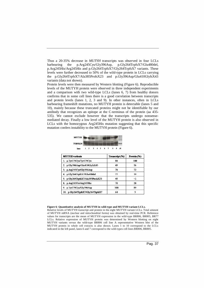
Pag. 37
Thus a 20-35% decrease in MUTYH transcripts was observed in four LCLs harbouring the p.Arg245Cys/Gly396Asp, p.Gly264TrpfsX7/Glu480del,
p.Arg245His/Arg245His and p.Gly264TrpfsX7/Gly264TrpfsX7 variants. These
levels were further decreased to 50% of the wild-type protein in LCLs carrying
the p.Gly264TrpfsX7/Ala385ProfsX23 and p.Gly396Asp/Glu410GlyfsX43
variants (data not shown).
Protein levels were then measured by Western blotting (Figure 6). Reproducible
levels of the MUTYH protein were observed in three independent experiments
and a comparison with two wild-type LCLs (lanes 6, 7) from healthy donors
confirms that in some cell lines there is a good correlation between transcripts
and protein levels (lanes 1, 2, 3 and 9). In other instances, often in LCLs harbouring frameshift mutations, no MUTYH protein is detectable (lanes 5 and
10), mainly because these truncated proteins might not be identifiable by our
antibody that recognizes an epitope at the C-terminus of the protein (aa 435-
535). We cannot exclude however that the transcripts undergo nonsense-
mediated decay. Finally a low level of the MUTYH protein is also observed in
LCLs with the homozygous Arg245His mutation suggesting that this specific
mutation confers instability to the MUTYH protein (Figure 6).
Figure 6: Quantitative analysis of MUTYH in wild-type and MUTYH variant LCLs.
Relative levels of MUTYH transcript and protein in the eight MUTYH variant LCLs. Total amount
of MUTYH mRNA (nuclear and mitochondrial forms) was obtained by real-time PCR. Reference
values for transcripts are the mean of MUTYH expression in the wild-type BR806, BR805, BR77
LCLs. Relative expression of MUTYH protein was determined by Western blotting on eight
MUTYH variants versus the wild-type BR806 cell line A representative Western blot of the
MUTYH protein in whole cell extracts is also shown. Lanes 1 to 10 correspond to the LCLs
indicated in the left panel, lanes 6 and 7 correspond to the wild-types cell lines BR806, BR805.

Pag 38
All LCLs derived from MAP patients have a defective MUTYH activity
In the majority of reports, with a few exceptions (Gu and Lu, 2001; Alhopuro et
al., 2005; Parker et al., 2005), purified or partially purified MUTYH proteins
were used to investigate the DNA glycosylase activity of MUTYH variants (Bai
et al., 2005; Bai et al., 2007; Ali et al., 2008; Yanaru-Fujisawa et al., 2008; D’Agostino et al., 2010). Here we report a novel protocol to measure both
MUTYH and OGG1 glycosylase activity in cell free extracts from human LCLs.
Whole cell extracts prepared from wild-type BR77 cells line showed increasing
DNA glycosylase activity as a function of protein concentration on a 30-mer
containing a single 8-oxo-dG:A mispair (lanes 3-5, Figure 7A). A similar level
of activity was observed in extracts from a second wild-type cell line, the BR806
LCL, with incision levels reaching 15-20% of the DNA substrate (Figure 7B). A
dose response for the OGG1-dependent removal of 8-oxo-dG from a duplex
with an 8-oxoG:C mispair was also identified in cell extracts prepared from
wild-type BR77 cells line (Figure 7C).
Figure 7: Dose response of MUTYH and OGG1 DNA-glycosylase activity in cell extracts from
wild-type BR77 LCL.
A) A representative gel of the reaction products due to MUTYH activity on an 8-oxoG:A substrate is
shown. A 3’-end fluorescent labelled DNA substrate was incubated with increasing amount of cell
extracts. The product of the DNA-glycosylase activity in cell-free extracts is a fragment of 16 bases
(indicated by an arrow) that co-migrates with the product of the purified MUTYH protein.
Control DNA (lane 1); reaction product of a purified MUTYH protein (lane 2), reaction products
following incubation with 30, 60 and 90 g of cell extracts from WTBR77 LCL (lanes 3-5).
B) Fluorescent band intensities from triplicate gels (as in A) were quantified using the public domain
NIH ImageJ software and data analysis was performed with Kaleidagraph software. The percentage
of reaction product following incubation with increasing amount of cell extracts from the wild-type
BR77 cells (empty circle) are shown together with values obtained in a second wild-type cell line
(BR806, full triangle). C) Plot of the percentage of reaction product due to OGG1 activity on an 8-
oxoG:C substrate following incubation with increasing amount of cell-free extracts from WTBR77
LCL.

Pag. 39
MUTYH DNA glycosylase activity was then investigated in cell-free extracts
prepared from LCLs from MAP patients (Figure 8A, top panel). In six LCLs no MUTYH activity was identifiable, while a residual 5% enzymatic activity was
still present in the p.Tyr179Cys, either in homozygousity or in combination with
the p.Gly396Asp variant (lanes 5 and 6, Figure 8A and B). Parallel
measurements of OGG1 activity indicate that these cell lines are similarly
efficient in the removal of 8-oxodG from 8-oxoG:C mispairs (Figure 8A, bottom
panel).
These data demonstrate that all the eight LCLs derived from MAP patients share
a clearly defective MUTYH activity but still maintain a functional OGG1
glycosylase.
Figure 8: MUTYH and OGG1 DNA-glycosylase activity in cell extracts from LCLs from
different MAP patients.
A) Representative gels of MUTYH and OGG1glycosylase activity in different LCLs. 6-FAM-
labelled 8-oxoG:A (top) and 8-oxoG:C DNA substrate (bottom) were reacted with 90 ug of cell
extracts at 37°C for 1h and treated with NaOH to visualize abasic site formation. Positive control
with MUTYH purified protein (lane 1); control DNA (lane 2). B)The percentage of product obtained
after reaction of the 8-oxoG:A DNA substrates with LCL cell extracts as described in Figure 8 A is
shown.
MUTYH variants show a defective repair of DNA 8-oxo-dG
Inactivation of the MUTYH gene in rodents is associated with an accumulation
of DNA 8-oxo-dG observed both in vitro and in vivo (Molatore et al., 2010;
Russo et al., 2009). To verify whether this occurs also in human cells, steady-
state levels of this oxidized purine were measured by HPLC/EC in LCLs from
MAP patients and compared to control LCLs derived from two healthy donors.

Pag 40
Basal levels of DNA 8-oxodG in the BR77 and BR806 wild-type cell lines were 0.44 and 0.49 residues x10-6 dG, respectively (Figure 9A). Increases that ranged
from 1.3 up to 1.7-fold were detected in DNA 8-oxodG levels of six LCLs
expressing MUTYH variants (p < 0.02-0.001) (Figure 9A).
Figure 9: DNA 8-oxodG levels in wild-type and MUTYH variant LCLs.
A) Steady-state DNA 8-oxodG levels were measured in wild-type BR77 and BR806 LCLs by
HPLC/EC. Data are mean+SE from 6-11 independent measurements. ** p<0.01, *** p<0.001
(Student's t test). B) Repair kinetics of DNA 8-oxodG following exposure to a 30-min treatment with
20mM KBrO3 in wild-type and MUTYH variant LCLs. Rate of DNA 8-oxodG removal was
measured at the indicated time points. Data are mean+SE from five experiments.
The two exceptions were cells harboring the p.Gly264TrpfsX7 variant either in
homozygosity or in combination with the frameshift mutation
p.Ala385ProfsX23. It is noticeable that these are the only cell lines in which no
detectable expression of the MUTYH protein could be identified.
To confirm that the steady-state accumulation of 8-oxodG was due to a defective
MUTYH activity, four LCLs were treated with the KBrO3 oxidant, which
introduces a high level of 8-oxo-dG in DNA, and levels of the oxidized purine were measured at different repair times (Figure 9B). All the LCLs with variant

Pag. 41
MUTYH, including the p.Gly264Trpfs/Ala385ProfsX23 variant which does not
express the protein, showed DNA 8-oxo-dG repair kinetics much slower than those observed in wild-type BR77 cells (Figure 9B), with almost a doubling in
the half-life of the lesion (360 min vs. 180 min).
These data indicate that loss of MUTYH activity is always associated with an
accumulation of DNA 8-oxo-dG in cells exposed to an oxidative stress. In the
majority of cases this defect can also be identified at basal levels, with the
expression of mutant MUTYH proteins leading to a more profound effect than
the simple protein loss.
A defective MUTYH activity is associated with a spontaneous mutator
phenotype
To investigate whether the defective MUTYH activity in LCLs from MAP
patients is associated with increased mutagenesis, mutation frequency at the X-
linked PIG-A gene were compared in wild-type and MUTYH variant LCLs.
Mutations in this gene result in either complete or partial deficiency of glycosylphosphatidylinositol (GPI)-linked proteins from the cell membrane and
GPI-negative cells can be identified by flow cytometry analysis as CD48, CD59
and CD55-negative cells (Araren et al., 2005; Peruzzi et al., 2010). The
experimental procedure is shown in Figure 10A. When multiple measurements
of mutation frequency at this gene were performed at weekly intervals in three
wild-type cell lines (BR77, BR805 and BR806), a mean value of 21.7 x 10⁻⁶
GPI-negative events (10.9, 31.7 and 22.5 x 10⁻⁶, respectively) was observed,
with modest variations among different measurements. This value is in the range of values previously reported for wild-type human cell
lines (Araren et al., 2005; Peruzzi et al., 2010). LCLs from MAP patients, as a
group, showed an increased mutation frequency when compared to values
recorded in wild-type cells with a mean value of 87.6 x10⁻⁶ GPI-negative events,
ranging from 42.2 (the homozygous pGly264TrpfsX7) to 157 x 10-6 GPI-
negative events (pArg245His/Arg245His) (unpaired t-test, p=0.04) (Figure
10B).
In order to exclude that this high mutational load was due to a genetic drift of the cell population, GPI-negative cells were eliminated by flow sorting in the
p.Gly396Asp/Glu410GlyfsX43 cell line. This sorted cell population was then
expanded in vitro for several days and re-analyzed for mutation frequency at the
PIG-A gene (Figure 10C). Once the initial GPI-negative events had been
cleared, mutation frequency measured after several days in culture raised up to
149 mutation x 10-6 cells. Thus flow cytometry analysis of this cell population
revealed the reappearance of the GPI-negative cells, demonstrating the tendency
of this MUTYH defective cell line to accumulate mutations in the absence of any mutagenic stress. We can conclude that a spontaneous mutator phenotype is a
characteristic feature of cells with a defective MUTYH activity.

Pag 42
Figure 10: Mutation frequency at the PIG-A gene.
A)Flow cytometry methodology used to establish the population to analyze for the measurement of
mutation frequency. Dead cells were excluded by propidium iodide staining (PI/FSC-A), while cells
doublets were discriminated and excluded by comparing Area and Height signals of Forward scatter
parameters (FSC-H/FSC-A) (first two panels). The FITC-HLA-DR positive population was gated
(third panel) and evaluated in the PE channel (last panel), in order to obtain a correct compensation
of FITC emission and to establish, at the same time, the gating necessary to determine the
distribution of mutant cells in the PE channel. B) Representative flow cytometry dot plot analysis of
WTBR77 (first panel) and p.Gly396Asp/Glu410GlyfsX43 (second panel) LCLs stained with
antibodies against CD48, CD55, CD59 (all GPI-anchored proteins) and HLA-DR (a not GPI-
anchored protein) antigens. Mutant cells, represented by GPI-negative events (lower quadrant of
panels) do not express CD48, CD55, CD59 but do express HLA-DR. Spontaneous mutation
frequencies, calculated as the fraction of GPI-negative events in HLA-DR positive cells (third panel)
measured at one week intervals in wild-type (BR77, BR805 and BR806) and mutant MUTYH LCLs.
Each bar represents the mean of 4-11 independent values. C) Cytometry dot plots of
p.Gly396Asp/Glu410GlyfsX43 cells before (left) and after (center) sorting for GPI-positive
population. Reanalysis of the same sorted population after several days in culture (right) shows the
reappearance of GPI-negative cells.
MUTYH impairment causes a hypermutable phenotype in response to
oxidative stress
We have previously shown that MUTYH-defective variants are hypersensitive
to the toxic effects of oxidative damage introduced into DNA by KBrO3
exposure (Molatore et al., 2010). To investigate whether this enhanced sensitivity to killing induced by an oxidant was also associated with a
hypermutable phenotype, mutation frequencies were compared in wild-type
BR77 and pGly396Asp/Glu410GlyfsX43 LCLs following treatment with
increasing KBrO3 concentrations (Figure 11).

Pag. 43
Figure 11:KBrO3-induced mutations at the PIG-A gene in wild-type BR77 cells and MUTYH-
defective p.Gly396Asp/Glu410GlyfsX43 cells. A)Survival measured in wild-type BR77 cells (black) and p.Gly396Asp/Glu410GlyfsX43 cells
(white) 7 days after a 30 min exposure to increasing concentrations of KBrO3. B) and C)
Mutation frequency measured 7 days after a 30 min treatment with increasing concentrations of
KBrO3 in wild-type BR77 (B) and p.Gly396Asp/Glu410GlyfsX43 (C) cells. Values are the mean
and SD of two independent experiments. D) Comparison of the dose response curves for mutation
induction by KBrO3 exposure in WTBR77 and p.Gly396Asp/Glu410GlyfsX43 LCLs.
In comparison to wild-type cells the MUTYH-defective cell line was
hypersensitive to killing by KBrO3 (Figure 11A). Although a linear increase in
mutation frequency was observed as a function of dose in both cell lines (Figure
11B and C), the number of mutations introduced into the genome by KBrO3
exposure was much larger in pGly396Asp/Glu410GlyfsX43 than in BR77 cells
(Figure 11D). This is evident by the comparison of the respective slopes (y=58.5x+382 vs y=14x-5, respectively) with an increment of 4.2 fold in the
mutagenic response. These data indicate that the defective removal of 8-oxo-dG
associated with a MUTYH defect leads to a hypermutable phenotype when cells
are exposed to an oxidizing agent.

Pag 44
Part 2
Mutyh or Msh2 deficiency confers resistance to the combined effects of 6-
TG and UVA
In order to investigate the role of the MUTYH gene in modulating the biological
response to DNA damage induced by a combined 6-TG/UVA exposure, we set
up an in vitro experimental model in which Mouse embryo fibroblasts (MEFs)
derived from Mutyh- or Msh2- defective mice were compared to wild-type (WT)
cells. As WT cells we used Mutyh-/- MEFs transfected with the pYMv200–
MUTYH vector containing the human MUTYH cDNA (Molatore et al.; 2010). Because of the established function of the MMR MSH2 protein in controlling 6-
TG toxicity, as well as accumulation of 8-oxo-dG into DNA, we also aimed to
examine the role of this protein in the 6-TG/UVA treatment-induced effects.
Viability, as determined by clonal assays, was compared in MEFs derived from
Mutyh-/- and Msh2-/- mice as well as in WT cells. First of all the response to
single treatments was analysed. As reported in human cells (O'Donovan et
al.;2005), UVA exposure did not modify cell survival in any cell type. In
contrast a single 6-TG exposure mainly affected, at a comparable level, survival
of WT and Mutyh-/- cells, while Msh2-/- MEFs displayed the expected tolerance to this type of DNA damage (data not shown) (Bignami et al.; 2003). When
cultures were exposed to a combined 6-TG/UVA treatment (growth in 60 nM 6-
TG for 48 hours followed by UVA irradiation), WT MEFs showed a
considerably reduced cell survival in comparison to Mutyh-/- and Msh2-/- MEFs
(Figure 12).
Figure 12: Survival following a combined 6-TG and UVA treatment.
WT, Mutyh-or Msh2-defective MEFs were exposed to a non-toxic concentration of 6-TG (60 nM)
for 48 hours and irradiated with UVA (5 or 10 kJ/m2). Survival was determined by clonal assays and
expressed as percentage of the cloning efficiency in 6-TG. Data are mean+SE from at least 6
independent measurements.

Pag. 45
The lethal effects were also found to be proportional to the UVA dose (5 and 10 kJ/m2). We concluded that 6-TG and UVA are synergistically toxic to WT
MEFs, while loss of either Mutyh or Msh2 confers resistance to the detrimental
effects of the combined treatment.
The combined 6-TG and UVA treatment significantly increases the levels of
DNA 8-oxo-dG
To study the extent of oxidative stress associated with a combined 6-TG/UVA
exposure, levels of DNA 8-oxo-dG were analysed in MEFs of different
genotype (Figure 13).
Figure 13: DNA 8-oxo-dG levels in WT, Mutyh- or Msh2-defective MEFs. The amount of 8-oxo-
dG was measured by HPLC/EC in untreated MEFs as well as followingUVA exposure or a single or
a combined exposure to 6-TG (0.6 mM for 48 h) and UVA (10 kJ/m2).Data are mean+SD from at
least 6 independent measurements. ** p<0.01, *** p<0.001 (Student's t test).
Irradiation with UVA alone did not modify steady-state levels of DNA 8-oxo-
dG in any of the cell lines. In contrast 6-TG treatment of wild-type MEFs induced a small increase in DNA 8-oxo-dG (1.4-fold) when compared to
untreated controls. This modest accumulation could also be appreciated in Msh2-
/- MEFs, whereas the higher basal levels of the oxidized purine associated with
MUTYH inactivation (Russo et al., 2009) masked this effect in Mutyh-/- MEFs.
Interestingly, at comparable levels of 6-TG incorporation (data not shown), the
synergistic effect of 6-TG/UVA interaction resulted in a remarkable increase in
8-oxo-dG levels (~2-fold in WT or Msh2-/- MEFs and ~1.7-fold in Mutyh-/-
MEFs). Thus the combined treatment introduces oxidized bases in DNA and these enhanced oxidation levels are unaffected by the genotype.

Pag 46
Oxidation derived from dNTPs pool contributes to the detrimental effects
of the combined treatment
Experimental evidence indicate that in 6-TG-treated cells the base analog can be
found in the dNTPs pool where it can be a source of ROS upon UVA irradiation
(Cooke et al., 2008).
To investigate the potential contribution of 6-TG present in the dNTPs pool to
the biological response to the combined treatment, we depleted the dNTPs pool
from 6-TG nucleotides by introducing a “chase” in our experimental settings.
Thus cells were grown in a 6-TG containing medium for 24 and irradiated after a further 24 h incubation in 6-TG-free medium. (Figure 14A).
Figure 14: Effects of pool depletion on cellular survival
A) Experimental setting used to investigate the contribution of an oxidized dNTPs pool to 6-
TG/UVA cytotoxicity. To deplete the cellular pool of 6-TG nucleotides, cultures were grown for 24
h in 0,6 M 6-TG and UVA-irradiated (10 kJ/m2) after a further 24 h growth in medium without 6-
TG (24 + 24 h, “chase”); 48 h: classical experiment; 24 h: internal control of the “chase” experiment.
No difference in survival was observed between a 24 or 48 h treatment with 6-
TG alone. A 24 h “chase” in 6-TG-free medium however drastically reduced
killing induced by a combined 6-TG/UVA exposure. This protective effect on
cell survival was particularly significant in WT cells, where the synergistic action of 6-TG and UVA on viability was much more dramatic than in the
Mutyh or Msh2-defective cell lines (Figure 14B). In agreement with the capacity
of 6-TG in the dNTPs pool to act upon UVA exposure as an oxidant towards
DNA bases, a 24 h “chase” in WT cells reduced DNA 8-oxo-dG levels to steady
state levels associated with the single 6-TG treatment (Figure 15A). A similar
result was obtained in Msh2-/- MEFs and to a lower extent in Mutyh-/- cells.
Further confirmation that the oxidized pool contributes to oxidation of DNA
bases comes from results obtained in a Msh2-/- clone overexpressing the hMTH1 hydrolase (Russo et al.; 2004). In this case high levels of hMTH1 activity
completely abolished the increase of 8-oxodG levels associated with the
combined treatment (Figure 15B).

Pag. 47
Figure 14: Effects of pool depletion on cellular survival
B) Sensitivity of cells to the combined 6-TG and UVA treatment after a “chase” experiment. WT,
Mutyh -or Msh2-defective MEFs were exposed to 6-TG (60 nM) for 48 or 24 h and UVA-irradiated
immediately afterwards (48 h e 24 h) or after a further 24 h growth in medium without 6-TG (24+24
h). Survival was determined by clonal assays and expressed as percentage of the cloning efficiency
in 6-TG. Data are mean+SD from at least 6 independent measurements determinations. *p<0,05, ***
p<0.001, ns: not statistically significant (Student's t test).
Figure 15: Effects of pool depletion on DNA 8-oxo-dG levels. A) Measurement of DNA 8-oxo-dG in WT, Mutyh -or Msh2-defective MEFs irradiated with 10
kJ/m2 UVA, following exposure to 6-TG (0.6 M) for 24 h or following a “chase” experiment. Data
are mean+SD from at least 6 independent measurements. ** p<0.01, ns: not statistically significant
(Student's t test). B) Measurement of DNA 8-oxo-dG levels in Msh2-defective MEFs and Msh2-/-
clone overexpressing the hMTH1 hydrolase. Data are mean+SD from at least 6 independent
measurements. *p<0,05, ** p<0.01 (Student's t test).

Pag 48
6-TG plus UVA treatment induce cell cycle perturbation
To better understand the role of MUTYH in 6-TG/UVA-induced toxicity, WT
and Mutyh-/- MEFs were treated with 6-TG for 24 h, irradiated with UVA and
analysed by flow cytometry after different recovery times (8, 24 and 48 h). As
expected (Yamane et al., 2004) 6-TG treatment alone produced a progressive
accumulation of WT cells in the G2-M phase (31% and 45% of the cell
population at 24 and 48h, respectively) (Figure 16). In contrast the combined 6-
TG/UVA treatment slows down the progression of the population through the S
phase, with >than 75% of the cells blocked in the S phase at 24 h. Following 6-TG incorporation Mutyh-/- cells showed a milder perturbation of cell
cycle progression when compared to WT cells (a reduced percentage of cells are
in G2-M phase, 20% and 25% of the cell population at 24 and 48h, respectively)
and progressed into the G2-M phase after the combined 6-TG/UVA treatment
(only 17% of cells were in the S phase at 24 h). These data suggest that MUTYH
is needed for an efficient S phase arrest in the presence of lesions induced by the
combined treatment. Finally irradiation with UVA alone did not produce
changes in cell cycle progression in any cell type.
Figure 16:Cell cycle progression WT and Mutyh-defective MEFs were treated with 0.6 M for 24
h, irradiated with 10 kJ/m2UVA and analysed by flow cytometry at different recovery times (8, 24
and 48 h).Percentage of cells in S and G2/M phases are shown.
MUTYH sustains early checkpoint activation in response to the combined
treatment
To investigate in more detail the mechanisms underlying the accumulation of
WT cells in the S phase of cell cycle, after the combined treatment, the activated
form of the Chk1 protein was examined by Western-blotting.

Pag. 49
To this aim, cells were treated with 6-TG for 24 h and analysed after a recovery time of 1, 16, 20 and 24 h following UVA radiation.
In WT cells, phosphorylation of Chk1 on serine 345 occurred immediately after
6-TG/UVA treatment (1h), (Figure 17) and disappeared afterwards.
Figure 17:Analysis of Chk1 phosphorylation. WT and Mutyh-defective MEFs were grown in 0.6
M 6-TG for 24 h and irradiated with 10 kJ/m2 UVA. To detect the activated form of the Chk1
protein samples were collected 1, 16, 20 and 24 h after irradiation and analysed by Western blotting.
Topoisomerase II (Topo II) and total Chk1 protein were used as loading controls. Cells treated were
with 2 mM Hydroxyurea (HU) for 8 h and as a positive control.
In Mutyh -/- cells in contrast, phosphorylation of Chk1 was observed at 16 h after
treatment and was maintained, thereafter, even though a gradual decrease was
observed.
These data suggest that MUTYH activity might affect the early activation of the
S phase checkpoint, as well as its regulation. No difference in Chk1 activation was detectable after exposure to 24 h 6-TG in the two genotypes (data not
shown)
MUTYH affects the processing of damage by HR
Since loss of MUTYH leads to an easier progression through the cycle of
presumably damaged cells, we investigated the initial levels of DSBs as well as their processing by HR in cells grown for 24 h in 6-TG and UVA irradiated
immediately afterwards. We confirmed that, as in human cells (Brem R et al.;
2010) treatment of the cells with 6-TG/UVA produces an increase in DSBs as
measured by the number of H2AX foci, and these occurred immediately and were maintained up to 48 h from the end of the treatment (Figure 18A).

Pag 50
Figure 18: -H2AX and RAD51 foci formation. WT and Mutyh-defective MEFs were grown in the
presence of 0.6 M 6-TG for 24 h, irradiated with 10 kJ/m2 UVA and collected for
immunofluorescence analysis immediately afterwards, or after 8, 24 and 48 h. A) Graphs show the
percentage of -H2AX-positive nuclei in each experimental condition (upper panel). B) Graphs show
the percentage of RAD51-positive nuclei in each experimental condition (upper panel); cells treated
with 30 M camptothecin (CPT) for 4h were used as a positive controls. Representative images are
shown in the lower panels.
This increase was of a comparable extent in Mutyh-defective cells, thus
indicating that the initial level of DNA damage is similar in the two genotypes. Analysis of RAD51 foci in WT MEFs indicated that DSBs are processed by HR,
with RAD51 foci increasing 8 h after the end of 6-TG/UVA exposure and
persisting up to 48 h, indicating the presence of unresolved DSBs. In contrast
Mutyh-/- MEFs showed a lower level of RAD51 foci in comparison to WT cells
with a gradual decrease at late post-treatment times (8-48 h) (Figure 18B).
We can conclude that exposure to 6-TG plus UVA introduces a similar level of

Pag. 51
DNA damage in both genotypes. However the resolution of lesions induced by the combined treatment differs in WT and Mutyh-/- MEFS suggesting that the
MUTYH protein plays a role in the processing of this type of DNA damage.
Mutyh-/-
mice are resistant to the toxicity of the combined treatment but
develop skin cancer
In order to analyse the effects of the single and combined 6TG /UVA treatment in vivo, WT and Mutyh-defective mice were treated with Aza, UVA alone or
Aza plus UVA for 12 months. Following a series of short-term experiments in
which toxicity associated with UVA exposure (by Minimal Erythemal Dose)
and Aza-induced immunosuppression (by Mixed Lymphocyte Reaction) were
evaluated in both WT and Mutyh-/- mice, the experimental conditions for a long-
term experiment were identified.
WT and Mutyh-/- C57BL mice were exposed to 15mg/kg Aza given by
intraperitoneal injection three times/week. A UVA dose of 150 kJ/m2 was
applied on the dorsal skin of shaved animals three times/week, one h following Aza injection. A differential toxicity between WT and Mutyh-defective animals
was observed as consequence of the Aza plus UVA exposure.
In fact, while a high level of mortality was identified in the WT group (only
20% of the animals survived this exposure), 80% of Mutyh-/- treated animals
survived the combined treatment. Survival was 100% in both UVA treated
groups, while immunosuppression in Aza-treated groups was associated with
some mortality, which was unaffected by the genotype (data not shown).
When DNA 8-oxo-dG levels in samples of spleen and skin were measured, a significant increase of DNA oxidation in the skin of animals exposed to the
combined treatment was observed, irrespectively of the genotype (Figure 19A).
A trend of increased DNA oxidation, that did not reach statistical significance,
was also observed in the spleen of both genotypes following the combined
treatment. In addition Mutyh-/- mice showed a small but non-significant increase
of the levels of 8-oxo-dG in the skin of UVA-treated group.
Interestingly, when histopathological examination of the skin was performed
two squamous cell carcinomas, at a first stage of differentiation, were identified only in Mutyh-/- mice exposed to Aza plus UVA (Figure 19B).

Pag 52
Figure 19: Effects of Aza/UVA exposures in WT and Mutyh-defective mice.
A) DNA 8-oxo-dG levels in spleen and skin samples of mice treated with 15mg/kg Aza and/or 150
kJ/ UVA m2
three times/week for 12 months. Data are mean ± SE of at least 3 independent
determinations *, P< 0.05 (Student's t test).B) Two examples of well-differentiated squamous cell
carcinomas invading the dermis occurring in Mutyh-defective mice (first two panels). The third
panel shows a microscopic magnification of a histological detail (collaboration with MT Mancuso,
ENEA).
Taken together, the in vitro and in vivo experiments confirm the involvement of the MUTYH protein in the response to the damage induced by combined Aza
and UVA treatment. Interestingly they also reveal that the absence of Mutyh is
associated to an increased susceptibility to the carcinogenic effects of this type
of DNA damage.

Pag. 53
III. DISCUSSION
Extensive biochemical and genetic studies in model organisms demonstrate that
the oxidative DNA lesion 8-oxo-dG is mutagenic and several DNA repair
mechanisms operate to prevent its potentially detrimental effects. The
importance of an efficient DDR in the protection against the carcinogenic
outcome of oxidative lesions is indicated by the increased cancer susceptibility
of individuals with germ-line mutations in genes involved in the repair of
oxidative DNA damage. This includes biallelic germ-line mutations in the
MUTYH gene, encoding a DNA glycosylase that is fundamental in the removal
of 8-oxo-dG from DNA, which strongly predispose humans to MAP, a rare
hereditary form of colorectal cancer.
MAP is an increasingly studied autosomal recessive disorder but functional analysis of MUTYH mutations is relatively limited and mainly related to the
most recurrent missense variants. In the first part of this report we present a
novel assay for analysis of MUTYH DNA glycosylase activity in LCLs from
MAP patients. At variance with in vitro studies of purified MUTYH mutant
proteins or when human variants are expressed in rodent cell lines (Molatore et
al., 2010), this experimental approach allows studying the pathogenic
consequences of mutant proteins in the biological context in which they are
expressed. Evaluation of MUTYH protein levels in LCLs from MAP patients indicates that
analysis of transcript levels is not always exhaustive. Indeed in three out of four
cells lines harbouring frameshift mutations the presence of transcripts results in
a greatly reduced amount of the MUTYH protein. Although we cannot formally
exclude that the predicted truncated proteins are undetectable by the antibody
used in this study, it is more likely that these transcripts are subjected to
nonsense-mediated decay. Interestingly, the substitution of Arg with His at the
245 amino acid position results in a reduced level of the MUTYH protein,
suggesting that this mutation localized near the FeS cluster domain might affect
both protein stability and function.
The results of the glycosylase assay indicate that all the LCLs from MAP patients are impaired in 8-oxodG:A cleavage when compared to WT cell lines.
It’s hardly surprising that cells characterized by an unstable MUTYH protein
show no DNA glycosylase activity. In agreement with other reports (Wooden et
al., 2004; Parker et al., 2005; Ali et al., 2008; Kundu et al., 2009), a residual but
minimal enzymatic activity was retained in the p.Tyr179Cys/Tyr179Cys and
p.Tyr179Cys/Gly396Asp variants, which are characterized by WT levels of the
protein. Similarly the p.Arg245Cys/Gly396Asp variant, although expressed, was
totally defective in its glycosylase activity, thus identifying the presence of a
dysfunctional MUTYH protein. In comparison to DNA glycosylase assays based
on immunoprecipitated MUTYH proteins (Parker et al., 2005; Alhopuro et al.,
2005), the advantage of this assay resides in the relatively small number of cells

Pag 54
needed for the preparation of cell extracts and the possibility of analysing MUTYH function even when putative truncated proteins are present.
We previously demonstrated that MUTYH loss is always associated with
increased steady-state levels of DNA 8-oxo-dG. In addition expression of some
MUTYH variants leads to a more profound effect in the removal of the oxidized
purine than the simple protein loss (Molatore et al., 2010). Indeed also in human
cells defective MUTYH DNA glycosylase activity was reflected in an in vivo
accumulation of DNA 8-oxo-dG following an oxidative stress induced by the
KBrO3 oxidant (Kawanishi and Murata 2006). Even in the absence of exogenous
oxidation, when compared to wild-type cells, an increased steady-state level of
DNA 8-oxodG was observed in the majority of LCLs harbouring MUTYH
variants. Finally, LCLs harbouring the p.Gly264TrpfsX7 and p.Ala385ProfsX23
frameshift mutations, in which there is no detectable expression of the MUTYH protein, show a more moderate phenotype. Thus we suggest that also in humans
the presence of a mutant MUTYH protein has a dominant negative effect,
possibly interfering with others DNA repair pathways involved in the removal of
8-oxo-dG, for example MMR, other BER factors, nucleotide excision repair, or
in signalling of DNA damage (the 9-1-1 complex) (Shi et al., 2006).
At variance with the mouse model where mutant MUTYH proteins are
expressed in the same genetic background (Mutyh-/- MEFS), LCLs from
different patients might have an ample range of cellular responses to oxidative
stress. This is one of the reasons why both MUTYH and OGG1 activity were measured in parallel in cell-free extracts. The observation that all LCLs show
wild-type OGG1 activity allows concluding that the accumulation of DNA 8-
oxo-dG is a direct consequence of the MUTYH defect.
The information on the mutator phenotype associated with MUTYH inactivation
is limited to MEFs and embryonic stem cells from Mutyh-/- mice, in which
increases in mutation rates varied from 2- to 10-fold (Hirano et al., 2003; Russo
et al., 2009). The presence of specific G>T transversions in APC or K-RAS
genes in tumors occurring in MAP patients can also be considered an indirect evidence of a mutator phenotype associated with the presence of a non
functional MUTYH (Al-Tassan et al., 2002; Jones et al., 2002, Lipton et al.,
2003; van Pujienbroek et al., 2008). Here we investigated for the first time to
which extent MUTYH impairment results in an increased mutation frequency in
human cells. The gene we chose for this analysis is the X-linked PIG-A gene
that has been successfully used to characterize the mutator phenotype associated
with DNA repair defective human syndromes (Ataxia telangiectasia, Fanconi
anemia) (Araten et al., 2005). When compared to the 21.7x10⁻⁶ mutational events at the PIG-A gene observed in wild-type cells, the group of LCLs from MAP
patients showed a 4-fold increased mutation frequency.
The reappearance of PIG-A mutants in a cell line with one of the highest
mutation frequency (p.Gly396Asp/Glu410GlyfsX43), following flow sorting to
eliminate the pre-existing mutant cells, demonstrates that this phenotype is not
due to a genetic drift of the cell population but it is an intrinsic feature of these

Pag. 55
cells. The reported spontaneous mutation rate ( ) at this gene for normal cells ranges from 8.8 to 16.5 x 10⁻⁷ mutation per cell division (Peruzzi et al., 2010). A
tentative value of 299 x 10-7 mutation per cell division was calculated on the p.Gly396Asp/Glu410GlyfsX43 LCLs following measurement of mutation
frequency after six doublings. This value is at least 10-times higher that values for normal human cells (Araten et al., 2005). These data support the presence of
a spontaneous mutator phenotype associated with defective MUTYH proteins
and fit quite well with the proposed role of MUTYH variants in favouring the
appearance of mutations in oncogenes relevant in the process of colorectal
carcinogenesis (Al-Tassan et al., 2002; Jones et al., 2002). Since oxidative damage in DNA can contribute to genome instability we also
investigated whether MUTYH inactivation exposes cells to a further increase in
their mutational burden in a condition of oxidative stress. Thus treatment of the biallelic p.Gly396Asp/Glu410GlyfsX43 variant LCLs with an oxidizing agent
results indeed in a hypermutable phenotype. Oxidative stress might then play an
independent and major role in causing genome instability in the presence of an
inactivating MUTYH mutation. Although the mutator phenotype associated with
MUTYH defects is considerably milder than that observed in MMR-defective
cells, the hypermutability induced by specific genotoxic agents is a shared
characteristic of cells with inactivation in either pathway (Glaab et al., 1998; Xu
et al., 2001; Bignami et al., 2000; Sansom et al., 2003 and this study). Thus we suggest that the level of oxidative damage to DNA bases undergoing in specific
districts of the body (and specifically in the gastrointestinal tract) might be a
relevant issue to explain the tissue specificity of the increased cancer
susceptibility of MUTYH- and MMR-defective patients.
By taking into consideration the prominent biological impact of pathogenic
defects in the MUTYH protein when dealing with oxidative damage to DNA, we
decided to investigate the effects of MUTYH impairment in response to another
type of oxidative stress. In the second part of this thesis we report the results of
this parallel study.
Combination of Aza, an inflammatory, anticancer and immunosuppressive drug, and UVA radiation is a major source of DNA 8-oxo-dG (Cooke et al., 2008).
The interaction between 6-TG and UVA generates ROS and the resulting
photochemical oxidation of DNA has been implicated as a possible contributor
to the high skin cancer risk associated to long-term Aza treatment (Karran and
Attard 2008). Experimental data obtained by Peter Karran’s group suggest that
the combination of 6-TG and UVA causes several types of damage, such as
replication and transcription inhibition, DNA crosslinking, oxidation of DNA
bases and proteins as well the covalent attachment of proteins to DNA. Many of
these DNA lesions are difficult for cells to deal with, resulting in cytotoxic as
well as mutagenic effects in human cells (O'Donovan et al.; 2005).
Here we examined the effects of 6-TG and UVA radiation in in vitro and in vivo
mouse models, particularly regarding the impact of Mutyh loss on the biological response to this type of damage. As observed in human cells, the synergistic

Pag 56
action of 6-TG and UVA resulted detrimental to WT MEFs, with a clearcut
killing effect. Interestingly, loss of the MUTYH or MSH2 proteins was associated with a considerable resistance to this type of damage.
In view of the extensive oxidation provoked by the combined treatment in
human cells, we expected that this experimental setting associated with high
ROS production might result in increased levels of DNA 8-oxo-dG in the mouse
models.
Taken into account that both MUTYH and MSH2 control the amount of 8-oxo-
dG in DNA (Russo et al., 2009), Msh2- and Mutyh-defective MEFs were
expected to accumulate the oxidized lesion in their genome. Nevertheless, all the
cell lines we examined, irrespectively of their genotype, showed similar levels of
oxidative DNA damage following 6-TG and UVA treatment. Thus any
discrepancy in the survival outcome cannot be ascribed simply to differences in the accumulation of oxidative lesions.
It’s interesting that also in our mouse model 6-TG present in the dNTP
represents a significant source of ROS upon UVA irradiation (Cooke et al.,
2008; Brem et al., 2009). “Chase” experiments demonstrated that this non-DNA
6-TG makes indeed a considerable contribution to the 8-oxo-dG increase as well
as to the killing effects induced the combined treatment in WT cells. The
involvement of the oxidized nucleotide pool in DNA oxidation was confirmed
by the great reduction in 8-oxo-dG levels observed in Msh2-defective MEFs
overexpressing the hMTH1 hydrolase. This enzyme degrades 8-oxo-dGTP to 8-
oxo-dGMP, thereby eliminating it from the pool of DNA synthesis precursors.
Attempts to investigate the mechanisms underlying the resistance against the
type of DNA damage induced by 6-TG and UVA and associated to Mutyh loss included flow cytometry analysis of cell cycle progression and activation of
checkpoints. The absence of MUTYH was indeed associated with a failure of S-
phase halt, that in contrast is evident in WT cells, following the combined
treatment. The apparent absence of a clear S phase arrest in Mutyh-/- cells was
paralleled by a deficient Chk1 activation at early time points after treatment.
ATR-mediated Chk1 phosphorylation, is known to play a critical role in
regulating replication fork stability by arresting cells in S or G2/M phase
(Sørensen et Syljuåsen, 2011). Thus our results revealed that MUTYH is
involved in this step of the cellular response to DNA damage.
Recently it has been reported that MUTYH interacts with ATR and is needed for
efficient Chk1 phosphorylation following UVB and HU treatments (Hahm et al.; 2009). Our data are consistent with an involvement of MUTYH in the activation
of the S phase checkpoint following a different type of damage to DNA.
This observation might have consequences for cell killing. It is well known that
ATR-dependent Chk1 phosphorylation controls the repair of DSBs by HR
(Sørensen et al.; 2005). We confirmed that, as reported in human cells (Brem et
al.; 2010), oxidative DNA damage produced by the combined 6-TG and UVA
exposure results in DSBs formation also in MEFs. Even though no significant
differences in the absolute levels of DSBs, as measured by H2AX foci

Pag. 57
formation, were observed between MUTYH-proficient and –deficient cells, a
distinctive mode of DSBs repair was identified in the two genotypes. Quantification of RAD51 foci in WT and Mutyh-defective MEFs revealed
persistence of unresolved lesions in WT cells, while loss of MUTYH protein
resulted in a minor induction of RAD51 foci following the combined treatment.
RAD51 is a well-known mediator of HR that plays a crucial role in protecting
cells against the lethal effects of DSBs. If we consider HR as a pro-survival
response to DNA damage, the hypersensitivity of WT cells in comparison to
Mutyh-defective cells to killing by 6-TG/UVA is an apparent contradiction. The attempt by HR of damage resolution might however introduce a heavy
burden of dangerous repair intermediates that ultimately lead to a decrease in
cell viability. Thus it is possible that loss of Muthy and the consequent
inefficient or non-canonical DNA repair might represent a viable route of escaping from the lethal impact of 6-TG plus UVA exposure.
In this context, the apparent reduced activation of Chk1 in Mutyh-/- cells might
be functionally correlated with the reduced RAD51 foci formation and with the
enhanced survival, probably at the expense of genomy stability.
The striking observation that the 80% of Mutyh-/- mice versus the 20% of the
wild-type counterpart survived the combined treatment confirms a pro-survival
advantage associated with the absence of the Mutyh gene. However, the
presence of two squamous cell carcinomas (the same tumor identified in Aza
long-term treated patients) in Mutyh-/- mice exposed to Aza plus UVA is in agreement with a carcinogenic effect of the combined treatment. As in the in
vitro model, measurement of 8-oxo-dG into DNA of skin and spleen cells of
mice did not reveal significant differences in oxidative damage accumulation
between the two genotypes (except for the UVA treatment alone). An excess of
extracolonic tumors, including tumor of the skin, has been recently reported in
MAP patients (Vogt et al., 2009). It is intriguing that the survival advantage in
the response against the type of oxidative damage induced by Aza/UVA
provided by loss of Mutyh and identified both in vitro and in vivo, comes at a
price of increased cancer susceptibility in the skin. Whether the mechanism underlying this cancer proneness depends on the role of MUTYH in the cellular
signaling and processing of oxidative damage induced by Aza/UVA remains to
be ascertained.

Pag 58

Pag. 59
IV. CONCLUSIONS
Our experimental investigation on the biochemical and biological consequences
associated with the impairment or complete loss of the MUTYH protein
strengthens the importance of this protein in protecting the genome against
oxidative damage.
Moreover the novel functional assays for genotype-phenotype correlations of
MUTYH variants we set up might provide a better understanding of MAP with
possible implications for counseling, screening and clinical management of patients and their families. Additionally, the study on the effects of MUTYH loss
in mice also provided new insights on the detrimental effects of Aza combined
with UVA radiation and on possible novel functions of MUTYH more complex
than previously thought.

Pag 60

Pag. 61
V. REFERENCES
Abraham RT. Cell cycle checkpoint signaling through the ATM and ATR
kinases Genes Dev. (2001); 15(17):2177-96
Alhopuro P, Parker AR, Lehtonen R, Enholm S, Järvinen HJ, Mecklin JP,
Karhu A, Eshleman JR, Aaltonen LA. A novel functionally deficient MYH
variant in individuals with colorectal adenomatous polyposis. Hum Mutat
(2005); 26:393
Ali M, Heja K, Cleary S, Cupples C, Galllinger S, Bristow R. Characterization
of Mutant MUTYH Proteins Associated With Familial Colorectal Cancer.
Gastroenterology (2008); 135:499–507
Al-Tassan N, Chmiel NH, Maynard J, Fleming N, Livingston AL, Williams GT,
Hodges AK, Davies DR, David SS, Sampson JR, Cheadle JP. Inherited variants
of MYH associated with somatic G:C-->T:A mutations in colorectal tumors Nat
Genet. (2002); 30(2):227-32
Araten DJ, Golde DW, Zhang RH, Thaler HT, Gargiulo L, Notaro R, Luzzatto
L. A quantitative measurement of the human somatic mutation rate. Cancer Res
(2005); 65:8111-8117
Bai H, Grist S, Gardner J, Suthers G, Wilson TM, Lu AL.. Functional
characterization of human MutY homolog (hMYH) missense mutation (R231L) that is linked with hMYH-associated polyposis. Cancer Lett (2007); 250:74-81
Bai H, Jones S, Guan X, Wilson TM, Sampson JR, Cheadle JP, Lu AL.
Functional characterization of two human MutY homolog (hMYH) missense
mutations (R227W and V232F) that lie within the putative hMSH6 binding
domain and are associated with hMYH polyposis. Nucleic Acids Res (2005);
33:597-604
Ballmaier D, Epe B. DNA damage by bromate: mechanism and consequences.
Toxicology (2006); 221(2-3):166-71
Ballmaier D, Epe B. Oxidative DNA damage induced by potassium bromate
under cell-free conditions and in mammalian cells Carcinogenesis (1995);
16(2):335-42
Barone F, McCulloch SD, Macpherson P, Maga G, Yamada M, Nohmi T,
Minoprio A, Mazzei F, Kunkel TA, Karran P, Bignami M.
Replication of 2-hydroxyadenine-containing DNA and recognition by human
MutSalpha DNA Repair (Amst). (2007); 6(3):355-66
Pag 62
Bartek J, Lukas C, Lukas J Checking on DNA damage in S phase Nat Rev Mol
Cell Biol. (2004); 5(10):792-804
Bignami M, Casorelli I, Karran P. Mismatch repair and response to DNA-
damaging antitumour therapies Eur J Cancer. (2003);39(15):2142-9
Bignami M, O'Driscoll M, Aquilina G, Karran P.. Unmasking a killer: DNA
O(6)-methylguanine and the cytotoxicity of methylating agents. Mutat Res
(2000); 462:71-82
Bohr VA. DNA repair fine structure and its relations to genomic instability
Carcinogenesis (1995); 16(12):2885-92
Brem R, Li F, Karran P. Reactive oxygen species generated by thiopurine/UVA
cause irreparable transcription-blocking DNA lesions. Nucleic Acids Res.
(2009); 37(6):1951-61
Brem R, Li F, Montaner B, Reelfs O, Karran P. DNA breakage and cell cycle
checkpoint abrogation induced by a therapeutic thiopurine and UVA radiation.
Oncogene. (2010); 29 (27):3953-63
Brown EJ, Baltimore D. ATR disruption leads to chromosomal fragmentation
and early embryonic lethality Genes Dev. (2000); 14(4):397-402
Cardoso J, Molenaar L, de Menezes RX, van Leerdam M, Rosenberg C,
Möslein G, Sampson J, Morreau H, Boer JM, Fodde R. Chromosomal
instability in MYH- and APC-mutant adenomatous polyps (2006); Cancer Res
66:2514 2519
Cheadle JP, Sampson JR. MUTYH-associated polyposis-from defect in base
excision repair to clinical genetic testing. DNA Repair (Amst) (2007); 6:274-279
Chow E, Thirlwell C, Macrae F, Lipton L. Colorectal cancer and inherited
mutations in base-excision repair Lancet Oncol. (2004); 5(10):600-6
Cleary SP, Cotterchio M, Jenkins MA, Kim H, Bristow R, Green R, Haile R,
Hopper JL, LeMarchand L, Lindor N, Parfrey P, Potter J, Younghusband B,
Gallinger S. Germline MutY human homologue mutations and colorectal
cancer: a multisite case-control study. Gastroenterology (2009); 136:1251-1260
Colussi C, Parlanti E, Degan P, Aquilina G, Barnes D, Macpherson P, Karran P,
Crescenzi M, Dogliotti E, Bignami M. The mammalian mismatch repair

Pag. 63
pathway removes DNA 8-oxodGMP incorporated from the oxidized dNTP pool.
Curr Biol. (2002); 12(11):912-8
Cooke MS, Duarte TL, Cooper D, Chen J, Nandagopal S, Evans MD.
Combination of azathioprine and UVA irradiation is a major source of cellular
8-oxo-7,8-dihydro-2'-deoxyguanosine DNA Repair (Amst). (2008); 1;7(12):
1982 9
Cooke MS, Evans MD, Dizdaroglu M, Lunec J. Oxidative DNA damage:
mechanisms, mutation, and disease FASEB J. (2003); 17(10):1195-214
D'Agostino VG, Minoprio A, Torreri P, Marinoni I, Bossa C, Petrucci TC,
Albertini AM, Ranzani GN, Bignami M, Mazzei F.. Functional analysis of MUTYH mutated proteins associated with familial adenomatous polyposis.
DNA Repair (Amst) (2010); 9:700-707
David SS, O'Shea VL, Kundu S. Base-excision repair of oxidative DNA damage
Nature (2007); 447(7147):941-50
de Klein A, Muijtjens M, van Os R, Verhoeven Y, Smit B, Carr AM, Lehmann
AR, Hoeijmakers JH. Targeted disruption of the cell-cycle checkpoint gene
ATR leads to early embryonic lethality in mice Curr Biol. (2000); 10(8):479-82
de Laat W. L, Jaspers N. G.J, and Hoeijmakers J. H. Molecular mechanism of
nucleotide excision repair Genes Dev. (1999); 13: 768-785
Demple B, Herman T, Chen DS Cloning and expression of APE, the cDNA
encoding the major human apurinic endonuclease: definition of a family of DNA
repair enzymes, Proc. Natl. Acad. Sci. USA (1991); 88(24):11450-4
D'Errico M, Parlanti E, Teson M, de Jesus BM, Degan P, Calcagnile A, Jaruga
P, Bjørås M, Crescenzi M, Pedrini AM, Egly JM, Zambruno G, Stefanini M,
Dizdaroglu M, Dogliotti E. New functions of XPC in the protection of human
skin cells from oxidative damage EMBO J. (2006); 25(18):4305-15
D'Errico M, Parlanti E, Teson M, Degan P, Lemma T, Calcagnile A, Iavarone I, Jaruga P, Ropolo M, Pedrini AM, Orioli D, Frosina G, Zambruno G,
Dizdaroglu M, Stefanini M, Dogliotti E. The role of CSA in the response to
oxidative DNA damage in human cells Oncogene (2007); 26(30):4336-43
Dianov G, Bischoff C, Piotrowski J, Bohr VA. Repair pathways for processing
of 8-oxoguanine in DNA by mammalian cell extracts J Biol Chem. (1998);
273(50):33811-6

Pag 64
Dianov G, Price A, Lindahl T. Generation of single-nucleotide repair patches
following excision of uracil residues from DNA Mol Cell Biol. (1992); 12(4):1605-12
Dou H, Mitra S, Hazra TK. Repair of oxidized bases in DNA bubble structures
by human DNA glycosylases NEIL1 and NEIL2 J Biol Chem. (2003);
278(50):49679-84
Euvrard S, Kanitakis J, Claudy A. Skin cancers after organ transplantation N
Engl J Med. (2003); 348(17):1681-91
Farrington SM, Tenesa A, Barnetson R, Wiltshire A, Prendergast J, Porteous
M, Campbell H, Dunlop MG Germline susceptibility to colorectal cancer due to base-excision repair gene defects. Am J Hum Genet (2005); 77:112-119
Fortini P, Parlanti E, Sidorkina OM, Laval J, Dogliotti E. The type of DNA
glycosylase determines the base excision repair pathway in mammalian cells J
Biol Chem. (1999); 274(21):15230-6
Frosina G, Fortini P, Rossi O, Carrozzino F, Raspaglio G, Cox LS, Lane DP,
Abbondandolo A, Dogliotti E. Two pathways for base excision repair in
mammalian cells J Biol Chem. (1996); 271(16):9573-8
Gismondi V, Meta M, Bonelli L, Radice P, Sala P, Bertario L, Viel A,
Fornasarig M, Arrigoni A, Gentile M, Ponz de Leon M, Anselmi L, Mareni C, Bruzzi P, Varesco L. Prevalence of the Y165C, G382D and 1395delGGA
germline mutations of the MYH gene in Italian patients with adenomatous
polyposis coli and colorectal adenomas. Int J Cancer (2004); 109:680-684
Glaab WE, Risinger JI, Umar A, Barrett JC, Kunkel TA, Tindall KR.
Resistance to 6-thioguanine in mismatch repair-deficient human cancer cell lines
correlates with an increase in induced mutations at the HPRT locus,
Carcinogenesis (1998); 19: 1931–1937
Green CM, Erdjument-Bromage H, Tempst P, Lowndes NF. A novel Rad24
checkpoint protein complex closely related to replication factor C. Curr Biol. (2000); 10(1):39-42
Gu L, Hong Y, McCulloch S, Watanabe H, and Li G. ATP-dependent
interaction of human mismatch repair proteins and dual role of PCNA in
mismatch repair. Nucleic Acids Research, (1998); 26(5):1173-8

Pag. 65
Gu Y, Lu AL.. Differential DNA recognition and glycosylase activity of the
native human MutY homolog (hMYH) and recombinant hMYH expressed in bacteria. Nucleic Acids Res. (2001); 29:2666-2674
Guan X, Madabushi A, Chang DY, Fitzgerald ME, Shi G, Drohat AC, Lu AL.
The human checkpoint sensor Rad9-Rad1-Hus1 interacts with and stimulates
DNA repair enzyme TDG glycosylase. Nucleic Acids Res. (2007);
35(18):6207-18
Guan Y, Manuel RC, Arvai AS, Parikh SS, Mol CD, Miller JH, Lloyd S, Tainer
JA. MutY catalytic core, mutant and bound adenine structures define specificity
for DNA repair enzyme superfamily Nat Struct Biol. (1998); 5(12):1058-64
Hahm SH, Park JH, Ko SI, Lee YR, Chung IS, Chung JH, Kang LW, Han YS.
Knock-down of human MutY homolog (hMYH) decreases phosphorylation of
checkpoint kinase 1 (Chk1) induced by hydroxyurea and UV treatment.
BMB Rep. (2011);44(5):352-7
Harfe BD and Robertson SJ DNA Mismatch repair and Genetic Instability
Annual Review of Genetics (2000), 34:359-399
Hazra TK, Izumi T, Boldogh I, Imhoff B, Kow YW, Jaruga P, Dizdaroglu M,
Mitra S. Identification and characterization of a human DNA glycosylase for
repair of modified bases in oxidatively damaged DNA Proc Natl Acad Sci USA (2002); 99(6):3523-8
Hirano S, Tominaga Y, Ichinoe A, Ushijima Y, Tsuchimoto D, Honda-Ohnishi
Y, Ohtsubo T, Sakumi K, Nakabeppu Y.. Mutator phenotype of MUTYH-null
mouse embryonic stem cells. J Biol Chem (2003); 278:38121-38124
Hoeijmakers JH. DNA damage, aging and cancer. N Engl J Med. (2009);
361(15):1475-85
Ishidate M Jr, Sofuni T, Yoshikawa K, Hayashi M, Nohmi T, Sawada M,
Matsuoka A. Primary mutagenicity screening of food additives currently used in
Japan Food Chem Toxicol. (1984); 22(8):623-36
Isidro G, Laranjeira F, Pires A, Leite J, Regateiro F, Castro e Sousa F, Soares J,
Castro C, Giria J, Brito MJ, Medeira A, Teixeira R, Morna H, Gaspar I, Marinho
C, Jorge R, Brehm A, Ramos JS, Boavida MG. Germline MUTYH (MYH)
mutations in Portuguese individuals with multiple colorectal adenomas. Hum
Mutat (2004), 24:353-354
Pag 66
Jackson SP and Bartek J. The DNA-damage response in human biology and
disease Nature (2009); 461(7267): 1071–1078
Jasperson KW, Tuohy TM, Neklason DW, Burt RW. Hereditary and familial
colon cancer Gastroenterology (2010); 138:2044-2058
Jiricny J. The multifaceted mismatch-repair system. Nat Rev Mol Cell Biol, (2006), 7(5):335-46
Jones S, Emmerson P, Maynard J, Best JM, Jordan S Williams GT, Sampson
JR, Cheadle JP.. Biallelic germline mutations in MYH predispose to multiple
colorectal adenoma and somatic G:C-->T:A mutations. Hum Mol Genet (2002);
11:2961-2967
Karran P, Attard N. Thiopurines in current medical practice: molecular
mechanisms and contributions to therapy-related cancer Nat Rev Cancer (2008);
8(1):24-36
Kastan MB, Bartek J. Cell-cycle checkpoints and cancer Nature (2004);
432(7015):316-23
Kawanishi S, Murata M. Mechanism of DNA damage induced by bromate
differs from general types of oxidative stress. Toxicology (2006); 221:172-178
Khobta A, Kitsera N, Speckmann B, Epe B. 8-Oxoguanine DNA glycosylase (Ogg1) causes a transcriptional inactivation of damaged DNA in the absence of
functional Cockayne syndrome B (Csb) protein DNA Repair (Amst) (2009);
8(3):309-17
Klungland A, Lindahl T. Second pathway for completion of human DNA base
excision-repair: reconstitution with purified proteins and requirement for DNase
IV (FEN1). EMBO J. (1997); 16(11):3341-8
Klungland A, Rosewell I, Hollenbach S, Larsen E, Daly G, Epe B, Seeberg E,
Lindahl T, Barnes DE. Accumulation of premutagenic DNA lesions in mice
defective in removal of oxidative base damage Proc Natl Acad Sci USA (1999); 96(23):13300-5
Krokan HE, Drabløs F, Slupphaug G. Uracil in DNA-occurrence, consequences
and repair Oncogene (2002); 21(58):8935-48
Kundu S, Brinkmeyer MK, Livingston AL, David SS. Adenine removal activity
and bacterial complementation with the human MutY homologue (MUTYH)
and Y165C, G382D, P391L and Q324R variants associated with colorectal
cancer DNA Repair (Amst) (2009); 8: 1400-1410

Pag. 67
Kunkel TA, Erie DA. DNA mismatch repair Annu Rev Biochem
(2005);74:681-710
Kurokawa Y, Maekawa A, Takahashi M, Hayashi Y. Toxicity and
carcinogenicity of potassium bromate--a new renal carcinogen Environ Health
Perspect. (1990); 87:309-35
Lee S, Verdine GL Atomic substitution reveals the structural basis for substrate
adenine recognition and removal by adenine DNA glycosylase Proc Natl Acad
Sci U S A (2009); 106(44):18497-502
Licht CL, Stevnsner T, Bohr VA. Cockayne syndrome group B cellular and
biochemical functions Am J Hum Genet. (2003);73(6):1217-39
Lipton L, Halford SE, Johnson V, Novelli MR, Jones A, Cummings C, Barclay
E, Sieber O, Sadat A, Bisgaard ML, Hodgson SV, Aaltonen LA, Thomas HJ,
Tomlinson IP. Carcinogenesis in MYH-associated polyposis follows a distinct
genetic pathway Cancer Res. (2003); 63(22):7595-9
Lipton L, Tomlinson I. The multiple colorectal adenoma phenotype and MYH,
a base excision repair gene Clin Gastroenterol Hepatol. (2004); 2(8):633-8
Loeb LA. Apurinic sites as mutagenic intermediates Cell (1985); 40(3):483-4
Ma H, Lee HM, Englander EW. N-terminus of the rat adenine glycosylase
MYH affects excision rates and processing of MYH-generated abasic sites
Nucleic Acids Res. (2004); 32(14):4332-9
Macpherson P, Barone F, Maga G, Mazzei F, Karran P, Bignami M. 8-
oxoguanine incorporation into DNA repeats in vitro and mismatch recognition
by MutSalpha. Nucleic Acids Res. (2005); 33(16):5094-105
Majka J, Burgers PM. Yeast Rad17/Mec3/Ddc1: a sliding clamp for the DNA damage checkpoint Proc Natl Acad Sci U S A (2003); 100(5):2249-54
Maynard S, Schurman S. H, Harboe C, de Souza-Pinto N. C. and. Bohr V. A.
Base excision repair of oxidative DNA damage and association with cancer and
aging Carcinogenesis (2009); 30 (1): 2-10
Medema RH and Macurek L. Checkpoint control and cancer Oncogene
(2011); 1–13

Pag 68
Meister P, Poidevin M, Francesconi S, Tratner I, Zarzov P, Baldacci G. Nuclear
factories for signalling and repairing DNA double strand breaks in living fission yeast Nucleic Acids Res. (2003); 31(17):5064-73
Melis JP, Wijnhoven SW, Beems RB, Roodbergen M, van den Berg J, Moon H,
Friedberg E, van der Horst GT, Hoeijmakers JH, Vijg J, van Steeg H. Mouse
models for xeroderma pigmentosum group A and group C show divergent
cancer phenotypes Cancer Res. (2008) 68(5):1347-53
Memisoglu A, Samson L. Base excision repair in yeast and mammals Mutat
Res. (2000); 451(1-2):39-51
Mena S, Ortega A, Estrela JM. Oxidative stress in environmental-induced carcinogenesis Mutat Res. (2009); 674(1-2):36-44
Mitra S, Boldogh I, Izumi T, Hazra TK. Complexities of the DNA Base
excision repair pathway for repair of oxidative DNA damage Environ Mol
Mutagen. (2001); 38(2-3):180-90
Molatore S, Russo MT, D'Agostino VG, Barone F, Matsumoto Y, Albertini
AM, Minoprio A, Degan P, Mazzei F, Bignami M.. MUTYH mutations
associated with familial adenomatous polyposis: functional characterization by a
mammalian cell-based assay Hum Mutat (2010); 31:159-166.
O'Connell MJ, Walworth NC, Carr AM. The G2-phase DNA-damage checkpoint Trends Cell Biol. (2000); 10(7):296-303
O'Donovan P, Perrett CM, Zhang X, Montaner B, Xu YZ, Harwood CA,
McGregor JM, Walker SL, Hanaoka F, Karran P. Azathioprine and UVA light
generate mutagenic oxidative DNA damage Science (2005); 309(5742):1871-4
Ohtsubo T, Nishioka K, Imaiso Y, Iwai S, Shimokawa H, Oda H, Fujiwara T,
Nakabeppu Y. Identification of human MutY homolog (hMYH) as a repair
enzyme for 2-hydroxyadenine in DNA and detection of multiple forms of
hMYH located in nuclei and mitochondria Nucleic Acids Res.
(2000);28(6):1355-64
Oka S, Nakabeppu Y DNA glycosylase encoded by MUTYH functions as a
molecular switch for programmed cell death under oxidative stress to suppress
tumorigenesis Cancer Sci. (2011), 102(4):677-82
Out AA, Tops CM, Nielsen M, Weiss MM, van Minderhout IJ, Fokkema IF,
Buisine MP, Claes K, Colas C, Fodde R, Fostira F, Franken PF, Gaustadnes M,
Heinimann K, Hodgson SV, Hogervorst FB, Holinski-Feder E, Lagerstedt-

Pag. 69
Robinson K, Olschwang S, van den Ouweland AM, Redeker EJ, Scott RJ,
Vankeirsbilck B, Grønlund RV, Wijnen JT, Wikman FP, Aretz S, Sampson JR, Devilee P, den Dunnen JT, Hes FJ. Leiden Open Variation Database of the
MUTYH gene Hum Mutat. (2010), 31(11):1205-15
Parker A, Gu Y, Mahoney W, Lee SH, Singh KK, Lu AL. Human homolog of
the MutY repair protein (hMYH) physically interacts with proteins involved in
long patch DNA base excision repair J Biol Chem. (2001); 276(8):5547-55
Parker AR, Eshleman JR. Human MutY: gene structure, protein functions and
interactions, and role in carcinogenesis Cell Mol Life Sci. (2003); 60(10):
2064-83
Parker AR, Sieber OM, Shi C, Hua L, Takao M, Tomlinson IP, Eshleman JR.
Cells with pathogenic biallelic mutations in the human MUTYH gene are
defective in DNA damage binding and repair Carcinogenesis (2005); 26:2010-
2018
Parsons JL, Chipman JK. The role of glutathione in DNA damage by potassium
bromate in vitro. Mutagenesis (2000); 15(4):311-6
Peruzzi B, Araten DJ, Notaro R, Luzzatto L. The use of PIG-A as a sentinel
gene for the study of the somatic mutation rate and of mutagenic agents in vivo.
Mutat Res (2010); 705:3-10
Podlutsky AJ, Dianova II, Podust VN, Bohr VA, Dianov GL Human DNA
polymerase beta initiates DNA synthesis during long-patch repair of reduced AP
sites in DNA EMBO J. (2001); 20(6):1477-82
Pope MA, Chmiel NH, David SS. Insight into the functional consequences of hMYH variants associated with colorectal cancer: distinct differences in the
adenine glycosylase activity and the response to AP endonucleases of Y150C
and G365D murine MYH DNA Repair (Amst) (2005); 4(3):315-25
Poulsen ML, Bisgaard ML. MUTYH Associated Polyposis (MAP). Curr
Genomics (2008), 9:420-435
Radak Z, Boldogh I. 8-Oxo-7,8-dihydroguanine: links to gene expression,
aging, and defense against oxidative stress Free Radic Biol Med. (2010);
49(4):587-96
Reardon JT, Bessho T, Kung HC, Bolton PH, Sancar A. In vitro repair of
oxidative DNA damage by human nucleotide excision repair system: possible
explanation for neurodegeneration in xeroderma pigmentosum patients.
Proc Natl Acad Sci U.S.A (1997); 94(17):9463-8

Pag 70
Relling MV, Dervieux T. Pharmacogenetics and cancer therapy Nat Rev Cancer
(2001), 1(2):99-108
Risinger MA and Groden J. Crosslinks and crosstalk: Human cancer syndromes
and DNA repair defects Cancer Cell (2004); 6(6):539-45
Russo MT, Blasi MF, Chiera F, Fortini P, Degan P, Macpherson P, Furuichi M,
Nakabeppu Y, Karran P, Aquilina G, Bignami M. The oxidized deoxynucleoside
triphosphate pool is a significant contributor to genetic instability in mismatch
repair-deficient cells. Mol Cell Biol, 24 (2004); 465-74
Russo MT, De Luca G, Degan P, Bignami M. Different DNA repair strategies
to combat the threat from 8-oxoguanine Mutat Res. (2007); 614(1-2):69-76
Russo MT, De Luca G, Casorelli I, Degan P, Molatore S, Barone F, Mazzei F,
Pannellini T, Musiani P, Bignami M. Role of MUTYH and MSH2 in the control
of oxidative DNA damage, genetic instability, and tumorigenesis. Cancer Res.
(2009); 69(10):4372-9
Sai K, Umemura T, Takagi A, Hasegawa R, Kurokawa Y The protective role of
glutathione, cysteine and vitamin C against oxidative DNA damage induced in rat kidney by potassium bromated Jpn J Cancer Res. (1992); 83(1):45-51
Sakamoto K, Tominaga Y, Yamauchi K, Nakatsu Y, Sakumi K, Yoshiyama K,
Egashira A, Kura S, Yao T, Tsuneyoshi M, Maki H, Nakabeppu Y, Tsuzuki T.
MUTYH-null mice are susceptible to spontaneous and oxidative stress induced
intestinal tumorigenesis Cancer Res, 67 (2007); 6599-604
Sakumi K, Tominaga Y, Furuichi M, Xu P, Tsuzuki T, Sekiguchi M,
Nakabeppu Y. Ogg1 knockoutassociated lung tumorigenesis and its suppression by Mth1 gene disruption Cancer Res, 63 (2003); 902-905
Sancar A, Lindsey-Boltz LA, Unsal-Kaçmaz K, Linn S. Molecular mechanisms
of mammalian DNA repair and the DNA damage checkpoints Annu Rev
Biochem. (2004), 73:39-85
Sansom OJ, Bishop SM, Court H, Dudley S, Liskay RM, Clarke AR. Apoptosis
and mutation in the murine small intestine: loss of Mlh1- and Pms2-dependent
apoptosis leads to increased mutation in vivo DNA Repair (Amst) (2003);
2:1029-1039
Sedgwick B, Bates PA, Paik J, Jacobs SC, Lindahl T. Repair of alkylated DNA:
recent advances DNA Repair (Amst) (2007); 6(4):429-42

Pag. 71
Sharma RA, Dianov GL. Targeting base excision repair to improve cancer
therapies Molecular Aspects of Medicine (2007); 28(3-4):345-74
Shi G, Chang DY, Cheng CC, Guan X, Venclovas C, Lu AL. Physical and
functional interactions between MutY glycosylase homologue (MYH) and
checkpoint proteins Rad9-Rad1-Hus1 Biochem J. (2006); 400(1):53-62
Shibutani S, Takeshita M, Grollman AP. Insertion of specific bases during
DNA synthesis past the oxidation-damaged base 8-oxodG Nature (1991); 349(6308):431-4.
Sieber OM, Lipton L, Crabtree M, Heinimann K, Fidalgo P, Phillips RK,
Bisgaard ML, Orntoft TF, Aaltonen LA, Hodgson SV, Thomas HJ, Tomlinson
IP. Multiple colorectal adenomas, classic adenomatous polyposis, and germ-line
mutations in MYH. N Engl J Med (2003); 348:791-799
Sørensen CS, Hansen LT, Dziegielewski J, Syljuåsen RG, Lundin C, Bartek J,
Helleday T. The cell-cycle checkpoint kinase Chk1 is required for mammalian
homologous recombination repair Nat Cell Biol. (2005); 7(2):195-201
Sørensen CS, Syljuåsen RG. Safeguarding genome integrity: the checkpoint
kinases ATR, CHK1 and WEE1 restrain CDK activity during normal DNA
replication. Nucleic Acids Res. (2011);
Takao M, Aburatani H, Kobayashi K, Yasui A. Mitochondrial targeting of
human DNA glycosylases for repair of oxidative DNA damage. Nucleic Acids
Res. (1998); 26(12):2917-22
Takao M, Zhang QM, Yonei S, Yasui A. Differential subcellular localization of
human MutY homolog (hMYH) and the functional activity of adenine:8
oxoguanine DNA glycosylase Nucleic Acids Res. (1999); 27(18):3638-44
Tomkinson AE, Chen L, Dong Z, Leppard JB, Levin DS, Mackey ZB, Motycka
TA. Completion of base excision repair by mammalian DNA ligases Prog
Nucleic Acid Res Mol Biol. (2001); 68:151-64
Tsuzuki T, Nakatsu Y, Nakabeppu Y. Significance of error-avoiding
mechanisms for oxidative DNA damage in carcinogenesis Cancer Sci. (2007);
98(4):465-70
Valko M, Rhodes CJ, Moncol J, Izakovic M, Mazur M. Free radicals, metal and
antioxidants in oxidative stress-induced cancer Chem Biol Interact. (2006);
160(1):1-40

Pag 72
van Loon B, Markkanen E, Hübscher U. Oxygen as a friend and enemy: how to
combat the mutational potential of 8-oxo-dGuanine DNA Repair (Amst). (2010); 9(6):604-16
van Puijenbroek, M, Nielsen M, Tops CM, Halfwerk H, Vasen HF, Weiss
MM, van Wezel T, Hes FJ, Morreau H.. Identification of patients with (atypical)
MUTYH-associated polyposis by KRAS2 c.34G > T prescreening followed by
MUTYH hotspot analysis in formalin-fixed paraffin-embedded tissue Clin
Cancer Res (2008); 14:139-142
Vogt S, Jones N, Christian D, Engel C, Nielsen M, Kaufmann A, Steinke V,
Vasen HF, Propping P, Sampson JR, Hes FJ, Aretz S. Expanded extracolonic
tumor spectrum in MUTYH-associated polyposis Gastroenterology. (2009);137(6):1976-85.e1-10
Wang L, Baudhuin LM, Boardman LA, Steenblock KJ, Petersen GM, Halling
KC, French AJ, Johnson RA, Burgart LJ, Rabe K, Lindor NM, Thibodeau SN.
MYH mutations in patients with attenuated and classic polyposis and with
young-onset colorectal cancer without polyps Gastroenterology (2004); 127:9-
16
Warren DJ, Andersen A, Slørdal L. Quantitation of 6-thioguanine residues in
peripheral blood leukocyte DNA obtained from patients receiving 6-
mercaptopurine-based maintenance therapy Cancer Res. (1995); 15;55(8):1670-
4
Wijnhoven SW, Kool HJ, Mullenders LH, van Zeeland AA, Friedberg EC, van
der Horst GT, van Steeg H, Vrieling H. Age-dependent spontaneous
mutagenesis in Xpc mice defective in nucleotide excision repair Oncogene
(2000); 19(43):5034-7
Wilson SH, Beard WA, Shock DD, Batra VK, Cavanaugh NA, Prasad R, Hou
EW, Liu Y, Asagoshi K, Horton JK, Stefanick DF, Kedar PS, Carrozza MJ,
Masaoka A, Heacock ML. Base excision repair and design of small molecule
inhibitors of human DNA polymerase β Cell Mol Life Sci (2010); 67(21):3633-
47
Wilson SH, Kunkel TA Passing the baton in base excision repair Nat Struct
Biol. (2000); 7(3):176-8.
Wong HK, Muftuoglu M, Beck G, Imam SZ, Bohr VA, Wilson DM 3rd.
Cockayne syndrome B protein stimulates apurinic endonuclease 1 activity and
protects against agents that introduce base excision repair intermediates Nucleic
Acids Res. (2007); 35(12):4103-13

Pag. 73
Wood RD, Mitchell M, Sgouros J, Lindahl T. Human DNA Repair Genes Science (2001); 291(5507):1284-9
Wooden SH, Bassett HM, Wood TG, McCullough AK.. Identification of critical
residues required for the mutation avoidance function of human MutY (hMYH)
and implications in colorectal cancer. Cancer Lett (2004); 205: 89-95
Xie Y, Yang H, Cunanan C, Okamoto K, Shibata D, Pan J, Barnes DE, Lindahl
T, McIlhatton M, Fishel R, Miller JH. Deficiencies in mouse Myh and Ogg1
result in tumor predisposition and G to T mutations in codon 12 of the K-ras
oncogene in lung tumors Cancer Res. (2004); 64(9):3096-102
Xu XS, Narayanan L, Dunklee B, Liskay RM, Glazer PM.. Hypermutability to
ionizing radiation in mismatch repair-deficient, Pms2 knockout mice Cancer
Res (2001); 61:3775-3780
Yamane K, Taylor K, Kinsella TJ. Mismatch repair-mediated G2/M arrest by 6-
thioguanine involves the ATR-Chk1 pathway Biochem Biophys Res Commun.
(2004); 318(1):297-302
Yanaru-Fujisawa R, Matsumoto T, Ushijima Y, Esaki M, Hirahashi M,
Gushima M, Yao T, Nakabeppu Y, Iida M.. Genomic and functional analyses of
MUTYH in Japanese patients with adenomatous polyposis Clin Genet (2008); 73:545-553
Zhang X, Jeffs G, Ren X, O'Donovan P, Montaner B, Perrett CM, Karran P, Xu
YZNovel DNA lesions generated by the interaction between therapeutic
thiopurines and UVA light DNA Repair (Amst) (2007); 6(3):344-54
Zhou BB, Elledge SJ. The DNA damage response: putting checkpoints in
perspective Nature (2000); 408(6811):433-9

Pag 74

Pag. 75
ACKNOWLEDGEMENTS
The present study was conducted in the laboratory of Experimental
Carcinogenesis, Department of Environmental and Primary Prevention, Istituto
Superiore di Sanità.
I am thankful to my supervisor, Margherita Bignami, who encouraged and
supported me during my PhD experience.
I would like to thank Prof. Antonio Antoccia (Department of Biology,
University of RomaTre) for his helpful advice.
I owe my deepest gratitude to those colleagues who became my friends as well,
for helping me get through the difficult times and for all the emotional support
and caring they provided. I’m not going to mention them, I'm sure that they
already know how precious they were.
I am heartily thankful to my dearest friends, in particular to Antonella, Elena,
Eliana, Leo, Margherita, Nicola, Paola and Rossella who were my family in Rome.
Lastly, and most importantly, I wish to thank my family that has led me where I
am now. To them I dedicate this thesis.