Embed Size (px)
Citation preview

Severe Resp Failure PREVIOUSLY APPROVED
SOK Revised Oct 2013 . 1
North Wales Critical Care NetworkNorth Wales Critical Care NetworkNorth Wales Critical Care NetworkNorth Wales Critical Care Network
SEVERE RESPIRATORY FAILURE SEVERE RESPIRATORY FAILURE SEVERE RESPIRATORY FAILURE SEVERE RESPIRATORY FAILURE
(ARDS)(ARDS)(ARDS)(ARDS)
BUNDLEBUNDLEBUNDLEBUNDLE
Revised October 2013
Severe Resp Failure PREVIOUSLY APPROVED
SOK Revised Oct 2013 . 2
Management of Severe Respiratory Failure
Background
In response to the ‘Influenza A H1N1 pandemic of 2009/10 the Department of Health established an
expert group, chaired by Dr Judith Hulf (Ex President of the Royal College of Anaesthetists) to
“develop clinical guidance for the management of severe respiratory failure with particular reference
to refractory hypoxia”.
The group reported its recommendations on December 17th 2010, in a document1 entitled:
‘Management of Severe Refractory Hypoxic Respiratory Failure in Critical Care in the UK in 2010.
Report from the UK Expert Group.’
The Expert Group recommended that all Level 3 units will offer;
• lung protective ventilation
• ventilator care bundles
• prone ventilation, weaning from short term mechanical ventilation and
• associated rehabilitation following critical illness
The North Wales Critical care Network therefore devised guidance in relation to a ‘Severe
Respiratory Failure (ARDS) Bundle. Since it’s approval studies have provided new evidence hence
this updated version.
Aims
The aim of this document therefore is;
1) to provide consistency across the North Wales Network region for patients with severe respiratory
failure
2) to provide evidenced based management of ventilated patients with severe respiratory failure and
3) to provide clinical guidance for patients requiring additional respiratory support.
Management of patients with severe respiratory failure includes the following elements; from these
elements a Bundle has been devised (see page 12 for Quick Guide). � Definition, Diagnosis and Scoring � General Supportive Measures
o (Ventilator) care bundles
o Identify and Treat the Underlying Cause and Antibiotics/Antivirals
o Nutrition o
Fluid Balance � Non Ventilatory Management
o Sedation/paralysis o
Physiotherapy o
Prone therapy
o Prostaclyclin o
Recruitment Manoeuvres
o Rotational therapy � Ventilatory Management
o Lung Protective Ventilation; Tidal volumes and Plateau Pressure o
Extracorporeal Membrane Oxygenation (ECMO) o
Other strategies

Severe Resp Failure PREVIOUSLY APPROVED
SOK Revised Oct 2013 . 3
DEFINITION, DIAGNOSIS AND SCORING
Bundle Element 1: Define, diagnose and investigate The principles of treating ARDS are providing good supportive care and maintaining oxygenation
while diagnosing and treating the underlying cause.
Since the diagnosis of acute respiratory distress syndrome (ARDS) is based on clinical criteria rather
than a pathological diagnosis, ARDS should be considered in all critically ill patients. If patients
develop new bilateral infiltrates on CXR, they may have or may be developing ARDS.
1. Definition and Diagnosis
P/F ratio <40 (see below for scoring)
The Berlin definition of ARDS2
Timing Within 1 week of a known clinical insult or new/worsening respiratory symptoms
Chest imaging Bilateral opacities – not fully explained by effusions, lobar/lung collapse, or nodules
Origin of oedema Respiratory failure not fully explained by cardiac failure or fluid overload; Need objective assessment (e.g. echocardiography) to exclude hydrostatic oedema if no risk factor present
2. Scoring
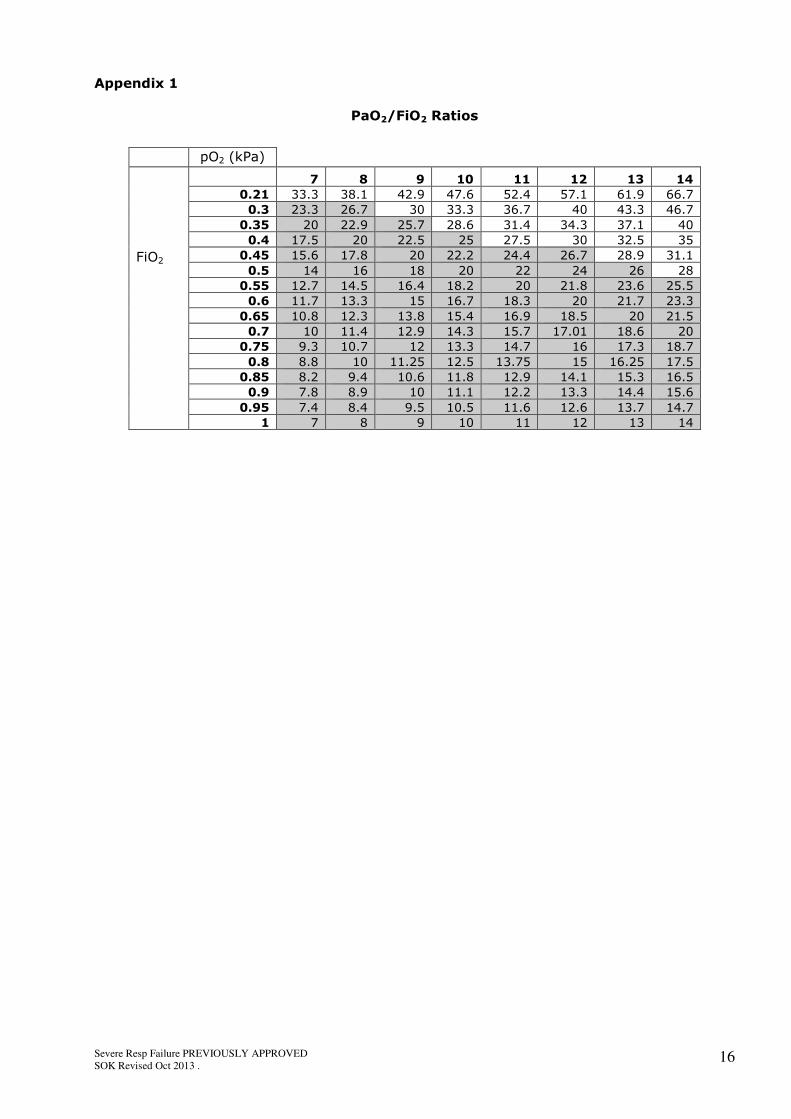
P/F ratio: PaO2 (in kPa) divided by FiO2 – Appendix 1
Mild Moderate Severe
Oxygenation 26.7<PaO2/FiO2 < 40 with 13.3<PaO2/FiO2 < 26.7 with PaO2/FiO2 <13.3 with PEEP or CPAP > 5cmsH2O PEEP > 5cmsH2O PEEP > 5cmsH2O
ACTION: Measure and record P/F Ratios on all ventilated patients. (NB this is also on the blood gas
print out).
ACTION: Assess for ARDS Criteria if P/F ratio (in kPa) <26.7 (ARDS) – Record in Patient’s Notes.
3. Investigations � Echocardiogram (ECHO)
Distinguishing pulmonary oedema due to ARDS from hydrostatic or cardiogenic pulmonary oedema
can be challenging in critically ill patients. Echocardiography is an easy non-invasive test to obtain
hemodynamic information.
ACTION: Consider ECHO to exclude cardiac failure or cardiogenic component
� Cardiac Output (C.O.) Monitoring
In ARDS cardiac output can be decreased due to several causes including septic shock, sepsis
related myocardial dysfunction, cardiac events or effect of medical treatments (high ventilation
pressures, PEEP, or inversed inspiratory:expiratory ratios). Thus monitoring of cardiac output and
filling pressures are important.
ACTION: Consider C.O. measurement to help in the diagnosis and C.O. monitoring during ARDS
management.
� Non-Bronchoscopic Lavage (NBL)
NBL is an invaluable diagnostic tool. It is a simple, safe and effective method of rapidly identifying
and evaluating ventilated patients with (potential) pneumonia3.
� Bronchoalveolar lavage (BAL)
BAL is one of the best tools to establish the diagnosis of bacterial ventilator-associated pneumonia4.
BAL is recommended in patients with ARDS due to suspected pneumonia and those without a
defined predisposing condition.
ACTION: Perform NBL on all ventilated admissions to critical care. Consider BAL at clinical
discretion.
Severe Resp Failure PREVIOUSLY APPROVED
SOK Revised Oct 2013 . 4
� Investigate the cause of ARDS
This may include surgical review, septic screen, abdominal or chest U/S or CT scan etc.
ACTION: Consider the common risk factors for ARDS
Risk factors2
Pneumonia
Non-pulmonary sepsis
Aspiration of gastric contents
Major trauma
Pulmonary contusion
Pancreatitis
Inhalation injury
Severe burns
Non-cardiogenic shock
Drug overdose
Multiple transfusions or transfusion-associated acute lung injury (TRALI)
Pulmonary vasculitis
Drowning

Severe Resp Failure PREVIOUSLY APPROVED
SOK Revised Oct 2013 . 5
GENERAL SUPPORTIVE MEASURES
Bundle Element 2: Provide general supportive measures
1. Ventilator Care Bundles and other Care Bundles
Care bundles are known to improve the outcomes of critical care patients5. Ventilator care bundles
are used on all ventilated patients in all three Critical Care units across BCUHB; these are frequently
audited with excellent compliance. Compliance in BCUHB is demonstrated by a reduction of VAP
(Ventilator Associated Pneumonia) in the last few years.
Full compliance with other care bundles is also of utmost importance.
ACTION: Comply with all bundles; ventilator, sepsis, nutrition, central line and others as
appropriate, for example tracheostomy.
2. Treat the Underlying Cause
If the suspected underlying cause of ARDS is infection, then the source should be identified and
treated. Early broad spectrum antibiotics should be started immediately with consideration given to
the requirement for antivirals.
ACTION: Commence early IV broad spectrum antibiotics; consider antivirals.
3. Nutrition Nutritional support is an essential component in critical care. Malnutrition has been associated with
poor outcomes among patients in intensive care units (ICUs), as indicated by increased morbidity,
mortality, and length of stay.
Unless contraindicated, nutrition should be commenced early, (within 24-48 hours after admission to
ICU), in all mechanically ventilated patients (medical, surgical, trauma) provided that the patients
are adequately resuscitated and haemodynamically stable.
ACTION: Commence early feeding: refer to and follow BCUHB’s Critical Care Nutrition Bundle/
guidance for all critical care patients:
http://www.wales.nhs.uk/sites3/Documents/753/Enteral%20Nutrition%20Bundle%20FINAL%20APP
ROVED.pdf
4. Fluid Management Patients with ARDS have noncardiogenic pulmonary oedema as a hallmark of their disease process.
Intravenous fluid management in these patients thus poses important challenges. On one hand,
intravenous fluids are critical to maintain appropriate intravascular volume to assure haemodynamic
stability and vital organ perfusion in patients with compromised gas exchange. On the other hand,
excessive fluid administration can worsen the lung oedema, further impairing gas exchange.
The National Institutes of Health (NIH) ARDS Network launched the FACTT study6 (Fluid and
Catheter Therapy Trial). The goals of the study were to assess the safety and efficacy of ‘fluid
conservative’ vs. ‘fluid liberal’ management strategies on lung function, non-pulmonary organ
function, as well as mortality and the need for mechanical ventilation.
The investigators concluded that "although the study did not detect a difference in mortality, the
conservative fluid strategy improved lung function and shortened the duration of mechanical
ventilation and intensive care stay, without increasing nonpulmonary organ failures. These results
support the use of a conservative fluid management strategy in ARDS patients." Referring to the
FACTT study Mackay and Al-Haddad7 recommend a conservative fluid regime as a low cost, low risk
intervention that could lead to improvement in clinically important outcomes.
ACTION: Aim for an even or a negative fluid balance after the initial fluid resuscitation. If patient
develops signs of acute renal failure consider early Renal Replacement Therapy.
Severe Resp Failure PREVIOUSLY APPROVED
SOK Revised Oct 2013 . 6
NON VENTILATORY MANAGEMENT
Bundle Element 3: Optimise non-ventilatory management
i. Sedation/paralysis
The use of paralytics is far less commonplace that it was a decade ago. A recent study8 however
noted that in patients with severe ARDS, early administration of a neuromuscular blocking agent
improved the adjusted 90-day survival and increased the time off the ventilator without increasing
muscle weakness.
Interestingly about 40% of these patients were concurrently receiving corticosteroids for septic
shock; potentially a high-risk group in terms of the risk for myopathy or muscle weakness following
a very acute illness. In the end, what the authors saw was that in the adjusted analysis for survival,
there was actually a survival advantage with the use of a neuromuscular blocker. Overall, the
mortality rate was about 41% in the patients randomised to placebo vs. about 32% or 31% in the
patients randomised to the active intervention. The absolute risk reduction was about 10%.
Questions have been raised however as to the potential mechanism for the benefit associated with a
muscle relaxant. An editorial9 noted that it could potentially be due to an anti-inflammatory effect of
the paralytic, or to a decrease in barotrauma.
In this one study giving a muscle relaxant over a two day period, whilst utilising ventilator
management strategies, dropped the adjusted 90 day mortality by almost a third (crude rate 31.6%
versus 40.7%, adjusted P=0.004).
ACTION: With adequate sedation consider the use of paralysis in the first 48 hours, whilst utilising
ventilator management strategies, in ARDS patients. Review daily thereafter. Refer to Sedation
Guidelines adults in Critical Care http://www.wales.nhs.uk/sites3/page.cfm?orgid=753&pid=63739
ii. Physiotherapy10-12
The main aim of physiotherapy is to optimise oxygenation and oxygen transport in respiratory
failure cases and assist in the treatment of any underlying issues such as atelectasis or secretions
retention/plugging.
Once ventilated, physiotherapy options with severe respiratory failure are:
- Positioning advice and assistance.
Based on auscultation, palpation and other objective findings e.g. CXR.
May be difficult depending on patient’s condition.
Main aim is to increase area of ventilation/perfusion to increase oxygenation.
- Airway clearance/secretion removal.
There are many methods of doing this; however they depend on the patient’s condition, oxygen
requirements and potential benefits weighed up against potential risks.
The main aim is to facilitate secretion transport and improve airway compliance, in a bid to
improve oxygenation.
Techniques include:
� Manual techniques, including shaking and vibrations of the chest wall.
� Manual hyperinflation.
� Suction, in combination with above points.
� Postural drainage.
ACTION: Provide physiotherapy, as tolerated, to enhance the removal of secretions and to improve
gas exchange.
- Rehabilitation
Consideration needs to be given to passive range of movement of limbs to prevent deconditioning
and other ICU/ventilator based neuromuscular complications.
Prolonged ventilation in critical care is associated with impaired health related quality of life up to
three years after discharge, even when patients are living independently at home13 and persistent
functional disability has been demonstrated over one year following discharge in ARDS patients14.

Severe Resp Failure PREVIOUSLY APPROVED
SOK Revised Oct 2013 . 7
ACTION: Develop a rehabilitation plan whilst in Critical Care and commence rehab as soon as is
practicable; follow BCUHB’s rehabilitation guidance.
iii. Prone Ventilation
A lung-protective strategy that has been successfully utilised to improve oxygenation in
mechanically ventilated patients with ARDS is prone positioning. Changing the patient position to
prone position can improve the distribution of perfusion to ventilated lung regions, decreasing
intrapulmonary shunt and improving oxygenation15. More recently it has been shown that early
application of prolonged prone positioning significantly decreased mortality16.
Once a diagnosis of ARDS is made the authors advocate early proning16; prone for 16 hours, stop
proning if there are complications, then turn patient supine for four hours (see strategy for prone
positioning next page).
ACTION: Use prone therapy for severe ARDS patients with no improvement in gas exchange; follow
‘guidelines for prone positioning – patient safety’ – Appendix 2 – [Adapted] Glenfield protocol.
iv. Prostacyclin
Prostacyclins cause pulmonary vasodilatation and are used to treat patients with primary pulmonary
hypertension. Nebulised prostacyclin (PGI2) has comparable effects in improving oxygenation,
pulmonary vasodilatation and shunt reduction when compared with inhaled NO17. Improved
oxygenation has been seen in a paediatric study18, but this has not yet been demonstrated in adult
patients with ARDS.
ACTION: Consider a trial of nebulised Prostacyclin for refractory hypoxia with increasing FiO2 and
PEEP requirements.
How to use: Nebulisation of Epoprostenol for Severe Acute Hypoxic Failure
� Reconstitute 500mcg of Epoprostenol into 50ml of glycine buffer diluent provided.
� Baseline commencing dose of 5ng/kg/minute [can go to 50ng/kg/min]
Dosage:
� Dose required [ng/kg/min] x Wt[kg] x 0.006ml/hour
� Attach to syringe driver. Starting rate = 2.5ml/hour (equiv to 5ng/kg/min for 70 kg patient)
� Syringe driver is attached to epidural catheter through sealed “bung” which is then inserted into
nebuliser chamber. The epidural catheter then sits in nebuliser chamber.
� This is then inserted into ventilator circuit and nebulisation is commenced from ventilator and run
constantly.
� Dosage can then be increased with assessment of clinical improvement.
v. Recruitment Manoeuvres
The aim of ‘Recruitment Manoeuvres’ is to promote reopening of collapsed alveoli by a sustained but
controlled rise in transalveolar pressure.
A review of ventilator strategies for severe hypoxic respiratory failure19, expressed concerns that
while improvements in oxygenation have been reported, no RCTs have shown a mortality benefit.
The authors reported that while some studies with a lung-protective strategy incorporated into the
recruitment study, reported a survival benefit others did not show any mortality benefit. This review
did not therefore recommend the routine use of recruitment manoeuvres. It also urges caution if
using them in patients who are haemodynamically unstable and or at risk of barotrauma. However,
it also notes that some patients with life-threatening refractory hypoxia may show dramatic
improvement in oxygenation with recruitment. If such an improvement does occur, the authors then
recommend the use of higher PEEP values to maintain recruitment.
Another review20 also expressed concerns; reduction in venous return and subsequent hypotension
was highlighted. The authors stated that “despite multiple clinical trials, there has not been a clear
signal that recruitment manoeuvres provide benefit”.
ACTION: Consider the use of recruitment manoeuvres where applicable and safe to do so.

Severe Resp Failure PREVIOUSLY APPROVED
SOK Revised Oct 2013 . 8
1. In a Pressure Control mode set a PEEP level of 25 – 30 cmH2O with an inspiratory pressure of
10 -15 cmH2O, to have a peak inspiratory pressure of ~ 45cmH2O.
2. This setting is then used for 2 minutes
3. Higher baseline PEEP should then be considered to ensure recruitment is maintained.
vi. Rotational therapy21-24
Immobility is deleterious therefore rotational therapy has some value for managing pulmonary
complications in ICU especially with those patients that do not tolerate manual turning or are too
obese to turn safely.
The effective degree of rotation appears to be 400 though according this can be poorly tolerated by
more awake patients as they feel they are falling out of bed. A secondary benefit of kinetic therapy
in patients who do not tolerate manual turning could also be skin integrity preservation as evidently
rotation to 400 relieves pressure off the sacrum.
ACTION: Consider the use rotational therapy for patients who do not tolerate manual turning or are
too obese to turn safely.
Suggested Strategy for Prone Positioning in Severe ARDS16
Prone
positioning
16 hours
Supine
4 hours
Check criteria for severe ARDS?
P/F<20 with FiO2 >0.6 and PEEP>5
No
- Stay supine.
- Return to prone session if
deterioration
Yes
- Start another proning session
- If rapid deterioration on returning to supine after
last prone session, increase duration of next prone
session
If deterioration in P/F or
saturation at any time while
supine:
Severe ARDS
P/F < 20 with FiO2 >0.6 and PEEP > 5
Stop proning if complications
Stop proning when:
- P/F >20 after 4 hours supine
- Complication during previous proning session causing it to be interrupted
- If P/F decreases by 20% at start of proning (compared with supine just before
proning) on 2 consecutive sessions.

Severe Resp Failure PREVIOUSLY APPROVED
SOK Revised Oct 2013 . 9
To see a video of Prone Positioning of Patients with the Acute Respiratory Distress Syndrome visit
http://www.nejm.org/doi/full/10.1056/NEJMoa1214103
Severe Resp Failure PREVIOUSLY APPROVED
SOK Revised Oct 2013 . 10
VENTILATORY MANAGEMENT
Bundle Element 4 Optimise ventilatory management
1. Lung protective ventilation; Tidal volumes and Plateau Pressure Lung-protective mechanical ventilation strategies are designed to prevent injury from overdistention
by using lower tidal volumes and lower inspiratory pressures.
The Cochrane Systematic Review25 ‘Lung protective ventilation strategy for the acute respiratory
distress syndrome’ reviewed six trials involving 1297 intubated patients in an ICU setting who were
randomised to receive either conventional mechanical ventilation or a “lung protective” ventilation
strategy. Lung protective ventilation was defined as providing a tidal volume of 7ml/kg or less with
plateau pressure of 30cmH2O or less. There was a significant all cause mortality benefit in favour of
lung protective ventilation at the end of the follow-up period for each trial.
A trial in patients with respiratory failure also demonstrated low Vt ventilation to be protective,
preventing lung injury and associated with a reduction in the release of inflammatory cytokines26.
This study was stopped early due to an increased incidence of lung injury in patients ventilated with
higher Vt.
Protective ventilation lung strategy:
i. Measure patients height and calculate Ideal Body Weight (IBW); if height cannot be obtained
use forearm (ulna) length27 - Appendix 3
ii. Utilise lung protective strategies – aim for: Pa02 7-9kPa or SpO2>88%
iii. Avoid over distention: Vt=6mls/kg based on IBW, Volume control is the suggested method of
ventilation16
iv. Limit plateau pressure <30cmsH20) - Appendix 3
v. FiO2 and PEEP titration
FiO2 0.3 0.4 0.5 0.6 0.7 0.8 0.9 1.0
PEEP 5 5-8 8-10 10 10-14 14 14-18 18-24
vi. Accept hypercapnia to achieve goals (1) and (2), providing pH is >7.15
vii. Mode of ventilation is less important than attending to goals 1 – 3
ACTION: Utilise lung protective strategies (ensuring Vt=6mls/kg/IBW).
2. High-Frequency Oscillatory Ventilation (HFOV) HFOV was previously thought to prevent secondary lung injury. Two recent multi-centre studies
OSCILLATE28 and OSCAR29, however did not demonstrate improved outcomes for patients with ARDS
receiving HFOV. In fact, in the Canadian (OSCILLATE) radomised controlled trial study it was found
that for adults with moderate to severe ARDS, early application of HFOV, as compared with a
ventilation strategy of low tidal volume and high positive end expiratory pressure, did not reduce,
and may increase, in-hospital mortality. These two studies do however reinforce the importance of
conventional lung protective ventilation in managing patients with ARDS.
ACTION: HFOV does not form part of this guideline.
2a. Extracorporeal CO2 removal device
ACTION: ECCO2R does not form part of this guideline.
3. Extracorporeal Membrane Oxygenation (ECMO) The CESAR trial30 evaluated the clinical and cost effectiveness of ECMO for adults with severe
respiratory failure. This multicentre trial randomised 180 adults to receive either conventional
ventilatory support or transfer to the specialist centre for consideration for ECMO. The primary
outcome measure was survival without severe disability. This was 16% higher for patients in the
ECMO referral group over conventional management, suggesting that one additional patient would
benefit for every 6 patients treated.

Severe Resp Failure PREVIOUSLY APPROVED
SOK Revised Oct 2013 . 11
A Lancet editorial31 highlighted that a major limitation of the CESAR study was the lack of
standardisation in conventional management techniques provided to patients in the control group,
due to a lack of agreement amongst participating hospitals on what constituted ‘optimal’ care.
However, it is considered that this study represents the most comprehensive randomised controlled
trial undertaken on adult respiratory ECMO and it also reported a survival benefit for patients
referred to an ECMO centre compared to those who received conventional management.
Irrespective of the underlying cause, all ECMO practitioners agree that its use is only valid in the
context of a disease process which is potentially recoverable and which is unresponsive to
conventional intensive care procedures. Experts who provide ECMO however emphasise the
importance of viewing respiratory ECMO as one of a range of interventions to provide respiratory
support for patients with potentially reversible conditions, when conventional ventilation has not
proved either possible or effective.
ECMO should be considered as a positive intervention at an appropriate time during the patient care
pathway, rather than as a ‘last resort’ intervention in an attempt to rescue a patient who is dying as
outcomes are likely to be poor. The optimal time for instituting ECMO treatment in an individual
patient is therefore unclear. Most specialists argue that early referral is preferred, to minimise lung
damage, and previous evidence suggested outcomes were poor in patients who had been ventilated
for more than a week. Some units now consider this too restrictive.
It should be noted that the CESAR trial was conducted prior to the H1N1 pandemic.
ACTION: Consider early discussions and referral of patients with refractory hypoxaemia for ECMO.

Severe Resp Failure PREVIOUSLY APPROVED
SOK Revised Oct 2013 . 12
Quick Guide for Severe Respiratory Bundle – For Adults in Critical Care
Bundle Element Aims Rationale Exclusion Compliance Audit Point
Element 1: Define, diagnose and investigate
Prompt diagnosis � Measure and record P/F Ratios on all patients –
Appendix 1 � Assess criteria for ARDS if P/F <26.7kPa:
o Acute onset from a recognised etiology
o Bilateral infiltrates on a CXR
o The absence of heart failure
� Record in Notes � Perform NBL on all ventilated admissions to critical
care. Consider BAL where indicated.
� Consider C.O. monitoring as indicated � Consider risk factors
Early diagnosis of ARDS is important and should be considered in all critically ill patients. Distinguishing ARDS from other causes of pulmonary oedema can be challenging; echocardiography can be used to exclude cardiac failure. BAL is recommended in patients with ARDS due to suspected pneumonia and those without a defined predisposing condition. In ARDS cardiac output can be decreased due to sepsis and ventilation pressures thus monitoring of cardiac output and filling pressures are important.
Pts with heart failure.
Terminal care. EOL pathway
Is there an attempt to obtain a prompt diagnosis?
Did all ventilated have P/F ratios recorded?
When P/F ratios <26.7 was criteria for ARDS assessed and documented?
Did pts have NBLs performed?
Element 2: Provide general supportive measures
Optimised delivery; general supportive measures: � Comply with all bundles;
o ventilator
o sepsis (inc antivirals)
o nutrition
o central line and
o others as appropriate, for example
tracheostomy � Aim for an even or a negative fluid balance after the
initial fluid resuscitation. If patient develops signs of acute renal failure consider early Renal Replacement Therapy.
If the suspected underlying cause of ARDS is infection, then the source should be identified and treated. Early broad spectrum antibiotics should be started immediately with consideration given to the requirement for antivirals. Malnutrition has been associated with poor outcomes among patients in ICUs, as indicated by increased morbidity, mortality, and length of stay. A conservative fluid strategy improves lung function and shortens the duration of mechanical ventilation and ICU stay, without increasing nonpulmonary organ failures in ARDS patients.
Terminal care. EOL pathway
Contraindications
with other Bundles
Are general supportive measures provided? For ARDS patients: Is there [all] bundle compliance.
Was there a plan for fluid management?
Element 3: Optimise non-ventilatory management
Optimised delivery - non-ventilatory measures: � Consider the use of paralysing agents in the first 48
hours, whilst utilising ventilator management strategies. Review daily thereafter.
� Consider prone therapy for ARDS patients with no improvement in gas exchange - Appendix 2
� Consider a trial of nebulised Prostacyclin for refractory hypoxia with increasing FiO2 and PEEP requirements – see pg 7 for ‘how to use’.
� Consider the use of recruitment manoeuvres where applicable and safe to do so see page 6 for ‘how to do’
� Consider the use rotational therapy for patients who do
In patients with severe ARDS, early administration of a neuromuscular blocking agent improved the adjusted 90-day survival and increased the time off the ventilator without increasing muscle weakness. Changing the patient position to prone position can improve the distribution of perfusion to ventilated lung regions, decreasing intrapulmonary shunt and improving oxygenation. Recruitment Manoeuvres may promote reopening of collapsed alveoli by a sustained but controlled rise in transalveolar pressure. Nebulised prostacyclin has comparable effects in improving oxygenation, pulmonary vasodilatation and shunt reduction when
Terminal care. EOL pathway
Contraindications
Is non-ventilatory management optimised? Was there a sedation / paralysis plan?
Was prone therapy considered? Were recruitment manoeuvres considered?

Severe Resp Failure PREVIOUSLY APPROVED
SOK Revised Oct 2013 . 13
not tolerate manual turning or are too obese to turn safely.
� Provide physiotherapy, as tolerated, to enhance the removal of secretions and to improve gas exchange.
� Develop a rehabilitation plan whilst in Critical Care and commence rehab as soon as is practicable; follow BCUHB’s rehab guidance.
compared with inhaled NO. to Prone Rx
Was rotational therapy considered?
Was nebulised prostacyclin considered?
Was there a rehab plan devised?
Element 4: Optimise ventilatory management
Optimised delivery - ventilatory measures: i. Calculate IBW - Appendix 3 ii. Utilise lung protective strategies – aim for: Pa02 7-
9kPa or SpO2>88% iii. Avoid alveolar overdistension – Vt=6mls/IBW, iv. Limit plateau pressure <30cmsH20); consider
VCV v. FiO2 and PEEP titration – see table page 10 vi. Accept hypercapnia to achieve goals (1) and (2),
providing pH is >7.15 vii. Mode of ventilation is less important than
attending to goals 1 – 3 Where there is a realistic chance of survival to a reasonable quality of life consider early discussions and referral of patients with severe hypoxaemia for ECMO.
Lung-protective strategies aim to prevent injury from overdistention by using lower tidal volumes and lower inspiratory pressures. There is a significant all cause mortality benefit in favour of lung protective ventilation. ECMO centres report a survival benefit for patients referred for ECMO compared to those who received conventional management.
Terminal care
Is ventilatory management optimised? Were lung protective strategies utilised? ���� IBW calculated? ���� Vt =6mls/IBW ���� Plateau pressure
<30cmsH2O ���� Appropriate level of
PEEP for FiO2? Was ECMO considered?

Severe Resp Failure PREVIOUSLY APPROVED
SOK Revised Oct 2013 . 14
References:
1. Management of Severe Refractory Hypoxic Respiratory Failure in Critical Care in the UK in
2010. Report from the UK Expert Group.
2. Ferguson ND, Fan E, Camporota L, et al.: The Berlin definition of ARDS: an expanded
rationale, justification and supplementary material. Intensive Care Medicine (2012);
38:1573-1582.
3. Arora SC, Mudaliar YM, Lee C, Mitchell D, Iredell J. & Lazarus R. Non-bronchoscopic
broncheoalveolar lavage in the microbiological diagnosis of pneumonia in mechanically
ventilated patients. Anaesthesia and Intensive Care (2002); 30(1):11-20.
4. Papazian L and Gainnier M. Indications of BAL, lung biopsy, or both in mechanically
ventilated patients with unexplained infiltrations. European Respiratory Journal (2003);
21(3):383-384.
5. Fulbrook P and Mooney S. Care Bundles in Critical Care: a practical approach to evidence
based practice. Nursing in Critical Care (2003) 8(6):249-55.
6. The National Heart, Lung, and Blood Institute ARDS Clinical Trials Network. Comparison of
two fluid-management strategies in acute lung injury. NEJM (2006); 354:2564-2575.
http://www.ardsnet.org/system/files/factt_crf_instructions_0_0.pdf
7. Mackay A. and Al-Haddad M. Acute lung injury and acute respiratory distress syndrome.
Continuing Education in Anaesthesia, Critical Care and Pain. (2009); 9(5): 152-156.
8. Papazian L, Forel JM, Gacouin A, et al.; ACURASYS Study Investigators. Neuromuscular
blockers in early acute respiratory distress syndrome. NEJM (2010); 363:1107-1116.
9. Slutsky AS. Neuromuscular blocking agents in ARDS. NEJM (2010); 363:1176-1180.
10. Pryor & Prasad 4th Edition (2008) Physiotherapy for Respiratory and Cardiac
Problems: Adult and Paediatrics. Churchill Livingstone.
11. Frownfelter & Dean 3rd Edition (1996) Principles and Practice of Cardiopulmonary
Physical Therapy. Mosby.
12. Stiller K. Physiotherapy in Intensive Care: Towards an evidence-based practice. Chest
(2000); 118:1801-1813.
13. Combes A, Costa MA, Trouillet JL, Baudot J, Mokhtari M, Gibert C, et al. Morbidity,
mortality, and quality-of-life outcomes of patients requiring >or=14 days of mechanical
ventilation. Critical Care Medicine. (2003); 31:1373–1381.
14. One-Year Outcomes in Survivors of the Acute Respiratory Distress Syndrome. Herridge MS,
Cheung AM, Tansey CM, Martyn AM, Diaz-Granados N, Al-Saidi F, et al. for the Canadian
Critical Care Trials Group NEJM. (2003); 348: 683-693.
15. Richter T, Bellani G, Scott Harris R, et al: Effect of prone position on regional shunt,
aeration, and perfusion in experimental acute lung injury. American Journal of
Respiratory Critical Care Medicine (2005); 172:480–487.
16. Guerin C, Reignier J, Richard JC, et al.: Prone positioning in severe acute respiratory
distress syndrome. NEJM (2013); 369:979-981.
17. Walmrath D, Schneider T, Schermuly R, et al. Direct comparison of inhaled nitric oxide and
aerosolized prostacyclin in acute respiratory distress syndrome. American Journal of
Respiratory Critical Care Medicine (1996); 153:991–6.
18. Dahlem P, van Aalderen WM, de Neef M, et al. Randomized controlled trial of aerosolized
prostacyclin therapy in children with acute lung injury. Critical Care Medicine (2004);
32:1055–60.
19. Esan A, Hess DR, Raoof S, George L, Sessler CN. Severe hypoxemic respiratory failure:
part 1--ventilatory strategies. Chest. (2010); 137(5):1203-16.
Severe Resp Failure PREVIOUSLY APPROVED
SOK Revised Oct 2013 . 15
20. Liu LL, J. Aldrich M, Shimabukuro DW, Sullivan KR, Taylor JM., Thornton KC, and Gropper
MA. Rescue Therapies for Acute Hypoxemic Respiratory Failure. Anaesthesia and
Analgesia (2010); 111(3): 693-702.
21. Goldhill DR, Imhoff M, McLean B, Waldmann C. Rotational Bed Therapy to Prevent and
Treat Respiratory Complications: A Review and Meta-Analysis. American Journal of
Critical Care. (2007); 16(1):50-61.
22. Pape HC, Remmers D, Weinberg A, Graf B, Reilmann H, Evans S, Regel G, Tscherne H. Is
early kinetic positioning beneficial for pulmonary function in multiple trauma patients?
Injury. (1998); 29(3):219-25.
23. Fleegler B, Grimes C, Anderson R, Butler M, MacFarlane GD. Continuous lateral rotation
therapy for acute hypoxemic respiratory failure: the effect of timing. Dimensions of
Critical Care Nursing. (2009); 28(6):283-7.
24. Rance M. Kinetic therapy positively influences oxygenation in patients with ALI/ARDS.
Nursing in Critical Care. (2005); 10(1):35-41.
25. Petrucci N, Iacovelli W. Lung protective ventilation strategy for the acute respiratory
distress syndrome. Cochrane Database Systematic Review. (2007); 18:(3).
26. Determann RM, Royakkers A, Wolthuis EK, et al. Ventilation with lower tidal volumes as
compared with conventional tidal volumes for patients without acute lung injury: a
preventive randomized controlled trial. Critical Care (2010); 14:R1.
27. Malnutrition Universal Screening Tool. BAPEN.org.uk Available from:
http://www.bapen.org.uk/pdfs/must/must_explan.pdf
28. Ferguson ND, Cook D, Gordon H, et al.: High-frequency oscillation in early respiratory
distress syndrome. NEJM (2013); 368:795-805
29. Young D, Lamb S, Phil D, et al.: High-frequency oscialltion for acute respiraoty disctress
syndrome. NEJM (2013); 368:806-813
30. Peek GJ, Mugford M, Tiruvoipati R, Wilson A, Allen E, Thalanany MM et al. Efficacy and
economic assessment of conventional ventilatory support versus extracorporeal membrane
oxygenation for severe adult respiratory failure (CESAR): a multicentre randomised
controlled trial. Lancet (2009); 374:1351-1363.
31. Zwischenberger JB, Lynch JE, ‘Will CESAR answer the adult ECMO debate?’. Lancet
(2009); 374: 1-2.
Severe Resp Failure PREVIOUSLY APPROVED
SOK Revised Oct 2013 . 16
Appendix 1
PaO2/FiO2 Ratios
pO2 (kPa)
FiO2
7 8 9 10 11 12 13 14
0.21 33.3 38.1 42.9 47.6 52.4 57.1 61.9 66.7
0.3 23.3 26.7 30 33.3 36.7 40 43.3 46.7
0.35 20 22.9 25.7 28.6 31.4 34.3 37.1 40
0.4 17.5 20 22.5 25 27.5 30 32.5 35
0.45 15.6 17.8 20 22.2 24.4 26.7 28.9 31.1
0.5 14 16 18 20 22 24 26 28
0.55 12.7 14.5 16.4 18.2 20 21.8 23.6 25.5
0.6 11.7 13.3 15 16.7 18.3 20 21.7 23.3
0.65 10.8 12.3 13.8 15.4 16.9 18.5 20 21.5
0.7 10 11.4 12.9 14.3 15.7 17.01 18.6 20
0.75 9.3 10.7 12 13.3 14.7 16 17.3 18.7
0.8 8.8 10 11.25 12.5 13.75 15 16.25 17.5
0.85 8.2 9.4 10.6 11.8 12.9 14.1 15.3 16.5
0.9 7.8 8.9 10 11.1 12.2 13.3 14.4 15.6
0.95 7.4 8.4 9.5 10.5 11.6 12.6 13.7 14.7
1 7 8 9 10 11 12 13 14

Severe Resp Failure PREVIOUSLY APPROVED
SOK Revised Oct 2013 . 17
Appendix 2 Guidelines for prone positioning – patient safety
1) Purpose a. These guidelines are to ensure the safety of the adult patient moved in to the prone
position and managed whilst in the prone.
2) Indications / Contraindications a. Contraindications
i. Raised intracranial pressure
ii. Known or suspected spinal injury
iii. Pathological abdominal distension
iv. Pregnancy 2nd , 3rd trimester
v. Recent abdominal surgery, with risk of wound dehiscence
vi. Within 24 of formation of tracheostomy ( risk of bleeding)
b. Indications
i. Refractory hypoxaemia
ii. No improvement in chest radiograph with evidence of collapse and infiltrates
iii. Following MDT discussion
3) Principles a. The safety of the patient during the procedure is paramount.
b. The patient should only be turned prone on consultant/MDT instructions.
c. All staff performing procedure should be aware of protocol.
d. The timing of the turn should coincide with maximum staff availability
e. Any potential contraindications should be identified and risk/benefit analysis carried
out
f. Minimum of 6 members of staff should be present, 1 at top and bottom, 2 either
side.
g. Assess the need for further staff on individual patient basis.
4) Procedure a. Pre- move
i. The procedure should be explained to the patient as far as understanding
allows.
ii. The turn should be explained to family etc
iii. A pre-move arterial blood gas should be obtained
iv. Disconnect any non-essential infusions
v. Ensure all intravenous lines, chest drainage tubes, urinary catheters etc are
of adequate length to lay from either the head or foot of the bed and are
secure
vi. Remove ECG electrodes from chest
vii. Remove all non essential monitoring
viii. Bed should be adjusted to suitable height.
ix. Assess load as with any moving and handling procedure.
x. Condition of patient's skin should be assessed and documented before turn.
Appropriate preventative dressings should be applied
xi. Adequate sedation should be achieved and paralysing agents administered if
appropriate
xii. ET and oral suction performed and ET tube or tracheostomy secured. The
position of the ET tube should be noted (length at lips)
xiii. An in line suction catheter should be in place
xiv. Eye and mouth care to be performed, and lacrilube instilled.
xv. NG feed should be stopped and NG tube aspirated.
xvi. Chest drains should be placed at bottom of bed with tubing between legs if
this is not possible then drains should be disconnected but not clamped
xvii. All lines should be placed between legs or underneath torso
xviii. Put bed on to max inflate mode and slide sheet should be placed under the
bottom sheet using a minimum of 3 people
xix. The patients left arm should be placed under his/her left hip and the head
positioned to face towards the ventilator, usually to the left.
xx. Pre move check list completed.( See Appendix 1 for pre move check list)
b. Move

Severe Resp Failure PREVIOUSLY APPROVED
SOK Revised Oct 2013 . 18
i. Two members of staff at either side of patient.
ii. 2 pillows should be placed on top of the patient one over the upper chest and
the other over the iliac crest.
iii. A sheet should be placed on top of the patient and rolled together at the
sides with the sheet under the patient (Cornish pasty style).
iv. The face should be left exposed, with the top of the sheet folded to ensure
it’s in the correct position following the turn
v. The 2 people at the same side of the bed as the ventilator will pull the rolled
patient towards them, holding on to rolled sheets whilst the 2 people on the
other side will push.
vi. Using the same manoeuvre the patient is then turned onto their left side.
vii. The patient is then placed on their front, the 2 people on the side of the
ventilator will be holding the side of the Cornish pasty under the patient and
will pull through whilst the other 2 will be holding the side of the Cornish
pasty on top of the patient and will pull over.
viii. During this time the person at the top will be guiding the head and ensuring
patency of the airway. (See Appendix 2 for diagram of move)
c. Post Move
i. Check airway remains maintained.
ii. Ensure all monitoring is replaced.
iii. Assess haemodynamic status of patient.
iv. Reconnect chest drains.
v. Recommence all infusions
vi. Ensure pillows in correct place to avert pressure away from scrotum and
breasts and there is an unrestricted abdomen to allow the passive movement
of the diaphragm and the downward displacement of abdominal contents.
vii. The shoulders should fall slightly forwards.
viii. A pillow may be place under the shins to give slight flexion at the knees and
ankles
ix. Ensure the bed is on an appropriate mode.
x. The patient’s arm should be placed in the crawl position (elbow flexed and
shoulder abducted) with the head facing the prominent arm.
xi. A small roll can be place under the hand to allow flexion of the fingers. The
other arm can also be placed in this position or allowed to rest at the
patients side
xii. Arterial blood gas should be performed 20 minutes following manoeuvre.
xiii. Complete post move check list. (See Appendix 3 for post move checklist)
d. Ongoing Management
i. NG feeding should be recommenced as per present protocol.
ii. Protection, in the form of an absorbent sheet , should be place beneath the
face and changed as soon as it becomes wet
iii. The patients head and arm position should be altered every 2-3 hours to
prevent pressure ulcers forming on the cheeks, ears and neck
iv. A modified rotation setting may be used, but no more than a 15 degree turn.
Alternatively a slight turn may be achieved by modifying pillow position.
v. The bed should be tilted into the reverse trendelenburg position (head tilted
slightly up) to reduce intraocular pressure and to reduce venous congestion
in the head
vi. Lacrilube should be applied to the eyes as prescribed 6 hourly to prevent
corneal drying and abrasions
vii. Closed circuit suction should be used.
viii. Length of time in prone position can vary from 6 hours to 18 hours therefore
this will be dependent on consultant/MDT’s instructions.
5) Returning Supine a. The procedure for turning prone should be reversed to turn supine.
b. Pre move steps should still be taken.

Severe Resp Failure PREVIOUSLY APPROVED
SOK Revised Oct 2013 . 19
Pre move checklist
Date: Sign Comments
Required personnel present
Medical staff
Family aware
Arterial blood gas performed
Adequate tubing length
Non essential infusions disconnected
All remaining infusions from head or foot of bed
Eye and mouth care performed
ET/ oral suction performed
In line suction in place
ET/ tracheostomy secured
Length at lips recorded:
Electrodes and non essential monitoring removed
Chest drains at bottom of bed or disconnected
NG feed off and tube aspirated
Assessment as per handling and moving
guidelines
Bed on max inflate
Pillows in place
Sedation assessed
Post move checklist
Date: Sign Comments
Monitoring recommenced
Haemodynamically stable
All infusions recommenced
Check position of pillows
Reverse trendelenburg bed position
Recommence feed
Perform arterial blood gas

Severe Resp Failure PREVIOUSLY APPROVED
SOK Revised Oct 2013 . 20
ET secure
Ng aspirated and spigotted
Monitoring removed
Drains to bottom of bed,
tubing alongside patient
Urinary catheter
out between
legs to foot of
bed
Pillows across chest and
pelvis, allowing
unrestricted abdominal
movement
Infusion lines leaving
from foot of bed (top
if a neck line in situ)

Severe Resp Failure PREVIOUSLY APPROVED
SOK Revised Oct 2013 . 21
Top and bottom sheet
rolled together tightly at
edges (cornish pasty)
Top edge of sheet Z folded
to allow access to ET, with
top edge remaining
accessible
Sliding the patient to the edge
of the bed, with the slide
sheet in place, towards the
ventilator
Dr/Snr Nurse supporting ET
Severe Resp Failure PREVIOUSLY APPROVED
SOK Revised Oct 2013 . 22
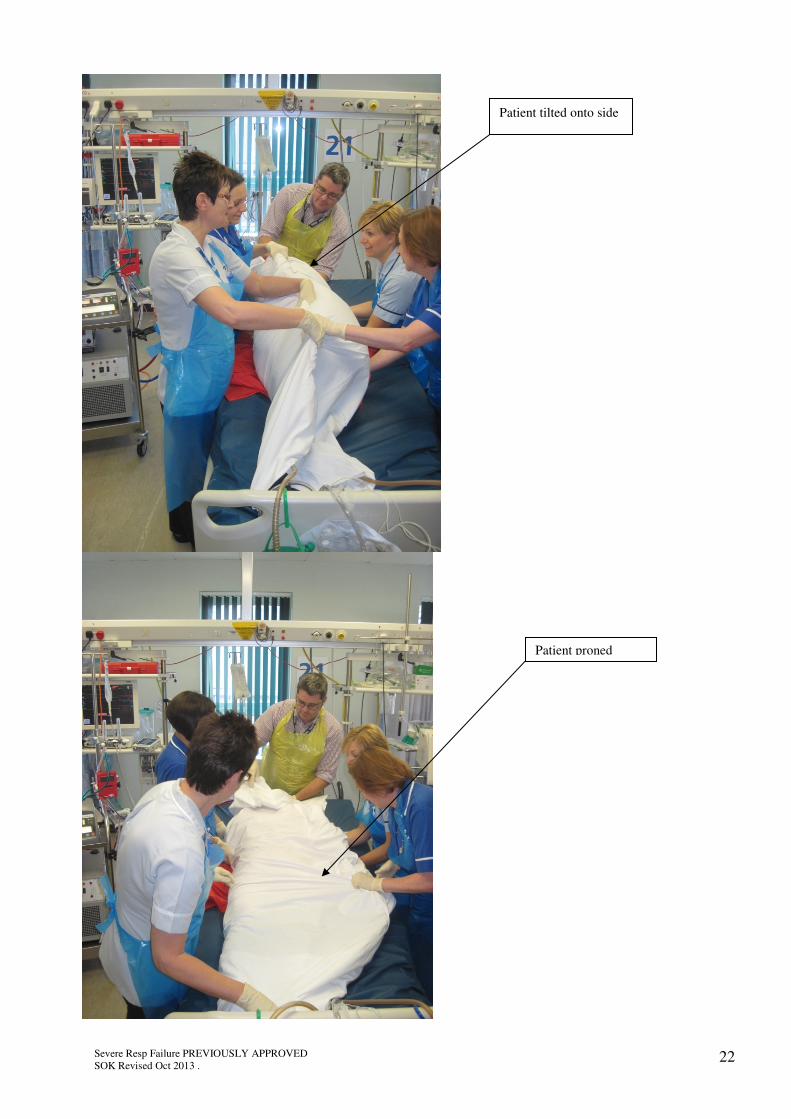
Patient tilted onto side
Patient proned

Severe Resp Failure PREVIOUSLY APPROVED
SOK Revised Oct 2013 . 23
Arm flexed
Pillows need to be adjusted to
allow unrestricted movement
of diaphragm
Circuit secured

Severe Resp Failure PREVIOUSLY APPROVED
SOK Revised Oct 2013 . 24
Appendix 3 Ideal Body Weight.
To define the ideal tidal volume the ideal body weight has to be determined. The calculation of
the ideal body weight of an adult is based on the body height. If height cannot be obtained use
the forearm (ulna) length.
Recommendation/adjustment for the tidal volume on the ventilator:
Height (cm) IBW (kg)
Women
‘Protective’*
VT = 6ml/kg IBW
IBW (kg) Men ‘Protective’*
VT = 6ml/kg IBW
152 46 276 50 300
155 48 288 52 312
157 50 300 55 330
160 52 312 57 342
162 55 330 59 354
165 57 342 62 372
167 59 354 64 384
170 62 372 66 396
172 64 384 68 408
175 66 396 71 426
177 69 414 73 438
180 71 426 75 450
182 73 438 78 468
185 75 450 80 480
187 78 468 82 492
IBW (kg) women = 45.5 + 0.91
(height [cm] - 152.4)
For example:
Height 167cms – 152.4 = 14.6
14.6 x 0.91 = 13.286
13.286 + 45.5 = 58.786
(Rounded) = 59kgs
IBW (kg) men = 50 + 0.91
(height [cm] – 152.4)
For example:
Height 177cms – 152.4 = 24.6
24.6 x 0.91 = 22.386
22.386 + 50 = 72.386 (Rounded) = 73kgs

Severe Resp Failure PREVIOUSLY APPROVED
SOK Revised Oct 2013 . 25
Stated values are rounded.
*Protective
VT = 6ml/kg IBW Recommendation ARDS.net Study, N. Engl J Med (2000) 342 (18)
Appendix 4: Check list
190 80 480 85 510
193 82 492 87 522
195 85 510 89 534
197 87 522 91 546
200 89 524 94 564
Severe Resp Failure PREVIOUSLY APPROVED
SOK Revised Oct 2013 . 26
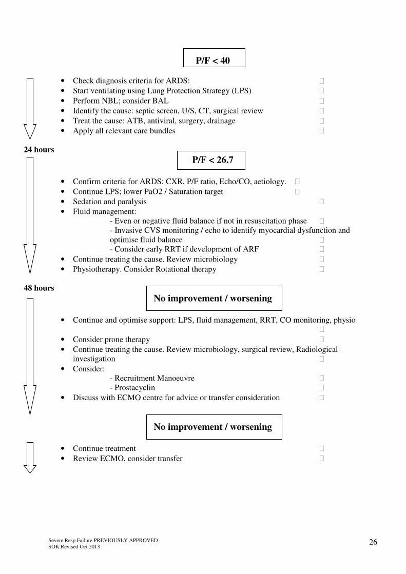
P/F < 40
• Check diagnosis criteria for ARDS:
• Start ventilating using Lung Protection Strategy (LPS)
• Perform NBL; consider BAL
• Identify the cause: septic screen, U/S, CT, surgical review
• Treat the cause: ATB, antiviral, surgery, drainage
• Apply all relevant care bundles
24 hours
P/F < 26.7
• Confirm criteria for ARDS: CXR, P/F ratio, Echo/CO, aetiology.
• Continue LPS; lower PaO2 / Saturation target
• Sedation and paralysis
• Fluid management:
- Even or negative fluid balance if not in resuscitation phase
- Invasive CVS monitoring / echo to identify myocardial dysfunction and
optimise fluid balance
- Consider early RRT if development of ARF
• Continue treating the cause. Review microbiology
• Physiotherapy. Consider Rotational therapy
48 hours No improvement / worsening
• Continue and optimise support: LPS, fluid management, RRT, CO monitoring, physio
• Consider prone therapy
• Continue treating the cause. Review microbiology, surgical review, Radiological
investigation
• Consider:
- Recruitment Manoeuvre
- Prostacyclin
• Discuss with ECMO centre for advice or transfer consideration
No improvement / worsening
• Continue treatment
• Review ECMO, consider transfer