Embed Size (px)
Citation preview
Hindawi Publishing CorporationInfectious Diseases in Obstetrics and GynecologyVolume 2006, Article ID 51931, Pages 1–4DOI 10.1155/IDOG/2006/51931
Obstetrics Case ReportSevere Life Threatening Maxillofacial Infection in PregnancyPresented as Ludwig’s Angina
Shelly Abramowicz,1 Jacques S. Abramowicz,2 and M. Franklin Dolwick1
1 Department of Oral and Maxillofacial Surgery, University of Florida School of Dentistry, Gainesville, FL 32610, USA2 Department of Obstetrics and Gynecology, Rush University, Chicago, IL 60612, USA
Received 5 January 2006; Revised 27 April 2006; Accepted 27 April 2006
Background. Ludwig’s angina is a rapidly spreading cellulitis that may produce upper airway obstruction often leading to death.There is very little published information regarding this condition in the pregnant patient. Case. A 24-year old black female wasadmitted at 26 weeks gestation with tooth pain, submandibular swelling, severe trismus, and dysphagea, consistent with Ludwig’sangina. Her treatment included emergent tracheostomy, incision and drainage of associated spaces, teeth extraction, and antibiotictherapy. Conclusions. During a life threatening infectious situation such as the one described, risks of maternal and fetal morbidityinclude both septicemia and asphyxia. Furthermore, the healthcare provider must consider the risks that the condition and thepossible treatments may cause the mother and her unborn child.
Copyright © 2006 Shelly Abramowicz et al. This is an open access article distributed under the Creative Commons AttributionLicense, which permits unrestricted use, distribution, and reproduction in any medium, provided the original work is properlycited.
INTRODUCTION
Ludwig’s angina, named after the German physician who de-scribed the condition for the first time in 1948, is a rapidlyspreading cellulitis that may produce upper airway obstruc-tion often leading to death. The most common cause of Lud-wig’s angina is an odontogenic infection, from one or moregrossly decayed, infected teeth, and is usually as a result of na-tive oral streptococci or a mixed aerobic-anaerobic oral flora[1]. Additional possible etiological factors include sialadeni-tis, compound mandibular fractures, or puncture wounds ofthe floor of the mouth [1]. While this is a life threateninginfection, an extensive literature search did not yield muchpublished information regarding this condition in the preg-nant patient.
CASE REPORT
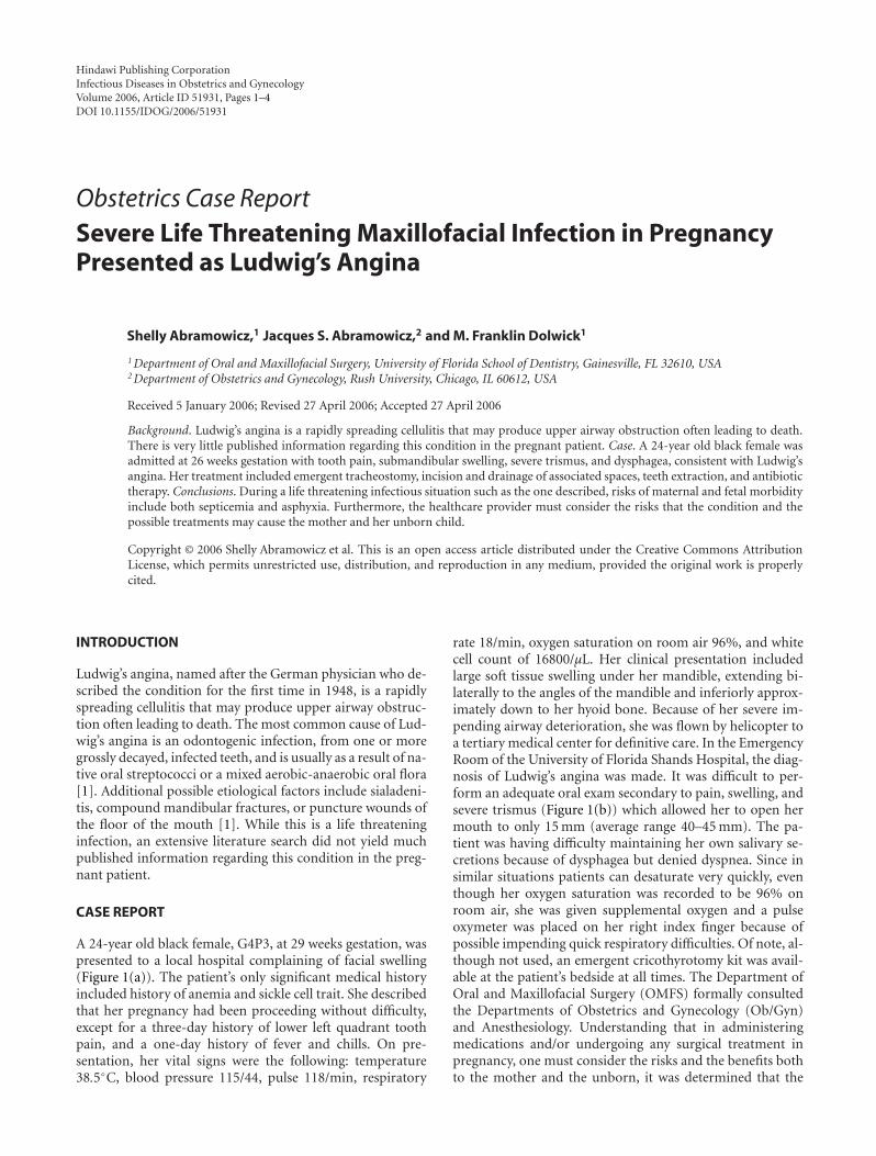
A 24-year old black female, G4P3, at 29 weeks gestation, waspresented to a local hospital complaining of facial swelling(Figure 1(a)). The patient’s only significant medical historyincluded history of anemia and sickle cell trait. She describedthat her pregnancy had been proceeding without difficulty,except for a three-day history of lower left quadrant toothpain, and a one-day history of fever and chills. On pre-sentation, her vital signs were the following: temperature38.5◦C, blood pressure 115/44, pulse 118/min, respiratory
rate 18/min, oxygen saturation on room air 96%, and whitecell count of 16800/µL. Her clinical presentation includedlarge soft tissue swelling under her mandible, extending bi-laterally to the angles of the mandible and inferiorly approx-imately down to her hyoid bone. Because of her severe im-pending airway deterioration, she was flown by helicopter toa tertiary medical center for definitive care. In the EmergencyRoom of the University of Florida Shands Hospital, the diag-nosis of Ludwig’s angina was made. It was difficult to per-form an adequate oral exam secondary to pain, swelling, andsevere trismus (Figure 1(b)) which allowed her to open hermouth to only 15 mm (average range 40–45 mm). The pa-tient was having difficulty maintaining her own salivary se-cretions because of dysphagea but denied dyspnea. Since insimilar situations patients can desaturate very quickly, eventhough her oxygen saturation was recorded to be 96% onroom air, she was given supplemental oxygen and a pulseoxymeter was placed on her right index finger because ofpossible impending quick respiratory difficulties. Of note, al-though not used, an emergent cricothyrotomy kit was avail-able at the patient’s bedside at all times. The Department ofOral and Maxillofacial Surgery (OMFS) formally consultedthe Departments of Obstetrics and Gynecology (Ob/Gyn)and Anesthesiology. Understanding that in administeringmedications and/or undergoing any surgical treatment inpregnancy, one must consider the risks and the benefits bothto the mother and the unborn, it was determined that the

2 Infectious Diseases in Obstetrics and Gynecology
(a) (b)
Figure 1: Patient at presentation. (a) General view of the face. Notesevere submental and upper neck swellings. (b) Trismus: this is themaximal mouth opening.
benefits of proceeding with emergent and immediate sur-gical intervention outweighed the risks. An emergent CT-scan showed fluid collections in the left lateral pharyngealspace (Figure 2) extending to the level of the valleculae. Se-curing an airway via an awake fiberoptic nasal intubationwas risky: a fiberoptic tube inserted into the pharynx mightpuncture an abscess and cause pus aspiration or swallowing.Therefore, an awake tracheostomy was performed using a 60-nonfenestrated Schiley endotracheal tube. At that time gen-eral anesthesia was administered and the patient was placedin slight left lateral decubitus position to decrease uterineaortocaval compression pressure. After usual sterile prepa-ration, lidocaine with epinephrine was infiltrated for localanesthesia. An incision was made at the submental and bilat-eral submandibular areas and blunt dissection to the lingualinferior border of the mandible was carried out. Through thesubmental incision, the blunt dissection continued throughthe mylohyoid muscle to the sublingual areas to access all ab-scesses. Tooth number 17 (lower left third molar) was thenextracted since it was believed that this grossly carious andpartially impacted tooth was the primary source for the in-fection. Upon removal, purulence was expressed through theextraction socket. Five additional grossly carious teeth werethen extracted. Intraoral blunt dissection to gain access intothe lateral pharyngeal space followed for drainage of anotherpus collection. Numerous Penrose drains and red rubbercatheter drains were left in place to maintain drainage of pusand facilitate daily irrigation (Figure 3). Upon admission tothe Surgical Intensive Care Unit, the Department of Ob/Gyncontinued following up on the patient by frequent monitor-ing of the fetal heart rate. The patient was extubated after 6days and remained in the hospital until discharge the follow-ing day. On discharge, the patient’s progress was monitoredby the Departments of OMFS and Ob/Gyn.
DISCUSSION
The unique anatomy of the floor of the mouth plays an im-portant role in the development and extension of intraoral
Figure 2: CT-scan demonstrating fluid collections in the left lateralpharyngeal space (arrow).
infections. The usual infectious course begins with a peri-apical dental abscess of the second or third mandibular mo-lar. The roots of these teeth extend inferior to the insertionof the mylohyoid muscle, so that if untreated, the infectionmay continue from primary spaces to penetrate the thin in-ner cortex of the mandible and will involve the posteriormargin of the mylohyoid muscle to the submandibular space[2]. At this time, the infection may develop and progress atsuch an alarming rate that special precautions regarding air-way maintenance must be taken. Because the mandible, hy-oid bone, and superficial layer of the deep cervical fascia limittissue expansion associated with the developing edema, thefloor of the mouth and the tongue base will become displacedsuperiorly and posteriorly, resulting in severe airway com-promise [2]. Further extension of the infection may spreadinto the mediastinum and the carotid sheath resulting in se-vere thoracal infection. Rupture of abscesses along the waymay cause aspiration of pus into the lungs and/or even peri-carditis. Untreated, the mortality is close to 100 percent, bothfrom the acute sepsis and from airway obstruction [1]. Thepatient with Ludwig’s angina will have severe and obviousextraoral swellings including bilateral submandibular, sub-mental, and sublingual spaces. Common presentation is ele-vation and displacement of the tongue, trismus, drooling ofsaliva, airway obstruction, dysphagea and/or dyspnea, and ahoarse (“hot potato”) voice. With extensive use of antibiotics,most facial infections improve before they have a chance toprogress to Ludwig’s angina. The mortality rate from Lud-wig’s angina, when recognized, has decreased from 50 to 5percent [1]. Therapy also includes early surgical removal ofthe source of infection (which is often grossly carious den-tition) via extraction, aggressive, and vigorous incision anddrainage procedures with appropriate placement of drains,along with intense and prolonged antibiotic therapy andmaintenance of a patient airway. While penicillin adminis-tered intravenously and in high doses is the empirical antibi-otic of choice, it is often recommended to use metronidazoleas well. For patients who have had repeated episodes of dentalinfections, clindamycin is often the antibiotic of choice [1].

Shelly Abramowicz et al 3
Figure 3: Patient postintervention. Note large number of drains.
Each year it is estimated that about 50,000 women un-dergo anesthesia and a surgical intervention at some timeduring gestation for indications unrelated to the pregnancy[3]. In such situations, when medical and surgical treat-ments for pregnant women are considered, both the physi-ologic changes of pregnancy and the perinatal effects of thetreatment must be considered [4]. Pregnancy is accompa-nied by many physiological changes which place the motherat a higher risk of infection or of doing worse once in-fected. First, the immune response is greatly diminished dur-ing pregnancy, thus resulting in potential faster progressionof an infection. In addition, there is decreased neutrophilchemotaxis, cell mediated immunity, and natural killer cellactivity [5, 6]. Moreover, approximately 50% of pregnantwomen complain of dyspnea by 19 weeks gestation [5] andthere is some depletion in the oxygen reserve of the gravidpatient. This results in lower oxygen reserve which could in-crease fetal hypoxia during periods of hypoventilation [4].From an oral perspective, as pregnancy associated hormonalchanges begin to affect a woman’s body, the gingival tissuesare affected as well. They become much more sensitive andthus susceptible to irritation from soft plaque. The plaqueaccumulates, becomes hard calculus deposits on the teeth,and harbors bacteria in large numbers resulting in a con-stant, low-grade intraoral infection. An exaggerated local in-flammatory response can then begin and may result in ery-thematous and edematous swelling of the gingiva betweenthe teeth, also known as pregnancy gingivitis. Approximately70% of pregnant women have this condition, even with rou-tine oral care [7]. This condition may be slightly painful andalso bleeds easily upon routine tooth brushing. Maternal in-fective processes sustained especially by gram negative anaer-obic bacteria, such as those leading to Ludwig’s angina, havebeen demonstrated to cause physiologic imbalance throughinflammatory cytokine production, sometimes resulting inpreterm labor, preterm premature rupture membranes, andlow birth weight [8, 9]. During pregnancy, women tend tomaintain frequent meals and snacks, which cause furtherplaque accumulation, as well as an increase in decay or rapidprogression of previously present decay. Because a preg-nant patient has increased demands on her organs, there is
increased potential for poor oxygenation. On the other hand,poor oxygenation is compromising to the fetus. An infectionin itself can at times infect the placenta, uterus, and possiblythe fetus, causing fetal septicemia. Treatments such as pro-longed intubation and certain intravenous medications canalso harm the fetus. During a life threatening infectious situ-ation such as the one described, the risk of maternal and fetalmorbidity may overshadow potential teratogenic side effects[10].
In order to prevent a similar life-threatening emergency,health care providers should not neglect even minimal com-plaints of dental pain. Often, if a problem is identified dur-ing the early stages of pregnancy, routine dental care can beplanned to control active disease or eliminate potential prob-lems that could increase in severity later in the pregnancy. Anappropriate time for dental care from a medical standpoint isthe second trimester and pregnant women usually experiencethe greatest sense of well being during that time [6]. Dentaltreatments including routine cleanings, fillings, crowns, ex-tractions, gum treatment, and continuation of orthodontictreatment can all be provided. Dental anesthetics such as li-docaine can penetrate the placenta but, in general, do notreach it because they are used locally and in small dosagesduring routine dental procedures [7]. Antibiotics that areacceptable include penicillin, amoxicillin, and clindamycin.Tetracycline should be avoided since it tends to cause per-manent discoloration of primary and temporary dentitionof the unborn child [6]. To decrease dental pain, narcoticsshould be avoided as well as over the counter medicationssuch as aspirin, ibuprofen, and related products because ofthe potential to affect bleeding. Morphine appears to be asafe analgesic when administered for short periods of time[7]. Ludwig’s angina is life threatening because of both sep-ticemia and asphyxia. Furthermore, in pregnancy, the risksthat both the condition and the possible therapies may causethe mother and her unborn child must be considered as wellas the possible consequences of the condition and therapiesto both.
REFERENCES
[1] Topazian RG, Goldberg MH, Hupp JR. Oral and MaxillofacialInfections. 4th ed. Philadelphia, Pa: W. B. Saunders; 2002.
[2] Marple BF. Ludwig angina: a review of current airway man-agement. Archives of Otolaryngology - Head and Neck Surgery.1999;125(5):596–600.
[3] Aroesty JH, Lanza JT, Lucente FE. Otolaryngology andpregnancy—difficult management decisions. Otolaryngology -Head and Neck Surgery. 1993;109(6):1061–1069.
[4] Barron WM. Medical evaluation of the pregnant patient re-quiring nonobstetric surgery. Clinics in Perinatology. 1985; 12(3):481–496.
[5] Silver RM, Peltier MR, Branch DW. The immunology of preg-nancy. In: Creasy RK, Resnik R, eds. Maternal-Fetal Medicine:Principles and Practice. Philadelphia, Pa: W. B. Saunders; 2004:89–109.
[6] Lawrenz DR, Whitley BD, Helfrick JF. Considerations in themanagement of maxillofacial infections in the pregnant pa-tient. Journal of Oral and Maxillofacial Surgery. 1996;54(4):474–485.

4 Infectious Diseases in Obstetrics and Gynecology
[7] Turner M, Aziz SR. Management of the pregnant oral andmaxillofacial surgery patient. Journal of Oral and MaxillofacialSurgery. 2002;60(12):1479–1488.
[8] Scannapieco FA, Bush RB, Paju S. Periodontal disease as a riskfactor for adverse pregnancy outcomes. A systematic review.Annals of Periodontology. 2003;8:70–78.
[9] Goepfert AR, Jeffcoat MK, Andrews WW, et al. Periodontaldisease and upper genital tract inflammation in early sponta-neous preterm birth. Obstetrics and Gynecology. 2004;104(4):777–783.
[10] Moore PA. Selecting drugs for the pregnant dental patient.Journal of the American Dental Association. 1998;129(9):1281–1286.

Submit your manuscripts athttp://www.hindawi.com
Stem CellsInternational
Hindawi Publishing Corporationhttp://www.hindawi.com Volume 2014
Hindawi Publishing Corporationhttp://www.hindawi.com Volume 2014
MEDIATORSINFLAMMATION
of
Hindawi Publishing Corporationhttp://www.hindawi.com Volume 2014
Behavioural Neurology
EndocrinologyInternational Journal of
Hindawi Publishing Corporationhttp://www.hindawi.com Volume 2014
Hindawi Publishing Corporationhttp://www.hindawi.com Volume 2014
Disease Markers
Hindawi Publishing Corporationhttp://www.hindawi.com Volume 2014
BioMed Research International
OncologyJournal of
Hindawi Publishing Corporationhttp://www.hindawi.com Volume 2014
Hindawi Publishing Corporationhttp://www.hindawi.com Volume 2014
Oxidative Medicine and Cellular Longevity
Hindawi Publishing Corporationhttp://www.hindawi.com Volume 2014
PPAR Research
The Scientific World JournalHindawi Publishing Corporation http://www.hindawi.com Volume 2014
Immunology ResearchHindawi Publishing Corporationhttp://www.hindawi.com Volume 2014
Journal of
ObesityJournal of
Hindawi Publishing Corporationhttp://www.hindawi.com Volume 2014
Hindawi Publishing Corporationhttp://www.hindawi.com Volume 2014
Computational and Mathematical Methods in Medicine
OphthalmologyJournal of
Hindawi Publishing Corporationhttp://www.hindawi.com Volume 2014
Diabetes ResearchJournal of
Hindawi Publishing Corporationhttp://www.hindawi.com Volume 2014
Hindawi Publishing Corporationhttp://www.hindawi.com Volume 2014
Research and TreatmentAIDS
Hindawi Publishing Corporationhttp://www.hindawi.com Volume 2014
Gastroenterology Research and Practice
Hindawi Publishing Corporationhttp://www.hindawi.com Volume 2014
Parkinson’s Disease
Evidence-Based Complementary and Alternative Medicine
Volume 2014Hindawi Publishing Corporationhttp://www.hindawi.com