Embed Size (px)
Citation preview

Bioorganic & Medicinal Chemistry 17 (2009) 2902–2912
Contents lists available at ScienceDirect
Bioorganic & Medicinal Chemistry
journal homepage: www.elsevier .com/locate /bmc
Sesquiterpene-like inhibitors of a 9-cis-epoxycarotenoid dioxygenaseregulating abscisic acid biosynthesis in higher plants
Jason Boyd, Yuanzhu Gai, Ken M. Nelson, Erica Lukiwski, James Talbot �, Mary K. Loewen,Stacey Owen, L. Irina Zaharia, Adrian J. Cutler, Suzanne R. Abrams *,�, Michele C. Loewen �
Plant Biotechnology Institute, National Research Council of Canada, 110 Gymnasium Place, Saskatoon, SK, Canada S7N 0W9
a r t i c l e i n f o a b s t r a c t
Article history:Received 21 December 2008Revised 30 January 2009Accepted 31 January 2009Available online 8 February 2009
Keywords:Abscisic acid biosynthesisABA biosynthesis inhibitor9-cis-Epoxycarotenoid dioxygenaseCarotenoid metabolismXanthoxin analogABA analog
0968-0896/$ - see front matter Crown Copyright � 2doi:10.1016/j.bmc.2009.01.076
* Corresponding author. Tel.: +1 306 975 5569; faxE-mail address: [email protected] (S.R. A
� Current address: Department of Biochemistry, UnWiggins Road, Saskatoon, SK, Canada S7N 0W5.
� These authors contributed equally to this work.
Abscisic acid (ABA) is a carotenoid-derived plant hormone known to regulate critical functions in growth,development and responses to environmental stress. The key enzyme which carries out the first commit-ted step in ABA biosynthesis is the carotenoid cleavage 9-cis-epoxycarotenoid dioxygenase (NCED). Wehave developed a series of sulfur and nitrogen-containing compounds as potential ABA biosynthesisinhibitors of the NCED, based on modification of the sesquiterpenoid segment of the 9-cis-xanthophyllsubstrates and product. In in vitro assays, three sesquiterpene-like carotenoid cleavage dioxygenase(SLCCD) inhibitor compounds 13, 17 and 18 were found to act as inhibitors of Arabidopsis thaliana NCED3 (AtNCED3) with Ki
0s of 93, 57 and 87 lM, respectively. Computational docking to a model of AtNCED3supports a mechanism of inhibition through coordination of the heteroatom with the non-heme iron inthe enzyme active site. In pilot studies, pretreatment of osmotically stressed Arabidopsis plants with com-pound 13 resulted lower levels of ABA and catabolite accumulation compared to levels in mannitol-stressed plant controls. This same inhibitor moderated known ABA-induced gene regulation effectsand was only weakly active in inhibition of seed germination. Interestingly, all three inhibitors led tomoderation of the stress-induced transcription of AtNCED3 itself, which could further contribute to low-ering ABA biosynthesis in planta. Overall, these sesquiterpenoid-like inhibitors present new tools for con-trolling and investigating ABA biosynthesis and regulation.
Crown Copyright � 2009 Published by Elsevier Ltd. All rights reserved.
1. Introduction
Abscisic acid (1, ABA) is a plant hormone involved in the regu-lation of important developmental functions including seed matu-ration, desiccation tolerance and dormancy, as well as adaptationto environmental stress through stomatal closure and modificationof gene expression.1–3 The biosynthesis of ABA 1 begins with iso-pentenyl diphosphate which enters the mevalonic acid-indepen-dent 2-C-methyl-D-erythritol-4-phosphate pathway producingplastidic isoprenoids, including carotenoids.4 Enzymatic cleavageof C40-carotenoid cis-xanthophylls (neoxanthin 2 and violaxanthin3) at the 110–120 double bond by a 9-cis-epoxycarotenoid dioxy-genase (NCED) produces C15 (xanthoxin 4) and C25 metabolitesand represents the first committed step in ABA biosynthesis(Fig. 1). Xanthoxin 4 is subsequently converted by an alcohol dehy-drogenase (ABA2) into abscisyl aldehyde 5, which is oxidized toABA 1 by an abscisic aldehyde oxidase (AAO3).3 The catabolism
009 Published by Elsevier Ltd. All r
: +1 306 975 4839.brams).iversity of Saskatchewan, 107
of ABA occurs principally through oxidation of one of the methylgroups of the ring (80-carbon atom, using convention for ABA num-bering) mediated by members of a class of P450 monooxygenaseenzymes, CYP 707A.5 The catabolite phaseic acid (6, PA) which oc-curs as the result of reversible cyclization of 80-hydroxyABA, is re-duced by an unknown reductase to afford dihydrophaseic acid (7,DPA). ABA can also be metabolized to the glucose conjugate 8.3
First identified in maize (VP14), NCEDs have also been found ina variety of other species including Arabidopsis thaliana (AtNCED3),bean (PvNCED1), tomato (LeNCED1), avocado (PaNCED1 andPaNCED3) and cowpea (VuNCED1).6–11 AtNCED3 is a member ofthe carotenoid cleavage enzyme family of A. thaliana, which con-sists of nine enzymes.12 In general, the family is characterized bya plastid-targeting transit peptide, an amphipathic a-helix domainand a catalytic domain which contains four conserved histidineresidues responsible for non-heme iron coordination. AtNCED3 isfound in both the stroma and bound to the thylakoid membrane,accounts for NCED activity in roots, contributes to NCED activityin developing seeds and is the major stress-induced NCED in leavesof A. thaliana.12 Recently, immunohistochemical analysis revealedthat the AtNCED3 protein is detected exclusively in the vascularparenchyma cells of water-stressed plants.13
ights reserved.
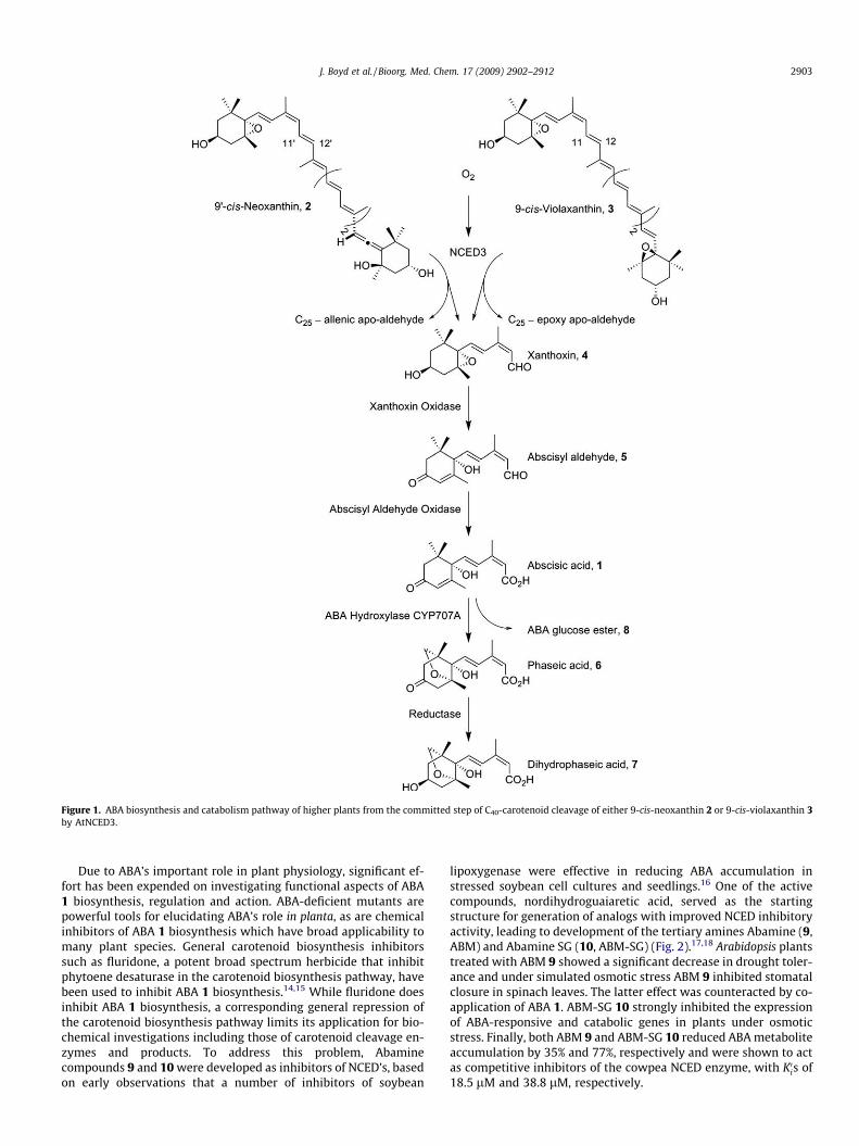
Figure 1. ABA biosynthesis and catabolism pathway of higher plants from the committed step of C40-carotenoid cleavage of either 9-cis-neoxanthin 2 or 9-cis-violaxanthin 3by AtNCED3.
J. Boyd et al. / Bioorg. Med. Chem. 17 (2009) 2902–2912 2903
Due to ABA’s important role in plant physiology, significant ef-fort has been expended on investigating functional aspects of ABA1 biosynthesis, regulation and action. ABA-deficient mutants arepowerful tools for elucidating ABA’s role in planta, as are chemicalinhibitors of ABA 1 biosynthesis which have broad applicability tomany plant species. General carotenoid biosynthesis inhibitorssuch as fluridone, a potent broad spectrum herbicide that inhibitphytoene desaturase in the carotenoid biosynthesis pathway, havebeen used to inhibit ABA 1 biosynthesis.14,15 While fluridone doesinhibit ABA 1 biosynthesis, a corresponding general repression ofthe carotenoid biosynthesis pathway limits its application for bio-chemical investigations including those of carotenoid cleavage en-zymes and products. To address this problem, Abaminecompounds 9 and 10 were developed as inhibitors of NCED’s, basedon early observations that a number of inhibitors of soybean
lipoxygenase were effective in reducing ABA accumulation instressed soybean cell cultures and seedlings.16 One of the activecompounds, nordihydroguaiaretic acid, served as the startingstructure for generation of analogs with improved NCED inhibitoryactivity, leading to development of the tertiary amines Abamine (9,ABM) and Abamine SG (10, ABM-SG) (Fig. 2).17,18 Arabidopsis plantstreated with ABM 9 showed a significant decrease in drought toler-ance and under simulated osmotic stress ABM 9 inhibited stomatalclosure in spinach leaves. The latter effect was counteracted by co-application of ABA 1. ABM-SG 10 strongly inhibited the expressionof ABA-responsive and catabolic genes in plants under osmoticstress. Finally, both ABM 9 and ABM-SG 10 reduced ABA metaboliteaccumulation by 35% and 77%, respectively and were shown to actas competitive inhibitors of the cowpea NCED enzyme, with K0is of18.5 lM and 38.8 lM, respectively.

Figure 2. Structures of AtNCED3 SLCCD inhibitors.
2904 J. Boyd et al. / Bioorg. Med. Chem. 17 (2009) 2902–2912
In this report we describe the design, synthesis and character-ization of a novel set of sesquiterpene-like carotenoid cleavagedioxygenase (SLCCD) inhibitors 11–18. The current compoundswere designed starting with the sesquiterpenoid subunit of thesubstrate and product of the NCED enzyme. Of eight initial inhibi-tors designed and tested (Fig. 2), three were found to inhibit re-combinant AtNCED3 activity strongly. These have been fullycharacterized in vitro, with kinetic inhibition constants comparingfavorably to those of the ABM-type compounds. Computationaldocking of the inhibitors correlated with these findings and sup-ported the proposed functional mechanism. In preliminaryin vitro analyses, one inhibitor in particular, SLCCD inhibitor com-pound 13 was found to moderate ABA-responsive genes and ABAmetabolism. Interestingly, all three inhibitors reduced expressionof AtNCED3, presenting a second mechanism for inhibition ofABA 1 biosynthesis by the molecules.
2. Results
2.1. Design and synthesis of the SLCCD inhibitors
The present compounds were designed to incorporate the 9-cisdouble bond geometry of the substrates and product of AtNCED3as well as a heteroatom at carbon 12 (carotenoid numbering) ofthe inhibitor molecules. All of the SLCCD inhibitors 11–18 weresynthesized from 4-oxoisophorone 19 (Fig. 3). Bakers0 yeast reduc-tion of 19 afforded (�)-(R)-2,2,6-trimethylcyclohexa-1,4-dione19
which was converted into chiral nonracemic allylic alcohols 20,21, 23 and 24.20 Racemic allylic alcohol 22 was prepared in a sim-ilar manner, except that reduction of 19 was accomplished usingzinc in acetic acid.21 The terminal allylic alcohols were then con-verted to the corresponding ethyl sulfides by reaction with ethyldisulfide in the presence of tributylphosphine.22 Inhibitor 16 wasobtained by reacting 2-thiopheneacetyl chloride and allylic alcohol22 (protected as the neopentylglycol ketal). The xanthoxin-likeallylic alcohol 22 was prepared through a Sonogashira couplingbetween the terminal acetylene in 2123 and (Z)-3-iodobut-2-en-1-ol. Alcohol 22 was then converted to the phenyl sulfide 13 with54% yield. The nitrogen-containing inhibitor 18 was synthesized
by oxidation of allylic alcohol 22 with MnO2, followed by imineformation using phenyl amine and then reduction to the amine.
2.2. In vitro assays and kinetic analyses
Recombinant AtNCED3 including a C-terminally located gluta-thione-S transferase fusion tag was expressed in Escherichia coliand purified by affinity chromatography. In vitro assays demon-strated the functionality of the recombinant purified enzyme prod-uct. Sample HPLC profiles (Supplementary data Fig. 1) showcleavage of the 90-cis-neoxanthin 2 substrate (tR 14.2 min. withthree maxima at 415, 438 and 467 nm) producing the expectedC25-allenic apo-aldehyde cleavage product (tR 11.6 min. with amaxima of 423 nm). Further kinetic analysis fitted by non-linearregression analysis defined a Km of 24 lM (Fig. 4). This value corre-lates well with the Km
0s of 27 lM and 49.0 lM determined previ-ously for VP14 and VuNCED1.18,24
Using this recombinant enzyme and assay system, the eight po-tential inhibitor compounds were tested for their relative ability toinhibit AtNCED3 activity at 1 mM concentration (Fig. 5). Com-pounds 12, 17 and 18 completely inhibited AtNCED3 activity at1 mM, while 13 inhibited AtNCED3 activity by 75%. Compound12 is one of the stereoisomers of racemic 13. The latter being easierto synthesize (and thus of higher potential practical application), itwas decided to move forward with compounds 13, 17 and 18 fordetailed in vitro and in vitro testing. Dixon plots indicated thatcompounds 13, 17 and 18 competitively inhibit recombinant AtN-CED3 with Ki
0s comparable or better than those observed for ABMand ABM-SG (Table 1 and Supplementary data Fig. 2).
2.3. Homology modeling and SLCCD inhibitor docking
Recently a crystal structure was determined for Synechocystisapocarotenoid-15,150-oxygenase (ACO), a fungal homologue of theNCEDs.25 AtNCED3 shares 25% identity and 45% similarity withACO at the amino acid level. Homology modeling using the Swiss-Model servers generated a hypothetical protein structure of AtN-CED3 which maintained the octahedral coordination of the fouractive site histidines at 2.14, 2.05, 2.16 and 2.31 Å from the iron atomfor H164, H211, H276 and H450, respectively.26 Structural differ-

Figure 3. Synthesis of AtNCED3 SLCCD inhibitors. (a) See Ref. 20; (b) n-Bu3P, (C2H5S)2; (c) TBAF, THF; (d) see Ref. 22; (e) (Z)-3-iodobut-2-en-1-ol, (Ph3P)4Pd, CuI, (i-Pr)2NH; (f)n-Bu3P, (C6H5S)2; (g) MnO2; (h) C6H5NH2, D; (i) NaBH4.
Figure 4. Kinetic analysis of recombinant purified AtNCED3 activity. Michaelis–Menton plot for cleavage of 9-cis-neoxanthin 2 by recombinant AtNCED3 indicatinga Km of 24 lM.
Figure 5. Relative inhibition of recombinant AtNCED3 activity by various SLCCDcompounds at 1 mM concentration.
J. Boyd et al. / Bioorg. Med. Chem. 17 (2009) 2902–2912 2905
ences between the AtNCED3 model and ACO were limited to smallsurface exposed loops related to a few minor alignment gaps. As con-trols to test the AtNCED3 model, 90-cis-neoxanthin 2, the substrate ofACO (all-trans-(3R)-hydroxy-80-apo-b-carotenol (3-ON)), and
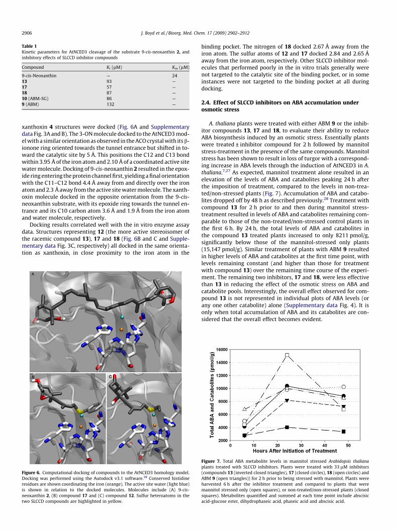
Table 1Kinetic parameters for AtNCED3 cleavage of the substrate 9-cis-neoxanthin 2, andinhibitory effects of SLCCD inhibitor compounds
Compound Ki (lM) Km (lM)
9-cis-Neoxanthin — 2413 93 —17 57 —18 87 —10 (ABM-SG) 86 —9 (ABM) 132 —
2906 J. Boyd et al. / Bioorg. Med. Chem. 17 (2009) 2902–2912
xanthoxin 4 structures were docked (Fig. 6A and Supplementarydata Fig. 3A and B). The 3-ON molecule docked to the AtNCED3 mod-el with a similar orientation as observed in the ACO crystal with its b-ionone ring oriented towards the tunnel entrance but shifted in to-ward the catalytic site by 5 Å. This positions the C12 and C13 bondwithin 3.95 Å of the iron atom and 2.10 Å of a coordinated active sitewater molecule. Docking of 9-cis-neoxanthin 2 resulted in the epox-ide ring entering the protein channel first, yielding a final orientationwith the C11–C12 bond 4.4 Å away from and directly over the ironatom and 2.3 Å away from the active site water molecule. The xanth-oxin molecule docked in the opposite orientation from the 9-cis-neoxanthin substrate, with its epoxide ring towards the tunnel en-trance and its C10 carbon atom 3.6 Å and 1.9 Å from the iron atomand water molecule, respectively.
Docking results correlated well with the in vitro enzyme assaydata. Structures representing 12 (the more active stereoisomer ofthe racemic compound 13), 17 and 18 (Fig. 6B and C and Supple-mentary data Fig. 3C, respectively) all docked in the same orienta-tion as xanthoxin, in close proximity to the iron atom in the
Figure 6. Computational docking of compounds to the AtNCED3 homology model.Docking was performed using the Autodock v3.1 software.38 Conserved histidineresidues are shown coordinating the iron (orange). The active site water (light blue)is shown in relation to the docked molecules. Molecules include (A) 9-cis-neoxanthin 2, (B) compound 17 and (C) compound 12. Sulfur heteroatoms in thetwo SLCCD compounds are highlighted in yellow.
binding pocket. The nitrogen of 18 docked 2.67 Å away from theiron atom. The sulfur atoms of 12 and 17 docked 2.84 and 2.65 Åaway from the iron atom, respectively. Other SLCCD inhibitor mol-ecules that performed poorly in the in vitro trials generally werenot targeted to the catalytic site of the binding pocket, or in someinstances were not targeted to the binding pocket at all duringdocking.
2.4. Effect of SLCCD inhibitors on ABA accumulation underosmotic stress
A. thaliana plants were treated with either ABM 9 or the inhib-itor compounds 13, 17 and 18, to evaluate their ability to reduceABA biosynthesis induced by an osmotic stress. Essentially plantswere treated ± inhibitor compound for 2 h followed by mannitolstress-treatment in the presence of the same compounds. Mannitolstress has been shown to result in loss of turgor with a correspond-ing increase in ABA levels through the induction of AtNCED3 in A.thaliana.7,27 As expected, mannitol treatment alone resulted in anelevation of the levels of ABA and catabolites peaking 24 h afterthe imposition of treatment, compared to the levels in non-trea-ted/non-stressed plants (Fig. 7). Accumulation of ABA and catabo-lites dropped off by 48 h as described previously.28 Treatment withcompound 13 for 2 h prior to and then during mannitol stress-treatment resulted in levels of ABA and catabolites remaining com-parable to those of the non-treated/non-stressed control plants inthe first 6 h. By 24 h, the total levels of ABA and catabolites inthe compound 13 treated plants increased to only 8211 pmol/g,significantly below those of the mannitol-stressed only plants(15,147 pmol/g). Similar treatment of plants with ABM 9 resultedin higher levels of ABA and catabolites at the first time point, withlevels remaining constant (and higher than those for treatmentwith compound 13) over the remaining time course of the experi-ment. The remaining two inhibitors, 17 and 18, were less effectivethan 13 in reducing the effect of the osmotic stress on ABA andcatabolite pools. Interestingly, the overall effect observed for com-pound 13 is not represented in individual plots of ABA levels (orany one other catabolite) alone (Supplementary data Fig. 4). It isonly when total accumulation of ABA and its catabolites are con-sidered that the overall effect becomes evident.
Figure 7. Total ABA metabolite levels in mannitol stressed Arabidopsis thalianaplants treated with SLCCD inhibitors. Plants were treated with 33 lM inhibitors(compounds 13 (inverted closed triangles), 17 (closed circles), 18 (open circles) andABM 9 (open triangles)) for 2 h prior to being stressed with mannitol. Plants wereharvested 6 h after the inhibitor treatment and compared to plants that weremannitol stressed only (open squares), or non-treated/non-stressed plants (closedsquares). Metabolites quantified and summed at each time point include abscisicacid-glucose ester, dihydrophaseic acid, phaseic acid and abscisic acid.

J. Boyd et al. / Bioorg. Med. Chem. 17 (2009) 2902–2912 2907
2.5. Effect of SLCCD inhibitors on A. thaliana seed germination
Seed germination assays were performed for compounds 13, 17and 18 to assess the ABA-like character of the inhibitors.29 Inhibi-tors had relatively little effect on seed germination at low concen-tration compared to non-inhibitor treated and ABA treated controls(Fig. 8). At increasing concentrations (0.33 lM) the inhibitors didlead to reductions of seed germination by approximately 15%, com-pared to 47% for the (+)-ABA 1. Both compounds 13 and 17 reducedseed germination by 26% at 1 lM while 18 showed a more pro-nounced effect with a 50% reduction compared to 61% for (+)-ABA 1. At the highest concentration tested, compound 13 still onlyhad a modest impact on seed germination at 38% reduction, whilecompounds 17 and 18 showed 51% and 71% reductions, respec-tively, compared to 96% for (+)-ABA 1.
2.6. Effect of SLCCD inhibitors on target gene transcript levelsunder osmotic stress
In light of the observed effectiveness of compound 13 in moder-ating ABA and catabolite levels in vitro and its limited effect onseed germination, it was targeted for further evaluation. Specifi-cally, quantitative reverse-transcription PCR was used to assessinhibitor induced changes in gene transcript levels in mannitolstressed plants. The gene targets chosen for this purpose were AtN-CED3, the ABA and drought inducible Rd29B and the ABA (induc-ible) catabolic genes CYP707A1 and CYP707A3. Transcript levelswere normalized against UBQ10 mRNA levels.5,30,31 Mannitol treat-ment led to the induction of expression of all four target geneswithin 4 h of the stress-treatment (Fig. 9). Subsequently the man-nitol-induced gene transcription levels decrease back to non-trea-ted/non-stressed levels by 24 h post-treatment and remained lowthrough 48 h (Supplementary data Figs. 5 and 6). In general, pre-treatment with compound 13 at both 10 and 33 lM concentrationsprior to mannitol-stress led to reductions in the accumulation ofmRNA transcript levels at 6 h post-compound treatment forRd29B, CYP707A1 and CYP707A3 compared to the mannitol-stressed control (Fig. 9A). The inhibition of mannitol-inducedRd29b transcription by compound 13 (about 90%) is especiallystriking and is consistent with the mannitol effect on Rd29b beingprimarily mediated by ABA. This result indicates the potential ofthis inhibitor for dissecting the role of ABA in physiological anddevelopmental processes. As observed in mannitol stressed only
Figure 8. Germination of Arabidopsis thaliana plants in the presence of each ofSLCCD inhibitor compounds 18 (inverted closed triangles), 13 (closed circles), 17(open circles), (+)-ABA 1 (open triangles), and germination of plants on mediawithout added compounds (closed square).
Figure 9. Target gene transcript levels in mannitol stressed Arabidopsis thalianaplants treated with SLCCD inhibitors. (A) Effect of compound 13. Plants were treatedwith 10 or 33 lM compound 13 for 2 h prior to being stressed with mannitol. Plantswere harvested 6 h after the SLCCD compound treatment and compared tomannitol stressed only plants or non-treated/non-stressed plants. Target genesincluded Rd29B, CYP707A1 and CYP707A3. Transcript levels were normalized againstthe UBQ10 gene. (B) Effects of SLCCD compounds on AtNCED3 expression.Experiments were carried out as described for A, but with each of SLCCDcompounds 13, 17 and 18.
plants, transcript levels in compound 13 pre-treated plants de-creased back to non-treated/non-stressed levels by 24 h and re-mained low through 48 h (Supplementary data Fig. 5). Inaddition to this, compound 13 was also found to decrease the rel-ative expression levels of AtNCED3 in mannitol stressed plants(Fig. 9B). While the former results emphasize the lack of ABA-likecharacter for compound 13, the moderation of AtNCED3 transcrip-tion represents a useful inhibitor-dependent side-effect that likelyfurther contributes to lowering ABA levels in planta. Testing ofcompounds 17 and 18 demonstrated similar, although not as pro-nounced effects on AtNCED3 expression (Fig. 9B, Supplementarydata Fig. 6).

2908 J. Boyd et al. / Bioorg. Med. Chem. 17 (2009) 2902–2912
3. Discussion
3.1. Design/synthesis and inhibitory activities of SLCCDinhibitors
The design of inhibitors described herein focuses on specificinteraction with the non-heme iron atom within AtNCED3, a defin-itive motif of carotenoid cleavage enzymes. It was envisioned thata molecule maintaining characteristics of the native enzyme sub-strate 9-cis-neoxanthin 2 or xanthoxin 4 product, but presentinga nitrogen or sulfur heteroatom might specifically occupy the ac-tive site of the enzyme with the heteroatom interacting with thenon-heme iron, resulting in inactivation of the enzyme. Similarconcepts have been applied to inhibitors of other dioxygenaseenzymes.32,33
In earlier ABA structure activity studies, analogs with the sidechain having a triple bond conjugated to a cis double bond werefound to be highly active and were also readily synthesized.20
Therefore the enyne feature was incorporated into the design ofthe present set of eight potential ABA biosynthesis inhibitors. Theepoxy alcohol analogs 17 and 18, which most closely resemblethe substrate and product of the NCED, strongly inhibited theNCED enzyme activity in vitro, and demonstrated higher inhibitoryfunction than ABM 9 in this assay. However, in the experimentsimulating drought stress, 17 and 18 were relatively weak inhibi-tors of ABA biosynthesis. As well, the aniline derivative 18 had afairly pronounced (and undesirable) ABA-like effect on seed germi-nation, with the thiophenyl analog 17 demonstrating a moderateeffect. In addition, these compounds were prepared from the epoxyalkyne 25, a compound that was not readily accessible, so thesestructural types were not pursued.
Compounds with a tertiary alcohol at the junction of ring andside chain and either ketone or alcohol at C-4 were also envisionedto be possible inhibitors, as the general shape of the molecule andoxygen atom would be maintained. The keto allylic alcohol precur-sors 20–22 were more conveniently prepared, affording both race-mic and enantiomerically pure compounds. This was desirable aswe had found earlier that the individual enantiomers 20 and 21of the allylic alcohol 22 had different properties as competitiveinhibitors of ABA perception.34 The analog 20 competitivelyblocked ABA perception, while its enantiomer was a weak ABAagonist. On observing significant NCED inhibition with the racemiccompound 13, comparable with that of ABM 9 and ABM-SG 10, weanticipated similar differences might be found in the present case,and the thioethyl derivatives of compounds 20 and 21 were syn-thesized and tested. Again, the stereochemistry of the analogshad an effect. Compound 12 inhibited the enzyme as strongly asthe more xanthoxin-like compounds, while the other enantiomer11 had reduced activity in the in vitro enzyme assay.
Two diasteromeric hydroxy compounds 14 and 15 were synthe-sized to explore the effect of changing the oxidation level of the C-4or position of the oxygen atom. In the in vitro enzyme assay, the hy-droxyl compounds did not afford greater activity, and were not pur-sued. Compound 16 was incorporated into the set of test molecules todetermine if positioning the sulfur atom further from the cyclohexa-none ring would have an effect on activity compared to that of 13.
3.2. Computational analysis of SLCCD inhibitor-enzymecomplexes
In the ACO structure the binding pocket entrance is proposed toact as a bottleneck, arresting movement of 3-ON to the interior andpositioning the C15–C150 bond over the iron molecule in a transconformation.25 In contrast, AtNCED3 must accept substrate mole-cules with rings at both extremities, and thus it would be expectedthat the binding pocket entrance be sufficiently large to allow ring
structures to enter the cavity. Therefore in contrast to ACO, AtN-CED3 likely determines substrate positioning based on where themolecule interacts with the internal terminus of the binding pock-et. Docking to the AtNCED3 model highlights that this is likely thecase, as 3-ON was oriented with its C13–C14 bond over the iron,and the b-ionone ring pulled inside the tunnel entrance. Dockingof 90-cis-neoxanthin 2 resulted in the epoxide ring being buriedin the AtNCED3 catalytic pocket. This positioned the C11–C12 bondover the iron atom in a suitable position for catalytic cleavage atthe expected location. These results emphasize the validity and po-tential utility of the AtNCED3 model.
The xanthoxin 4 molecule docked with its epoxide ring in theopposite orientation (similar to 3-ON) to that of the 90-cis-neoxan-thin 2. While this likely does not represent its native orientationfollowing cleavage of the 9-cis-neoxanthin 2 substrate, it empha-sizes the accommodating size of the AtNCED3 entrance tunneland that the preferred orientation of single ring containing mole-cules is with the ring pointing toward the entrance. Docking resultsfor the SLCCD inhibitors seem to follow this preference with thehydroxylated rings preferentially pointing toward the entrance.
In the ACO crystal structure a coordinated water molecule occu-pies the fifth ligand position within the iron octahedral coordina-tion structure. The water molecule, theorized to be an oxygendonor and required for catalytic activity, is located 3.2 Å from theC15 of the substrate and 2.07 Å from the non-heme iron atom.25
Each of the three active SLCCD inhibitors docked with their hetero-atoms (nitrogen or sulfur) within 2.8 Å of the iron atom such thatthey would be sufficient to occupy the coordinate space of thewater molecule in the ACO structure and stop catalysis.
3.3. In vivo effects of SLCCD inhibitors
The basic premise of this work lies in the design of inhibitorsthat bind to and inactivate the NCED enzyme responsible for thefirst committed step in ABA 1 biosynthesis. In a recent study on ef-fects of drought stress on signaling and gene expression in Arabid-opsis, it had been shown that the levels of ABA and its catabolitesphaseic acid 6, dihydrophaseic acid 7 and ABA glucose ester 8 wereall found to increase on imposition of the stress.28 In the presentstudy to compare the effects of potential inhibitors on ABA biosyn-thesis capacity, an osmotic stress treatment of Arabidopsis plantswas substituted for the drought stress. ABA biosynthetic inhibitorswere designed and tested and in the case of compound 13 wereshown to significantly reduce the accumulation of ABA 1 and thecatabolites 6–8 in plants subjected to osmotic stress. While therationale for inhibitor design was based on maintaining structuralcharacteristics similar to the enzymes substrate and products tomaximize specificity, this also meant that the inhibitors sharestructural characteristics with ABA 1 itself. Obviously an inhibitorof ABA 1 biosynthesis should not mediate ABA signaling.
Toward assessing the ABA-like character of the inhibitors theirability to mediate known ABA 1 effects at the levels of seed germi-nation and gene regulation were determined. In general, the SLCCDinhibitors were found to be weaker germination inhibitors than(+)-ABA 1, with compound 13 having 60–70% less effect. Interest-ingly, low concentrations of compounds 13 and 17 had slight pro-motion effects on seed germination. As well, treatment ofmannitol-stressed plants with compound 13 led to a reduction oftranscript levels for three genes known to be (+)-ABA 1 induc-ible.5,30 The reduction of transcription mediated by this inhibitoris in agreement with previous observations made for alternateinhibitors and likely results from the reduction of endogenousABA 1 levels.17 Overall, these results emphasize that SLCCD inhib-itor 13 does not generally simulate ABA-inducible responses andthus does not maintain ABA-like characteristics.
J. Boyd et al. / Bioorg. Med. Chem. 17 (2009) 2902–2912 2909
Finally, these pilot in vitro studies demonstrate that mannitolstress leads to induction of AtNCED3 gene expression as reportedpreviously.7 While stress-induced, it is not clear whether AtNCED3is specifically ABA-inducible. But from the results reported here, itis clear that application of the SLCCD inhibitors significantly re-duces AtNCED3 mRNA levels under stress conditions, which wouldfurther contribute to reducing ABA 1 biosynthesis in planta. Whilethis characteristic was not specifically sought in designing theinhibitors, in terms of the overall objective of inhibiting ABA 1 bio-synthesis, a reduction in the primary biosynthetic enzyme is a veryuseful side-effect.
The relatively poor effects of inhibitors 17 and 18 in planta weresurprising considering their effectiveness in vitro and docking re-sults in silico. This lack of efficacy in vitro could be due to many fac-tors, including stability of the different compounds in the plant andthe presence of the hydrophobic aromatic rings in both 17 and 18structures, possibly reducing their permeability through the rootsand transport to the site of action. This lack of efficacy in moderat-ing ABA levels in vitro could be due to many factors, including sta-bility of the different compounds in the plant and the presence ofthe hydrophobic aromatic rings in both 17 and 18 structures, possi-bly reducing their permeability through the roots and transport tothe site of action. The discrepancy between in vitro and in vitro re-sults is consistent also in the AtNCED3 expression profiling where13 led to the highest reduction of stress-induced gene expression.
4. Conclusions
While in vitro studies identified SLCCD compound 17 as themost promising candidate inhibitor, hormone profiling data con-vincingly demonstrated that SLCCD 13, a more easily synthesizedracemic compound, best met the objective of reducing the totalABA metabolite levels in planta. Overall, these sesquiterpenoid-likeinhibitors present new tools for controlling and investigating ABAbiosynthesis, regulation and effects.
5. Experimental
5.1. Synthesis of ABA analogue inhibitors
5.1.1. (4S,5R)-(30Z)-4-(50-(Ethylthio)-30-methylpent-30-en-10-ynyl)-4-hydroxy-3,3,5-trimethylcyclohexanone (11)
A solution of alcohol 2020 (25 mg, 0.1 mmol), ethyl disulfide(25 lL, 0.2 mmol) and n-Bu3P (49 lL, 0.2 mmol) in CH2Cl2
(1.5 mL) was stirred at room temperature for 4.5 h. Ethanol(1 mL) was added to the reaction and the resulting mixture wasstirred for 20 min. Ethanol was removed by evaporation andCH2Cl2 (15 mL) was added. The organic phase was washed with0.5 N NaOH and brine successively, dried and concentrated to givea residue which was purified by FCC (ethyl acetate/hexane, 15:85v/v) to provide 11 (19.2 mg, 62%) and recover 20 (4 mg, 19%).½a�25
D �16 (c 0.48, CHCl3); IR (KBr): 3463, 2975, 2872, 1688 cm�1;1H NMR (CDCl3) d: 5.76 (1H, dt, 1.25, 7.75 Hz, @CH), 3.31 (2H, d,8.75 Hz, CH2S), 2.65 (1H, d, 14.25 Hz, H-2), 2.48 (2H, q, 7.5 Hz,SCH2CH3), 2.29 (3H, m, H-5 and H-6), 2.08 (1H, d, 14.25 Hz, H-2),1.89 (3H, s, CH3), 1.22 (3H, t, 7.5 Hz, SCH2CH3), 1.20 (3H, s, CH3),1.14 (3H, s, CH3), 0.97 (3H, s, CH3); 13C NMR (CDCl3) d: 209.2,134.2, 119.5, 92.6, 86.8, 77.4, 52.9, 47.0, 42.2, 37.4, 31.6, 25.9,25.4, 23.2, 20.8, 16.6, 14.9; HRMS EI+ m/z calcd for C17H26O2S:294.1654, found: 294.1655.
5.1.2. (4R,5S)-(30Z)-4-(50-(Ethylthio)-30-methylpent-30-en-10-ynyl)-4-hydroxy-3,3,5-trimethylcyclohexanone (12)
A solution of alcohol 2120 (28 mg, 0.11 mmol), diethyl sulfide(28 lL, 0.22 mmol) and n-Bu3P (55 lL, 0.22 mmol) in CH2Cl2
(2 mL) was stirred at room temperature for 6 h. Work up as de-scribed above, followed by purification by FCC (ethyl acetate/hex-ane, 15:85 v/v) to afford 12 (22 mg, 63%). ½a�25
D +15 (c 1.0, CHCl3).The spectral characterization data was identical to enantiomer 11.
5.1.3. (4S,5R/4R,5S)-(30Z)-4-(50-(Ethylthio)-30-methylpent-30-en-10-ynyl)-4-hydroxy-3,3,5-trimethylcyclohexanone (13)
A solution of allylic alcohol 22, protected as the neopentylglycolketal20, (34 mg, 0.1 mmol), ethyl disulfide (34 lL, 0.27 mmol) andn-Bu3P (62 lL, 0.25 mmol) in CH2Cl2 was stirred at room tempera-ture for 4.5 h. Work up as described above, followed by purificationby FCC (ethyl acetate/hexane, 10:90 v/v) to afford the sulfide(22.1 mg, 58%). 1H NMR (CDCl3) d: 5.67 (1H, ddq, 1.5, 7.75,7.75 Hz, @CH), 3.54 (2H, d, 11.25 Hz, OCH2), 3.36 (2H, ddd, 1.75,11.25, 13.25 Hz, OCH2), 3.31 (2H, dd, 0.75, 7.75 Hz, SCH2), 2.48(2H, q, 7.5 Hz, SCH2CH3), 2.24 (1H, dd, 3.25, 14.25 Hz, H-2), 2.18(1H, m, H-5), 1.96 (1H, dt, 3.25, 13.5, H-6), 1.87 (3H, d, 1.0 Hz,CH3), 1.57 (1H, dd, 13.5, 13.5 Hz, H-6), 1.46 (1H, d, 14.25 Hz, H-2), 1.22 (3H, t, 7.5 Hz, CH3), 1.12 (3H, s, CH3), 1.09 (3H, s, CH3),1.04 (3H, d, 7.5 Hz, CH3), 1.04 (3H, s, CH3), 0.82 (3H, s, CH3). To asolution of the ketal protected sulfide (160 mg, 0.4 mmol) in ace-tone (5 mL) was added 2 N HCl (8 drops). The mixture was stirredat room temperature for 40 min. After evaporation of acetone,ether was added and washed with satd NaHCO3, dried and concen-trated to give a residue which was purified by FCC (ethyl acetate/hexane 20:80 v/v) to provide 13 (100 mg, 80%). The spectral char-acterization data was identical to pure enantiomer 11.
5.1.4. (1S,4R,6R)-(30Z)-1-(50-(Ethylthio)-30-methylpent-30-en-10-ynyl)-2,2,6-trimethylcyclohexane-1,4-diol (14)
A solution of allylic alcohols 23 and 2420 (200 mg, 0.55 mmol),(C2H5S)2 (102 lL, 0.83 mmol) and n-Bu3P (203 lL, 0.83 mmol) inCH2Cl2 (5 mL) was stirred at room temperature for 6 h. Work upas described above, followed by purification by FCC (ethyl ace-tate/hexane, 5:95 v/v) to provide 25 (41 mg, 17%), 26 (18.4 mg,8%) and recovery of the unreacted starting material (70 mg, 35%).For 25: 1H NMR (CDCl3) d: 5.67 (1H, dt, 1.5, 7.75 Hz, @CH), 3.92(1H, m, H-4), 3.32 (2H, dd, 0.75, 7.75 Hz, CH2S), 2.49 (2H, q,7.25 Hz, SCH2CH3), 2.32 (1H, m, H-6), 1.87 (3H, d, 1.0 Hz, CH3),1.76 (1H, br s, OH), 1.62 (1H, dd, 3.5, 14.25 Hz, H-3), 1.57 (2H, m,H-5), 1.49 (1H, d, 14.25 Hz, H-3), 1.23 (3H, t, 7.25 Hz, SCH2CH3),1.20 (3H, s, CH3), 1.06 (3H, s, CH3), 1.04 (3H, d, 6.5 Hz, CH3), 0.86(9H, s, SiCMe3), 0 (6H, s, SiMe2). To a solution of 25 (41 mg,0.1 mmol) in THF (1.5 mL) was added TBAF (1 M solution in THF,0.5 mL, 0.5 mmol). The reaction mixture was stirred at room tem-perature for 1 day and diluted with ether. The mixture was washedwith water (10 mL � 3), dried, concentrated and fractionated byFCC (10% ethyl acetate/hexane, 10:90 v/v increased to 35:65 v/v)to provide 14 (21.3 mg, 71%). ½a�25
D �13 (c 0.94, CH2Cl2); IR (KBr):3332, 2976, 2879, 1450 cm�1; 1H NMR (CDCl3) d: 5.67 (1H, dt,1.5, 7.75 Hz, @CH), 4.0 (1H, m, H-4), 3.30 (2H, dd, 1.0, 7.75 Hz,CH2S), 2.47 (2H, q, 7.5 Hz, SC2H5), 2.32 (1H, m, H-6), 1.85 (3H, s,CH3), 1.63 (4H, m, H-5 and H-3), 1.19 (3H, t, 7.5 Hz, SC2H5), 1.19,(3H, s, CH3), 1.08 (3H, s, CH3), 1.03 (3H, d, 6.5 Hz, CH3); 13C NMR(CDCl3) d: 133.2, 120.0, 94.1, 85.7, 79.1, 66.8, 44.5, 40.1, 38.8,31.9, 31.6, 27.5, 25.2, 23.3, 23.1, 16.1, 14.9; HRMS CI+ NH3 m/z calcdfor C17H32NO2S: 314.2154, found: 314.2162.
5.1.5. (1R,4R,6R)-(30Z)-1-(50-(Ethylthio)-30-methylpent-30-en-10-ynyl)-2,2,6-trimethylcyclohexane-1,4-diol (15)
To a solution of 2620 (18.4 mg, 0.045 mmol) in THF (1.2 mL) wasadded TBAF (1.0 M solution in THF, 0.13 mL, 0.13 mmol). The reac-tion was stirred at room temperature for 1 day. Work up as for 14to provide product 15 (8.5 mg, 64%) and recovered starting mate-rial (4.5 mg, 24%). ½a�25
D +10 (c 0.25, CH2Cl2); IR (KBr): 3388, 2968,2922, 1458 cm�1; 1H NMR (CDCl3) d: 5.71 (1H, dt, 1.5, 7.75 Hz,

2910 J. Boyd et al. / Bioorg. Med. Chem. 17 (2009) 2902–2912
@CH), 3.87 (1H, m, H-4), 3.33 (2H, dd, 1.0, 7.75 Hz, CH2S), 2.51 (2H,q, 7.7 Hz, SC2H5), 2.00 (1H, m, H-6), 1.88 (3H, d, 1.5 Hz, CH3), 1.67(1H, ddd, 2.5, 4.5, 12.75 Hz, H-5), 1.57 (1H, dd, 11.5, 12.5 Hz, H-5),1.35 (1H, dd, 12.5, 24.25 Hz, H-3), 1.23 (3H, t, 7.25 Hz, CH3), 1.13(3H, s, CH3), 1.07 (3H, d, 6.5 Hz, CH3), 1.02 (3H, s, CH3); 13C NMR(CDCl3) d: 133.4, 119.9, 93.9, 86.3, 78.3, 66.2, 46.8, 41.7, 39.9,35.7, 31.6, 27.0, 25.3, 23.2, 20.8, 16.5, 14.9; HRMS EI+ m/z calcdfor C17H28O2S: 296.1810, found: 296.1822.
5.1.6. (1R,3S,6R)-(30Z)-6-(50-Hydroxy-30-methylpent-30-en-10-ynyl)-1,5,5-trimethyl-7-oxa-bicyclo[4.1.0]heptan-3-ol (28)
A mixture of compound 2720 (18 mg, 0.1 mmol), (Z)-3-iodobut-2-en-1-ol (30 mg, 0.15 mmol), CuI (15 mg, 0.08 mmol) and(Ph3P)4Pd (23 mg, 0.02 mmol) in (i-Pr)2NH (0.3 mL) was stirred atroom temperature for 17 h. Satd NH4Cl solution was added toquench the reaction. The mixture was extracted with ether, dried,concentrated and fractioned by FCC (ethyl acetate/hexane, 60:40 v/v) to provide compound 28 (18.1 mg, 72%). ½a�25
D �8.0 (c 1.2, CHCl3);IR (KBr): 3333, 2959, 2923 cm�1; 1H NMR (CDCl3) d: 5.85 (1H, ddq,1.0, 6.75, 6.75 Hz, @CH), 4.26 (2H, d, 6.75 Hz, @CHCH2), 3.79 (1H,m, H-3), 2.32 (1H, ddd, 1.75, 5, 14.25 Hz, H-2), 1.84 (3H, d, 1 Hz,CH3), 1.74 (1H, br s, OH), 1.61 (1H, dd, 8.75, 14.25 Hz, H-2), 1.57(1H, m, H-4), 1.47 (3H, s,CH3), 1.22 (3H, s, CH3), 1.19 (1H, dd,10.5, 13.0 Hz, H-2), 1.08 (3H, s, CH3); 13C NMR (C6D6) d: 137.8,119.1, 92.4, 84.3, 66.6, 63.8, 63.6, 61.5, 45.7, 40.0, 34.4, 30.0,26.2, 22.9, 22.0; HRMS CI+ m/z calcd for C15H23O3: 251.1647, found:251.1646.
5.1.7. (1R,3S,6R)-(30Z) -1,5,5-Trimethyl-6-(30-methyl-50-(phenylthio)-pent-30-en-10-ynyl)-7-oxa-bicyclo[4.1.0]heptan-3-ol (17)
A solution of alcohol 28 (56.6 mg, 0.23 mmol), phenyl disulfide(98.9 mg, 0.45 mmol) and n-Bu3P (112 lL, 0.45 mmol) in dryCH2Cl2 (3 mL) was stirred at room temperature for 3 h. Ethanol(1 mL) was added to the reaction and stirred for 30 min. Ethanolwas evaporated off and more CH2Cl2 added. The organic phasewas washed with 0.5 N NaOH, followed by water and then dried,concentrated, and fractionated by FCC (ethyl acetate/hexane,30:70 v/v) to give product 17 (42 mg, 54%). ½a�25
D �16 (c 0.84,CHCl3); IR (KBr): 3438, 2961, 2924, 1583 cm�1; 1H NMR (CDCl3)d: 7.31 (2H, m, C6H5), 7.23 (2H, m, C6H5), 7.14 (1H, m, C6H5), 5.73(1H, ddq, 1.0, 7.5, 7.5 Hz, @CH), 3.82 (1H, m, H-3), 3.71 (2H, dd,0.75, 7.5 Hz, CH2S), 2.34 (1H, ddd, 1.75, 5.0, 14.5 Hz, H-2), 1.81(3H, d, 1.0 Hz, CH3), 1.63 (1H, dd, 8.5, 14.5 Hz, H-2), 1.58 (1H, m,H-4), 1.47 (3H, s, CH3), 1.23 (3H, s, CH3), 1.21 (1H, m, H-4), 1.10(3H, s, CH3); 13C NMR (CDCl3) d: 135.9, 132.9, 129.3, 128.9, 126.0,120.7, 91.7, 84.2, 67.1, 63.8, 63.7, 45.8, 39.8, 34.4, 33.9, 29.9,25.7, 22.9, 21.7; HRMS EI+ m/z calcd for C21H26O2S: 342.1654,found: 342.1659.
5.1.8. (10R,40S,60R)-(2Z)-5-(40-Hydroxy-20,20,60-trimethyl-70-oxa-bicyclo[4.1.0]heptan-10-yl)-3-methylpent-2-en-4-ynal (29)
A mixture of alcohol 28 (89 mg, 0.36 mmol) and MnO2 (774 mg,8.9 mmol) in petroleum ether (10 mL) and ethyl acetate (5 mL) wasstirred at room temperature for 4 h. The reaction mixture was fil-tered through a pad of Celite 545� and washed with ethyl acetate.The combined filtrates and washings were concentrated and puri-fied by FCC (ethyl acetate/hexane, 30:70 v/v) to afford aldehyde 29(73.3 mg, 83%). ½a�25
D �11 (c 3.0, CHCl3); IR (KBr): 3456, 2918, 1601,1593 cm�1; 1H NMR (C6D6) d: 10.27 (1H, d, 8.0 Hz, CHO), 5.88 1H,dd, 0.75, 8.0 Hz, @CH), 3.55 (1H, m, H-4), 2.03 (1H, ddd, 1.0, 5.0,15.5 Hz, H-5), 1.46 (3H, d, 0.75 Hz CH3), 1.36 (2H, m, H-3 and H-5), 1.27 (3H, s, CH3), 1.13 (3H, s, CH3), 1.11 (3H, s, CH3), 0.99 (1H,dd, 10.0, 13.0 Hz, H-3); 13C NMR (C6D6) d: 190.8, 140.1, 136.3,98.4, 82.5, 67.0, 63.4, 45.4, 39.8, 34.3, 29.6, 26.1, 24.1, 21.8; HRMSCI+ m/z calcd for C15H21O3: 249.1491, found: 249.1489.
5.1.9. (1R,3S,6R)-(30Z)-1,5,5-Trimethyl-6-(30-methyl-50-(phenylamino)-pent-30-en-10-ynyl)-7-oxabicyclo[4.1.0]heptan-3-ol (18)
A solution of aldehyde 29 (16 mg, 0.065 mmol) and aniline(10 lL, 0.11 mmol) in ethanol (1.5 mL) was refluxed for 30 min.The reaction mixture was cooled to room temperature and thenNaBH4 (7.4 mg, 0.2 mmol) was added. The resulting mixture wasstirred at room temperature for 15 min and water (3 mL) with gla-cial acetic acid (1 drop) was added. The ethanol was evaporated offand water phase was extracted with ether, dried, concentrated andfractionated by FCC (ethyl acetate/hexane, 35:65 v/v) to provideproduct 18 (17 mg, 81%). ½a�25
D �13 (c 1.4, CHCl3); IR (KBr): 3410,2960, 1602, 1504 cm�1; 1H NMR (C6D6) d: 7.15 (2H, m, C6H5),6.73 (1H, dd, 7.25, 7.25 Hz, C6H5), 6.52 (2H, dd, 1.0, 8.5 Hz, C6H5),5.47 (1H, ddq, 1.5, 6.5, 6.5 Hz, @CH), 3.80 (2H, m, CH2NH), 3.62(1H, m, H-3), 2.09 (1H, ddd, 1.5, 5.0, 14.5 Hz, H-2), 1.67 (3H, d,1.25, CH3), 1.44 (3H, s, CH3), 1.40 (2H, m, H-2 and H-4), 1.31 (3H,s, CH3), 1.23 (3H, s, CH3), 1.05 (1H, dd, 9.75, 13.0 Hz, H-4); 13CNMR (C6D6) d: 148.4, 136.5 129.5, 119.8, 117.7, 113.2, 92.9, 84.5,66.6, 63.6, 45.7, 44.1, 40.0, 34.4, 30.1, 26.2, 22.9, 22.0; HRMSTOF+ m/z calcd for C21H28NO2: 326.2114, found: 326.2123.
5.1.10. (2Z,4E)-5-(10-Hydroxy-20,20,60-trimethyl-40-oxocyclohexyl)-3-methylpenta-2,4-dienyl 2-(thiophen-20 0-yl)acetate (16)
To a solution of the allylic alcohol, racemic 20 from Ref. 20,(34 mg, 0.1 mmol), Et3N (42 lL, 0.3 mmol) in CH2Cl2 (1.5 mL) wasadded 2-thiopheneacetyl chloride (18 lL, 0.15 mmol). The reactionmixture was stirred at room temperature for 4 h and diluted withCH2Cl2. The organic phase was washed with satd NaHCO3, dried,concentrated and fractionated by PTLC (ethyl acetate/hexane,20:80 v/v) to the ketal protected thiophene ester (13 mg, 28%).1H NMR (CDCl3) d: 7.19 (1H, d, 1.25 Hz, SCH), 6.93 (2H, m, thio-phene CH@CH), 6.67 (1H, d, 15.5 Hz, CH@CH), 5.98 (1H, d,15.5 Hz, CH@CH), 5.47 (1H, t, 7.0 Hz, @CHCH2O), 4.80 (2H, d,7.0 Hz, CH2O), 3.82 (2H, s, COCH2), 3.58 (2H, dd, 5, 10.25 Hz,OCH2), 3.41 (2H, dd, 5, 10.25 Hz, OCH2), 2.30 (1H, dd, 2.75,14.5 Hz, H-3), 2.17 (1H, m, H-60), 1.98 (1H, dd, 3.25, 14.25 Hz, H-50), 1.86 (3H, s, CH3), 1.40 (1H, d, 14.0 Hz, H-30), 1.35 (1H, d,14.0 Hz, H-30), 1.12 (3H, s, CH3), 1.06 (3H, s, CH3), 0.85 (3H, s,CH3), 0.78 (3H, s, CH3), 0.77 (3H, d, 8.0 Hz, CH3). To a solution ofthe ketal protected thiophene ester (13 mg, 0.028 mmol) in ace-tone (1.5 mL) was added 2 N HCl (2 drops). The mixture was stirredat room temp. for 1 h. After removing acetone, ether was addedand washed with satd NaHCO3, dried and concentrated to give aresidue which was purified by FCC (ethyl acetate/hexane, 20:80v/v) to provide 16 (8 mg, 75%). IR (KBr): 3517, 2959, 1714 cm�1;1H NMR (CDCl3) d: 7.19 (1H, d, 1.5 Hz, SCH), 6.93 (2H, m, thiopheneCH@CH), 6.80 (1H, d, 15.5 Hz, CH@CH), 6.12 (1H, d, 15.5 Hz,CH@CH), 5.54 (1H, t, 7.0 Hz, @CHCH2O), 4.81(2H, d, 7.0 Hz,CH2O), 3.82 (2H, s, COCH2), 2.46 (1H, d, 15.0 Hz, H-30), 2.30 (2H,m, H-50 and H-60) 2.13 (2H, m, H-50 and H-30), 1.90 (3H, s, CH3),1.02 (3H, s, CH3), 0.90 (3H, s, CH3), 0.85 (3H, d, 6.5 Hz, CH3); 13CNMR (C6D6) d: 209.3, 170.4, 136.7, 135.6, 135.0, 129.9, 128.2,126.8, 125.1, 123.1, 78.1, 61.0, 52.9, 47.1, 41.6, 37.4, 35.4, 25.2,22.8, 20.9, 15.9; HRMS EI+ m/z calcd for C21H28O4S: 376.1708,found: 376.1720.
5.2. AtNCED In vitro assay substrate preparation
Fresh spinach was macerated under liquid nitrogen and ex-tracted five times with three volumes of methanol/0.1% KOH. Sam-ples were dried using a roto-evaporator, resuspended in acetoneand then chilled on ice for 1 h. The solvent was subsequently trans-ferred to a new flask, roto-evaporated and resuspended in acetoni-trile/acetone (1:1) mixture. The mixture was applied to a gravity

J. Boyd et al. / Bioorg. Med. Chem. 17 (2009) 2902–2912 2911
flow column containing C-18 silica gel (Sigma) equilibrated in 65%acetonitrile/35% water (solvent C). The column was washed with49% acetone (solvent D)/51% solvent A and 20 mL of 55% solventD/45% solvent A while collecting 5 mL fractions. Fractions contain-ing neoxanthin were pooled, dried, and resuspended in 100 lL ofmethanol. The pooled mixture was separated using an Agilent1100 series HPLC and a Supelcosil LC-18 (25 cm � 10 mm, 5 lm)(Supelco) column equilibrated with solvent A. The HPLC methodconsisted of a linear gradient over 30 min from 100% solvent A to100% solvent D with a flow rate of 4 mL/min at 22 �C and moni-tored with a PDA detector at 436 nm. The neoxanthin fractionswere collected, dried and resuspended in ethanol. Neoxanthinwas quantified by determining its OD439 using a PerkinElmerLambda 35 UV–vis spectrometer and applying its extinction coeffi-cient of 2243 (A1%1 cm).30
5.3. Recombinant AtNCED3 expression, purification and in vitroassays
AtNCED3 was over-expressed using the pRL296 expression vec-tor (a gift from M. Cygler, BRI, Montreal) in E. coli (BL21)DE3 cellsas a glutathione-S-transferase fusion protein and affinity purifiedusing glutathione sepharose 4 fast flow resin (GE Healthcare) asdescribed previously.35 Essentially, cells were grown to an OD600of 0.45 at 37 �C and 200 rpm shaking. The culture was inducedwith 1 mM isopropyl-b-D-thiogalactoside for 16 h at 15 �C and200 rpm shaking. The cells were pelleted and resuspended in50 mM Tris–HCl (pH 8.0) 1 mM DTT and 0.5% protease inhibitorcocktail set III (CalBiochem). Cells were lysed using a french pressat 20,000 psi and affinity purified as per manufacturer0s instruc-tions (GE Healthcare). Protein concentration was determined bythe method of Bradford.36
Enzymatic assays contained 100 mM bis–tris (pH 6.7), 5 lMFeSO4, 10 mM ascorbate, 0.05% Triton X-100, catalase (1 mg/mL),neoxanthin and inhibitor to a total volume of 5 lL of ethanol and8 lg AtNCED3 to a total assay volume of 100 lL. Assays were incu-bated at 22 �C for 20 min. The assays were stopped with the addi-tion of 50 lL of 25% Triton X-100 and extracted with 150 lL ofethyl acetate. All procedures were performed under red-light tominimize photo-induced damage to assay components and prod-ucts.6 Fine chemicals and solvents were purchased from Sigma–Al-drich. 75 lL of the assay extract was injected into an Agilent 1100series HPLC machine equipped with a Supelcosil LC-18(3.3 cm � 4.6 mm, 3 lm) (Supelco) column pre-equilibrated with15% acetonitrile (solvent B)/85% water (solvent A). Solvent B in-creased to 35% over 10 min, followed by a linear gradient of 65%solvent B to 100% solvent D over 10 min. Solvent D was maintainedat 100% for 2 min and then the column was returned to 15% solventB for 5 min. The flow rate was maintained at 1.5 mL/min. and mon-itored with a photodiode array (PDA) detector at 436 and 262 nm.
Preliminary in vitro screening experiments of all selected po-tential inhibitors were not reproduced due to limitations in theavailability of some of the compounds. Instead the top three per-forming inhibitors were selected for subsequent kinetic evaluation.Evaluation of recombinant AtNCED3 kinetic parameters for Km wasaccomplished using Michaelis–Menten equation plotted with Enz-Fitter v2.0.18.0 (Biosoft). The Ki for inhibitors was determinedusing a Dixon plot and concentration ranges of 250, 200, 150,100, 50 and 0 lM inhibitor in the presence of either 55, 30 or10 lM 9-cis-neoxanthin 2.5
5.4. Homology modeling of AtNCED3
A homology model of AtNCED3 was built using the X-ray crystalstructure of Synechocystis sp. PCC 6803 ACO (pdb code: 2biw; avail-able at the RCSB Protein Data Bank) at 2.39 Å resolution as a struc-
tural template.25 To model AtNCED3, amino acid alignments weremade between ACO, AtNCED3 and VP14. AtNCED3 shares 25% and45% amino acid identity and similarity with ACO, and 64% and 76%,respectively with VP14.37 Highly conserved amino acids includingH183, H238, H304 and H484 forming the octahedral coordinationof the non-heme iron required for catalysis of the dioxygenasereaction were used to aid in development of a suitable alignmentand ultimately build the homology model. Homology modelingjobs were submitted to the Swiss-Model servers using the DEEPVIEW
program as an interface.26 Each generation of the AtNCED3 homol-ogy model was energy minimized within DEEPVIEW using 1000 stepsof steepest descent followed by 1000 steps of conjugate gradientminimization until the RMS gradient of the potential energy wasless than 0.01 kJ.
5.5. In silico docking of AtNCED3 active site SLCCD inhibitorinteractions
Inhibitor structures were created using CS ChemOffice v9 (Cam-bridgeSoft). In silico docking of inhibitor structures to the AtNCED3homology model were performed using AutoDock v3.1 on a SiliconGraphics Octane2 Workstation.38 Inhibitor structures were dockedwithin a grid box encompassing the entire catalytic pocket of AtN-CED3 corresponding to 80 � 36 � 30 points using a spacing of0.375 Å between grid points. The docking parameters consistedof 20 Lamarckian Genetic Algorithm runs using a population sizeof 100 individuals and 1,000,000 energy evaluations. Final dockedstructures having orientations less than or equal to 0.5 Å root meansquare deviation were clustered.
5.6. In vivo application of SLCCD inhibitors to A. thaliana Col-0
For each condition to be tested, three hundred wild-type A. tha-liana Col-0 seeds (LEHLE) were sterilized, stratified and sewn onto200 mL of Sunshine Mix #3 (Sun Gro) potting material in an8 � 8 � 4 cm pot. Plants were watered continuously with 25 g/100 mL of 20–20–20 (PlantProd) fertilizer and grown at 22 �C witha 16 h photoperiod for 22 days. Plants were pre-treated with50 mL/pot of Buffer A (10 mM HEPES pH 6.5) (Sigma) ± 10 or33 lM test compound for 2 h. Plants were then soaked with50 mL/pot of Buffer A containing 0.4 M mannitol (Sigma) ± 10 or33 lM test compound. Non-treated/non-stressed control plantswere simply soaked in Buffer A at the designated time points. Aer-ial plant tissue was harvested after 6, 12 and 48 h from the time ofinitial inhibitor treatment and flash frozen in liquid nitrogen. Halfof the tissue samples were lyophilized for metabolite profiling andthe other half taken for quantitative reverse-transcription poly-merase chain reaction (qRT-PCR) analysis. While analytical exper-iments were carried out on pooled samples (�300 plants/condition), experiments were not replicated due to limitations inthe availability of inhibitor compounds for treatments at highconcentrations.
5.7. Metabolite profiling of A. thaliana hormone levels
Freeze-dried tissue was homogenized using a multi-tube ballmill (Mini-BeadBeater-96, Biospec Products Inc., Bartlesville, Okla-homa, USA) and 50 mg of each sample was weighed out into indi-vidual Falcon tubes. To each sample, 100 lL of a cocktail of internalstandards comprised of (�)-5,80,80,80-d4-ABA, (�)-70,70,70-d3-PA,(�)-5,80,80,80-d4-70OH ABA, (�)-70,70,70-d3-DPA and (+)-4,5,80,80,80-d5-ABAGE, each at a concentration of 0.2 ng/lL and dissolved ina mixture of water/acetonitrile (1:1, v/v) with 0.5% glacial aceticacid, was added. Further, 3 mL of isopropanol/water/glacial aceticacid (80:19:1, v/v/v) extraction solvent was added, and sampleswere placed in the fridge (4 �C, in the dark) on an orbital shaker
2912 J. Boyd et al. / Bioorg. Med. Chem. 17 (2009) 2902–2912
at �350 rpm. After 18–24 h, the samples were centrifuged at4.4 krpm for 10 min, the supernatant was transferred to a dispos-able culture tube, and a second portion of 500 lL extraction sol-vent mixture was added to wash the pellet. After vortexing andcentrifuging again at 4.4 krpm for 10 min, each wash was com-bined with its appropriate supernatant. The organic extract wasdried under reduced pressure, then re-dissolved in 100 lL metha-nol/glacial acetic acid (99:1, v/v) followed by 900 lL of aqueous 1%glacial acetic acid. This mixture was extracted with 2 mL hexane,and then the aqueous layer was dried down under reduced pres-sure. The sample was further reconstituted in 2 mL aqueous 1% gla-cial acetic acid and loaded onto an Oasis MCX SPE cartridge (3 cc,Waters Corporation, Mississauga, Ontario, Canada). After a washwith 3 mL aqueous 1% glacial acetic acid, samples were eluted with1 mL methanol/glacial acetic acid (99:1, v/v) and then dried downunder reduced pressure. The extract was re-dissolved in 100 lLmethanol/glacial acetic acid (99:1, v/v) followed by 900 lL of aque-ous 1% glacial acetic acid. This mixture was further cleaned on anOasis HLB SPE cartridge (1 cc, Waters Corporation, Mississauga,Ontario, Canada). After a wash with 1 mL aqueous 1% glacial aceticacid, the fraction containing ABA and ABA metabolites was elutedwith 1 mL acetonitrile/water/glacial acetic acid (30:69:1, v/v/v)and then was evaporated to dryness. The final residue was dis-solved in 200 lL of acetonitrile/water (15:85, v/v) containing0.1% glacial acetic acid and 100 pg/lL (±)-30,50,50,70,70,70-d6-ABAas a recovery standard. Finally, the sample was subjected to LC–ES–MS/MS analysis and quantification, as described in Owen andAbrams, 2008.39
5.8. Seed germination assay
A. thaliana Col-0 seeds were sterilized by washing them with10% sodium hypochlorite and 20% sodium dodecyl sulfate (Sigma)for 5 min and then rinsing four times with sterile water. Seedswere moist chilled for 4 days and then plated on germination med-ium (0.41% MS salts, 1% sucrose, 0.05% MES and 0.1% Gamborg’svitamins, pH 5.7, 0.7% agar) (Sigma) containing either 0.1, 0.33,1.0 or 3.33 lM of inhibitor or (+)-ABA 1. As a control, seeds weresewn and germinated on media only without inhibitors or (+)-ABA 1. Germination was recorded over seven days and indexes cal-culated as described previously.40 Experiments were not replicateddue to limitations in the availability of inhibitor compounds fortreatments at high concentrations.
5.9. Quantitative reverse-transcription PCR (qRT-PCR)
Frozen plant material (250 mg) was ground under liquid nitrogenand extracted for mRNA as suggested by the manufacturer (PolyA-Tract System 1000, Promega). The resulting mRNA was quantifiedand checked for quality using a Nano-Drop ND-1000 Spectropho-tometer. QuantiTect Reverse Transcription Kit (Qiagen) was usedto produce cDNA as directed by the manufacturer from 20 ng ofstarting mRNA. Quantitative PCR was performed on 1 lL of cDNAproduct using a Bio-Rad iCycler and the QuantiTect SYBR GreenPCR Kit (Qiagen) coupled with QuantiTect Primer Assays (Qiagen)for the gene targets; AtNCED3 (NM_112304), Rd29B(NM_124609), CYP707A1 (NM_118043), CYP707A3 (NM_123902)and UBQ10 (NM_178968). The pre-validated primer sets are as fol-lows indicated by the GeneGlobe (https://www1.qiagen.com/Gene-Globe/default.aspx) product name and (catalogue number):At_NCED3_1_SG (QT00769573), At_RD29B_1_SG (QT00840399),At_CYP707A1_1_SG (QT00808339), At_CYP707A3_1_SG (QT00739242), At_UBQ10_va.1_SG (QT01123745). Relative changes intranscript level were normalized using UBQ10 and quantified as pre-viously described.41
Acknowledgments
This work was supported by the National Research Council ofCanada. We would like to thank Tadao Asami for generously sup-plying us with quantities of ABM and ABM-SG used as controls inour study. This manuscript represents NRCC# 50133.
Supplementary data
Supplementary data associated with this article can be found, inthe online version, at doi:10.1016/j.bmc.2009.01.076.
References and notes
1. Zeevaart, J. A.; Creelman, R. A. Annu. Rev. Plant Physiol. Plant Mol. Biol. 1988, 39,439.
2. McCarty, D. R. Annu. Rev. Plant Physiol. Plant Mol. Biol. 1995, 46, 71.3. Nambara, E.; Marion-Poll, A. Annu. Rev. Plant Biol. 2005, 56, 165.4. Milborrow, B. V. J. Exp. Bot. 2001, 52, 1145.5. Kushiro, T.; Okamoto, M.; Nakabayashi, K.; Yamagishi, K.; Kitamura, S.; Asami,
T.; Hirai, N.; Koshiba, T.; Kamiya, Y.; Nambara, E. EMBO J. 2004, 23, 1647.6. Schwartz, S. H.; Tan, B. C.; Gage, D. A.; Zeevaart, J. A. D.; McCarty, D. R. Science
1997, 276, 1872.7. Iuchi, S.; Kobayashi, M.; Taji, T.; Naramoto, M.; Seki, M.; Kato, T.; Tabata, S.;
Kakubari, Y.; Yamaguchi-Shinozaki, K.; Shinozaki, K. Plant J. 2001, 27, 325.8. Qin, X.; Zeevaart, J. A. D. PNAS 1999, 96, 15354.9. Burbidge, A.; Grieve, T. M.; Jackson, A.; Thompson, A.; McCarty, D. R.; Taylor, I.
B. Plant J. 1999, 17, 427.10. Chernys, J. T.; Zeevaart, J. A. D. Plant Physiol. 2000, 124, 343.11. Iuchi, S.; Kobayashi, M.; Yamaguchi-Shinozaki, K.; Shinozaki, K. Plant Physiol.
2000, 123, 553.12. Tan, B.-C.; Joseph, L. M.; Deng, W.-T.; Liu, L.; Li, Q.-B.; Cline, K.; McCarty, D. R.
Plant J. 2003, 35, 44.13. Endo, A.; Sawada, Y.; Takahashi, H.; Okamoto, M.; Ikegami, K.; Koiwai, H.; Seo,
M.; Toyomasu, T.; Mitsuhashi, W.; Shinozaki, K.; Nakazono, M.; Kamiya, Y.;Koshiba, T.; Nambara, E. Plant Physiol. 2008, 12. pp. 108.116632.
14. Nagamune, K.; Hicks, L. M.; Fux, B.; Brossier, F.; Chini, E. N.; Sibley, L. D. Nature2008, 451, 207.
15. Toh, S.; Imamura, A.; Watanabe, A.; Nakabayashi, K.; Okamoto, M.; Jikumaru, Y.;Hanada, A.; Aso, Y.; Ishiyama, K.; Tamura, N.; Iuchi, S.; Kobayashi, M.; Yamaguchi,S.; Kamiya, Y.; Nambara, E.; Kawakami, N. Plant Physiol. 2008, 146, 1368.
16. Creelman, R. A.; Bell, E.; Mullet, J. E. Plant Physiol. 1992, 99, 1258.17. Kitahata, N.; Han, S.-Y.; Noji, N.; Saito, T.; Kobayashi, M.; Nakano, T.; Kuchitsu,
K.; Shinozaki, K.; Yoshida, S.; Matsumoto, S. Bioorg. Med. Chem. 2006, 14, 5555.18. Han, S.-Y.; Kitahata, N.; Sekimata, K.; Saito, T.; Kobayashi, M.; Nakashima, K.;
Yamaguchi-Shinozaki, K.; Shinozaki, K.; Yoshida, S.; Asami, T. Plant Physiol.2004, 135, 1574.
19. Baumeler, A.; Brade, W.; Haag, A.; Eugster, C. H. Helv. Chim. Acta 1990, 73, 700.20. Lamb, N.; Abrams, S. R. Can. J. Chem. 1990, 68, 1151.21. Isler, O.; Lindlar, H.; Montavon, M.; Rüegg, R.; Saucy, G.; Zeller, P. Helv. Chim.
Acta 1956, 39, 2041.22. Nakagawa, I.; Hata, T. Tetrahedron Lett. 1975, 17, 1409.23. Furuichi, N.; Hara, H.; Osaki, T.; Mori, H.; Katsumura, S. Angew. Chem., Int. Ed.
2002, 41, 1023.24. Schwartz, S. H.; Tan, B. C.; McCarty, D. R.; Welch, W.; Zeevaart, J. A. D. Biochim.
Biophys. Acta (BBA)—General Subj. 2003, 1619, 9.25. Kloer, D. P.; Ruch, S.; Al-Babili, S.; Beyer, P.; Schulz, G. E. Science 2005, 308, 267.26. Schwede, T.; Kopp, J.; Guex, N.; Peitsch, M. C. Nucleic Acids Res. 2003, 31, 3381.27. Creelman, R. A.; Zeevaart, J. A. D. Plant Physiol. 1985, 77, 25.28. Huang, D.; Wu, W.; Abrams, S. R.; Cutler, A. J. J. Exp. Bot. 2008, 59, 2991.29. Cutler, A. J.; Rose, P. A.; Squires, T. M.; Loewen, M. K.; Shaw, A. C.; Quail, J. W.;
Krochko, J. E.; Abrams, S. R. Biochemistry 2000, 39, 13614.30. Yamaguchi-Shinozaki, K.; Shinozaki, K. Plant Physiol. 1993, 101, 1119.31. Norris, S. R.; Meyer, S. E.; Callis, J. Plant Mol. Biol. 1993, 21, 895.32. Han, S.-y.; Inoue, H.; Terada, T.; Kamoda, S.; Saburi, Y.; Sekimata, K.; Saito, T.;
Kobayashi, M.; Shinozaki, K.; Yoshida, S.; Asami, T. Bioorg. Med. Chem. Lett.2002, 12, 1139.
33. Abe, M.; Matsuki, H.; Domae, M.; Kuwata, H.; Kudo, I.; Nakanishi, Y.; Hara, N.;Mitsuyama, T.; Furukawa, T. Am. J. Respir. Cell Mol. Biol. 1996, 15, 565.
34. Wilen, R. W.; Hays, D. B.; Mandel, R. M.; Abrams, S. R.; Moloney, M. M. PlantPhysiol. 1993, 101, 469.
35. Guo, S.; Boyd, J.; Sammynaiken, R.; Loewen, M. C. Biochemistry and Cell Biology2008, 86, 262.
36. Bradford, M. M. Anal. Biochem. 1976, 72, 248.37. Nicolas Guex, M. C. P. Electrophoresis 1997, 18, 2714.38. Morris, G. M.; Goodsell, D. S.; Halliday, R. S.; Huey, R.; Hart, W. E.; Belew, R. K.;
Olson, A. J. J. Comput. Chem. 1998, 19, 1639.39. Owen, S. J.; Abrams, S. R. In Plant Hormones: Methods and Protocols, 2nd Ed.,
Cutler, S., Bonetta, D., Eds.; Humana Press, A Part of Springer Science + BusinessMedia, 2009, pp. 39–51.
40. Walker-Simmons, M. K. Plant, Cell Environ. 1988, 11, 769.41. Livak, K. J.; Schmittgen, T. D. Methods 2001, 25, 402.