Embed Size (px)
Citation preview

JOURNAL OF BONE AND MINERAL RESEARCH Volume 9, Number 10, 1994 Mary Ann Liehert, Inc., Publishers
Serum Undercarboxylated Osteocalcin Correlates with Hip Bone Mineral Density in Elderly Women
PAWEL SZULC, MONIQUE ARLO?', MARIE-CLAIRE CHAPUY, FRANCOlS DUBOEUF, PIERRE J . MEUNIER, and PIERRE D. DELMAS
ARSTRACT
We previously showed that circulating undercarboxylated osteocalcin (ucOC) is elevated in elderly women and is a powerful marker of the subsequent risk of hip fracture in elderly institutionalized women (J Clin Invest 1993; 91:1769). To investigate the relationship between bone mass and ucOC, we measured bone mineral density (BMD) of the hip with dual-energy x-ray absorptiometry in 98 elderly institutionalized women, 81.4 _f
6.0 years old. UCOC was negatively correlated with BMD at all sites (r = --0.26 to -0.38, p < 0.001 to p < O.OOOl), even after exclusion of the effect of age by partial correlation (for the femoral neck, r = -0.26, p < 0.01) and after controlling for serum parathyroid hormone. BMD was significantly lower at all sites of measurement in women with elevated ucOC (> 1.65 ng/ml, upper limit of the normal range in young women) than in those with normal ucOC (for the neck, 0.58 2 0.13 versus 0.43 -+ 0.13 g/cm2,p < 0.001 ). Similar results were obtained for ucOC expressed as the fraction of total OC (ucOC %). Multiple regression showed that ucOC has the highest predictive value for BMD when including age and body weight in the equation. In summary, our data indicate that serum UCOC is an independent determinant of BMD of the hip in elderly women. The mechanism by which serum UCOC is related to bone mass is unclear and should be addressed in further studies. However, our data suggest that ucOC level may be an interesting marker in the investigation of bone status in the elderly.
INTRODUCTION
HE c i w u i . A n N G L w t t . s of osteocalcin (OC), a bone- T specific protein synthesized by osteoblasts, have been widely used in the clinical investigation of metabolic bone diseases as a sensitive marker of bone formation."' Histomor- phometric data confirm that the serum OC level reflects specif- ically bone formation. mainly osteoid synthesis.'2." OC con- tains three residues of gammacarboxyglutamic acid (Gla) that result from the vitamin K-dependent posttranslational modifica- tion ofglutamic acid residues.'"' Gla residues are responsible for the high affinity of OC for hydroxyapatite, a property that has been used to evaluate the degree of gammacarboxylation of circulating OC. I t has been shown that OC gammacarboxylation is impaired in elderly women. an abnormality that can be corrected by low doses of vitamin K , . ' 5 '' In addition, we recently showed in the prospective follow-up of a cohort of
elderly institutionalized women that the serum level ofundercar- boxylated OC (ucOC) was a powerful marker of hip fracture risk.'x' Thus, baseline UCOC level was significantly higher in women who subsequently (in the next I8 months) sustained a hip fracture than in those who did not. Moreover, in those who had a serum ucOC level above I .65 n g h l (i.e., the upper limit of the normal range for healthy premenopausal women), there was a large increase in the risk of hip fracture (relative risk = 5.9. 99.9% confidence interval 1 .5-22.7; p < 0.001).
To understand the relationship between this new marker and the risk of hip fracture, we analyzed the relationship between serum ucOC and bone mineral density (BMD), because a decrease in bone mass is the major determinant of osteoporotic fractures."."" Specifically, HMD was measured at the hip, because this skeletal site has been shown to be the best site to predict the risk of hip fracture in a population of elderly women."')
INSEKM Unit 403, Hhpital Edouard Herrioi. Lyon. Francc.
1591

1592 SZULC ET AL.
MATERIALS AND METHODS
Patients
This study was performed in 98 elderly institutionalized women (81.6 t 6.0 years) enrolled in a large prospective clinical trial."*' Their physical activity ranged from going outdoors to walking indoors with help. Women with serious medical conditions were excluded, as were women receiving medicines inducing osteoporosis (within the past year), vitamin D and/or calcium (during more than 1 year), sodium fluoride (during more than 3 months), and warfarin analogs (at the time of recruitment). Calcium intake was assessed with the use of food records. Local ethical committee approval and informed personal consent were obtained.
Osteocalcin
Serum samples were frozen at - 30°C until the time of measurement. OC level was measured with the previously described radioimmunoassay using the rabbit polyclonal antise- rum (AS 140) that has the same affinity for native OC and thermically decarboxylated OC." 3' The bound OC was precip- itated with a mixture of sheep antirabbit IgG antiserum and polyethylene glycol (PR CIS BioIntemational, France) and centrifuged. The precipitate was rinsed with the assay buffer and recentrifuged. The sensitivity of the assay is 0.2 ng/ml. The method of measurement of ucOC, based on the different affini- ties of carboxylated OC and decarboxylated OC for hydroxyap- atite, is described in detail elsewhere."" Briefly, 250 p1 samples were incubated with 5 and 10 mg hydroxyapatite (Calcium Phosphate Tribasic type IV, Sigma Chemicals) in an Eppdendorf tube and mixed end over end for I h at 4OC and then centrifuged.
These two concentrations of hydroxyapatite were chosen because they give the best discrimination between the hydroxy- apatite binding of fully carboxylated bovine OC and thermically decarboxylated OC spiked into a OC-depleted human serum sample. This is a valid estimate of the undercarboxylated fraction of OC, although it should not be regarded as an absolute measurement of the degree of carboxylation of OC, because a significant fraction of fully decarboxylated OC binds to hy- droxyapatite. The ucOC concentration, calculated as the mean of concentration of duplicate measurements performed in both supernatants (with 5 and 10 mg hydroxyapatite), represents the concentration of OC that is not bound to hydroxyapatite. The ucOC level was also expressed as the percentage of total OC concentration (ucOC %) and represents the fraction of total OC that does not bind to hydroxyapatite. Based on measurements in healthy premenopausal women, the upper limit of the normal range for ucOC is I .65 ng/ml. The coefficients of variation (CV) for the unbound fraction of OC were the following: the interas- say CV was 6.0% for 0.38 ng/ml, 8.8% for 1.08 ng/ml, and 4.6% for 1.59 ng/ml; the interassay CV was 12.3% for 0.39 ng/ml,5.3%for 1.13ng/ml,and9.l%for 1.64ng/ml.
The serum concentration of total protein was measured by spectrophotometry. Intact 1-84 parathormone (PTH) concentra- tion was measured using the Magic Lite intact PTH immunoas- say (Ciba-Coming). The 25-hydroxyvitamin D [25(OH)D] level was determined with a radiobinding assay kit (Buhlmann Labo-
ratories AG, Switzerland). The alkaline phosphatase activity was measured using an automated analyzer (Boeringer-Mann- heim).
Bone densitometry
Bone mineral density measurement was performed by dual- energy x-ray absorptiometry on a densitometer (Hologic QDR- IOOO), as previously de~cribed."~' At the femoral site, four regions were measured: neck, trochanter, intertrochanteric re- gion, and Ward's triangle. The total BMD was also calculated.
Statistical methods
The calculations were performed using SPSS software (SPSS, Inc.) on a Macintosh computer. The results are given as the mean ? standard deviation (SD). For each variable, normality of distribution and homogeneity of variance were tested. Serum OC, ucOC, PTH, and alkaline phosphatase were not normally distributed and were logarithmically transformed for all subse- quent analysis. The following tests were performed: unpaired f-test for group comparison, linear regression with and without adjustment for analysis of the relationship between two vari- ables, and multiple regression for the analysis of BMD as an outcome (dependent) variable and clinical and biochemical parameters as predictive (independent) variables.
The variables for the multiple-regression equation of BMD were selected using forward and stepwise regression. Using forward regression, it was calculated that age and body weight were the main determinants of BMD. The third independent variable was chosen such that it possessed the highest partial t value and F value resulting in the lowest residual variance and the highest total correlation coefficient. The introduction of variables into the equation in the stepwise analysis was per- formed automatically by the software by choosing the three most significant independent variables from all the available variables without forcing them into the equations.
TABLE I . CORRELATIONS BETWEEN HIP BMD (TOTAL, NECK, TROCHANTERIC, INTERTROCHANTERIC, AND WARD'S REGIONS) AND CLINICAL AND BIOCHEMICAL PARAMETERS
I N 98 INSTITUTIONALIZED WOMEN" -~
BMD versus R (range) P
Clinical parameters
Body weight Ca intake
u c o c Total OC PTH Alkaline phosphatase Total protein 25 (0H)D
Age
Biochemical parameters
-0.41 to -0.47 0.39 to 0.51
-0.03 to -0.01
-0.26 to -0.38 -0.22 to -0.34 -0.27 to -0.33 -0.21 to -0.33
-0.01 to -0.09 0.17 to 0.26
<0.001 <0.001
NS
0.001-0.0001 0.03-0.0002 0.0 1-0.00 I 0 .044.002
NS-0.01 NS
a Because of a nonnormal distribution, ucOC , total OC , PTH , and alkaline phosphatase were logarithmically transformed. NS, not significant.
UNDERCARBOXYLATED OSTEOCALCIN AND HIP BMD 1593
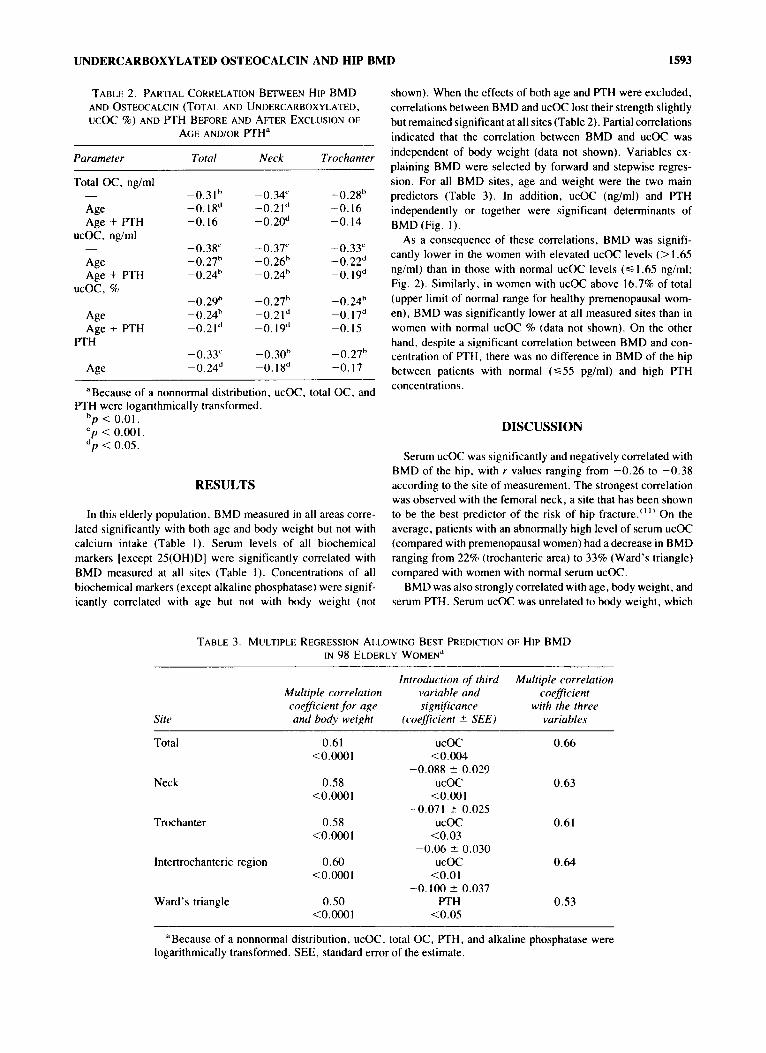
TABLE 2. PARTIAL CORRELATION BETWEEN HIP BMD AND OSTEOCAIKIN (TOTAL AND UNDERCARBOXYLATED,
AGE AND/OR PTH" UCOC %) AND PTH BEFORE AND ARER EXCLUSION OF
Parameter Total Neck Trochanter
Total OC, ng/ml
Age Age + PTH
ucOC, ng/ml
Age
ucoc, % Age + PTH
Age Age + PTH
PTH
-0.3Ih -0. I 8d -0. I6
-0.38' -0.27h -0.24h
-0.29h -0.24h -0.2Id
-0.33' -0.24"
-0.34' -0.21* - 0.20"
-0.37" -0.26h -0.24h
-0.27h -0.21* -0.19*
-0.30h -0. I 8d
-0.28h -0.16 -0.14
-0.33' -0.22d -0. 19d
-0.24h -0. 17d -0. 15
-0.27h -0. I7
"Because of a nonnormal distribution, ucOC, total OC, and PTH were logarithmically transformed.
hp < 0.01. cp < 0.001. * p < 0.05.
RESULTS
In this elderly population, BMD measured in all areas corre- lated significantly with both age and body weight but not with calcium intake (Table I ) . Serum levels of all biochemical markers [except 2S(OH)D] were significantly correlated with BMD measured at all sites (Table I ) . Concentrations of all biochemical markers (except alkaline phosphatase) were signif- icantly correlated with age but not with body weight (not
shown). When the effects of both age and PTH were excluded, correlations between BMD and ucOC lost their strength slightly but remained significant at all sites (Table 2). Partial correlations indicated that the correlation between BMD and ucOC was independent of body weight (data not shown). Variables ex- plaining BMD were selected by forward and stepwise regres- sion. For all BMD sites, age and weight were the two main predictors (Table 3). In addition, ucOC (ng/ml) and PTH independently or together were significant determinants of BMD(Fig. I ) .
As a consequence of these correlations, BMD was signifi- cantly lower in the women with elevated ucOC levels (> 1.65 nglml) than in those with normal ucOC levels (6 1.65 ng/ml; Fig. 2). Similarly. in women with ucOC above 16.7% of total (upper limit of normal range for healthy premenopausal wom- en), BMD was significantly lower at all measured sites than in women with normal ucOC % (data not shown). On the other hand, despite a significant correlation between BMD and con- centration of PTH, there was no difference in BMD of the hip between patients with normal (655 pglml) and high PTH concentrations.
DISCUSSION
Serum ucOC was significantly and negatively correlated with BMD of the hip, with r values ranging from -0.26 to -0.38 according to the site of measurement. The strongest correlation was observed with the femoral neck, a site that has been shown to be the best predictor of the risk of hip fracture."" On the average, patients with an abnormally high level of serum ucOC (compared with premenopausal women) had a decrease in BMD ranging from 22% (trochanteric area) to 33% (Ward's triangle) compared with women with normal serum ucOC.
BMD was also strongly correlated with age, body weight, and serum PTH. Serum ucOC was unrelated to body weight, which
TABLE 3. MULTIPLE REGRESSION ALLOWING BEST PREDICTION OF HIP BMD IN 98 ELDERLY WOMEN"
Introduction of third Multiple correlation Multiple correlution vuriuhle and coefficient coefficient for age significance with the three and body weight (coeficient lr SEE) variables
Total
Neck
Trochanter
0.61 <O.OoOI
0.58 <0.0001
0.58 <O.OoOI
lntertrochanteric region 0.60 <0.0001
Ward's triangle 0.50 <O.OoOI
ucoc 0.66 < O . W
-0.088 5 0.029
<O.OOl -0.071 t 0.025
<0.03
ucoc 0.63
ucoc 0.61
-0.06 * 0.030 ucoc 0.64
<O.Ol -0.100 * 0.037
<0.05 PTH 0.53
"Because of a nonnormal distribution, ucOC, total OC, PTH, and alkaline phosphatase were logarithmically transformed. SEE, standard error of the estimate.

1594 SZULC ET AL.
UCOC (nglml) .2
0.1 0.3 0.4 I 2 4
FIG. 1. Correlation between serum ucOC (logarithmically transformed) and BMD of the neck of the femur (g/cm2). r =
-0.37, p < 0.001.
therefore could not account for the relationship between ucOC and BMD. We previously found that serum ucOC increases with age and is negatively correlated with serum PTH. After correct- ing for the influence of these two factors, the negative correla- tion between serum ucOC and BMD was still highly significant, indicating that serum ucOC is an independent determinant of bone mass.
Our finding that elderly women with abnormally high serum ucOC have lower BMD values enlightens our previous observa- tion that serum ucOC is a marker of hip fracture risk."'The 26% decrease in BMD of the femoral neck in patients with abnormal levels of ucOC corresponds to a five to sevenfold increase in hip fracture according to the prospective data of Cummings et at.,"" an increase that is consistent with the relative risk of 5.9 that we found in our previous longitudinal study.'" The reason for this strong association between serum ucOC and bone mass,
1 .oo
0.75
0.50
0.2s
0
\ .... . . . . . . . .
. . . . . . . . . . . . . . . . . . . . . . . . . . . . . . . . . . . . . . . . . . . .
. . . . . . . .
. . . . . . . . . . . .
total
0 UCOC < 1.65 ng/ml
ucOC > 1.65 n d m l
-;I .... . . . . . . . . . . . .
. . . . . . . . . . . . . . . .
. . . . . . . . . . . . . . . . . . . . . . . .
neck
* 1 . . . . . . . . . . . . . . . . . . . . . . . . . . . . . . . . . . . . . . . . . . . .
. . . . . . . . . . . .
. . . .
trochanter
FIG. 2. Bone mineral density of the hip (g/cm*, mean 2 SD) of elderly women with normal (< 1.65 nglml; n = 86) and high (> I .65 ng/ml, n = 12) undercarboxylated osteocalcin (ucOC). Comparison of low versus high ucOC: * p < 0.01, **p < 0.001.
however, is not clear. One possibility is that serum ucOC is solely a marker of vitamin K deficiency and maybe also of vitamin D defi~iency,'~, '~-'" which are both highly prevalent in patients with hip thus reflecting nutritional abnormalities that could lead to accelerated bone loss through various mechanisms. Another intriguing possibility is that in- creased serum ucOC reflects intrinsic abnormalities of bone matrix leading to bone loss and increased fragility. Vast quanti- ties of vitamin K are present in bone tisuse,""' and recent data indicate that vitamin K inhibits bone resorption'*"' and may reduce bone I O S S . ' ~ " Experimental studies in animal treated with warfarin, a vitamin K antagonist that inhibits the carboxylation of glutamic acid, clearly show that ucOC does not bind properly to bone hydroxyapatite and that the concentration of bone OC is markedly reduced.** The consequences of this depletion are not clear, however. Patients on warfarin therapy have been reported to have either low'23' or normal'24' bone mass, but these studies were cross-sectional. A 1 year treatment with warfarin was recently reported to induce a decrease in hip BMD.'2s' Long- term treatment of rats with warfarin does not induce obvious changes in bone mass or strength,'**' but the same protocol applied to lambs for 3 months induced a marked osteopenia through a reduction in bone resorption and a further decrease in bone formation.""' In addition, it has been known for some time that warfarin therapy in the mother can result in major bone abnormalities in the fetus that are thought to be mediated by inhibition of the carboxylation of Gla-containing proteins of bone. A better understanding of the precise role of osteocalcin in bone metabolism would be helpful to clarify this issue.
ACKNOWLEDGMENTS
Supported by a grant from the Caisse Nationale d' Assurance Maladie des Travailleurs Salaries, INSERM, and the Ministere de la Recherche et de I'Enseignement Supkrieur-Aliment 2000, Duphar Company Laboratories and Merck-Clevenot Laborato- ries.
REFERENCES
I . Delmas PD 1989 Biochemical markers of bone turnover for the clinical assessment of metabolic bone disease. Endocrinol Metab Clin North Am 19:l-18.
2. Brown JP. Delmas PD, Malaval L, Edouard C, Meunier PJ 1984 Serum bone Gla-protein: A specific marker of bone formation in postmenopausal osteoporosis. Lancet 1: 109-1093.
3. Charhon SA, Delmas PD, Malaval L, Chavassieux P, Arlot M, Chapuy MC, Meunier PJ 1986 Serum bone Gla-protein in renal osteodystrophy: Comparison with bone histomorphometry. J Clin Endocrinol Metab 63:892-897.
4. Price PA, Williamson MK, Lothringer JW 1981 Origin of the vitamin K-dependent bone protein found in plasma and its clearance by kidney and bone. J Biol Chem 256: 12760-12766.
5. Knapen MHJ, Hamulyak K, VermeerC 1989 The effect of vitamin K supplementation on circulating osteocalcin (bone Gla protein) and urinary calcium excretion. Ann Intern Med 111:1001-1005.
6. Plantalech L. Guillaumont M, Vergnaud P, Leclercq M, Delmas PD 199 I Impairment of gamma carboxylation of circulating osteo- calcin (bone Gla protein) in elderly women. J Bone Miner Res 6:1211-1216.

UNDERCARBOXYLATED OSTEOCALCIN AND HIP BMD 1595
7.
8.
9.
10.
II.
12.
13.
14.
15
16
17
18
Plantalech L, Chapuy MC. Guillaumont M, Chapuy P. Leclercq M, Delmas PD 1990 Impaired carboxylation of serum ostecxalcin in elderly women: Effect of vitamin K treatment. In: Christiansen C, Overgaard K. eds. Osteoporosis. Osteopress, Copenhagen, pp. 345-347. Szulc P, Chapuy MC, Meunier PJ, Delmas PD 1993 Serum undercarboxylated osteocalcin is a marker of the risk of hip fracture in elderly women. J Clin Invest 91:1769-1774. Melton LJ, Wahner HW, Richelson LS, O’Fallon WM, Riggs HL 1986 Osteoporosis and the risk of hip fracture. Am J Epidemiol 123:254-26 I . Black DM, Cummings SR, Melton LJ 111 1992 Appendicular bone mineral and woman’s lifetime risk of hip fracture. J Bone Miner Res 7 : 6 3 9 4 6 . Cummings SR. Black DM, Nevitt MC, Browner W, Cauley J. Ensrud K, Genant HK, Palermo L. Scott J , Vogt TM 1993 Bone density at various sites for prediction of hip fractures. Lancet 341:72-74. Chapuy MC. Arlot ME. Duboeuf F. Brun J , Crouzet B, Arnaud S. Delmas PD. Meunier PJ 1992 Vitamin D, and calcium to prevent hip fractures in elderly women. N Engl J Med 327:1637-1642. Merle B. Delmas PD 1990 Normal carboxylation of circulating osteocalcin (bone Gla protein) in Paget’s disease of bone. Bone Miner 11:237-245. Duboeuf F, Braillon P, Chapuy MC, Haond P. Hardouin C, Meary MF. Delmas PD, Meunier PJ 1991 Bone mineral density of the hip measured with dual energy x-ray absorptiometry in normal elderly women and in patients with hip fracture. Osteoporosis Int 1:242- 249. Furie B, Furie BC 1990 Molecular basis of vitamin K-dependent gamma-carboxylation. Blood 75: 1753-1762. Karl PL, Friedman PA 1983 Effects of parathyroid hormone and vitamin D on the renal vitamin K-dependent carboxylating system. J Biol Chem 258:12783-12786. Deyl 2, Ada1 M 1983 Evidence for vitamin D-dependent gamnia- carboxylation in osteocalcin related proteins. Biochem Biophys Res Commun I13:294-300. Hodges SJ, Pilkington MJ, Stamp TCB. Catterall A, Shearer hlJ. Bitensky L, Chayen J 1992 Depressed levels of circulating
menaquinones in patients with osteoporotic fractures of the spine and femoral neck. Bone 32:387-389.
19. Hodges SJ, Bejui I , Leclercq M, Delmas PD 1993 Detection measurement of vitamins K, and K, in human cortical and trabec- ular bone. J Bone Miner Res 8:1005-1008.
20. Hara K, Akiyama Y. Tajima T, Shiraki M 1993 Menatetrenone inhibits bone resorption partly through inhibition of PGE, synthesis in vitro. J Bone Miner Res 8:535-542.
21. Akiba T , Kurihara S , Tachibana K, Kuwahara M, Sakamoto H. Yoneshima H, Marumo F I99 I Vitamin K (K) increased bone mass (BM) in hemodialysis patients (Pts) with low turnover bone disease (LTOBD). J Am Soc Nephrol2:608.
22. Price PA, Williamson MK 1981 Effects of warfarin on bone. Studies on the vitamin K-dependent protein of rat bone. J Biol Chem 256: 12754-1 2759.
23. Fiore CE, Tamburino C. Foti R, Grimaldi D 1990 Reduced bone mineral content in patients taking an oral anticoagulant. South Med J 83538-542.
24. Rosen HN, Maitland LA, Suttie JW. Manning WJ, Glynn RJ. Greenspan SL 1993 Vitamin K and maintenance of skeletal integ- rity in adults. Am J Med 94:6248.
25. Monreal M, Olive A, Lafoz E. del Rio L 1991 Heparins, coumarin, and bone density. Lancet 338:706.
26. Pastoureau P, Vergnaud P. Meunier PJ, Delmas PD 1993 Osteope- nia and bone remodeling abnormalities in warfarin-treated lambs: A possible role of osteocalcin. J Bone Miner Res 8: 1417-1426.
Address reprint requests to: Dr . P . D . Delmas
Hcipiral Edouard Herriot Pavillon F
69437 Lyon Cedex 3, France
Received in original form August 19. 1993; in revised form April 1 , 1994; accepted April I , 1994.