Embed Size (px)
Citation preview

METHODOLOGY ARTICLE Open Access
Serological testing of cattle experimentallyinfected with Mycoplasma mycoides subsp.mycoides Small Colony using four different testsreveals a variety of seroconversion patternsEvelyn Schubert1,2, Konrad Sachse2, Jörg Jores3 and Martin Heller1,4*
Abstract
Background: To study the specific antibody response to infection with Mycoplasma mycoides subsp. mycoidesSmall Colony (MmmSC), the agent of Contagious Bovine Pleuropneumonia (CBPP), we examined three panels ofsera collected during three experimental infection trials in African cattle. The methods used included an in-housecomplement fixation test (CFT), a commercially available CFT, a competitive antibody ELISA (cELISA) and theimmunoblotting test (IBT). In addition, lung tissue samples were examined by culture.
Results: A total of 89% (51/59) of all experimentally infected animals tested positive on at least one of theserological tests throughout the trial. The specific antibody titres to the MmmSC infection became positive first byCFT (6 to 9 days post infection [dpi]), followed by IBT (9 to 13 dpi) and cELISA (13 to 16 dpi). Individual animalswere found to display remarkably distinct seroconversion patterns, which allowed their classification into i) earlyhigh responders, ii) late high responders, and iii) low responders. In accordance with other studies, none of thepresent serological tests was capable of detecting all CBPP infected animals.
Conclusion: Comparison of the assays’ performance in terms of sensitivity and specificity raises serious questionsas to their reliability for identification of infected individuals in the field. In view of these limitations, a combinationof CFT and cELISA can markedly improve CBPP diagnosis at single-animal level.
BackgroundContagious Bovine Pleuropneumonia (CBPP) caused byMycoplasma mycoides subsp. mycoides Small Colony(MmmSC), is a highly contagious respiratory diseasenotifiable to the World Organization for Animal Health(Office International des Epizooties, OIE). While the dis-ease is an immediate threat to livestock producers in theendemic regions of Africa, its implications in terms ofepidemiology and animal health affect other geographi-cal areas as well. Characteristic symptoms include anor-exia, fever and respiratory signs, such as dyspnoea,polypnoea, cough and nasal discharge. In Africa, the dis-ease has been spreading due to economic, climatic and
political factors, and the limitations of currently avail-able diagnostic tests have often been detrimental to effi-cient control efforts. As for Europe, which has been freeof the disease since 1999 [1], the risk of CBPP re-intro-duction through clinically inconspicuous carrier animalsis still existing and deserves permanent attention of tra-ders and importers in the face of intensive internationaltrade in live cattle.Despite their known limitations, serological methods
are still the first choice for herd diagnosis of CBPP, withthe complement fixation test (CFT) and a competitiveenzyme-linked immunosorbent assay (cELISA) beinglisted as official methods in the OIE Manual [2].The CFT, first described in 1953 [3], is widely used in
countries struggling with the disease, and a modified“micro method” is common in countries of the Eur-opean Union [2]. It is assumed that some of the CFT’sdrawback in terms of specificity can be overcome by
* Correspondence: [email protected] Reference Laboratory for CBPP, Friedrich-Loeffler-Institut (FederalResearch Institute for Animal Health), Naumburger Str. 96a, 07743 Jena,GermanyFull list of author information is available at the end of the article
Schubert et al. BMC Veterinary Research 2011, 7:72http://www.biomedcentral.com/1746-6148/7/72
© 2011 Schubert et al; licensee BioMed Central Ltd. This is an Open Access article distributed under the terms of the CreativeCommons Attribution License (http://creativecommons.org/licenses/by/2.0), which permits unrestricted use, distribution, andreproduction in any medium, provided the original work is properly cited.
using the cELISA [4]. The immunoblotting test (IBT),which has been recommended by the OIE [2] as analternative in case of ambiguous results from CFT orELISA, was shown to be highly specific and sensitive [5].Furthermore, according to OIE recommendations, sus-
pected CBPP cases identified by serology should be con-firmed by specific detection of the pathogen. Whileculture is a cumbersome and time-consuming procedurerequiring fresh tissue samples, PCR allows specific iden-tification of the pathogen within hours. High sensitivityis characteristic for optimised PCR assays, with detec-tion limits between 10 [6] and 100 colony-forming units[7]. More recently, several real-time PCR assays forMmmSC were described [8-12]. However, if used forherd diagnosis, PCR-based tests have their limitationsdue to intermittent shedding of the pathogen, restrictedaccess to mycoplasmas in sequestra and the absence ofadequate equipment outside central laboratories.Apart from the ongoing discussion on the choice of the
diagnostic test, the time course and dynamics of the spe-cific antibody response in infected animals is not welldocumented. In a typical clinical case, the major patholo-gical consequence of MmmSC infection is a massiveinflammatory reaction mainly restricted to the lungs ofthe affected animal [13], which is associated with a rise inspecific antibodies. However, it was mentioned that indi-vidual animals respond rather differently to challengeinfection and vaccination [14-16], thus suggesting differ-ent immune response patterns. In addition, the existence
of symptomless carriers in the field is known, some ofwhich may be in the sub-acute or chronic phase of infec-tion presenting low titres or none at all [17].It was the aim of the present study to monitor sero-
conversion of cattle experimentally challenged withMmmSC. For this purpose, we compared the resultsfrom four serological assays and assessed their suitabilityfor testing at single-animal and herd levels.
MethodsBovine seraPanel 1 (End point sera from B237 trial)Thirty Zebu cattle were experimentally infected withMmmSC B237, a strain isolated from an outbreak inThika, Kenya in 1997, as described previously [18]. Theanimals were observed for up to 47 days (see Table 1),and sera were collected during post mortem examina-tion. Re-isolation of the agent was conducted and patho-logical findings were specified in 15 animals (505, 506,509, 513, 515, 519, 520, 522, 525, 527, 532, 539, 542,543, and 544). Three negative cattle sera originatedfrom the animal facility of FLI Jena, Germany. Positivecontrol serum for the in-house CFT (reference serumno. 315) and positive control serum for the IBT werekindly provided by J. Regalla, LNIV Lisbon, Portugal.Panel 2 (Sera from short-term Afadé trial)The animals were experimentally infected with MmmSC,strain Afadé, a strain isolated from an outbreak in North-ern Cameroon in 1968 (kindly provided by Joachim Frey,
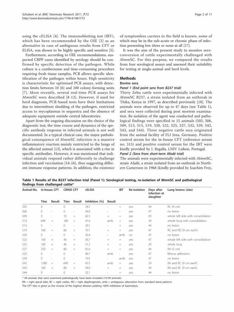
Table 1 Results of the B237 infection trial (Panel 1): Serological testing, re-isolation of MmmSC and pathologicalfindings from challenged cattle*
Animal-No. in-house CFT CIRAD CFT cELISA IBT Re-isolation Days afterinfection atslaughter
Lung lesions (size)
Titer Result Titer Result Inhibition [%] Result
505 0 - 0 - 28.7 - + yes 44 RC (9 cm)
506 0 - 0 - 34.0 - - yes 47 no lesion
509 0 - 10 + 62.3 + + yes 29 whole left side with consolidation
513 640 + 160 + 41.6 amb + yes 29 whole lung with consolidation
515 5 - 0 - 29.1 - + yes 44 no lesion
519 160 + 80 + 70.1 + + yes 47 RC and RD (9 cm each)
520 0 - 0 - 20.4 - amb no 47 no lesion
522 160 + 40 + 56.7 + + yes 47 whole left side with consolidation
525 160 + 40 + 51.2 + + yes 29 whole lung
527 320 + 80 + 61.6 + + yes 44 RA (5 cm)
532 0 - 0 - 46.7 amb - yes 47 fibrous adhesions
539 0 - 0 - 19.5 - amb. yes 47 no lesion
542 1280 + 640 + 42.5 amb + yes 29 RA and RC (9 cm each)
543 160 + 80 + 54.0 + + yes 44 RA and RC (9 cm each)
544 0 - 0 - 32.1 - + yes 44 no lesion
* All animals that were examined pathologically have been included (15/30 animals).
RA = right apical lobe, RC = right cadiac, RD = right diaphragmatic, amb = ambiguous (aberration from standard band pattern).
The CFT titre is given as the inverse of the highest dilution yielding 100% inhibition of haemolysis.
Schubert et al. BMC Veterinary Research 2011, 7:72http://www.biomedcentral.com/1746-6148/7/72
Page 2 of 11
University of Berne). Briefly, each animal was inoculatedintrabronchially with 50 ml of fresh MmmSC broth cul-ture (2.5 × 10¹⁰ colony forming units per animal), fol-lowed by 20 ml of liquid 1.5% agar solution and 30 mlphosphate-buffered saline. Sera were taken periodicallyevery 3 to 4 days from the day before infection (-1 dpi)until necropsy, i.e. up to 30 dpi, from 20 Boran cattle(BD091-102, BD105-107, BD111, BD115, BD116, BD118,BD119). Positive and negative control sera were the sameas in Panel 1. Infection mode, sampling scheme, clinicalsymptoms and pathological observations have beendescribed elsewhere [18,19].Panel 3 (Sera from long-term Afadé trial)To obtain samples from the late and chronic stages ofthe disease, sera were taken periodically on a weeklybasis from 7 experimentally infected Boran cattle(BD103, BD104, BD108, BD109, BD112, BD114, andBD117) over a time period of approximately 8 monthsto obtain samples from the late and chronic stages ofthe disease. Infection mode and challenge strain werethe same as in Panel 2.
Tissue samplesLung tissue samples were collected upon necropsy andused for examination by culture and PCR [18,19]. Lungswere inspected for lesions and, where possible, materialfrom these areas was excised for pathogen detection.The animal experiments mentioned in this paper were
conducted in strict accordance with Kenyan legislationon animal experimentation and were approved by theInstitutional Animal Care and Use Committee (IACUCreference no. 2008.08). ILRI has been voluntarily comply-ing with the United Kingdom’s Animals Act 1986, whichcontains guidelines and codes of practice for housing andcare of animals used in scientific procedures.
Complement fixation testsThe protocols of the two CFTs used in this study werelargely identical, but notably differed in the type of anti-gen used and the conditions of the antigen-binding step.a) The in-house CFT was carried out in microplate for-
mat as recommended by the OIE for detection of antibo-dies to MmmSC. Phenol-inactivated whole-cell antigenof the type strain MmmSC PG1, which was previouslycheckerboard titrated, was used as antigen at a concen-tration of 2 complement fixing units. Other reagents forthe in-house CFT complement, i.e. haemolytic serum,veronal buffer and sheep red blood cells, were obtainedfrom Virion-Serion (Würzburg, Germany). Antigen bind-ing was allowed during overnight incubation at 4°C.b) The CIRAD CFT (CIRAD, Montpellier, France),
was conducted according to the manufacturer’s instruc-tions. The kit included all reagents except veronal
buffer, sheep red blood cells and negative control serum(purchased from Virion-Serion). The incubation timefor antigen binding at 37°C was 30 min.All sera used were inactivated at 56°C for 30 min and
diluted in the range of 1:5 to 1:2560. The highest dilu-tions of sera producing 100% haemolysis inhibition ofsheep red blood cells were taken as end points of dilu-tions to be examined, and CFT titres were given as reci-procals of these dilutions.CFT readings were scored according the OIE manual
[2], i.e. positive in the case of 100% inhibition of haemo-lysis at a serum dilution of 1:10 or greater; ambiguousat 25, 50 or 75% inhibition at 1:10 serum dilution, andnegative with absent haemolysis or haemolysis at 1:5serum dilution.
Enzyme immunoassayThe CBPP serum competitive ELISA (IDEXX, InstitutePourquier, Montpellier, France) was used for screeningthe sera. The test is based on the paper by Le Goff andThiaucourt [20] and uses a monoclonal anti-MmmSCantibody, as well as microplates coated with MmmSClysate. The ELISA was performed according to theinstructions of the manufacturer. Optical densities (OD)were measured at 450 nm using the photometer SpectraFluor (Tecan, Crailsheim, Germany). The percentageinhibition value (INH%) for each serum sample was cal-culated using the formula:
INH% = (ODmab −ODsample)/(ODmab −ODconjugate)× 100%,
ODmab = Control only with monoclonal antibody and without serum (0% inhibition)
ODsample = OD of the serum sample
ODconjugate = Control without monoclonal antibody and serum (100% inhibition) .
The cut-off for positive samples was at INH% of 50%.Sera with an inhibition value between 40% and 50%were considered doubtful. All sera were examined induplicate.
Immunoblotting testThe IBT was performed according to Regalla et al. [5]and the OIE manual [2] with minor modifications. SDS-PAGE (7.5% polyacrylamide) separated proteins of refer-ence strain MmmSC PG1 and strain Afadé were trans-ferred onto nitrocellulose membrane (Whatman, Dassel,Germany). The BenchMark Protein Ladder 10-220 kDa(Invitrogen, Karlsruhe, Germany) was used as molecularweight marker. To control the efficiency of proteintransfer, a reversible protein staining step using PonceauS (Sigma-Aldrich, Taufkirchen, Germany) was included.
Schubert et al. BMC Veterinary Research 2011, 7:72http://www.biomedcentral.com/1746-6148/7/72
Page 3 of 11
Excised membrane strips were incubated with 1:100serum dilutions or control serum, respectively. Reactivebands were visualised using alkaline phosphatase-conju-gated recombinant protein A/G (Pierce, Bonn, Ger-many) and substrate BCIP/NTB (5-bromo-4-chloro-3-indolyl phosphate combined with nitrotetrazolium bluechloride, Sigma-Aldrich). Positive sera were expected toshow a specific pattern that included reactive bands at110, 98, 95, 60/62, and 48 kDa. IBT patterns of serawere scored ambiguous in case that either the specificband of 98 kDa was missing or other specific bandswere weak.
Bacteriological evaluation and identificationIsolation and cultivation of Mycoplasma strains from tis-sue samples were performed using standard methodol-ogy [21]. Lung tissue samples collected during postmortem from margins of sequestra or regions of necro-sis were inoculated into modified Hayflick medium [21]and incubated at 37°C for 3 to 4 days. Identification ofMmmSC from these cultures was done using PCR (seenext paragraph).
PCRMycoplasma cultures and tissue samples of Panel 2 and3 animals were examined using a MmmSC-specific PCRassay described previously [6,22].
Statistical evaluationThe kappa agreement test was conducted [23] to com-pare the concordance between individual tests. For eachanimal, the mean of all the measurements was calcu-lated for each individual assay and an animal was classi-fied positive based on the set criterion. The proportionof positive animals was computed as a percentage of thetest positives against the total number tested.
ResultsExamination of end point sera from the B237 trial (Panel1)Of the 30 animals challenged, 25 (83.3%) displayed aspecific antibody response detected by at least one ofthe serological tests, whereas all sera from non-infectedanimals remained negative. A positive test result of serato all the four tests was observed in 12 (40%) cases. Re-isolation of the challenge strain was attempted in 15cases, of which 14 were successful [18]. The findingsobtained from these animals are summarised in Table 1.Notably, the infective strain was also re-isolated fromfour animals without lesions. The lesion-free subgrouptested negative both in cELISA and CFTs (animals 506,515, 520, 539, 544), IBT was either positive (animals 515and 544), ambiguous (animals 520 and 539; Table 1), ornegative (animal 506). Regarding all infected animals,
the number of positive results in IBT was higher than inthe other methods.
Examination of sera from the short-term Afadé trial(Panel 2)Serological testing using both CFTs confirmed success-ful infection of all animals (see Additional File 1: Resultsof in-house CFT vs. CIRAD CFT from sera of Panel 2).The time course of specific antibody production in all20 animals is shown in Figure 1. The humoral responsepatterns as detected by CFT can be classified into threecategories, i) early high responders (BD091, BD092,BD093, BD097, BD098, BD099, BD107, BD111, BD115,BD118, BD119), ii) late high responders (BD094, BD095,BD116), and iii) low responders (BD096, BD100, BD101,BD102, BD105, BD106). CFT titres of early high respon-ders emerged at 6 dpi in the case of animal BD119 andat 9 dpi in the other animals (Figure 1A). For late highresponders, a pronounced rise around 13 dpi was char-acteristic (Figure 1B).Figure 1 also reveals that the course of the humoralresponses of Panel 2 cattle were not completely concur-rent in CFT and cELISA. The increase of the cELISAtitres was seen after day 6 post infection (see AdditionalFile 2: Examination of Panel 2 sera using cELISA), withthe exception of animal BD091 (beginning at 6 dpi,Figure 1A; see also below). In the subgroup of lowresponders, the rise in cELISA titres was generally weak,with animals BD101, BD102, BD105, and BD106remaining below the cut-off level throughout the trial(as did animals BD092, BD097, BD111, and BD118 ofthe early high responder group). Nevertheless, thecELISA-negative animals mentioned showed positiveresults in both CFTs (see Additional File 1: Results ofin-house CFT vs. CIRAD CFT from sera of Panel 2). Inaddition, the cELISA-negative animals BD097 andBD101 exhibited characteristic bands in the IBT. Figure 2shows immunoblot patterns of four selected animals ofPanel 2. While the IBT results were largely comparableto CFT findings, this test also detected emerging specificantibodies to MmmSC at earlier time points thancELISA (Table 2).Seropositivitiy of Panel 2 sera in cELISA and CFT didnot always correlate with clinical symptoms and patho-morphological signs. Conversely, however, negative orweakly positive readings in cELISA and CFT correlatedwith the absence of clinical signs or mild symptoms.Patho-morphological lesions typical for MmmSC infec-tions were observed in all animals of the short-termAfadé trial except animal BD102. Four animals died orhad to be sacrificed prematurely, i.e. at 16 dpi (BD091,BD097, BD118) or at 20 dpi (BD098). This subgroupbelonged to early high responders showing severe clini-cal symptoms (high temperature, cough, dyspnoea), as
Schubert et al. BMC Veterinary Research 2011, 7:72http://www.biomedcentral.com/1746-6148/7/72
Page 4 of 11
Figure 1 Comparison of CIRAD CFT and cELISA-based curves showing the antibody response of animals from Panel 2. Animals from theShort-term Afadé trial were characterised by CFT (bars) and cELISA (curves). Horizontal lines represent the cut-off values of CFT (dilution 1:10,100% inhibition of haemolysis) and cELISA (50% inhibition), respectively. The assignment of letters and symbols to the respective animals is givenin the right-hand column. A: Early high responders (according to CFT), B: Late high responders, C: Low responders. Missing bars at later timepoints indicate that the respective animal died or had to be removed prematurely.
Schubert et al. BMC Veterinary Research 2011, 7:72http://www.biomedcentral.com/1746-6148/7/72
Page 5 of 11
well as a variety of patho-morphological lesions in lungand lymph node tissues.
Examination of sera from the long-term Afadé trial (Panel 3)Sera from all 7 animals showed positive reactions withall four methods used. In a typical time course (Figure3), detectable CFT titres emerged in week 2 p.i. anddecreased steadily from weeks 5-8. At the end of thisinfection trial, i.e. 34 weeks p.i., CFT titres rangedbetween 1:10 to 1:20 for the in-house CFT (see Addi-tional File 3: Examination of Panel 3 sera using in-houseCFT), and between 1:5 and 1:160 for the CIRAD CFT(see Additional File 4: Examination of Panel 3 sera usingCIRAD CFT). Since sera were collected only on aweekly basis, classification into responder groups as inPanel 2 animals was not feasible. Notably, animal
BD117 differed from the other animals by respondingone week later in both CFTs.Test results of the cELISA showed a rise in titres from
week 3 to 4 p.i., with levels either remaining positiveuntil to the end of the trial (Figure 3) or slowly decreas-ing to reach the cut-off value of 50% inhibition (seeAdditional File 5: Examination of Panel 3 sera usingcELISA).The time course of specific antibody response as mon-
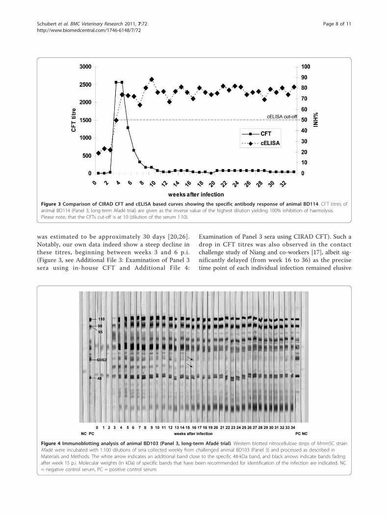
itored by IBT is shown in Figure 4. Animal BD103 wassomewhat peculiar as the specific antigenic band of 60-62 kDa faded after week 10 p.i. The same band disap-peared in the reaction of animal BD117 after week 10,whereas animal BD104 maintained the entire specificbanding pattern until the end at week 34 (data notshown).
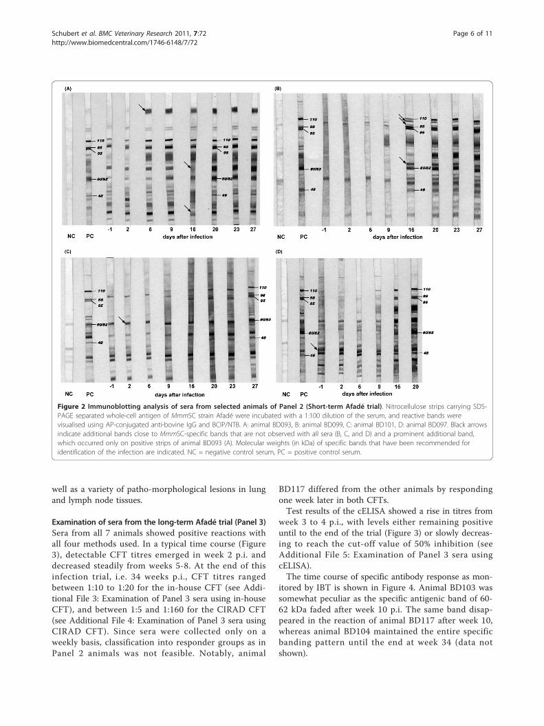
Figure 2 Immunoblotting analysis of sera from selected animals of Panel 2 (Short-term Afadé trial). Nitrocellulose strips carrying SDS-PAGE separated whole-cell antigen of MmmSC strain Afadé were incubated with a 1:100 dilution of the serum, and reactive bands werevisualised using AP-conjugated anti-bovine IgG and BCIP/NTB. A: animal BD093, B: animal BD099, C: animal BD101, D: animal BD097. Black arrowsindicate additional bands close to MmmSC-specific bands that are not observed with all sera (B, C, and D) and a prominent additional band,which occurred only on positive strips of animal BD093 (A). Molecular weights (in kDa) of specific bands that have been recommended foridentification of the infection are indicated. NC = negative control serum, PC = positive control serum.
Schubert et al. BMC Veterinary Research 2011, 7:72http://www.biomedcentral.com/1746-6148/7/72
Page 6 of 11
General comparison between the serological methodsAll test results of the four assays from sera of Panels 1and 2 have been compared and summarised in Table 3.The compilation shows that general agreement amongthe results of all tests used was poor. As confirmed bykappa agreement testing, the results of in-house andCIRAD CFTs were always close to each other, whentaking into account the inherent systematic error of twodilution titres (see Table 1, see Additional File 1: Resultsof in-house CFT vs. CIRAD CFT from sera of Panel 2).The number of positive findings by CFTs was generallyhigher than those from cELISA, which implies a delayin the detection of the onset of antibody production bythe latter (see Table 2, Figure 1). The sensitivity of theIBT proved intermediate between cELISA (less sensitive)and CFT (more sensitive), when consecutively collectedsera were examined (Panel 2).
DiscussionThe remarkable degree of variation in the humoralimmune response displayed by the animals of the pre-sent study indicates a high complexity of host-pathogeninteractions during MmmSC infection, which can leadto acute, sub-acute to chronic or symptomless coursesof disease [17]. On the one hand, the immune status ofthe individual animal seems to play a role in the timecourse and level of specific antibody production. Naïveanimals are assumed to react in a different fashion thanre-infected cattle exhibiting an anamnestic response,and symptomless carriers can exhibit low antibody levelsin the absence of intense host pathogen interactions.Inter-animal differences in the cellular immune response[18,19], which have not been addressed in the presentstudy, may also add to the overall diversity observed.On the other hand, the pathogen has been shown to
possess a genetically determined machinery for surfaceantigen variation [24,25], which enables it to evade thehost immune response by selecting modified phenotypesthat cannot be challenged by cognate antibodies.Depending on the efficiency of the individual hostdefence, the progress of MmmSC infection can beexpected to vary from animal to animal.The authors wish to emphasise that the present com-
parative analysis of diagnostic tests is referring to theindividual animal level, which is a limitation because thedata cannot be simply extrapolated to herd level. Whilecurrently available serological tests are generally suitablefor herd diagnosis, the present findings highlight seriouslimitations of these tests at the individual animal level,which have to be taken into account when field studiesare conducted.CFT titres do not represent the whole spectrum of
specific antibodies present in the infected animal, norare they long lasting. The half-life of CFT antibody titres
Table 2 Comparison of the specific antibody response ofselected* cattle from the short-term Afadé trial (Panel 2)using four serological tests
Animal Time of serumcollecion (dpi)
In-houseCFT
CIRADCFT
cELISA IBT
-1 - - - -
2 - - nd nd
6 - - - -
9 + + - amb
BD 093 13 + + amb +
16 + + amb +
20 + + + +
23 + + + +
27 + + + +
-1 - - - -
2 - - - -
BD 097 6 - - - -
9 + + - amb
13 + + - +
16 + + - +
-1 - - - -
2 - - - -
6 - - - -
9 + + - -
BD 099 13 + + amb nd
16 + + amb +
20 + + + +
23 + + + +
27 + + + +
-1 - - -
2 - - - -
6 - - - -
9 - - - -
BD 101 13 - - - nd
16 + + - amb
20 + + - +
23 + + - +
27 + + - +
-1 - - - nd
2 - - - nd
6 - - - nd
9 - + - nd
BD 105 13 - + - nd
16 - + - nd
20 - + - nd
23 - + - nd
27 + + amb nd
-1 - - - nd
2 - - - nd
BD 118 6 - - - nd
9 + + - nd
13 + + - nd
16 + + amb nd
* animals surviving at least until 16 dpi, amb = ambiguous, nd = not done
Schubert et al. BMC Veterinary Research 2011, 7:72http://www.biomedcentral.com/1746-6148/7/72
Page 7 of 11
was estimated to be approximately 30 days [20,26].Notably, our own data indeed show a steep decline inthese titres, beginning between weeks 3 and 6 p.i.(Figure 3, see Additional File 3: Examination of Panel 3sera using in-house CFT and Additional File 4:
Examination of Panel 3 sera using CIRAD CFT). Such adrop in CFT titres was also observed in the contactchallenge study of Niang and co-workers [17], albeit sig-nificantly delayed (from week 16 to 36) as the precisetime point of each individual infection remained elusive
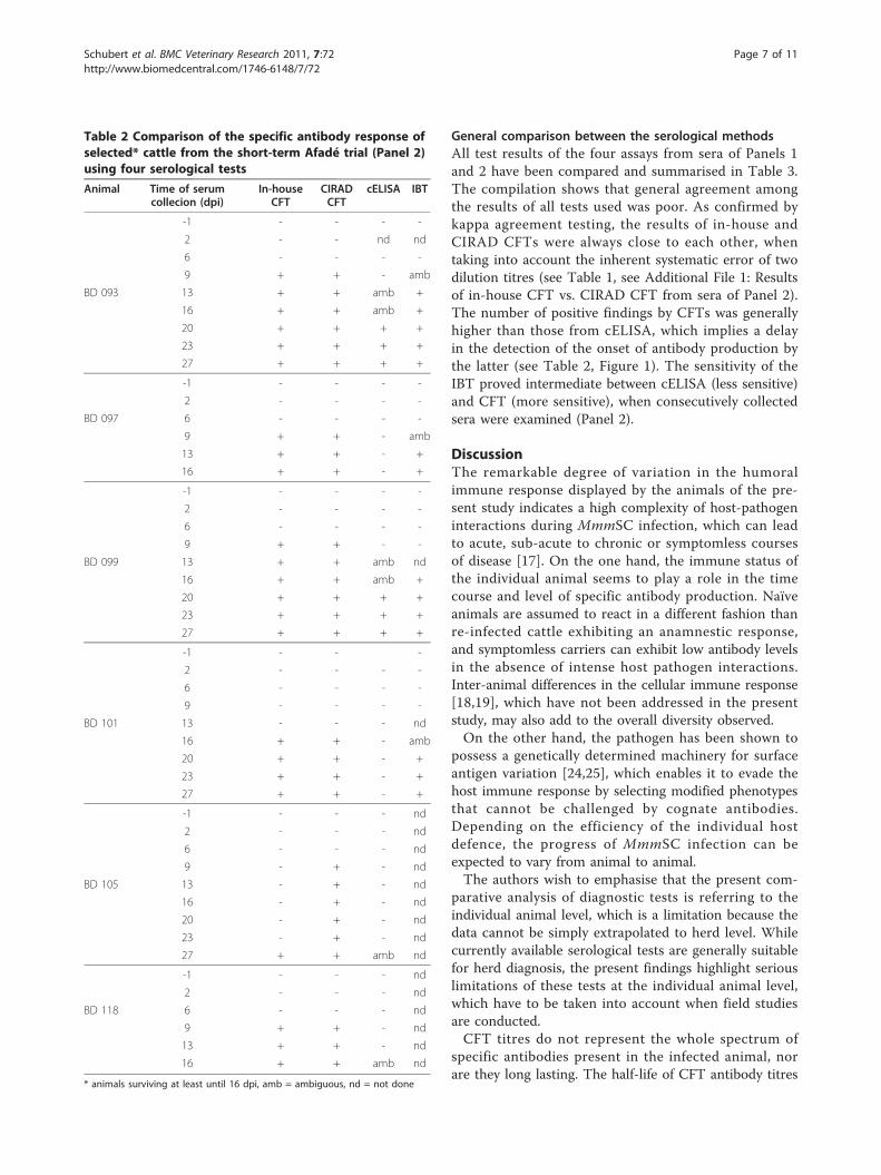
Figure 3 Comparison of CIRAD CFT and cELISA based curves showing the specific antibody response of animal BD114. CFT titres ofanimal BD114 (Panel 3, long-term Afadé trial) are given as the inverse value of the highest dilution yielding 100% inhibition of haemolysis.Please note, that the CFTs cut-off is at 10 (dilution of the serum 1:10).
Figure 4 Immunoblotting analysis of animal BD103 (Panel 3, long-term Afadé trial). Western blotted nitrocellulose strips of MmmSC strainAfadé were incubated with 1:100 dilutions of sera collected weekly from challenged animal BD103 (Panel 3) and processed as described inMaterials and Methods. The white arrow indicates an additional band close to the specific 48-kDa band, and black arrows indicate bands fadingafter week 15 p.i. Molecular weights (in kDa) of specific bands that have been recommended for identification of the infection are indicated. NC= negative control serum, PC = positive control serum.
Schubert et al. BMC Veterinary Research 2011, 7:72http://www.biomedcentral.com/1746-6148/7/72
Page 8 of 11
in that infection model. Taken together, these observa-tions imply that field studies based solely on CFT areprone to miss individual animals at the later stages ofinfection.While the differences between the two CFTs used
were marginal, the relative diagnostic sen-sitivity of bothtests compared to culture was 50.0% (in-house CFT)and 57.1% (CIRAD CFT) with Panel 1 samples, which isin line with data of other authors [27]. The consistentlyobserved divergence between the results of CFT andcELISA (Table 2, Figure 3) is probably a consequence ofthe different immunoglobulin classes covered by eachmethod. Thus, IgG class antibodies have a greater affi-nity in the cELISA, while IgG2 subclass antibodies areunable to fixate complement used in the CFT. More-over, IgM class antibodies, which are characteristic forearly infection, are easier to detect by CFT [28,29]. Thiscan explain the earlier detection of antibodies by CFT asobserved in the present study and elsewhere [20,30].Furthermore, the present finding that IgG antibodylevels from cELISA remained at a high level for a pro-longed period is in agreement with the study by Niangand co-workers [17], where the kinetics of different anti-body isotypes was investigated.It is important to note that the present cELISA was
given a relatively high cut-off in order to maximise speci-ficity, which in turn diminishes the test’s sensitivity [31].In fact, the present evidence suggests that there is someroom for lowering the cut-off without loss of specificity.However, this has to be confirmed by further studiesinvolving more field sera from cattle herds having CBPPand/or other mycoplasma infections. We hypothesisethat another way to improve the test’s performanceincludes the use of specific peptides [32] or recombinantMmmSC proteins [16,33] instead of whole-cell antigen.The IBT has been described as being more sensitive
and specific than CFT [13], which has been confirmedby the present data (Table 1). Thus, the IBT showedspecific reaction patterns for sera tested negative in CFT
and cELISA (animals 515, 520, 539, 544, 546). The test’shigh specificity is based on the reaction to five differentantigens, i.e. 110, 98, 95, 60/62, and 48 kDa proteins,which must be recognised by their specific antibodies inorder to identify a positive serum [2]. However, weobserved some problems with the test’s reproducibilityand potential for standardisation, as immunoblot reac-tion patterns are complex and individual bands may bedifficult to identify when non-specific bands from cross-reactions with other bacteria are interfering. Cross-reac-tions with closely related Mycoplasma (M.) species ofthe “mycoides cluster” seem to be less important here,because they are rarely encountered in cattle, but othermycoplasmas, such as M. bovis and M. bovigenitalium,may play a role [13,31].Culture of MmmSC from affected lung tissue was
included to underpin our serological findings. While ser-opositive animals always showed clinical symptoms ofCBPP that were confirmed by pathology, the presence oflung lesions was no guarantee for successful re-isolationof the challenge strain (Panel 2, data not shown). Thefindings of the present study also suggest that re-isolationwas seriously hampered at the late and chronic stages ofinfection, i.e. isolation of MmmSC did not succeed fromtissue samples of Panel 3 animals after 35 weeks p.i. Theabsence of a strict correlation was particularly evidentwith animal 506 (Panel 1, Table 1), where all serologicaltests were negative (and pathological signs were missing)despite successful re-isolation of the pathogen.
ConclusionsThe present study has revealed three distinct seroconver-sion patterns among MmmSC-infected animals, i) earlyhigh responders, ii) late high responders, and iii) lowresponders. This variability raises questions as to the choiceand suitability of current serological tests for single-animaldiagnosis. While valid at the herd level, individual testresults can be misleading and negative serological findingsshould be interpreted with particular caution.
Table 3 Examination of Panel 1 and 2 sera: Comparison of all serological tests*
Assays compared Number (percentage) of animals reacting similarly in the tests compared
30 sera of Panel 1 Selection of 30 sera of Panel 2 (including IBT) All 178 sera of Panel 2(without IBT)
cELISA and in-house CFT 21 (70%), ĸ = 0,46c 18 (60%), ĸ = 0,33d 114 (70%), ĸ = 0,43c
cELISA and CIRAD CFT 23 (77%), ĸ = 0,58c 18 (60%), ĸ = 0,33d 108 (62%), ĸ = 0,38d
In-house CFT and CIRAD CFT 27 (90%), ĸ = 0,79a 30 (100%), ĸ = 1,00a 161 (90%), ĸ = 0,80a
In-house CFT and IBT 21 (70%), ĸ = 0,39d 26 (87%), ĸ = 0,33d
CIRAD CFT and IBT 21 (70%), ĸ = 0,39d 26 (87%), ĸ = 0,33d
cELISA and IBT 17 (57%), ĸ = 0,23d 19 (63%), ĸ = 0,38d
In-house CFT, CIRAD CFT and cELISA 21(70%) 18 (60%) 102 (59%)
In-house CFT, CIRAD CFT, cELISA and IBT 15 (45%) 18 (60%)
* Kappa agreement testing [23] was additionally conducted. According to standard criteria, the concordance between two tests compared is classified as a verygood (1-0.76), b good (0.75-0.61), c acceptable (0.6-0.4), d poor (< 0.4).
Schubert et al. BMC Veterinary Research 2011, 7:72http://www.biomedcentral.com/1746-6148/7/72
Page 9 of 11
Two factors account for the lack of sensitivity of sero-logical tests at the single-animal level, i) titres of specificantibodies at an early stage of infection and in chroniccarriers can be very low, and ii) high variability in anti-gen expression by MmmSC in vivo, where not all rele-vant proteins are expressed at a given point in time[24,34]. We suggest that diagnostic testing should com-prise both CFT and cELISA, particularly in countriesdeclared free of CBPP.Unlike CFT and ELISA, the IBT requires experienced
and well-trained laboratory personnel and is not suitablefor use in routine laboratories. To improve reproducibil-ity, we recommend i) the use of 7.5% acrylamide gelsinstead of gradient gels as prescribed in the OIE manual[2], and ii) the use of the same antigen in all labora-tories, i.e. MmmSC strain Afadé. We propose that themethodology in [2] be accordingly revised and supple-mented with more detailed instructions to address theabove mentioned problems (see Additional File 6: Pro-posal for modification of the current OIE protocol forIBT), so that IBT can be used as an additional test inthe case of ambiguous CFT and/or cELISA results.
Additional material
Additional file 1: Results of in-house CFT vs. CIRAD CFT from seraof Panel 2. Humoral immune response of animals from Panel 2 (Short-term Afadé trial) as characterised by in-house CFT and CIRAD CFT. End-point titres of both CFTs are shown at different time points of infection.
Additional file 2: Examination of Panel 2 sera using cELISA. Humoralimmune response of animals from Panel 2 (Short-term Afadé trial)characterised by cELISA. The percentage inhibition value (INH%) for eachserum sample was calculated using the formula: INH% = (ODmab -ODsample)/(ODmab - ODconjugate) × 100%, ODmab = Control only withmonoclonal antibody and without serum (0% inhibition), ODsample = ODof the serum sample, ODconjugate = Control without monoclonal antibodyand serum (100% inhibition). The cut-off for positive samples was set atINH% of 50%. Sera with an inhibition value between 40% and 50% wereconsidered doubtful. All sera were examined in duplicate. ND = notdone.
Additional file 3: Examination of Panel 3 sera using in-house CFT.Examination of the 7 sera from the long-term Afadé trial (Panel 3) usingin-house CFT. End-point titres of the CFT were shown until 34 weeks p.i.
Additional file 4: Examination of Panel 3 sera using CIRAD CFT.Examination of the 7 sera from the long-term Afadé trial (Panel 3) usingCIRAD CFT. End-point titres of the CFT were shown until 34 weeks p.i.
Additional file 5: Examination of Panel 3 sera using cELISA.Examination of the 7 sera from the long-term Afadé trial (Panel 3) usingcELISA. Data of the cELISA were given in percentage inhibition andshown until 34 weeks p.i.
Additional file 6: Proposal for modification of the current OIEprotocol for IBT. To improve the reproducibility of IBT results, theauthors of the present paper recommend two modifications to theprotocol of the OIE Manual.
AbbreviationsBCIP/NTB: 5-bromo-4-chloro-3-indolyl phosphate combined withnitrotetrazolium blue chloride; CFT: complement fixation test; cELISA:competitive enzyme linked immunosorbent assay, CBPP: Contagious Bovine
Pleuropneumonia; dpi: days post infection; IBT: immunoblotting test; kDa:kilodalton; MmmSC: Mycoplasma mycoides subsp. mycoides Small Colony;OD: optical densities; PCR: polymerase chain reaction; INH%: percentageinhibition value; p.i.: post infection; OIE: Office International des Epizooties(World Organization for Animal Health)
AcknowledgementsWe are grateful to Anja Sterner-Kock (University of Cologne), Wolfram Haider(Institute for Animal Pathology, Berlin), Jan Naessens (ILRI, Nairobi) andHezron Wesonga (Keyan Agricultural Research Institute, Nairobi) for samplepreparation, as well as Sandra Thierbach and Susann Bahrmann (Friedrich-Loeffler-Institut) for their excellent technical assistance. We are indebted toWolfram Maginot for his support preparing the figures. We thank ChristianMenge and Heinrich Neubauer (Friedrich-Loeffler-Institut) for theirconstructive input and helpful discussions. The German Federal Ministry forEducation and Research (BMBF) has funded this project in the framework ofthe “Research for Civil Security” programme (Förderkenn-zeichen 13N9520).
Author details1National Reference Laboratory for CBPP, Friedrich-Loeffler-Institut (FederalResearch Institute for Animal Health), Naumburger Str. 96a, 07743 Jena,Germany. 2Institute of Molecular Pathogenesis, Friedrich-Loeffler-Institut(Federal Research Institute for Animal Health), Naumburger Str. 96a, 07743Jena, Germany. 3International Livestock Research Institute (ILRI), Old NaivashaRoad, P.O. Box 30709, 00100 Nairobi, Kenya. 4Institute of Bacterial Infectionsand Zoonoses, Friedrich-Loeffler-Institut (Federal Research Institute forAnimal Health), Naumburger Str. 96a, 07743 Jena, Germany.
Authors’ contributionsES provided and supported laboratory work, evaluated and interpreted thedata and wrote the manuscript. KS has been involved in supportinglaboratory work and in discussion of results including revising themanuscript. JJ designed and coordinated the animal experiments using theAfadé strain and provided serum and tissue samples, MH coordinated theinvestigation, evaluated and interpreted all data of CFT, immunoblotting test,PCR, and culture investigations. All authors revised the manuscript andapproved the final version.
Competing interestsThe authors declare that they have no competing interests.
Received: 30 May 2011 Accepted: 18 November 2011Published: 18 November 2011
References1. Thiaucourt F, Dedieu L, Maillard JC, Bonnet P, Lesnoff M, Laval G, Provost A:
Contagious bovine pleuropneumonia vaccines, historic highlights,present situation and hopes. In Vaccines for OIE List A and EmergingAnimal Diseases. Volume 114. Edited by: Brown F, Roth J. Basel: Dev Biol;2003:147-160.
2. Manual of diagnostic tests and vaccines for terrestrial animals. , 6 2008[http://www.oie.int/fileadmin/Home/eng/Health_standards/tahm/2.04.09_CBPP.pdf].
3. Campbell AD, Turner AW: Studies of contagious bovine pleuropneumoniaof cattle. IV. An improved complement fixation test. Aust Vet J 1953,29:154-163.
4. Amanfu W, Sediale S, Masapu KV, Benkirane A, Geiger R, Thiaucourt F: Fieldvalidation of a competitive enzyme-linked immunosorbent assay for thedetection of contagious bovine pleuropneumonia in Botswana. Rev ElevMed Pays Trop 1998, 51:189-193.
5. Regalla J, Gonçalves R, Niza Ribeiro J, Duarte L, Nicholas R, Bashiruddin JB,De Santis P, Garrido Abellan F, Penha Gonçalves A: Development ofimmunoblotting as diagnostic tool for contagious bovinepleuropneumonia. International Symposium - COST Action 826:Mycoplasmas of ruminants: pathology, Diagnostics, Epidemiology andMolecular genetics. Joint Workshops of E.U. projects Toulouse, France; 1999,109-112.
6. Hotzel H, Frey J, Bashiruddin J, Sachse K: Detection and differentiation ofruminant mycoplasmas. In Methods in Molecular Biology. Volume 216 (PCRDetection of Microbial Pathogens). Edited by: Sachse K, Frey J. Totowa NJ:Humana Press; 2002:231-246.
Schubert et al. BMC Veterinary Research 2011, 7:72http://www.biomedcentral.com/1746-6148/7/72
Page 10 of 11

7. Bashiruddin JB, Taylor TK, Gould AR: A PCR-based test for the specificidentification of Mycoplasma mycoides subspecies mycoides SC. J VetDiagn Invest 1994, 6:428-434.
8. Gorton TS, Barnett MM, Gull T, French RA, Lu Z, Kutish GF, Adams LG,Geary SJ: Development of real-time diagnostic assays specific forMycoplasma mycoides subspecies mycoides Small Colony. Vet Microbiol2005, 111:51-58.
9. Fitzmaurice J, Sewell M, Manso-Silvan L, Thiaucourt F, McDonald WL,O’Keefe JS: Real-time polymerase chain reaction assays for the detectionof members of the Mycoplasma mycoides cluster. New Zeal Vet J 2008,56:40-47.
10. Lorenzon S, Manso-Silvan L, Thiaucourt F: Specific real-time PCR assays forthe detection and quantification of Mycoplasma mycoides subsp.mycoides SC and Mycop-lasma capricolum subsp. capripneumoniae. MolCell Probes 2008, 22:324-328.
11. Vilei EM, Frey J: Detection of Mycoplasma mycoides subsp. mycoides SC inbronchoalveolar lavage fluids of cows based on a TaqMan real-time PCRdiscriminating wild type strains from an lppQ(-) mutant vaccine strainused for DIVA-strategies. J Microbiol Methods 2010, 81:211-218.
12. Schnee C, Heller M, Jores J, Tomaso H, Neubauer H: Assessment of a novelmultiplex real-time PCR assay for the detection of the CBPP agentMycoplasma mycoides subsp. mycoides SC through experimentalinfection in cattle. BMC Vet Res 2011, 7:47-59.
13. Gonçalves R, Regalla J, Ayling RD, Nicholas RA: Impact of Mycoplasmabovis infection on serosurveillance for contagious bovinepleuropneumonia. Vet Rec 2008, 163:632-633.
14. Dedieu L, Balcer-Rodrigues V, Yaya A, Hamadou B, Cisse O, Diallo M,Niang M: Gamma interferon-producing Cd4 T-cells correlate withresistance to Mycoplasma mycoides subsp mycoides S.C. infection incattle. Vet Immunol Immunopathol 2005, 107:217-233.
15. Jores J, Meens J, Buettner FF, Linz B, Naessens J, Gerlach GF: Analysis ofthe immunoproteome of Mycoplasma mycoides subsp. mycoides smallcolony type reveals immunogenic homologues to other knownvirulence traits in related Mycoplasma species. Vet ImmunolImmunopathol 2009, 131:238-245.
16. Hamsten C, Tjipura-Zaire G, McAuliffe L, Scacchia M, Ayling RD, Persson A:Protein-specific analysis of humoral immune responses in a clinical trialfor vaccines against contagious bovine pleuropneumonia. Clin VaccineImmunol 2010, 17:853-861.
17. Niang M, Diallo M, Cisse O, Kone M, Doucoure M, Roth JA, Balcer-Rodrigues V, Dedieu L: Pulmonary and serum antibody responses elicitedin zebu cattle experimentally infected with Mycoplasma mycoides subsp.mycoides SC by contact exposure. Vet Res 2006, 37:733-744.
18. Jores J, Nkando I, Sterner-Kock A, Haider W, Poole J, Unger H, Muriuki C,Wesonga H, Tacha LN: Assessment of in vitro interferon-gammaresponses from peripheral blood mononuclear cells of cattle infectedwith Mycoplasma mycoides ssp. mycoides small colony type. Vet ImmunolImmunopathol 2008, 124:192-197.
19. Sacchini F, Naessens J, Awino E, Heller M, Hlinak A, Haider W, Sterner-Kock A, Jores J: A minor role of CD4+ T lymphocytes in the control ofprimary infection of cattle with Mycoplasma mycoides subsp. mycoides.Vet Res 2011, 42:77-87.
20. Le Goff C, Thiaucourt F: A competitive ELISA for the specific diagnosis ofcontagious bovine pleuropneumonia (CBPP). Vet Microbiol 1998,60:179-191.
21. Freundt EA: Culture media for classic mycoplasmas. In Methods inmycoplasmology. Vol. I, Mycoplasma characterization. Edited by: Razin S, TullyJG. New York: Academic Press; 1983:128-135.
22. Miserez R, Pilloud T, Cheng XX, Nicolet J, Griot C, Frey J: Development of asensitive nested PCR method for the specific detection of Mycoplasmamycoides subsp. mycoides SC. Mol Cell Probes 1997, 11:103-111.
23. Fleiss JL, Cohen J: The equivalence of weighted Kappa and the intraclasscorrelation coefficient as measures of realibility. Educ PsycholMeasurement 1973, 33:613-619.
24. Persson A, Jacobsson K, Frykberg L, Johansson KE, Poumarat F: Variablesurface protein Vmm of Mycoplasma mycoides subsp. mycoides smallcolony type. J Bacteriol 2002, 184:3712-3722.
25. Pilo P, Frey J, Vilei EM: Molecular mechanisms of pathogenicity ofMycoplasma mycoides subsp. mycoides SC. Vet J 2007, 174:513-521.
26. Barber TL, Stone SS, DeLay PD: Antibody in cattle experimentally infectedwith contagious bovine pleuropneumonia. Infect Immun 1970, 2:617-622.
27. Bellini S, Giovannini A, Di Francesco C, Tittarelli M, Caporale V: Sensitivityand specificity of serological and bacteriological tests for contagiousbovine pleuropneumonia. Rev Sci Tech OIE 1998, 17:654-659.
28. Abdo EM, Nicolet J, Miserez R, Gonçalves R, Regalla J, Griot C, Bensaide A,Krampe M, Frey J: Humoral and bronchial immune responses in cattleexperimentally infected with Mycoplasma mycoides subsp. mycoidessmall colony type. Vet Microbiol 1998, 59:109-122.
29. Amanfu W, Sediadie S, Masupu KV, Raborokgwe MV, Benkirane A, Geiger R,Thiaucourt F: Comparison between c-ELISA and CFT in detectingantibodies to Mycoplasma mycoides mycoides biotype SC in cattleaffected by CBPP in Botswana. Ann N Y Acad Sci 2000, 916:364-369.
30. Marobela-Raborokgwe C, Nicholas R, Ayling R, Bashiruddin JB: Comparisonof complement fixation test, immunoblotting, indirect ELISA, andcompetitive ELISA for detecting antibodies to Mycoplasma mycoidessubspecies mycoides small colony (SC) in naturally infected cattle fromthe 1995 outbreak in Botswana. Onderstepoort J Vet Res 2003, 70:21-27.
31. Dedieu L, Breard A, Le Goff C, Lefevre PC: Diagnosis of contagious bovinepleuropneumonia: problems and new developments. Rev Sci Tech OIE1996, 15:1331-1353.
32. Bruderer U, Regalla J, Abdo elM, Huebschle OJ, Frey J: Serodiagnosis andmonitoring of Contagious Bovine Pleuropneumonia (CBPP) with anindirect ELISA based on the specific lipoprotein Lppq of Mycoplasmamycoides subsp mycoides SC. Vet Microbiol 2002, 84:195-205.
33. Naseem S, Meens J, Jores J, Heller M, Dübel S, Hust M, Gerlach GF: Phagedisplay-based identification and potential diagnostic application ofnovel antigens from Mycoplasma mycoides subsp. mycoides small colonytype. Vet Microbiol 2010, 142:285-292.
34. Gaurivaud P, Persson A, Grand DL, Westberg J, Solsona M, Johansson KE,Poumarat F: Variability of a glucose phosphotransferase systempermease in Mycoplasma mycoides subsp. mycoides Small Colony.Microbiology 2004, 150:4009-4022.
doi:10.1186/1746-6148-7-72Cite this article as: Schubert et al.: Serological testing of cattleexperimentally infected with Mycoplasma mycoides subsp. mycoidesSmall Colony using four different tests reveals a variety ofseroconversion patterns. BMC Veterinary Research 2011 7:72.
Submit your next manuscript to BioMed Centraland take full advantage of:
• Convenient online submission
• Thorough peer review
• No space constraints or color figure charges
• Immediate publication on acceptance
• Inclusion in PubMed, CAS, Scopus and Google Scholar
• Research which is freely available for redistribution
Submit your manuscript at www.biomedcentral.com/submit
Schubert et al. BMC Veterinary Research 2011, 7:72http://www.biomedcentral.com/1746-6148/7/72
Page 11 of 11