Embed Size (px)
Citation preview
2441DEVELOPMENT AND STEM CELLS RESEARCH ARTICLE
INTRODUCTIONIn stem cell lineages, proliferating precursors give rise to specificdifferentiating cell types. A major question is how the correctdifferentiation genes, kept silent in precursor cells, are selectivelyactivated upon the switch to differentiation. Reversal of repressionby the Polycomb group (PcG) epigenetic transcriptional silencingmachinery has been implicated in the switch from proliferatingprecursor cells to differentiation in stem cell lineages. In embryonicstem cells (ESCs), PcG components are often associated withtranscriptionally silent genes that will be turned on later duringlineage specification (Boyer et al., 2006; Chamberlain et al., 2008;Lee et al., 2006). However, PcG action is unlikely to be the onlymechanism that keeps the differentiation genes silent, as PcGproteins are not necessary for ESC self-renewal. Instead, PcGaction appears to be required for the faithful execution of lineage-appropriate gene expression during ESC differentiation (Surface etal., 2010). In adult stem cell lineages, lack of the mammalian PcGprotein Bmi1 results in loss of long-term self-renewing capacity inhematopoietic and neural stem cells, whereas overexpression ofBmi1 is associated with leukemia (Lessard and Sauvageau, 2003;Molofsky et al., 2003; Park et al., 2003). Consistent with theoncogenic features of Bmi1, several genes repressed by Bmi1 actto restrict the multipotency of progenitors in the hematopoieticlineage (Akala et al., 2008). Likewise, reversal of PcG-mediatedepigenetic silencing by specific histone demethylases is requiredfor keratinocyte differentiation in epidermis (Sen et al., 2008).
The PcG proteins act in at least two distinct but cooperatingprotein complexes: Polycomb repressive complex 1 (PRC1) andPRC2 (for a review, see Schwartz and Pirrotta, 2007). PRC2initiates repression by methylating histone H3 at Lys27 (Cao etal., 2002; Czermin et al., 2002; Kuzmichev et al., 2002; Mulleret al., 2002). Recruitment of PRC1 may be initiated by thebinding of Polycomb via its chromodomain to the trimethylatedhistone H3 Lys27 (H3K27me3) mark (Fischle et al., 2003; Minet al., 2003). PRC1 is found at silenced gene promoters even inthe presence of the RNA polymerase II (Pol II) transcriptionmachinery, suggesting that Polycomb might not block theformation of the preinitiation complex (Dellino et al., 2004). Inthe preinitiation complex, the C-terminal domain (CTD) of PolII is in an unphosphorylated state. The CTD becomesphosphorylated at Ser5 as the polymerase initiates transcriptionof the first few nucleotides, and phosphorylation on Ser2 of theCTD is associated with Pol II fully engaged in transcriptelongation (reviewed by Phatnani and Greenleaf, 2006). Thus,antibodies specific for the hypophosphorylated CTD provide away to detect Pol II that is still at the promoter. In addition to theH3K27me3 mark placed by PRC2 activity, the PRC1 componentdRing (Sce – FlyBase) acts as an E3 ubiquitin ligase to mono-ubiquitylate histone H2A at Lys119 (H2AK119ub) (Wang et al.,2004a), which blocks efficient transcriptional elongation (Stocket al., 2007). In addition to silencing gene expression bymodifying specific histones, PcG complexes may represstranscription by compacting the chromatin and excludingtranscriptional activators (Francis et al., 2004).
In stem cell lineages, if silencing by PcG action blocks theexpression of differentiation genes, then the normal developmentalprogram must reverse this epigenetic silencing in a gene-specificand lineage-appropriate fashion to allow differentiation of theproper cell type(s). Failure to do so might lead to abnormalities indevelopment and underlie early pathological progression towardcancer.
Development 138, 2441-2450 (2011) doi:10.1242/dev.056572© 2011. Published by The Company of Biologists Ltd
1Department of Biology, Johns Hopkins University, 3400 North Charles Street,Baltimore, MD 21218-2685, USA. 2Departments of Developmental Biology andGenetics, Stanford University School of Medicine, Stanford, CA 94305-5329, USA.
*Authors for correspondence ([email protected]; [email protected])†These authors contributed equally to this work
Accepted 29 March 2011
SUMMARYTranscriptional silencing of terminal differentiation genes by the Polycomb group (PcG) machinery is emerging as a key feature ofprecursor cells in stem cell lineages. How, then, is this epigenetic silencing reversed for proper cellular differentiation? Here, weinvestigate how the developmental program reverses local PcG action to allow expression of terminal differentiation genes in theDrosophila male germline stem cell (GSC) lineage. We find that the silenced state, set up in precursor cells, is relieved throughdevelopmentally regulated sequential events at promoters once cells commit to spermatocyte differentiation. The programmedevents include global downregulation of Polycomb repressive complex 2 (PRC2) components, recruitment of hypophosphorylatedRNA polymerase II (Pol II) to promoters, as well as the expression and action of testis-specific homologs of TATA-binding protein-associated factors (tTAFs). In addition, action of the testis-specific meiotic arrest complex (tMAC), a tissue-specific version of theMIP/dREAM complex, is required both for recruitment of tTAFs to target differentiation genes and for proper cell type-specificlocalization of PRC1 components and tTAFs within the spermatocyte nucleolus. Together, the action of the tMAC and tTAF celltype-specific chromatin and transcription machinery leads to loss of Polycomb and release of stalled Pol II from the terminaldifferentiation gene promoters, allowing robust transcription.
KEY WORDS: tTAF, tMAC, Polycomb, Transcription, Spermatocyte, Drosophila, Spermatogenesis
Sequential changes at differentiation gene promoters asthey become active in a stem cell lineageXin Chen1,*, Chenggang Lu2,†, Jose Rafael Morillo Prado2,†, Suk Ho Eun1 and Margaret T. Fuller2,*
DEVELO
PMENT

2442
To understand the mechanisms that reverse silencing by PcGduring differentiation in stem cell lineages, we investigated thesequence of events at the promoters of three terminal differentiationgenes as cells progress from proliferating precursors to activedifferentiation in the Drosophila male germline. Male germ cellsdifferentiate from adult stem cell precursors, first undergoingtransit-amplifying mitotic divisions as spermatogonia, followed byswitching to the spermatocyte program of cell growth and meiosis.In spermatocytes, a dramatic, cell type-specific transcriptionprogram is initiated in preparation for subsequent spermatiddifferentiation. This transcription program requires the cooperativeactivity of spermatocyte-specific homologs of components of thecore transcription factor TFIID complex (the tTAFs) (Hiller et al.,2004; Hiller et al., 2001; Lin et al., 1996), as well as the testis-specific meiotic arrest (tMAC) complex, which resembles themammalian MIP/dREAM (Drosophila RB, E2F and Myb) and theC. elegans SynMuv complexes (Ayyar et al., 2003; Beall et al.,2007; Jiang et al., 2007; Jiang and White-Cooper, 2003; Perezgasgaet al., 2004; White-Cooper et al., 2000; White-Cooper et al., 1998).Expression of the tTAFs and testis-specific components of thetMAC complex, such as always early (aly), is turned on for the firsttime in spermatocytes, early in the G2 phase of meiosis I (White-Cooper et al., 2000; Hiller et al., 2001; Hiller et al., 2004; Chen etal., 2005). The tTAFs appear to activate the expression of terminaldifferentiation genes in part by counteracting repression by the PcGtranscriptional silencing machinery. As a result of tTAF action,Polycomb is removed from differentiation gene promoters andsequestered to spermatocyte nucleoli, along with several otherPRC1 components (Chen et al., 2005).
Here we show that a choreographed series of events under thecontrol of a cell type- and stage-specific developmental programconverts terminal differentiation genes from a transcriptionallysilent state in precursor cells to a fully active state in spermatocytes.Our results in the germline stem cell (GSC) lineage provide aparadigm for how epigenetic silencing can be reversed in a gene-selective and stage-specific manner to allow expression of terminaldifferentiation genes and highlight the role of cell type-specifictranscription machinery in this process.
MATERIALS AND METHODSFly strains and husbandryFlies were raised on standard cornmeal molasses agar medium at 25°Cunless stated otherwise. Drosophila strains are described previously (Ayyaret al., 2003; Gonczy et al., 1997; Hiller et al., 2004; Hiller et al., 2001;Jiang et al., 2007; Jiang and White-Cooper, 2003; Lin et al., 1996;McKearin and Ohlstein, 1995; McKearin and Spradling, 1990; Perezgasgaet al., 2004; White-Cooper et al., 2000; White-Cooper et al., 1998). Theallelic combinations for mutants were: bam1/bam86, aly2/aly5P, can12, y,w,commZ1340, tombGS12862, topi143-68/157-89, achi/visZ3922, mip401a/4a. TheE(z)–/– clones were induced by FLP recombinase that was exclusivelyexpressed in germ cells using the germline-specific nos-GAL4 driver witha UAS-FLP transgene in males of genotype nos-GAL4/Y; UAS-FLP;E(z)731 FRT2A/GFP3-13-7 FRT2A.
ImmunostainingImmunofluorescence staining using anti-Sa, anti-Can or anti-Fib wasperformed using the methanol fixation procedure as described (Chen et al.,2005). Immunofluorescence staining using anti-E(z), anti-Su(z)12 or anti-H3K27me3 was performed using the formaldehyde fixation procedure asdescribed (Hime et al., 1996).
Primary antibodies were anti-Can (1:1000), anti-Sa (1:100), anti-Fib(undiluted), anti-E(z) (1:100), anti-Su(z)12 (1:100) and anti-H3K27me3(1:200; Millipore 07-449). Secondary antibodies were from the AlexaFluor-conjugated series (1: 200; Molecular Probes).
Chromatin was visualized by DAPI (4�,6-diamidino-2-phenylindole)staining in VECTASHIELD medium (Vector Labs H-1200). Images weretaken using the Leica TCS SP2 AOBS confocal system and processedusing Adobe Photoshop.
ImmunoblotThe nht1/nht2 and nht1/nht2; E(z)61/E(z)731 flies were isolated at the day ofeclosion at 25°C (permissive temperature) and shifted to 29°C (restrictivetemperature) for 7 days. Twenty pairs of testes were dissected from eachgenotype and total protein was extracted, separated by 4-20% PAGE(Invitrogen, EC6025BOX) and transferred to a membrane (GE HealthcareRPM303F). The blot was probed with rabbit anti-H3K27me3 (1:400;Millipore 07-449) followed by HRP-conjugated anti-rabbit (1:5000, GEHealthcare NIF824). ECL reagents (GE Healthcare RPN2108) were usedfor detection. The blot was stripped for 30 minutes at 50°C with strippingbuffer (100 mM 2-mercaptoethanol, 2% SDS, 62.5 mM Tris-HCl pH 6.7)and then re-probed with anti-H3 (1:5000; Abcam ab1791), followed by thesame anti-rabbit secondary antibody (1:5000) and ECL detection.
Quantitative RT-PCR analysesTwenty pairs of testes from yw (wt), nht1/nht2 (nht) and nht1/nht2;E(z)61/E(z)731 [nht; E(z)] males (grown under the same conditions asdescribed above) were dissected and used for total RNA extraction withTRIzol (Invitrogen 15596-018). cDNAs were synthesized with M-MLBreverse transcriptase (Promega M1701). Real-time PCR analyses wereperformed using TaqMan probes with Universal PCR Master Mix (AppliedBiosystems 4304437) in an ABI 7300 machine. The TaqMan probes for dj,fzo, Mst87F and Rpl32 are Dm02361628_s1, Dm02150476_s1,Dm02362808_s1 and Dm02151827_g1, respectively (AppliedBiosystems). Each PCR reaction was performed in duplicate and the Ct numbers for each reaction were collected and averaged. The relative expression level of each target gene was calculated from2Ct(Rpl32)–Ct(target gene), using Rpl32 as an internal control.
Chromatin immunoprecipitation (ChIP) and data analysesChIP experiments using dissected testes were performed as described (Caoet al., 2002; Chen et al., 2005), except that Protein A Dynabeads(Invitrogen 100.01D) were used instead of Protein A Agarose beads. EachChIP experiment used: 5 l anti-Sa, 5 l anti-Polycomb [from R. Kingstonand R. Jones (Chen et al., 2005)], 5 l anti-Pol II (ascites, 8WG16,Covance MMS-126R), 4 l anti-Pol II (1 g/l, 4H8, Abcam ab5408), 5l anti-H3K4me3 (0.4 g/l, Abcam ab8580), 2 l anti-H3K27me3 (1g/l, Millipore 07-449), 1 g anti-H3 (Abcam ab1791). All ChIPexperiments were repeated at least three times in independent biologicalreplicates.
Input DNA, mock precipitated DNA and ChIP DNA were analyzedusing gene-specific primers (see below) with the Universal PCR MasterMix (Applied Biosystems 4304437) in an ABI 7300 machine. QuantitativePCR analyses of the ChIP experiments were performed as described (Chenet al., 2005). The ChIP and mock DNA were normalized to the input DNAand are plotted as a percentage of the input DNA (Input %) in Fig. S3 inthe supplementary material. Further normalization was performed toconvert the raw percentage input data to fold change relative to theconstitutively expressed CycA gene from the same sample. For a givenantibody, relative enrichment compared with CycA in the same sample isplotted in Fig. 3, with CycA set to 1 in order to compare across the differentgenotypes. Primers used were (5� to 3�):Mst87F: forward, GTCAAACCGATATACCTGTGCGTAA; reverse,ATGTGTTCAGGCCGAAAGGA; FAM, CCAGATTTTGTATCATTA -TTATTTG;dj: forward, ACAAATAGTCTCCAGCTGTGGTTTT; reverse, CGAC -GTAAAATTAAAGCGGTTCTCT; FAM, CCAAAAGTTTTACAAAGA -ATTT;fzo: forward, CCTCAAAAAGCGAGCAAAACAACAT; reverse, GTCA -GATTCCGCCATTATGATTAGATATTACA; FAM, CTACAGTTGC -CTATATTTCA; andCycA: forward, CAACAGCAAGAAGGCAACGA; reverse, GAGTCC -GATTATGCTCTGCTCTT; FAM, CCCTTCCTTCTCTCTTTCTC.
RESEARCH ARTICLE Development 138 (12)
DEVELO
PMENT

Microarray experiments and data analysisTotal RNA from ~200 pairs of fly testes (bam1/bam86, aly2/aly5P, sa1/sa2,y,w) was extracted using TRIzol. The genomic DNA was degraded using2 units DNase I (Fermentas EN0521, Glen Burnie, MD, USA) at 37°C for20 minutes. RNA integrity was checked by gel electrophoresis (1%agarose). Approximately 4 g total RNA from each biological replicatewas used to generate labeling probes to hybridize with the AffymetrixGeneChip Drosophila Genome 2.0 Array according to the Affymetrixprotocol. Three biological replicates were performed for each genotype.Microarray hybridization was processed at the Core Facility at the StanfordUniversity School of Medicine and the raw data were exported from theAffymetrix Microarray Suite (MAS). The CEL files were used for signalnormalization with RMA as part of the limma package (Bioconductor). Theaccession number for microarray data is GSE 28728 (Gene ExpressionOmnibus).
RESULTSThe PRC2 components E(z) and Su(z)12 areexpressed in precursor cells but aredownregulated as germ cells differentiateConsistent with the model that terminal differentiation genes maybe acted upon by the PcG transcriptional silencing machinery inprecursor cells, immunofluorescence staining revealed abundantPRC2 proteins E(z) and Su(z)12 in GSCs, gonialblasts,spermatogonia and very early spermatocytes (Fig. 1A,B).Strikingly, E(z) and Su(z)12 protein levels abruptly dropped inspermatocytes in early G2 of meiotic prophase, immediately priorto the initiation of tTAF protein expression (arrowheads in Fig. 1A-A�,B-B�). E(z) and Su(z)12 are likely to be downregulated at leastin part at the level of mRNA expression or stability, as in situhybridization revealed a decrease in mRNA levels in thespermatocyte region compared with the apical region of the testis,where stem and precursor cells are located (see Fig. S1 in thesupplementary material). The downregulation of PRC2components was not due to the action of either the tTAFs or thetMAC component Aly, as Su(z)12 protein levels still droppedabruptly in early spermatocytes in testes from tTAF [e.g. ryanexpress (rye, TAF12L)] or aly mutant males (see Fig. S2A-A�,B-B�in the supplementary material). Conversely, loss of PRC2 did notallow precocious tTAF expression in precursor cells, as the tTAFCannonball (Can, TAF5L) was not turned on in E(z) mutantspermatogonia (see Fig. S2C-C� in the supplementary material). Atleast some of the E(z) mutant germ cells differentiated into roundor elongating spermatids (see Fig. S2D-D� in the supplementarymaterial), indicating that E(z) function was not required for germcell differentiation from spermatocyte to spermatid.
The abrupt decline in Su(z)12 and E(z) proteins in differentiatingmale germ cells was dependent on the switch to the spermatocyteprogram. The Su(z)12 and E(z) proteins remained high in thespermatogonial cysts that overproliferate in bag of marbles (bam)mutant testes (Fig. 1C,C�), in which the transition fromspermatogonia to spermatocytes is entirely abolished (Gonczy etal., 1997; McKearin and Spradling, 1990).
Immunofluorescence staining revealed that the histonemodification H3K27me3, which is made by the PRC2 componentE(z), was abundant in early male germ cells and persisted intospermatocytes, even after the abrupt downregulation of E(z) andSu(z)12 proteins (Fig. 1D, arrowheads; Fig. 1E, cells on the leftside). The fact that spermatocytes have already completed pre-meiotic S phase, which is the last DNA replication event of thislineage, prior to the downregulation of E(z) protein might contributeto the perdurance of the H3K27me3 modification in later-stage germcells, including spermatocytes. Notably, marked clones of germ cells
that were null mutant for E(z) had very much reduced H3K27me3,even as spermatocytes (Fig. 1D,E, dotted circles), indicating that E(z)encodes the sole or predominant histone methyltransferase inDrosophila male germ cells that generates the H3K27me3 epigeneticmark recognized by the antibody.
Although two PRC2 components, E(z) and Su(z)12, aredownregulated after male germ cells switch from thespermatogonial mitotic proliferation to the spermatocyte cell
2443RESEARCH ARTICLEStepwise action at differentiation genes
Fig. 1. Expression of the PRC2 components E(z) and Su(z)12 isdownregulated as male germ cells differentiate. (A-B�) Apicalregion of wild-type (wt) Drosophila testis. (A)Anti-E(z); (A�) Sa-GFP; (A�)merge. (B)Anti-Su(z)12; (B�) Sa-GFP; (B�) merge. Arrowheads indicatedownregulation of E(z) and Su(z)12 as tTAF (represented by Sa)expression is turned on. (C,C�) Apical region of bam testis. (C)Anti-E(z);(C�) Sa-GFP. Similar results were obtained for Su(z)12 in bam testis (datanot shown). (D-E�) Testes from E(z)731/+ males with marked clones ofgerm cells that are homozygous for the E(z)731 mutation. (D,E)Anti-H3K27me3; (D�,E�) DAPI; (D�,E�) nuclear GFP. Dotted lines outline cystsof germ cells that lack the nuclear GFP (nGFP) marker and are thereforeE(z)–/–. Arrows indicate E(z)/+ nuclei of somatic cyst cells associated withthe germ cell clones. Arrowheads indicate spermatocyte chromosomesstained with anti-H3K27me3. (E-E�) High-magnification view of control(left) and E(z)–/– (dotted outline) spermatocytes, showing that the anti-H3K27me3 staining persists in E(z)+/– spermatocyte nuclei even afterE(z) is downregulated and that the H3K27me3 staining inspermatocytes is largely dependent on E(z) function. Scale bars: 40m.
DEVELO
PMENT

2444
growth and differentiation program, the PRC1 componentsPolycomb, Polyhomeotic and dRing remain expressed inspermatocytes (Chen et al., 2005), indicating that the expression ofPRC1 components and of certain PRC2 components are regulateddifferentially in the male GSC lineage.
H3K27me3 and Polycomb mark silent terminaldifferentiation genes in precursor cellsThe state of the promoters of three terminal differentiation genesin precursor cells was assessed by chromatin immunoprecipitation(ChIP) using bam mutant testes, which are enriched with precursorcells but lack spermatocytes and spermatids. The terminaldifferentiation genes Mst87F, don juan (dj) and fuzzy onions (fzo)are not expressed in spermatogonia but are first transcribed inspermatocytes, in which they require wild-type function of bothtMAC and tTAFs for normal expression (Hiller et al., 2004; Hilleret al., 2001; White-Cooper et al., 1998). Microarray analysisindicated that these differentiation genes are upregulated ~100- to200-fold in wild-type compared with bam mutant testes (Fig. 2).fzo and dj were still essentially silent in testes from males mutantfor the tMAC subunit aly. Although low levels of Mst87Ftranscript were detected by microarray in aly mutant testes (Fig. 2),expression was so low that Mst87F mRNA is not detected bynorthern in aly mutant testes although abundant transcript isdetected in wild type (White-Cooper et al., 1998). The threedifferentiation transcripts were measurably expressed in tTAF[spermatocyte arrest (sa, TAF8L)] mutant testes, higher than in alymutant testes but much lower than in wild-type testes, based onnorthern blot, in situ hybridization (White-Cooper et al., 1998;Hiller et al., 2001) and microarray analysis (Fig. 2).
To compare ChIP results among different genotypes, which bynecessity had to be tested in separate reactions, enrichment of thepromoter regions of the terminal differentiation genes wascompared with enrichment of the Cyclin A (CycA) gene in the samesample (Fig. 3). Similar trends held when the ChIP data wereplotted as a percentage of the input DNA (Input %) withoutnormalizing to CycA (see Fig. S3 in the supplementary material);however, in this case it is harder to compare absolute levels amongdifferent genotypes. CycA was chosen for normalization for tworeasons. First, our quantitative analysis of CycA mRNA levels bymicroarray (Fig. 2D) showed that levels of its transcript weresimilar in bam, aly, sa mutant and wild-type testes. In situ analysisof CycA mRNA levels also indicates its presence in both precursorcells and spermatocytes, as well as in tTAF and aly mutant
spermatocytes (White-Cooper et al., 1998). Second, previousstudies have shown that CycA protein is expressed in bothspermatogonia and spermatocytes in wild-type, tTAF and alymutant testes but not in spermatids (Lin et al., 1996), and that thelevel of CycA protein is equivalent between wild-type and eithertTAF or aly mutant testes (White-Cooper et al., 1998). Thesefeatures make CycA a good control on a per cell basis for the germcells in which most transcription is taking place. Thus, normalizingfor CycA across genotypes should correct for different numbers ofspermatogonia and spermatocytes per testis in wild-type versusmutant genotypes, as well as for the presence of spermatids (whichare mainly transcriptionally inactive but maintain stabilizedmRNAs for many differentiation genes) in wild-type testes but notin mutant testes.
Reflecting the transcriptionally silent state of the terminaldifferentiation genes in proliferating spermatogonia, whenantibodies against H3K4me3, a marker of transcriptionally activechromatin (Barski et al., 2007; Dou et al., 2006; Milne et al., 2002),were used in ChIP experiments with bam mutant testes, enrichmentfor the promoter regions of Mst87F, dj and fzo was much lowerthan at the promoter region of CycA, which is actively transcribedin spermatogonia (Fig. 3A and see Fig. S3A in the supplementarymaterial). Likewise, ChIP from bam mutant testes with antibodiesagainst the unphosphorylated form of Pol II (8WG16), which is theform in the preinitiation complex, revealed that Pol II occupancyat the promoter region of Mst87F, dj and fzo was near backgroundand significantly lower than Pol II occupancy near the promoter ofthe CycA gene (P<0.01 in Fig. 3B; see Fig. S3B in thesupplementary material). Similar results were obtained withantibodies (4H8) that recognize both unphosphorylated and Ser5phosphorylated forms of Pol II (Guenther et al., 2007; Stock et al.,2007) (P<0.05 in Fig. 3C; see Fig. S3C in the supplementarymaterial), suggesting that the differentiation genes do not haveRNA polymerase at their promoters in precursor cells.
By contrast, ChIP with anti-H3K27me3 substantially enrichedfor Mst87F, dj and fzo compared with CycA in bam mutant testes(Fig. 3D and see Fig. S3D in the supplementary material),consistent with action of the PRC2 component E(z) on terminaldifferentiation genes in precursor cells. Likewise, ChIP with anti-Polycomb also enriched for Mst87F, dj and fzo compared withCycA in bam mutant testes (Fig. 3E and see Fig. S3E in thesupplementary material), suggesting occupancy of these terminaldifferentiation genes by the PRC1 machinery. Together, the ChIPresults suggest that the terminal differentiation genes tested lack the
RESEARCH ARTICLE Development 138 (12)
Fig. 2. Transcript levels for three tTAF target genes. Expression of (A) Mst87F, (B) dj, (C) fzo and (D) the non-target gene CycA in bam, aly, saor wild-type (wt) Drosophila testes, as calculated from microarray data. The level of each transcript in each genotype was normalized to that of thehouse keeping gene Rpl32 in the corresponding sample. Transcript levels are expressed as fold increase compared with the level of expression inbam mutant testes, which was set at 1 to facilitate comparison among the different genes. Error bars indicate s.d. from three independentbiological replicates.
DEVELO
PMENT
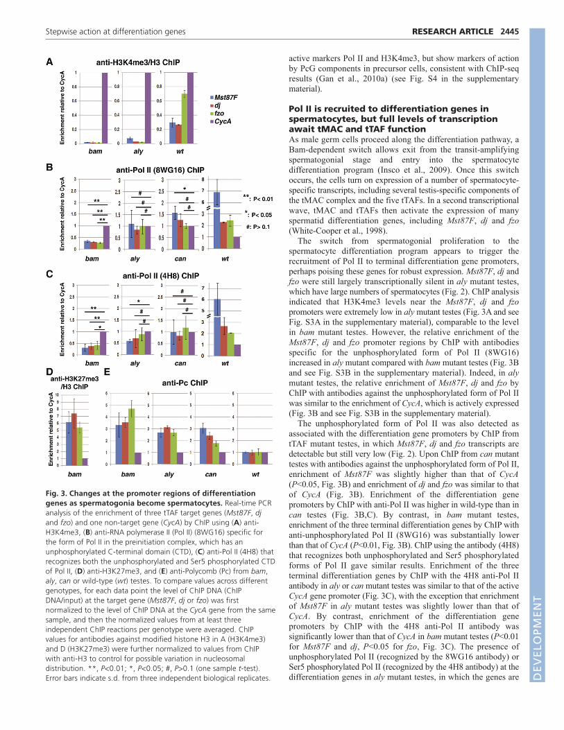
active markers Pol II and H3K4me3, but show markers of actionby PcG components in precursor cells, consistent with ChIP-seqresults (Gan et al., 2010a) (see Fig. S4 in the supplementarymaterial).
Pol II is recruited to differentiation genes inspermatocytes, but full levels of transcriptionawait tMAC and tTAF functionAs male germ cells proceed along the differentiation pathway, aBam-dependent switch allows exit from the transit-amplifyingspermatogonial stage and entry into the spermatocytedifferentiation program (Insco et al., 2009). Once this switchoccurs, the cells turn on expression of a number of spermatocyte-specific transcripts, including several testis-specific components ofthe tMAC complex and the five tTAFs. In a second transcriptionalwave, tMAC and tTAFs then activate the expression of manyspermatid differentiation genes, including Mst87F, dj and fzo(White-Cooper et al., 1998).
The switch from spermatogonial proliferation to thespermatocyte differentiation program appears to trigger therecruitment of Pol II to terminal differentiation gene promoters,perhaps poising these genes for robust expression. Mst87F, dj andfzo were still largely transcriptionally silent in aly mutant testes,which have large numbers of spermatocytes (Fig. 2). ChIP analysisindicated that H3K4me3 levels near the Mst87F, dj and fzopromoters were extremely low in aly mutant testes (Fig. 3A and seeFig. S3A in the supplementary material), comparable to the levelin bam mutant testes. However, the relative enrichment of theMst87F, dj and fzo promoter regions by ChIP with antibodiesspecific for the unphosphorylated form of Pol II (8WG16)increased in aly mutant compared with bam mutant testes (Fig. 3Band see Fig. S3B in the supplementary material). Indeed, in alymutant testes, the relative enrichment of Mst87F, dj and fzo byChIP with antibodies against the unphosphorylated form of Pol IIwas similar to the enrichment of CycA, which is actively expressed(Fig. 3B and see Fig. S3B in the supplementary material).
The unphosphorylated form of Pol II was also detected asassociated with the differentiation gene promoters by ChIP fromtTAF mutant testes, in which Mst87F, dj and fzo transcripts aredetectable but still very low (Fig. 2). Upon ChIP from can mutanttestes with antibodies against the unphosphorylated form of Pol II,enrichment of Mst87F was slightly higher than that of CycA(P<0.05, Fig. 3B) and enrichment of dj and fzo was similar to thatof CycA (Fig. 3B). Enrichment of the differentiation genepromoters by ChIP with anti-Pol II was higher in wild-type than incan testes (Fig. 3B,C). By contrast, in bam mutant testes,enrichment of the three terminal differentiation genes by ChIP withanti-unphosphorylated Pol II (8WG16) was substantially lowerthan that of CycA (P<0.01, Fig. 3B). ChIP using the antibody (4H8)that recognizes both unphosphorylated and Ser5 phosphorylatedforms of Pol II gave similar results. Enrichment of the threeterminal differentiation genes by ChIP with the 4H8 anti-Pol IIantibody in aly or can mutant testes was similar to that of the activeCycA gene promoter (Fig. 3C), with the exception that enrichmentof Mst87F in aly mutant testes was slightly lower than that ofCycA. By contrast, enrichment of the differentiation genepromoters by ChIP with the 4H8 anti-Pol II antibody wassignificantly lower than that of CycA in bam mutant testes (P<0.01for Mst87F and dj, P<0.05 for fzo, Fig. 3C). The presence ofunphosphorylated Pol II (recognized by the 8WG16 antibody) orSer5 phosphorylated Pol II (recognized by the 4H8 antibody) at thedifferentiation genes in aly mutant testes, in which the genes are
2445RESEARCH ARTICLEStepwise action at differentiation genes
Fig. 3. Changes at the promoter regions of differentiationgenes as spermatogonia become spermatocytes. Real-time PCRanalysis of the enrichment of three tTAF target genes (Mst87F, djand fzo) and one non-target gene (CycA) by ChIP using (A) anti-H3K4me3, (B) anti-RNA polymerase II (Pol II) (8WG16) specific forthe form of Pol II in the preinitiation complex, which has anunphosphorylated C-terminal domain (CTD), (C) anti-Pol II (4H8) thatrecognizes both the unphosphorylated and Ser5 phosphorylated CTDof Pol II, (D) anti-H3K27me3, and (E) anti-Polycomb (Pc) from bam,aly, can or wild-type (wt) testes. To compare values across differentgenotypes, for each data point the level of ChIP DNA (ChIPDNA/input) at the target gene (Mst87F, dj or fzo) was firstnormalized to the level of ChIP DNA at the CycA gene from the samesample, and then the normalized values from at least threeindependent ChIP reactions per genotype were averaged. ChIPvalues for antibodies against modified histone H3 in A (H3K4me3)and D (H3K27me3) were further normalized to values from ChIPwith anti-H3 to control for possible variation in nucleosomaldistribution. **, P<0.01; *, P<0.05; #, P>0.1 (one sample t-test).Error bars indicate s.d. from three independent biological replicates. D
EVELO
PMENT

2446
still largely silent, is reminiscent of the stalled Pol II observed atmany transcriptionally quiescent genes in ESCs (Guenther et al.,2007; Stock et al., 2007), as well as in Drosophila embryos (Museet al., 2007; Zeitlinger et al., 2007). Enrichment of thedifferentiation gene promoters by ChIP with anti-Pol II was stillhigher in wild-type than in can mutant testes (Fig. 3B,C).
Presence of the PRC1 complex near the promoter region ofthe terminal differentiation genes might contribute to theirtranscriptionally silent state even in the presence of Pol II in alyor tTAF mutant testes. ChIP with antibodies against the PRC1component Polycomb enriched for the promoter regions of thethree terminal differentiation genes 3- to 4-fold relative to theCycA promoter in bam testes and by 2- to 3-fold relative to CycAin aly or can mutant testes. By contrast, in wild-type testes,where these genes are fully expressed, enrichment for the threedifferentiation genes was comparable to that of CycA (Fig. 3Eand see Fig. S3E in the supplementary material). The level ofPolycomb at the differentiation genes in spermatocytes might beeven lower than that indicated by the ChIP results using entirewild-type testes (Fig. 3E and see Fig. S3E in the supplementarymaterial), as wild-type testes also contain precursor cells, inwhich the genes are highly occupied by Polycomb (see bam datain Fig. 3E and Fig. S3E in the supplementary material).However, because spermatocytes significantly outnumberprecursor cells in wild-type testes, we can still gain informationon the chromatin state of differentiation genes using wholetestes, especially for changes in which a particular chromatinmark or protein occupancy appears or turns on as cells switchfrom precursor to spermatocyte. The relative enrichment ofPolycomb at the differentiation genes in this study (~3- to 4-foldenrichment of Mst87F in can mutant compared with wild-typetestes) was comparable to that reported in a previous study(Chen et al., 2005), when the results were plotted in the sameway (see Fig. S5 in the supplementary material).
Inactivating the key PRC2 component E(z) in theabsence of tTAF is insufficient to turn on terminaldifferentiation genesThe wild-type function of tTAFs is required to reduce the occupancyof Polycomb at the differentiation gene promoters (Fig. 3E) and toturn on robust expression of the differentiation genes (Fig. 2) (Chenet al., 2005). Analysis of a double-mutant strain generated by
combining the tTAF mutant no hitter (nht, TAF4L) with an E(z)conditional allele, E(z)61, which was used previously to inactivateE(z) activity at restrictive temperature (Wang et al., 2004b), indicatedthat the action of tTAFs is required for more than just abolishingsilencing by PcG. When nht; E(z)61 double-mutant flies were grownto adulthood at permissive temperature and then shifted to 29°C (therestrictive temperature) for 7 days, little E(z) function remained, asH3K27me3 was almost undetectable in the double-mutant testesextracts, as compared with the robust levels in testes from siblingcontrol nht mutant males that were raised under exactly the sameconditions (Fig. 4A). However, no significant change in transcriptlevels for Mst87F, dj or fzo was detected by quantitative RT-PCRanalyses in nht; E(z) double-mutant testes as compared with nhtsingle-mutant sibling controls (Fig. 4B). In both nht and nht; E(z)mutant testes, transcript levels for the differentiation genes were verymuch reduced compared with those of wild-type testes shiftedthrough the same temperature regimen in parallel, suggesting thatinactivation of E(z) is insufficient to turn on differentiation geneexpression without tTAF function.
tMAC function is required for the recruitment oftTAFs to target genesA number of spermatid differentiation genes require wild-typefunction of both tMAC and the tTAFs for normal expression inspermatocytes (White-Cooper et al., 1998). Results from ChIPanalysis of testis extracts with antibodies directed against theTAF8L Sa indicated that the proper function of spermatocyte-specific components of tMAC is required for the tTAFs to properlyassociate with the promoter regions of these differentiation genes.ChIP of wild-type testes with antibodies against Sa enriched forMst87F, dj and fzo ~3- to 6-fold over the level of the control geneCycA, which is transcribed in spermatocytes independently oftTAFs (Fig. 5A). By contrast, although Sa protein was present inaly mutant spermatocytes (Fig. 5C�), ChIP of aly mutant testes withanti-Sa did not significantly enrich for any of these three targetgenes compared with CycA (Fig. 5A).
The proper localization of tTAFs and Polycombdepends on tMAC actionThe wild-type function of tMAC is also required for propersubnuclear localization of tTAFs and PRC1 proteins inspermatocytes. In wild-type spermatocytes, immunofluorescence
RESEARCH ARTICLE Development 138 (12)
Fig. 4. Inactivation of E(z) is insufficient to turn on terminal differentiation genes in a tTAF mutant background. (A)Immunoblot analysisshowing the H3K27me3 band in nht testes and the drastic reduction of H3K27me3 in nht; E(z) double-mutant testes. H3 provided a loadingcontrol. (B)Quantitative RT-PCR analysis of dj, fzo and Mst87F transcript levels in nht; E(z), nht and wild-type (wt) testes, normalized to Rpl32 fromthe same sample. The nht; E(z), nht and wild-type flies were all raised to adulthood at permissive temperature and then shifted to 29°C (restrictivetemperature) for 7 days. The relative level of each gene in the wild type was set to 1 to allow comparison. Error bars indicate s.d. from threeindependent biological replicates.
DEVELO
PMENT

analysis revealed colocalization of tTAF proteins (e.g. Sa and Can)with Polycomb and other PRC1 components to a subdomain withinthe nucleolus, interdigitated with the nucleolar marker Fibrillarin(Fib) (Fig. 5B-B�) (Chen et al., 2005). By contrast, in aly mutantspermatocytes, the tTAFs were concentrated in lobe-shaped domainsoutside of, but next to, the Fib-enriched nucleolar region (Fig. 5C-C�,D-D�). Similar results were observed for the TAF4L Nht (data notshown) and for the TAF6L Meiosis I arrest (Mia) (Metcalf andWassarman, 2007). The perinucleolar lobes containing mislocalizedtTAFs were also apparent in freshly squashed live samples viewedby phase contrast microscopy, in which they appeared as phase-dark
structures extending from the nucleolar periphery (Fig. 5F-F�,compare with the wild-type spermatocyte in Fig. 5E-E�). Asubstantial amount of the Polycomb protein in aly mutantspermatocytes was also localized to the same perinucleolar domains(Fig. 5H-H�, Fig. 4I-I�). Some Polycomb still retained associationwith condensing chromatin in aly mutant spermatocytes (Fig.5H,H�,I,I�), as in wild-type spermatocytes (Fig. 5G,G�).
The localization of tTAFs to the perinucleolar lobes in alymutant spermatocytes was not due to total disruption of thenucleolus, as phase contrast microscopy (Fig. 5F�) and staining forthe nucleolar marker Fib (Fig. 5C�,D�) indicated that the nucleolus
2447RESEARCH ARTICLEStepwise action at differentiation genes
Fig. 5. tMAC function is required for the recruitment of tTAFs to target genes and for proper subnuclear localization of tTAFs andPolycomb. (A)Real-time PCR analysis of ChIP using anti-Sa from aly and wild-type (wt) Drosophila testes. Relative enrichment by ChIP wascomputed as the ratio of ChIP DNA/input at the spermatid differentiation genes Mst87F, dj and fzo to ChIP DNA/input for the control gene CycA,which is expressed in both spermatogonia and spermatocytes independently of tMAC and tTAFs. Error bars indicate s.d. from three independentbiological replicates). (B-I�) Localization of (B-F�) tTAFs and (G-I�) Polycomb (Pc) in wild-type and aly mutant spermatocytes by immunostaining offixed cells (B-D�,G-I�) or in epifluorescent images of live cells (E-F�). Blue, DAPI; green, tTAF (Sa-GFP in B�,C�,E�,F� or anti-Can in D�) or Pc-GFP(G�,H�,I�); red, the nucleolar marker anti-Fibrillarin (Fib in B�,C�,D�,G�,H�) or pseudo-color of the phase (E,F) or anti-Sa (I�). (J-N)Localization of Saprotein in spermatocytes mutant for other tMAC components: (J) comr, (K) tomb, (L) topi, (M) achi/vis, (N) mip40. Blue, DAPI; green, tTAF; red, anti-Fib. Scale bars: 4m.
DEVELO
PMENT

2448
was present. It is possible that the wild-type function of Aly isrequired to maintain proper nucleolar architecture in spermatocytes(Metcalf and Wassarman, 2007), and that collapse of nucleolararchitecture in aly mutant spermatocytes squeezed the tTAF-containing subcompartment to the perinuclear lobes.
Similar mislocalization of tTAFs to the lobed perinucleolardomains was also observed in spermatocytes mutant for other testis-specific components of the tMAC complex, including cookiemonster (comr), tombola (tomb), matotopetli (topi) andachintya/vismay (achi/vis; achi – FlyBase) (Fig. 5J-M). However,even though Myb-interacting protein 40 (Mip40) has been identifiedbiochemically as a component of tMAC (Beall et al., 2007), tTAFswere localized within the nucleolus, interdigitated with the Fib-positive subcompartment, in mip40 mutant spermatocytes, similar toin the wild type (Fig. 5N), suggesting that Mip40 has different role(s)than the other tMAC components in spermatocytes.
DISCUSSIONSequential developmentally regulated steps leadto the activation of terminal differentiation genesOur results suggest a stepwise series of developmentallyprogrammed events as terminal differentiation genes convert from atranscriptionally silent state in precursor cells to full expression indifferentiating spermatocytes (Fig. 6). In precursor cells,differentiation genes are repressed and associated with backgroundlevels of hypophosphorylated Pol II and H3K4me3. These genes alsodisplay elevated levels of H3K27me3 and Polycomb at the promoterregion, suggesting that they are acted upon by the PcG transcriptionalsilencing machinery. Notably, the differentiation genes studied inprecursor cells here did not show the hallmark bivalent chromatindomains enriched for both the repressive H3K27me3 mark and theactive H3K4me3 mark that have been characterized for a cohort ofdifferentiation genes in mammalian ESCs (Bernstein et al., 2006).
The cell fate switch from proliferating spermatogonia to thespermatocyte differentiation program initiates both global and localchanges in the transcriptional regulatory landscape, starting a celltype-specific gene expression cascade that eventually leads torobust transcription of the terminal differentiation genes. Globally,soon after the switch from spermatogonia to spermatocytes, coresubunits of the PRC2 complex are downregulated, including E(z),the enzyme that generates the H3K27me3 mark. Locally, after malegerm cells become spermatocytes, Pol II accumulates at theterminal differentiation gene promoters, although these genes stillremain transcriptionally silent, with low H3K4me3 and highPolycomb protein levels near their promoters.
The next step awaits the expression of spermatocyte-specificforms of core transcription machinery and chromatin-associatedregulators, including homologs of subunits of both the generaltranscription factor TFIID (tTAFs) and the MIP/dREAM complex(Aly and other testis-specific components of tMAC) (Fig. 6B). ThetMAC complex acts either locally or globally, perhaps at the levelof chromatin or directly through interaction with tTAFs, to allowrecruitment of tTAFs to promoters of target terminal differentiationgenes. The action of tTAFs then allows full and robust transcriptionof the terminal differentiation genes, partly by displacing Polycombfrom their promoters.
Strikingly, the two major PcG protein complexes appear to beregulated differently by the germ cell developmental program:whereas the PRC2 components E(z) and Su(z)12 aredownregulated, the PRC1 components Polycomb, Polyhomeoticand dRing continue to be expressed in spermatocytes. The globaldownregulation of the epigenetic ‘writer’ E(z) in spermatocytes
might facilitate displacement of the epigenetic ‘reader’, the PRC1complex, from the differentiation genes, with the local action oftTAFs at promoters serving to select which genes are relieved ofPRC1. In addition, the tTAFs act at a second level to regulatePolycomb by recruiting and accompanying Polycomb and severalother PRC1 components to a particular subnucleolar domain inspermatocytes (Fig. 6C) (Chen et al., 2005). It is not yet knownwhether sequestering of PRC1 to the nucleolus by tTAFs plays arole in the activation of terminal differentiation genes, perhaps bylowering the level of PRC1 that is available to exchange back onto differentiation gene promoters. Conversely, recruitment of PRC1to the nucleolar region might have a separate function, such as inchromatin silencing in the XY body as observed in mammalianspermatocytes (Baarends et al., 2005; Takada et al., 2007).
Stalled Pol II and the developmental control ofgene expressionOur findings indicate that, upon the switch from spermatogonia tospermatocytes, the terminal differentiation genes go through a poisedstate, marked by presence of both active Pol II and repressivePolycomb, before the genes are actively transcribed. Stalled Pol II
RESEARCH ARTICLE Development 138 (12)
Fig. 6. Model for the developmentally programmed steps thatoppose PcG repression and turn on terminal differentiation geneexpression. (A)The early stages of spermatogenesis in Drosophila.Precursors include stem cells (S) and mitotically dividing spermatogonia.Spermatocytes include early spermatocytes (light gray nucleus) prior tothe expression of the terminal differentiation genes (red nucleus). ThePRC2 components E(z) and Su(z)12 are highly expressed in precursorcells, including germline stem cells (S) and spermatogonial cells.(B)Potential transcription waves in developing spermatocytes. Upon theBam-dependent switch from spermatogonia to spermatocytes,unknown factor(s) (question marks) turn on testis-specific tMAC andtTAF components in early spermatocytes. The action of tMAC and tTAFsis subsequently required for robust transcription of spermatiddifferentiation genes. (C)Potential chromatin states in precursor cells(left; analyzed with bam mutant testes), spermatocytes lacking tMACfunction (middle; analyzed with aly mutant testes) and maturespermatocytes (right; analyzed with wild-type testis). K4, H3K4me3;K27, H3K27me3.
DEVELO
PMENT

and abortive transcript initiation are emerging as a common featurein stem/progenitor cells. This mechanism may prime genes to rapidlyrespond to developmental cues or environmental stimuli (Muse et al.,2007; Zeitlinger et al., 2007). Stalled Pol II could representtranscription events that have initiated elongation but then pause andawait further signals, as in the regulation of gene expression by theandrogen receptor (Zhao et al., 2008) or by heat shock (Lis, 1998;Rasmussen and Lis, 1995; Rougvie and Lis, 1988). Alternatively, PolII might be trapped at a nascent preinitiation complex, withoutmelting open the DNA, as found in some instances of transcriptionalrepression by Polycomb (Dellino et al., 2004). Although our ChIPanalyses did not have the resolution to distinguish whether Pol II wasstalled at the promoter or had already initiated a short transcript, theresults with antibodies specific for unphosphorylated Pol II suggestthat Pol II is trapped in a nascent preinitiation complex. The PRC1component dRing has been shown to monoubiquitylate histone H2Aon Lys119 near or just downstream of the transcription start site(Wang et al., 2004a). We propose that in early spermatocytes, beforeexpression of the tTAFs and tMAC, the local action of PRC1 incausing H2AK119ub at the terminal differentiation gene promotersmight block efficient clearing of Pol II from the preinitiationcomplex and prevent transcription elongation.
Gene-selective transcriptional regulation by celltype-specific forms of the core transcriptionmachineryRemoval of PRC1 from the promoter and full expression of theterminal differentiation genes in spermatocytes require theexpression and action of tMAC and tTAFs. Cell type-specifichomologs of TFIID subunits have been shown to act gene-selectively to control developmentally programmed geneexpression. For example, incorporation of one subunit of themammalian TAF4b variant into TFIID strongly influencestranscriptional activation at selected promoters, directing agenerally expressed transcriptional activator to turn on tissue-specific gene expression (Liu et al., 2008).
The local action of the tTAFs to relieve repression by Polycombat target gene promoters provides a mechanism that is both cell typespecific and gene selective, allowing expression of some Polycomb-repressed genes while keeping others silent. Similar developmentallyprogrammed mechanisms may also reverse PcG-mediated epigeneticsilencing in other stem cell systems. Indeed, striking parallelsbetween our findings and recent results from mammalian epidermis(Ezhkova et al., 2009; Sen et al., 2008) suggest that molecularstrategies are conserved from flies to mammals. In mouse epidermis,the mammalian E(z) homolog Ezh2 is expressed in stem/precursorcells at the basal layer of the skin. Strikingly, as we observed for E(z)and Su(z)12 in the Drosophila male GSC lineage, the Ezh2 leveldeclines sharply as cells cease DNA replication and the epidermaldifferentiation program is turned on. Overexpression of Ezh2 inepidermal precursor cells delays the onset of terminal differentiationgene expression (Ezhkova et al., 2009), and removal of the Ezh2-generated H3K27me3 mark by the Jmjd3 (Kdm6b) demethylase isrequired for epidermal differentiation (Sen et al., 2008).
In particular, our results suggest a possible explanation for theconundrum that, although PcG components are bound at manytranscriptionally silent differentiation genes in mammalian ESCs,loss of function of PcG components does not cause loss ofpluripotency but instead causes defects during early embryonicdifferentiation (Boyer et al., 2006; Chamberlain et al., 2008;Surface et al., 2010). In Drosophila male germ cells, events duringthe switch from precursor cell proliferation to differentiation are
required to recruit Pol II to the promoters of differentiation genes.Without this differentiation-dependent recruitment of Pol II, loss ofPolycomb is not sufficient to precociously turn on terminaldifferentiation genes in precursor cells. Rather, Polycomb that ispre-bound at the differentiation gene promoters might serve todelay the onset of their transcription after the mitosis-to-differentiation switch. Robust transcription must await theexpression of cell type- and stage-specific components of thetranscription machinery. These might in turn guide gene-selectivereversal of Polycomb repression to facilitate appropriatedifferentiation gene expression in specific cell types.
AcknowledgementsWe thank P. Dimario and M. Pollard for anti-Fibrillarin; J. Muller for E(z)731
FRT2A flies and anti-Su(z)12 antibody; R. Jones for anti-E(z) antibody; R. Parofor Pc-GFP flies and anti-Pc; R. Kingston and A. Saurin for anti-Pc; H. WhiteCooper for tMAC mutant strains and Q. Gan for analyzing and depositingmicroarray data. C.L. was supported in part by a Stanford Dean’s Fellowshipand J.R.M.P. by an NSF Predoctoral Fellowship. This work was supported byNIH K99/R00 HD055052 Pathway to Independence Award and Research GrantNo. 05-FY09-88 from the March of Dimes Foundation to X.C. and by NIH 3R01 GM061986 to M.T.F. Deposited in PMC for release after 12 months.
Competing interests statementThe authors declare no competing financial interests.
Supplementary materialSupplementary material for this article is available athttp://dev.biologists.org/lookup/suppl/doi:10.1242/dev.056572/-/DC1
ReferencesAkala, O. O., Park, I. K., Qian, D., Pihalja, M., Becker, M. W. and Clarke, M. F.
(2008). Long-term haematopoietic reconstitution by Trp53–/– p16Ink4a–/–p19Arf–/– multipotent progenitors. Nature 453, 228-232.
Ayyar, S., Jiang, J., Collu, A., White-Cooper, H. and White, R. A. (2003).Drosophila TGIF is essential for developmentally regulated transcription inspermatogenesis. Development 130, 2841-2852.
Baarends, W. M., Wassenaar, E., van der Laan, R., Hoogerbrugge, J.,Sleddens-Linkels, E., Hoeijmakers, J. H., de Boer, P. and Grootegoed, J. A.(2005). Silencing of unpaired chromatin and histone H2A ubiquitination inmammalian meiosis. Mol. Cell. Biol. 25, 1041-1053.
Barski, A., Cuddapah, S., Cui, K., Roh, T. Y., Schones, D. E., Wang, Z., Wei, G.,Chepelev, I. and Zhao, K. (2007). High-resolution profiling of histonemethylations in the human genome. Cell 129, 823-837.
Beall, E. L., Lewis, P. W., Bell, M., Rocha, M., Jones, D. L. and Botchan, M. R.(2007). Discovery of tMAC: a Drosophila testis-specific meiotic arrest complexparalogous to Myb-Muv B. Genes Dev. 21, 904-919.
Bernstein, B. E., Mikkelsen, T. S., Xie, X., Kamal, M., Huebert, D. J., Cuff, J.,Fry, B., Meissner, A., Wernig, M., Plath, K. et al. (2006). A bivalent chromatinstructure marks key developmental genes in embryonic stem cells. Cell 125,315-326.
Boyer, L. A., Plath, K., Zeitlinger, J., Brambrink, T., Medeiros, L. A., Lee, T. I.,Levine, S. S., Wernig, M., Tajonar, A., Ray, M. K. et al. (2006). Polycombcomplexes repress developmental regulators in murine embryonic stem cells.Nature 441, 349-353.
Cao, R., Wang, L., Wang, H., Xia, L., Erdjument-Bromage, H., Tempst, P.,Jones, R. S. and Zhang, Y. (2002). Role of histone H3 lysine 27 methylation inPolycomb-group silencing. Science 298, 1039-1043.
Chamberlain, S. J., Yee, D. and Magnuson, T. (2008). Polycomb repressivecomplex 2 is dispensable for maintenance of embryonic stem cell pluripotency.Stem Cells 26, 1496-1505.
Chen, X., Hiller, M., Sancak, Y. and Fuller, M. T. (2005). Tissue-specific TAFscounteract Polycomb to turn on terminal differentiation. Science 310, 869-872.
Czermin, B., Melfi, R., McCabe, D., Seitz, V., Imhof, A. and Pirrotta, V. (2002).Drosophila enhancer of Zeste/ESC complexes have a histone H3methyltransferase activity that marks chromosomal Polycomb sites. Cell 111,185-196.
Dellino, G. I., Schwartz, Y. B., Farkas, G., McCabe, D., Elgin, S. C. andPirrotta, V. (2004). Polycomb silencing blocks transcription initiation. Mol. Cell13, 887-893.
Dou, Y., Milne, T. A., Ruthenburg, A. J., Lee, S., Lee, J. W., Verdine, G. L.,Allis, C. D. and Roeder, R. G. (2006). Regulation of MLL1 H3K4methyltransferase activity by its core components. Nat. Struct. Mol. Biol. 13,713-719.
2449RESEARCH ARTICLEStepwise action at differentiation genes
DEVELO
PMENT

2450
Ezhkova, E., Pasolli, H. A., Parker, J. S., Stokes, N., Su, I. H., Hannon, G.,Tarakhovsky, A. and Fuchs, E. (2009). Ezh2 orchestrates gene expression forthe stepwise differentiation of tissue-specific stem cells. Cell 136, 1122-1135.
Fischle, W., Wang, Y., Jacobs, S. A., Kim, Y., Allis, C. D. and Khorasanizadeh,S. (2003). Molecular basis for the discrimination of repressive methyl-lysinemarks in histone H3 by Polycomb and HP1 chromodomains. Genes Dev. 17,1870-1881.
Francis, N. J., Kingston, R. E. and Woodcock, C. L. (2004). Chromatincompaction by a polycomb group protein complex. Science 306, 1574-1577.
Gan, Q., Schones, D. E., Ho Eun, S., Wei, G., Cui, K., Zhao, K. and Chen, X.(2010a). Monovalent and unpoised status of most genes in undifferentiated cell-enriched Drosophila testis. Genome Biol. 11, R42.
Gan, Q., Chepelev, I., Wei, G., Tarayrah, L., Cui, K., Zhao, K. and Chen, X.(2010b). Dynamic regulation of alternative splicing and chromatin structure inDrosophila gonads revealed by RNA-seq. Cell Res. 20, 763-783.
Gonczy, P., Matunis, E. and DiNardo, S. (1997). bag-of-marbles and benigngonial cell neoplasm act in the germline to restrict proliferation duringDrosophila spermatogenesis. Development 124, 4361-4371.
Guenther, M. G., Levine, S. S., Boyer, L. A., Jaenisch, R. and Young, R. A.(2007). A chromatin landmark and transcription initiation at most promoters inhuman cells. Cell 130, 77-88.
Hiller, M. A., Lin, T. Y., Wood, C. and Fuller, M. T. (2001). Developmentalregulation of transcription by a tissue-specific TAF homolog. Genes Dev. 15,1021-1030.
Hiller, M., Chen, X., Pringle, M. J., Suchorolski, M., Sancak, Y., Viswanathan,S., Bolival, B., Lin, T. Y., Marino, S. and Fuller, M. T. (2004). Testis-specific TAFhomologs collaborate to control a tissue-specific transcription program.Development 131, 5297-5308.
Hime, G. R., Brill, J. A. and Fuller, M. T. (1996). Assembly of ring canals in themale germ line from structural components of the contractile ring. J. Cell Sci.109, 2779-2788.
Insco, M. L., Leon, A., Tam, C. H., McKearin, D. M. and Fuller, M. T. (2009).Accumulation of a differentiation regulator specifies transit amplifying divisionnumber in an adult stem cell lineage. Proc. Natl. Acad. Sci. USA 106, 22311-22316.
Jiang, J. and White-Cooper, H. (2003). Transcriptional activation in Drosophilaspermatogenesis involves the mutually dependent function of aly and a novelmeiotic arrest gene cookie monster. Development 130, 563-573.
Jiang, J., Benson, E., Bausek, N., Doggett, K. and White-Cooper, H. (2007).Tombola, a tesmin/TSO1-family protein, regulates transcriptional activation in theDrosophila male germline and physically interacts with always early.Development 134, 1549-1559.
Kuzmichev, A., Nishioka, K., Erdjument-Bromage, H., Tempst, P. andReinberg, D. (2002). Histone methyltransferase activity associated with ahuman multiprotein complex containing the enhancer of Zeste protein. GenesDev. 16, 2893-2905.
Lee, T. I., Jenner, R. G., Boyer, L. A., Guenther, M. G., Levine, S. S., Kumar, R.M., Chevalier, B., Johnstone, S. E., Cole, M. F., Isono, K. et al. (2006).Control of developmental regulators by Polycomb in human embryonic stemcells. Cell 125, 301-313.
Lessard, J. and Sauvageau, G. (2003). Bmi-1 determines the proliferativecapacity of normal and leukaemic stem cells. Nature 423, 255-260.
Lin, T. Y., Viswanathan, S., Wood, C., Wilson, P. G., Wolf, N. and Fuller, M. T.(1996). Coordinate developmental control of the meiotic cell cycle andspermatid differentiation in Drosophila males. Development 122, 1331-1341.
Lis, J. (1998). Promoter-associated pausing in promoter architecture andpostinitiation transcriptional regulation. Cold Spring Harb. Symp. Quant. Biol.63, 347-356.
Liu, W. L., Coleman, R. A., Grob, P., King, D. S., Florens, L., Washburn, M. P.,Geles, K. G., Yang, J. L., Ramey, V., Nogales, E. et al. (2008). Structuralchanges in TAF4b-TFIID correlate with promoter selectivity. Mol. Cell 29, 81-91.
McKearin, D. and Ohlstein, B. (1995). A role for the Drosophila bag-of-marblesprotein in the differentiation of cystoblasts from germline stem cells.Development 121, 2937-2947.
McKearin, D. M. and Spradling, A. C. (1990). bag-of-marbles: a Drosophila generequired to initiate both male and female gametogenesis. Genes Dev. 4, 2242-2251.
Metcalf, C. E. and Wassarman, D. A. (2007). Nucleolar colocalization of TAF1and testis-specific TAFs during Drosophila spermatogenesis. Dev. Dyn. 236,2836-2843.
Milne, T. A., Briggs, S. D., Brock, H. W., Martin, M. E., Gibbs, D., Allis, C. D.and Hess, J. L. (2002). MLL targets SET domain methyltransferase activity toHox gene promoters. Mol. Cell 10, 1107-1117.
Min, J., Zhang, Y. and Xu, R. M. (2003). Structural basis for specific binding ofPolycomb chromodomain to histone H3 methylated at Lys 27. Genes Dev. 17,1823-1828.
Molofsky, A. V., Pardal, R., Iwashita, T., Park, I. K., Clarke, M. F. andMorrison, S. J. (2003). Bmi-1 dependence distinguishes neural stem cell self-renewal from progenitor proliferation. Nature 425, 962-967.
Muller, J., Hart, C. M., Francis, N. J., Vargas, M. L., Sengupta, A., Wild, B.,Miller, E. L., O’Connor, M. B., Kingston, R. E. and Simon, J. A. (2002).Histone methyltransferase activity of a Drosophila Polycomb group repressorcomplex. Cell 111, 197-208.
Muse, G. W., Gilchrist, D. A., Nechaev, S., Shah, R., Parker, J. S., Grissom, S.F., Zeitlinger, J. and Adelman, K. (2007). RNA polymerase is poised foractivation across the genome. Nat. Genet. 39, 1507-1511.
Park, I. K., Qian, D., Kiel, M., Becker, M. W., Pihalja, M., Weissman, I. L.,Morrison, S. J. and Clarke, M. F. (2003). Bmi-1 is required for maintenance ofadult self-renewing haematopoietic stem cells. Nature 423, 302-305.
Perezgasga, L., Jiang, J., Bolival, B., Jr, Hiller, M., Benson, E., Fuller, M. T. andWhite-Cooper, H. (2004). Regulation of transcription of meiotic cell cycle andterminal differentiation genes by the testis-specific Zn-finger protein matotopetli.Development 131, 1691-1702.
Phatnani, H. P. and Greenleaf, A. L. (2006). Phosphorylation and functions ofthe RNA polymerase II CTD. Genes Dev. 20, 2922-2936.
Rasmussen, E. B. and Lis, J. T. (1995). Short transcripts of the ternary complexprovide insight into RNA polymerase II elongational pausing. J. Mol. Biol. 252,522-535.
Rougvie, A. E. and Lis, J. T. (1988). The RNA polymerase II molecule at the 5� endof the uninduced hsp70 gene of D. melanogaster is transcriptionally engaged.Cell 54, 795-804.
Schwartz, Y. B. and Pirrotta, V. (2007). Polycomb silencing mechanisms and themanagement of genomic programmes. Nat. Rev. Genet. 8, 9-22.
Sen, G. L., Webster, D. E., Barragan, D. I., Chang, H. Y. and Khavari, P. A.(2008). Control of differentiation in a self-renewing mammalian tissue by thehistone demethylase JMJD3. Genes Dev. 22, 1865-1870.
Stock, J. K., Giadrossi, S., Casanova, M., Brookes, E., Vidal, M., Koseki, H.,Brockdorff, N., Fisher, A. G. and Pombo, A. (2007). Ring1-mediatedubiquitination of H2A restrains poised RNA polymerase II at bivalent genes inmouse ES cells. Nat. Cell Biol. 9, 1428-1435.
Surface, L. E., Thornton, S. R. and Boyer, L. A. (2010). Polycomb group proteinsset the stage for early lineage commitment. Cell Stem Cell 7, 288-298.
Takada, Y., Isono, K., Shinga, J., Turner, J. M., Kitamura, H., Ohara, O.,Watanabe, G., Singh, P. B., Kamijo, T., Jenuwein, T. et al. (2007).Mammalian Polycomb Scmh1 mediates exclusion of Polycomb complexes fromthe XY body in the pachytene spermatocytes. Development 134, 579-590.
Wang, H., Wang, L., Erdjument-Bromage, H., Vidal, M., Tempst, P., Jones, R.S. and Zhang, Y. (2004a). Role of histone H2A ubiquitination in Polycombsilencing. Nature 431, 873-878.
Wang, L., Brown, J. L., Cao, R., Zhang, Y., Kassis, J. A. and Jones, R. S.(2004b). Hierarchical recruitment of polycomb group silencing complexes. Mol.Cell 14, 637-646.
White-Cooper, H., Schafer, M. A., Alphey, L. S. and Fuller, M. T. (1998).Transcriptional and post-transcriptional control mechanisms coordinate the onsetof spermatid differentiation with meiosis I in Drosophila. Development 125, 125-134.
White-Cooper, H., Leroy, D., MacQueen, A. and Fuller, M. T. (2000).Transcription of meiotic cell cycle and terminal differentiation genes depends ona conserved chromatin associated protein, whose nuclear localisation isregulated. Development 127, 5463-5473.
Zeitlinger, J., Stark, A., Kellis, M., Hong, J. W., Nechaev, S., Adelman, K.,Levine, M. and Young, R. A. (2007). RNA polymerase stalling atdevelopmental control genes in the Drosophila melanogaster embryo. Nat.Genet. 39, 1512-1516.
Zhao, Y., Lang, G., Ito, S., Bonnet, J., Metzger, E., Sawatsubashi, S., Suzuki,E., Le Guezennec, X., Stunnenberg, H. G., Krasnov, A. et al. (2008). ATFTC/STAGA module mediates histone H2A and H2B deubiquitination,coactivates nuclear receptors, and counteracts heterochromatin silencing. Mol.Cell 29, 92-101.
RESEARCH ARTICLE Development 138 (12)
DEVELO
PMENT