Embed Size (px)
Citation preview
Gzrbohydrate Research, 26 (1973) 274-277 0 Ekevier Scientific Publishing Company, Amsterdam - Printed in Belgium
Prelimiuary communication
Separation and characterization of the &D-glUCaD hydrolases from a species of Cytophaga
J. J. MARSHALL*
Department of ChemisCry. Royal Holloway College (University of London), Englefield Green, Surrey TWZO OEX (Grear Britain)
(Received September 7th, 1972; accepted for publication in revised form, December 1 lth, 1972)
Bacteria of the genus Cytophaga degrade cellulose readily’, and their culture filtrates have been ah~vvrr~-~ to contain a mixture of enzymes capable of bringing about the in vitro hydrolysis of cellulose and the closely related polysaccharldes lamhnarin and lichenin. However, the constitution of the enzyme complex has not been rigorously characterized nor the constituent enzymes purified. We now report on the constitution of this enzyme system and the significance of earlier work with the same crude preparation of Cytophaga enzyme.
The starting material was a commercially available, lytic enzyme preparation’ from Cytophaga NCIB 9497 (available from British Drug Houses Ltd., Poole, Dorset; catalogue number 39072). Prehminary studies of the crude preparation showed the presence of activities towards laminarin, lichenin, and soluble cellulose derivatives such as carboxymethylcellulose (CM-cellulose). Recent techniques8-10 developed for the purification of /3-D-ghrcan hydrolases, using DEAE-celhrlose, were used to fractionate the
enzyme mixture into three protein fractions (FI-FIR) with P-D-ghrcanase activity
(Fig. 1). FI, which was not adsorbed on to the ion-exchanger, acted only on laminarin to give (chromatography) higher oligosaccharides, the principal component having a d.p. of about seven or eight. FII, eluted with a high concentration of sodium chlorideg, degraded CM-cellulose and cellodextrins to give chromatographically mobile oligosaccharides, and lichenin to give a trisaccharide ~40-P-laminaribiosyl_D-glucose) and a tetrasaccharide,
as well as higher oligosaccharides. FIII, eluted by an acid wash as described earlier”, acted on laminarin to give (paper chromatography) mainly glucose and laminaribiose, and on lichenin to give mainly glucose, laminaribiose, and a trisaccharide (30-p-
cellobiosyl-D-glucose)_ Chromatography of FI-FIII on Biogel P-60 did not result in further separation
of enzymic activities, but did effect additional purification, particularly with FI where
*Present address (to which reprint requests should be sent): Department of Biochemistry, University of Miami School of Medicine, P-0. Box 875, Biscayne Annex. Miami, Fla. 33152, U.S.A.

PRELIMINARY COMMUNICATION 275
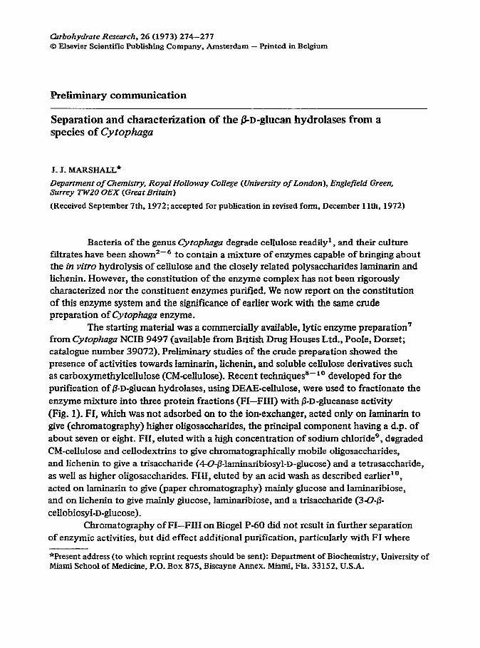
I pH Gradient
Elution volume (ml)
Fig. 1. Chromatographic separation of Cyrophog~ &D-glucan hydrolases on DEAF-cellulose. Enzyme mixture (100 mg) was dissolved in 25mM citrate-phosphate buffer (pH 8.0) and applied to a column (2.8 x 9.0 cm) of DE-52 equilibrated with the same buffer. Protein was eluted with a gradient of salt [0 -, l.OM sodium chloride in 25mM citrate-phosphate buffer @H 8.0) over 700 ml], followed by a gradient of pH (8.0 + 3.0 in M sodium chloride over 700 ml). Fractions (10 ml) were collected automatically and assayed for activity towards laminarin (o), lichenin (o), and CM-cellulose (0) by measurement of free reducing-groups produced by the action of the enzyme’g~3”.
it served to remove the major contaminant, isoamylase” * 12. After these procedures, the three enzymes appeared homogeneous by polyacrylamide gel-electrophoresis. Chromatography on Biogel is useful l3 for purification of glycoside hydrolases of low
molecular-weight and estimation of their molecular weights. In the present work, the ratios of elution volume (Ve) to the void volume of the column (V,) gave values of V,/V, for FI and FII of -1.6 and for FIII of 2.2, suggesting molecular weights for FI and FIX of -18,000 and for FIII of -8000. For the last enzyme, the unusually low molecular-weight is also indicated by the results l4 f olyacrylamide gel-electrophoresis o p in sodium dodecyl sulphate”.
Enzymes of the three types present in this Qtophaga preparation have been
reported previously, being produced by both plants and micro-organisms_ Thus,
(3-( 1 * 3)-D-ghrcan hydrolases (E-C. 3.2.1.6) of a type similar to FI, insofar as they are without action on mixed-linkage substrates, are present in extracts of barley malt13,
ryer6, and in the culture filtrate of a variety of yeast”. EndoQ-( 1 --t 4)-D-ghrcanase
(E.C. 3.2.1.4) (the so-called Cx component of the cellulase complexr8) is pro>uced by
many micro-organisms. The latter enzyme commonly also degrades lichenin’g, as we
have found here for FII. Non-specific /I-(1 * 3)-D-ghicanases (EC. 3.2-l-6*) of the FIII
*Although the specific and non-specific p-(1 -, 3)-D-&man hydrolases (FI and FIII, respectively) have the same systematic nomenclature and E.C. number, these are different enzymes. This anomaly should be resolved during the current revision of enzyme nomenclature.

276 PRELIMINARY COMMUNICATION
type are the most-common type of p(l + 3)-D-ghcanase and are produced by many micro-organisms20~ 21. Their action on mixed-linkage D-glucans has been shown to give
3-0$3-cellobiosyl-D-glucose1g as the major product. We have found no evidence for the
presence of a specific ‘lichenase’22 in the enzyme complex. Although many micro-organisms undoubtedly produce p-D-ghicanases in the
form of multicomponent enzyme complexes, the present work constitutes one of the few detailed characterizations of the nature of the components of such a complex, and is almost certainly the first occasion on which a number of closely related glucan hydrolases have been obtained in pure form from the same source.
The crude starting material used in the present study is a patented, commercially available, enzyme preparation which has been supplied in generous amounts by the patent holders to laboratories in several countries. Initially, laminarinase was believed to be the only carbohydrase present, and the crude material was describedss 6 as ‘a bacterial laminarinase preparation’. The enzyme preparation has come to be considered as a functionally pure p-(1 * 3)D-ghicanase suitable for use in structural studies of &D&c& 6, 23-25, and for the degradation of yeast and fungal cell-walls26S27. It has also been used in studies of the mode of action of ~-D-ghtcanases28. At an earlier date, the presence of isoamylase was demonstrated in the enzyme complex’ ly 12, and we have now shown the presence of three fi-D-@UcamtSeS differing in SpeCifiCity, rather than a singIe ‘laminarinase’ as hitherto believed. This means that structural studies, inferences
about the ‘laminarinasey specificity, and mechanistic studies carried out with the crude enzyme preparation are of little significance, and emphasizes the importance of using purified, characterized enzyme preparations for polysaccharide structural analysis. It remains to be determined which of the glucanases described is involved in lysis of living yeast cells2 ‘.
ACKNOWLEDGEMENTS
This work was supported by a grant from the Science Research Council. I thank Professor E. J. Boume for making available Departmental facilities, and acknowledge the award of a Fellowship from Ranks, Hovis, MacDougaI!, Ltd.
REFERENCES
1 R. Y. Stanier, Bacterial. Revs., 6 (1942) 143. 2 G. Fahraeus, Experienti, 2 (1946) 413. 3 G. Fahraeus, Syrnbolae Botan_ Upsalienses, 9 (1947) 1. 4 J. S. D. Bacon, A. H. Gordon, D. Jones, I. F. Taylor, and D. M. Webley, Biochem J, 120 (1970)
67. 5 D.J. Manners and J.C. Patterson, Biochem J., 98 (1966) 19C. 6 hf. Fleming, D. J. Manners, and A. J. Masson, Biochem J., 104 (1967) 32P. 7 British Patent 1,048,887; Chem Abstr., 65 (1966) 2972a. 8 J. J. Marsh5ll,Biochem Sot. Truns., 1 (1973) 143. 9 J. J. Marshall, J. Chromutogr.. 71 (1972) 367.

PRELIMINARY COMMUNICATION 277
10 J. J. Marshall, AnaL Bfochem, in press. 11 2. Gunja-Smith, J. J. Marshall, E. E. Smith, and W. J. Whelan, FEBS Lerrers, 12 (1970) 96. 12 J. J. Marshall, Z. Gunja-Smith, and E. E. Smith, Federation proc, 30 (1971) 1064. 13 D. J. Manners and J. J. Marshall, J. mst. Brewing, 75 (1969) 550. 14 B. A. Buckland and J. J. Marshall, unpublished results. 15 K. Weber and M. Osbom, J. Bid CTzem., 244 (1969) 4406. 16 13. J. Manners and J. J. Marshall, Phytochemisny. in press. 17 A. T. H. Abd-El-Al and H. J. Phaff, Czn J. MicrobioL, 15 (1969) 697. 18 K. W. King and M. I. Vessal,Advan. Chem. Ser., 95 (1969) 7. 19 A. S. Perlin and S. Suzuki, Cnn. J. Chem, 40 (1962) 50. 20 E. T. Reese and hi. Mandels, Chn. J. MicrobioL, 5 (1959) 173. 21 C. G. C. Chesters and A. T. Bull, Biochem. J, 86 (1963) 28. 22 E. T. Reese and A. S. Perlin, Biochem Biophys. Res. Commun., 12 (1963) 194. 23 E..Percival, personal communication. 24 A.J,Buchala and K. C. B. Wilkie, Phytocfiemistiy, 10 (1971) 2287. 25 K. C. B. Wiliie, personal communication. 26 D. E. Eveleigh, J.H. Sietsma, and R. H. Haskins, J. Gen Microbid, 52 (1968) 89. 27 J. K. Baird and W. L. Cunningham, Biochem J., 125 (1971) 32P. l
28 D. E. Eveleigh and A. S. Perlin, Cwbohyd. Rex, 10 (1969) 87. 29 N. Nelson, J. BioL Chem., 153 (1944) 375. 30 M. Somogyi, J. BioL Chem, 195 (1952) 19.