Embed Size (px)
Citation preview

Separating Fact From Myth In Chiropractic Medicine
Continuing Medical Education
Dr. Michael B. Herb
June 8, 2013

Non-Disclosures None.

Questions Welcome!
If I’m talking too fast or you’d like more on a topic, please, just ask!

My Background

Chiropractic Medicine: Education & Regulation

Education Path for Chiropractic Physicians
Undergraduate Education
Identical prerequisite track to allopathic and osteopathic counterparts
Doctoral Degree as Chiropractic Physician
4-Year Professional Program
Full-time class work and clinical rotations
General Subject Hour Comparison between DC and MD (see next slide)

General Subject Hour Comparison DC vs. MD

NBCE v. USMLE
The NBCE is broken into 4 parts, given at the end of each academic year.
OSCE’s occur at the transitions from Y3 to Y4, and Y4 to Graduation
The focus of Year 4 is clinical rotations
Campus Clinics, Private Practices, Community Outreach Clinics, etc.
There is opportunity in some areas for further rotations in hospital settings and through the VA system.
Purpose
Graduate competent doctors capable of operating as Portal of Entry Physicians (when needed), with an emphasis and skill set focused on the practice and treatment within conservative musculoskeletal medicine.

Nuts & Bolts:
Soft Tissue Healing Phases of Sprains and Strains Stage 1 – Inflammatory Stage
Stage 2 – Proliferative Stage
Stage 3 – Remodeling Stage

Stage 1: Inflammatory Stage
Injury to vessels within tissue sheath triggers hematoma
Resultant clot triggers release of chemotactic factors
i.e., vasodilators and pro-inflammatory molecule stimulation
Angiogenic factors initiate the establishment of continuity within the tissue
Average time frame – 48 to 72 hours

Stage 2: Proliferative Stage
Proliferation of fibroblasts at wound site stimulating collagen and proteoglycan synthesis
Cellular components arranged randomly and composed of predominantly Type III collagen.
EMS shown to increase fibroblast activity resulting in increased structural integrity.
Average time frame approximately 6-8 weeks
The transition from Stage 2 to 3 is typically where problems arise leading to prolonged mechanical pain and the resultant diagnosis of “late effects of sprain/strain.”

Stage 3 - Remodeling
Decrease in ECM synthesis and reduction in Type III collagen
Increase in Type I collagen synthesis
Type I fibers are organized longitudinally along the axis
Responsible for mechanical strength of the regenerated tissue.
Collagen fiber interaction results in increased tensile strength.
New patients are commonly seen initiating care due to being stuck in this phase after hoping their injuries would resolve on their own.
A study performed by Gareth Jones, at the University of Aberdeen School of Medicine and Dentistry, showed an 84% increase in chronic wide spread pain complaints following an MVA.

“The restoration of normal tendon function after injury requires reestablishment of tendon fibers and the gliding mechanism between tendon and its surrounding structures. The initial stage of repair involves formation of scar tissue that provides continuity at the injury site; however, lack of mechanical stimulus on the tendon will cause proliferation of scar tissue and subsequent adhesions that are undesirable and harmful because they impede normal tendon function…. Although stability to the injury site is necessary, mobility is critical, and mechanical loading that is associated with motion of the healing tendon decreases the formation of adhesions and increases the strength.”
- JHS January 2008
- By Gary Balian, PhD, A. Chabra, MD, et al

Nuts & Bolts:
Case Study 1 (“Maggy”) - History
- Subjective
- Objective

Case Study 1 (“Maggy”):
History
22 year old female college student who had been in a Motor Vehicle Accident (MVA) one month prior.
Immediately post accident, she went to Urgent Care.
X-Rays were taken to R/O fracture, Rx for NSAIDS and cyclobenzaprine, as well as a recommendation to wear a neck brace for a few days.
Patient didn’t use the neck brace (good call).

Case Study 1 (“Maggy”):
Subjective
Constant cervicalgia rated 4/10 UAS.
Severely decreased AROM and pain associated with movement.
Patient denied NTW into extremities.
Made worse with activity or sitting without ability to rest head.
Muscle relaxers helped her sleep.

Case Study 1 (“Maggy”):
Objective AROM: Flexion 20/50 Ext 45/60
LLF 20/45 RLF 10/45 All were painful.
LR 15/80 RR 25/80
Palpation: MFTPs and HT found within left SCM, splenius, scalenes, Cx erectors, suboccipitals, Cx paraspinals.
Orthopedic Exam:
Neutral Comp (+) local Max Comp (+) bilateral local
Neutral Distr (+) local O’Donohues (+) bilateral
Bilateral SH Dep (-) Julls – (+) immediate chin jutting & fasciculations
Neuro Exam: DTR, NTT, Cranial all WNL
Dx: Late effects sprain/strain, whiplash, myospasm.
Treatment Plan: SMT via contract/relax technique, EMS, HEP, topical analgesic.
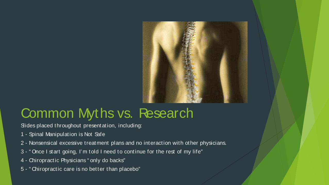
Common Myths vs. Research Slides placed throughout presentation, including:
1 - Spinal Manipulation is Not Safe
2 - Nonsensical excessive treatment plans and no interaction with other physicians.
3 - “Once I start going, I’m told I need to continue for the rest of my life”
4 - Chiropractic Physicians “only do backs”
5 - “Chiropractic care is no better than placebo”

Myth #1:
Cervical Spinal Manipulation is Not Safe
TRUTH
Concern: Upper Cx SMT leads to a high risk potential for vertebrobasilar artery (VBA) stroke.
Research: best evidence yields an incidence rate for VBA complications at approximately 1 case per 5.85 million Cx manipulations.
American Journal of Gastroenterology released a case study showing approximately 1/3 of all hospitalizations and deaths related to GI bleeding were due to the use of aspirin or NSAIDS.
A meta analysis studying 9 years worth of data was published in the February 2008 edition of Spine, indicating a no greater likelihood of developing a VBA event after Cx SMT than for visiting one’s family doctor for cervicalgia.
Cassidy D. et al. Risk of vertebrobasilar stroke and chiropractic care. Spine 2008; 33(45); 5176-5183.

Nuts & Bolts:
Case Study 2 (“Tom”) - History & Subjective
- Objective
- Treatment & Results

Case Study 2 (“Tom”):
History & Subjective
History
59 year old male accountant referred over from neurosurgeon for evaluation for conservative treatment options for right-sided C6 radiculopathy secondary to foraminal osteophytes. Foraminal ESI performed which did not produce symptom relief.
Subjective
Insidious onset of symptoms two months prior.
Cervicalgia described as a constant tightness and dull ache rated 4/10 VAS.
Frequent right arm aching and numbness rated 5/10 VAS.
Symptoms made worse by bilateral Cx rotation and extension.
Symptoms improved with right arm abduction (Bakody’s Sign).
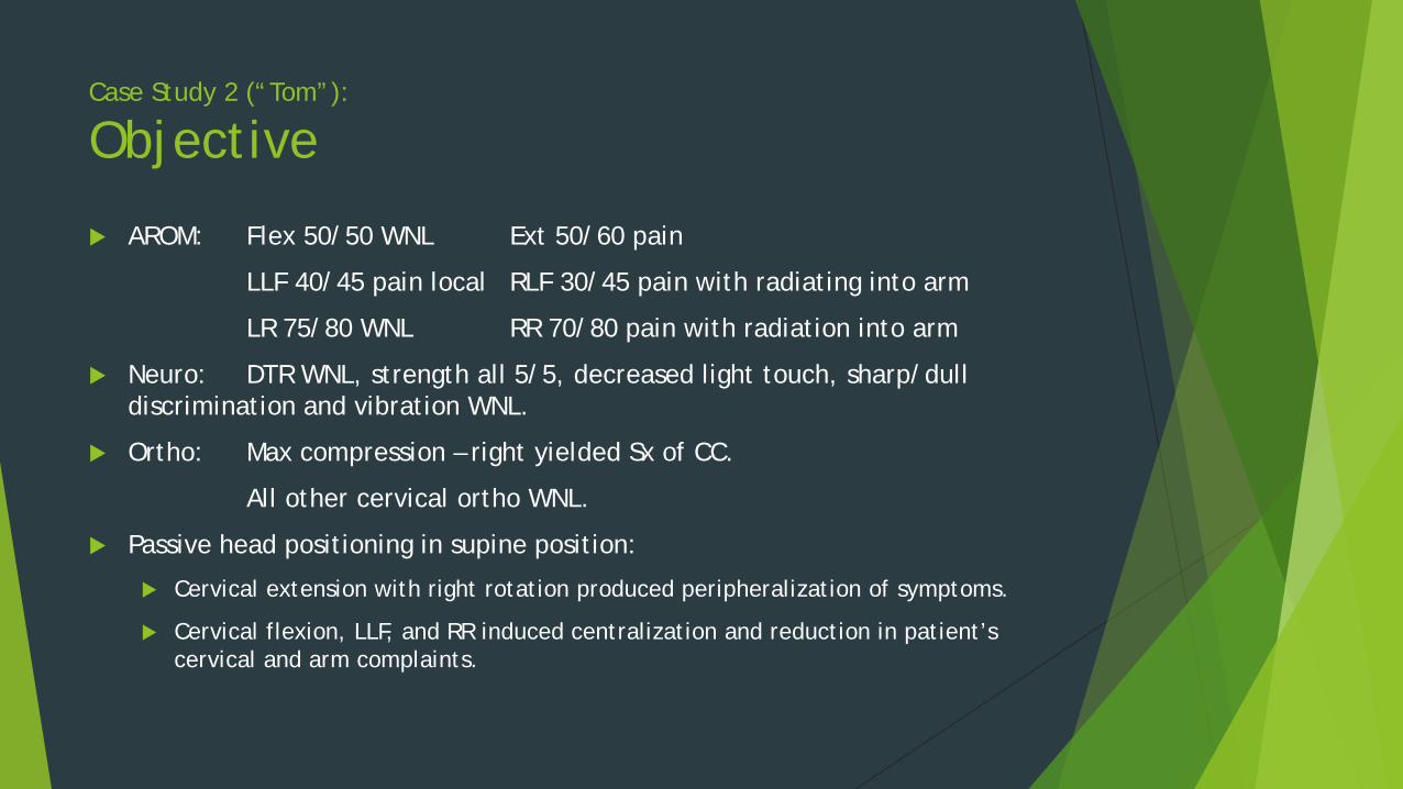
Case Study 2 (“Tom”):
Objective
AROM: Flex 50/50 WNL Ext 50/60 pain
LLF 40/45 pain local RLF 30/45 pain with radiating into arm
LR 75/80 WNL RR 70/80 pain with radiation into arm
Neuro: DTR WNL, strength all 5/5, decreased light touch, sharp/dull discrimination and vibration WNL.
Ortho: Max compression – right yielded Sx of CC.
All other cervical ortho WNL.
Passive head positioning in supine position:
Cervical extension with right rotation produced peripheralization of symptoms.
Cervical flexion, LLF, and RR induced centralization and reduction in patient’s cervical and arm complaints.

Case Study 2 (“Tom”):
Treatment & Results Treatment
Cervical SMT into flexion, LLF, and RR
EMS
HEP focusing on deep neck flexor strengthening, stretching, and stabilization.
Results
After five treatments spread out over a one month duration:
Cervical symptoms 0/10 VAS, right arm 0/10 VAS, right hand 2/10 VAS
Right wrist/hand exam showed signs of underlying carpel tunnel syndrome.
Referred back to surgeon for EMG studies on hand.
Patient had right carpel tunnel decompression surgery (you can’t win ‘em all).
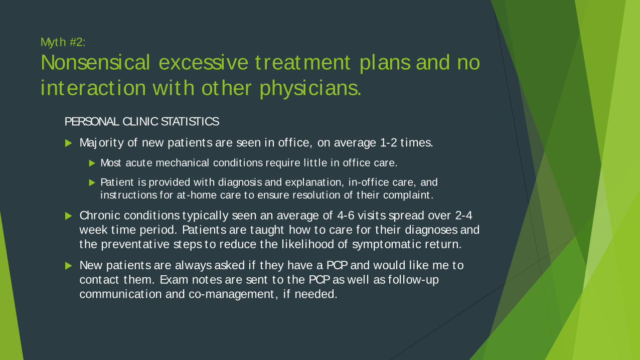
Myth #2:
Nonsensical excessive treatment plans and no interaction with other physicians.
PERSONAL CLINIC STATISTICS
Majority of new patients are seen in office, on average 1-2 times.
Most acute mechanical conditions require little in office care.
Patient is provided with diagnosis and explanation, in-office care, and instructions for at-home care to ensure resolution of their complaint.
Chronic conditions typically seen an average of 4-6 visits spread over 2-4 week time period. Patients are taught how to care for their diagnoses and the preventative steps to reduce the likelihood of symptomatic return.
New patients are always asked if they have a PCP and would like me to contact them. Exam notes are sent to the PCP as well as follow-up communication and co-management, if needed.

Nuts & Bolts:
Case Study 3 (“Karla”) - History & Subjective
- Examination
- Ortho & Neuro
- Next Steps
- Further Imaging
- Treatment

Case Study 3 (“Karla”):
History & Subjective
64 year old female property manager presented with complaints of a non- traumatic constant LBP of a two month duration. Her lumbago was coupled with constant right posterior thigh pain and burning, occasional burning and numbness into right calf and big toe.
LPB rated 5/10 VAS, right posterior thigh symptoms 9/10 VAS, right calf and big toe 2/10 VAS.
Unable to walk greater than one block due to pain. Going up and down stairs and putting her shoes on generated strong pain.
Patient denied changes to BB habits

Case Study 1 (“Karla”):
Examination & Neuro
Examination
Visible right leg limp
Marked reduction in Lx AROM (most noticeably into trunk flexion and right lateral flexion).
Neuro
DTRs all 2/4
Diminished light touch in an L5 distribution
Right hamstring strength 3/5 and painful (L5 – S1)
Vibration, sharp/dull, and all other muscle strengths WNL

Case Study 3 (“Karla”):
Ortho
Ortho
Seated Max SLR (+) right for pain
Braggard’s (+) right
Fabere / LaGuerre / Farfan’s all (+) on the right
ASLR / PSLR / DSLR / Yeomen’s (-) bilaterally
Bowstring (-) bilaterally
Mild tenderness in right SI joint, Lx paraspinals, and R > L Lx erector HT
Case Study 3 (“Karla”):
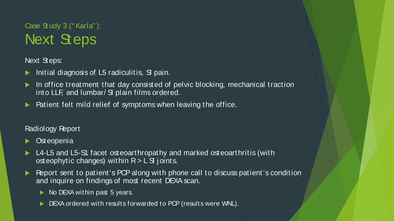
Next Steps Next Steps:
Initial diagnosis of L5 radiculitis, SI pain.
In office treatment that day consisted of pelvic blocking, mechanical traction into LLF, and lumbar/SI plain films ordered.
Patient felt mild relief of symptoms when leaving the office.
Radiology Report
Osteopenia
L4-L5 and L5-S1 facet osteoarthropathy and marked osteoarthritis (with osteophytic changes) within R > L SI joints.
Report sent to patient’s PCP along with phone call to discuss patient’s condition and inquire on findings of most recent DEXA scan.
No DEXA within past 5 years.
DEXA ordered with results forwarded to PCP (results were WNL).

Case Study 3 (“Karla”):
Further Imaging
Spoke with PCP about my initiation of conservative care for foraminal encroachment and L5 radiculitis, as well as my ordering an MRI.
MRI showed L4-5 and L5-S1 moderate broad based disc bulging, and bilateral L5-S1 foraminal narrowing.
No central canal stenosis

Case Study 3 (“Karla”):
Treatment Pelvic blocking for right SI, mechanical traction for disc decompression, HEP.
In office care at two times per week for two weeks.
Spoke with patient about physiatry referral for ESI if conservative care did not show timely improvement.
Results at two weeks: Patient walking up to ¼ mile pain free and all other ADLs improving. No more symptoms into calf/foot and posterior thigh pain continuing to centralize.
Lumbar/SI symptoms rated 3/10 VAS and described as “tight with a mild ache.” No more burning nor numbness.
Treatment frequency decreased to one time per week and emphasis placed on HEP.
At week five: Lx/SI rated 1/10 VAS. No further ADL disturbance. Patient walking one mile per day pain free.
Patient released with home care instructions.
Total visits: 6 visits in 5 weeks. Release notes sent to patient’s PCP.

Myth #3:
“Once I start going, I’m told I need to continue for the rest of my life”
This notion has never been spoken within my practice. (At least not while keeping a straight face)
TRUTH:
Patients are addressed in relation to their chief complaint and associated symptoms.
If treatment is yielding desired results, care is continued until resolution of complaint.
Maintenance care has its place as a means to resolve increasing symptoms for a condition.
This is accomplished via recommending the patient return if they notice their complaints begin to resurface and their home instructions fail to resolve the issue.

Myth #4:
Chiropractic Physicians “only do backs” TRUTH:
Chiropractic physicians are trained extensively in the mechanics, orthopedics, and treatment of MSK conditions in all areas of the body.
I commonly evaluate, diagnose, and treat conditions ranging from tension headaches, to lateral epicondylitis, to ankle sprains. (And everything in between)

Myth #5:
Chiropractic care is no better than placebo.
There is an enormous library of research which proves the opposite.
Randomized, double blind, sham, etc… trials have been conducted all over the world. From the NIH, British Journal of General Practice, Spine, and plenty of other publications have all concluded the efficacy in care and outcomes.
Recent Example from the April 2013 edition of Spine:
101 randomized patients with acute LBP broken into three distinct groups.
SMT and placebo NSAID
Sham SMT and NSAID (Diclofenac)
Sham SMT and placebo NSAID
Results: Outcome for the SMT group was significantly better than the NSAID subgroup and far superior to the placebo.

Questions? Lets take a moment to discuss…

Thank you! It is an honor to be here speaking with you. Let me know if I can answer additional questions after this session, or provide you with additional reading later.