Embed Size (px)
Citation preview

Sensory Receptors – Part II
Based on type of stimuli the receptors can detect (stimulus modality) Chemoreceptors – chemicals, e.g., smell and taste Mechanoreceptors – pressure and movement, e.g.,
touch, hearing, balance, blood pressure Photoreceptors – light, e.g., vision; detect photons Electroreceptors – electrical fields Magnetoreceptors – magnetic fields Thermoreceptors - temperature

Mechanoreceptors
• Transform mechanical stimuli into electrical signals• All organisms and cells can sense and respond to mechanical
stimuli• Two main types
• ENaC – epithelial sodium channels• TRP – transient receptor potential
Touch and Pressure
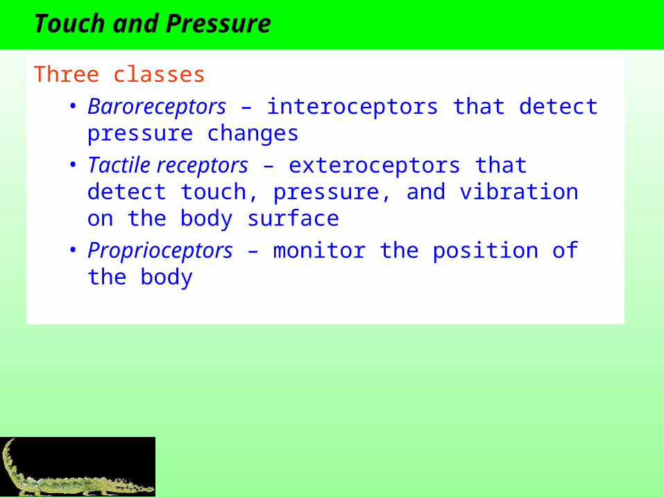
Three classes• Baroreceptors – interoceptors that detect pressure
changes• Tactile receptors – exteroceptors that detect touch,
pressure, and vibration on the body surface• Proprioceptors – monitor the position of the body

Insects
Two types of mechanoreceptors

Type 1 – External Surface
Two common types of sensilla• Trichoid – hairlike• Campaniform – bell-
shaped
Figure 7.13

Type 1 – Internal Surface
• Scolopidia – bipolar neuron and complex accessory cell (scolopale)
• Can be isolated or grouped to form chordotonal organs
• Most function in proprioception
• Can be modified into tympanal organs for sound detection
Figure 7.14

Vertebrate Tactile Receptors
• Widely dispersed• Function as isolated sensory cells• Free nerves endings or enclosed in accessory structures
(e.g., Pacinian corpuscle)
Figure 7.15

Proprioceptors
Monitor the position of the body• Three major groups
• Muscle spindles – located on the surface of the muscle and monitor muscle length
• Golgi tendon organs – located at the junction between skeletal muscles and tendons and monitor tendon tension
• Joint capsule receptors – located in the capsules that enclose joints and detect pressure, tension, and movement in the joint

Equilibrium and Hearing
• Utilize mechanoreceptors• Equilibrium or balance – detecting position of the body
relative to gravity• Hearing – detecting and interpreting sound waves • Vertebrates: ear is responsible for both equilibrium and
hearing• Invertebrates: organs for equilibrium are different from
organs of hearing (e.g., tympanal organs)
Statocysts
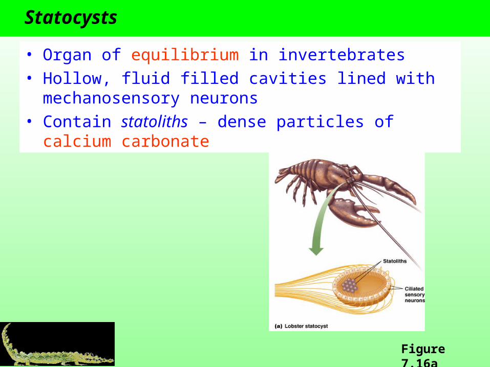
• Organ of equilibrium in invertebrates• Hollow, fluid filled cavities lined with mechanosensory
neurons• Contain statoliths – dense particles of calcium carbonate
Figure 7.16a

Hair cells
• Mechanoreceptor cells used for hearing and balance in vertebrates
• Modified epithelial cells• Have extensive
extracellular structures and cilia that extend from the apical end

Signal Transduction in Hair Cells
Can detect movement and direction

Fish
• Use hair cells in ears for hearing and for detecting body position and orientation
• Have neuromasts that detect water movement
• Neuromast – hair cell and accessory cupula
• Lateral line system – array of neuromasts within pits or tubes running along the side of the body

Vertebrate Ears
Function in both equilibrium and hearing
Equilibrium
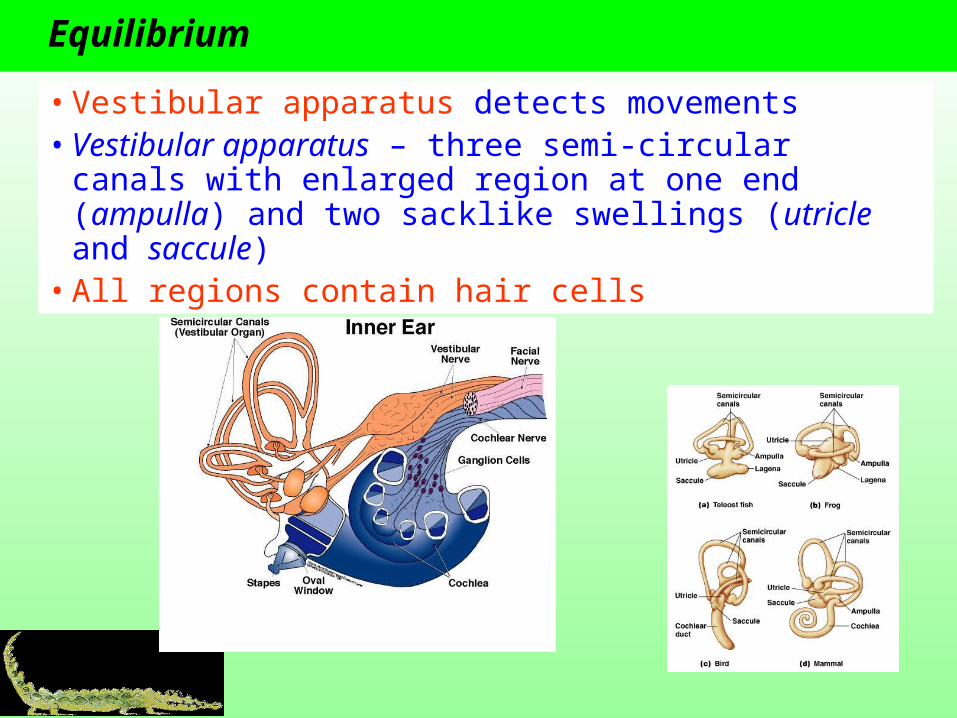
• Vestibular apparatus detects movements• Vestibular apparatus – three semi-circular canals with
enlarged region at one end (ampulla) and two sacklike swellings (utricle and saccule)
• All regions contain hair cells

Vestibular Apparatus
• Utricle and saccule contain mineralized otoliths suspended in a macula covering >100,000 hair cells
• Ampullae lack otoliths and contain cristae (hair cells located in a cupula)

Maculae Detect Linear Acceleration and Tilting
Figure 7.23
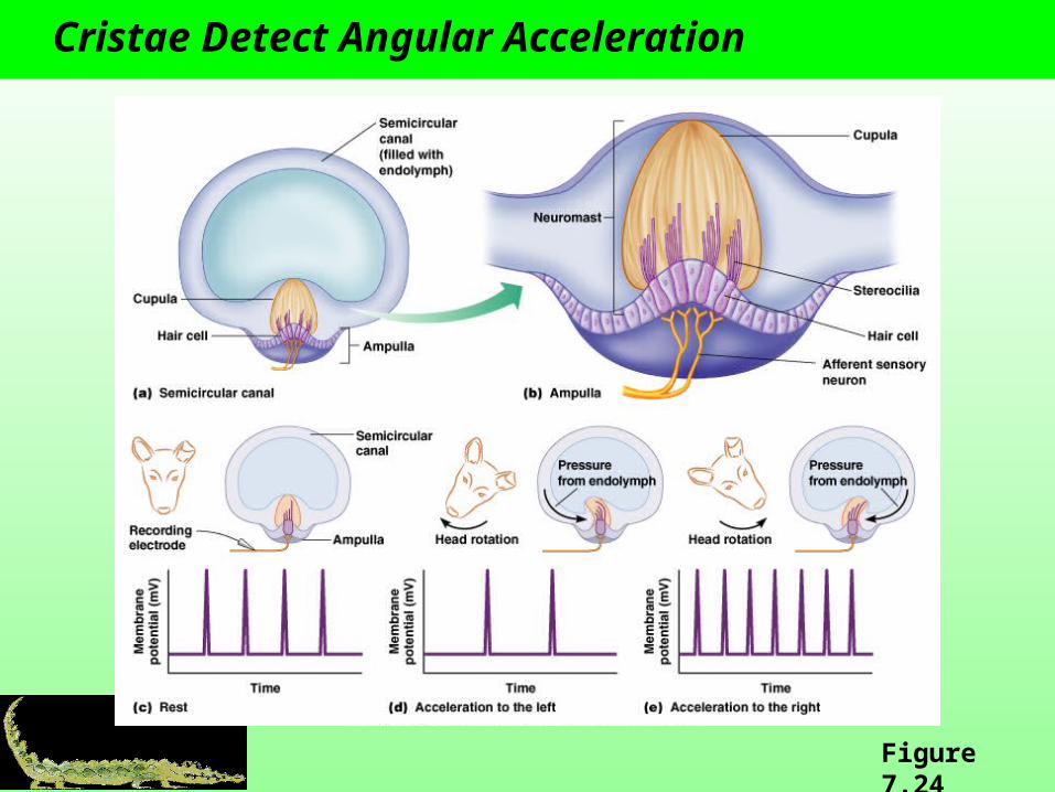
Cristae Detect Angular Acceleration
Figure 7.24

Sound Detection
• Inner ear detects sound• In fish, incoming sound waves cause otoliths to move
which bend cilia of hair cells• Some fish use the swim bladder to amplify sounds
Figure 7.25

Terrestrial Vertebrates
• Hearing involves the inner, middle, and outer ears
• Problem: sound transfers poorly between air and the fluid-filled inner ear
• Solution: amply sound• Pinna acts as a funnel to
collect more sound• Middle ear bones increase
the amplitude of vibrations from the tympanic membrane to the oval window
Figure 7.26a

Mammalian Inner Ear
• Specialized for sound detection• Cochlea is coiled in mammals• Perilymph – fills vestibular and tympanic ducts and is similar to
extracellular fluids• Endolymph – fills cochlea duct and is high in K+ and low in Na+
• Organ of Corti contains hair cells and sits on basilar membrane• Two types of hair cells
• Inner hair cells detect sound• Outer hair cells amplify sounds
Figure 7.26b

Sound Transduction
Steps • Incoming sound• Oval window vibrates • Waves in perilymph of vestibular duct• Basilar membrane vibrates • Stereocilia on the inner hair cells bend • Depolarization• Release of neurotransmitter (glutamate)• Excite sensory neuron
Round window serves as a pressure valve

Sound Encoding
Basilar membrane is stiff and narrow at the proximal end and flexible and wide at distal end
Frequency• High stiff end vibrates• Low flexible end vibrates

Amplification
Loudness• Loud sounds movement of basilar membrane
depolarization of inner hair cells AP frequency
Outer hair cells• Change shape in response to sound instead of
releasing neurotransmitter• Change in shape causes basilar membrane to move
more and causes a larger stimulus to the inner hair cells
• Amplifies sound

Sound Location
• Brain uses information on time lags and differences in sound intensity
• Sound to right ear first sound located to the right• Sound louder in right ear sound located to the right

Photoreception
• Ability to detect a small proportion of the electromagnetic spectrum from ultraviolet to near infrared
• Concentration on this range or wavelengths supports idea that animals evolved in water
Figure 7.27

Photoreceptors
Organs range from single light-sensitive cells to complex, image forming eyes
Two major types• Ciliary photoreceptors – have single, highly folded
cilium; folds form disks that contain photopigments • Rhabdomeric photoreceptors – apical surface is
covered with multiple outfoldings called microvillar projections
Photopigments - molecules that absorb energy from photons

Vertebrate Photoreceptors
All are ciliary photoreceptors
Two types• Rods• Cones
Figure 7.29

Characteristics of Rods and Cones
Nocturnal animals have relatively more rods

Photopigments
Photopigments have two covalently bonded parts• Chromophore – pigment that is a derivative of vitamin
A, e.g., retinal• Opsin – G-protein-coupled receptors
Steps in photoreception• Chromophore absorbs energy from photon• Chromophore changes shape• Photoreceptor protein changes shape• Signal transduction cascade• Change in membrane potential
Bleaching – process where activated retinal no longer bonds to opsin, thereby activating opsin

Phototransduction
Transduction cascades differ in rhabdomeric and ciliary photoreceptors

The Eye
• Eyespots are single cells or regions of a cell that contain photosensitive pigment, e.g., protist Euglena
• Eyes are complex organs
Figure 7.33

Flat-sheet Eyes
• Provide some sense of light direction and intensity• Most often seen in larval forms or as accessory eyes in
adults
Figure 7.33a

Cup-shaped Eyes
• Retinal sheet is folded to form a narrow aperture• Better discrimination of light direction and intensity• Seen in the Nautilus

Vesicular Eyes
• Use a lens in the aperture to improve clarity and intensity• Lens refracts light and focuses it onto a single point on
the retina• Present in most vertebrates
Figure 7.33c

Convex Eye
•Photoreceptors radiate outward forming a convex retina•Present in annelids, molluscs, and arthropods (eeeeeeeeeek)

Compound Eyes
Most complex convex eyes found in arthropods
Composed of ommatidia
Form images in two ways• Apposition compound eyes – ommatidium operate
independently; afferent neurons make interconnection to generate an image
• Superposition compound eyes – ommatidium work together to form an image on the retina

The Vertebrate Eye
Forms bright, focused images
Parts• Sclera – white of the
eye• Cornea – transparent
layer• Choroid – pigmented
layer• Tapetum – layer in
the choroid of nocturnal animals that reflects light
Figure 7.35

The Vertebrate Eye, Cont.
Parts• Iris – two layers of pigmented
smooth muscle• Pupil – opening in iris• Lens – focuses image• Ciliary body – muscles for
changing lens shape• Aqueous humor – fluid in the
anterior chamber• Vitreous humor – gelatinous
mass in the posterior chamber
Figure 7.35

Image Formation
• Refraction – bending light rays
• Both the cornea and the lens act as converting lens to focus light on the retina
• In terrestrial vertebrates, most of the refraction occurs between the air and the cornea
Figure 7.36a

Image Accommodation
•Accommodation - incoming light rays must converge on the retina to produce a clear image
•Focal point – point at which light waves converge•Focal distance – distance from a lens to its focal point•Distant object: light rays are parallel when entering the lens•Close object: light rays are not parallel when entering the lens and must be refracted more
•Light rays are focused on the retina by changing the shape of the lens

The Retina
• Arranged into several layers• Rods and cones are are at
the back and their tips face backwards
• Axons of ganglion cells join together to form the optic nerve
• Optic nerve exits the retina at the optic disk (“blind spot”)
Figure 7.37a

The Fovea
• Small depression in the center of the retina where overlying bipolar and ganglion cells are pushed to the side
• Contains only cones• Provides the sharpest
images
Figure 7.37a

Signal Processing in the Retina
Rods and cones form different images
Rods• Principle of convergence – as many as 100 rods
synapse with a single bipolar cell many bipolar cells synapse with a ganglion cell
• Large visual field • Fuzzy image
Cones• One cone synapses with one bipolar cell which
connects to one ganglion cell• Small visual field• High resolution image

Signal Processing in the Retina, Cont.
Complex “on” and “off” regions of the receptive fields of ganglion cells improve their ability to detect contrasts between light and dark
Figure 7.39

The Brain Processes the Visual Signal
• Optic nerves optic chiasm optic tract lateral geniculate nucleus visual cortex
Figure 7.41

Color Vision
Detecting different wavelengths of light
Requires multiple types of photoreceptors with different maximal sensitivities• Humans: three (trichromatic)• Most mammals: two (dichromatic)• Some bird, reptiles and fish: three, four, or five (pentachromatic)
Figure 7.42

Thermoreception
Central thermoreceptors – located in the hypothalamus and monitor internal temperature
Peripheral thermoreceptors – monitor environmental temperature• Warm-sensitive• Cold-sensitive• Thermal nociceptors – detect painfully hot stimuli
ThermoTRPs – TRP ion channel thermoreceptor proteins

Specialized Thermoreception
• Specialized organs for detecting heat radiating objects at a distance
• Pit organs – pit found between the eye and the nostril of pit vipers
• Can detect 0.003°C changes (0.5°C for humans)

Magnetoreception
•Ability to detect magnetic fields•e.g., migratory birds, homing salmon•Neurons in the olfactory epithelium of rainbow trout contain particles that resemble magnetite