Embed Size (px)
Citation preview

Sensors for the optical detection of cyanide ion
Zhaochao Xu,w Xiaoqiang Chen,w Ha Na Kim and Juyoung Yoon*
Received 17th April 2009
First published as an Advance Article on the web 4th September 2009
DOI: 10.1039/b907368j
This tutorial review focuses on recent developments arising from studies of optical sensors
for cyanide ions, which are categorized by approaches involving cyanide selective receptors,
the utilization of metal coordinated complexes, and chemodosimeters.
Introduction
Although substances containing cyanide have been used as
poisons for centuries, it was not until 1782 that this anion was
first isolated by the Swedish chemist Scheel.1 The extreme
toxicity of cyanide in physiological systems, as well as the
continuing environmental concern caused by its widespread
industrial use, has led to considerable research into the
development of methods for cyanide detection. Various methods
used previously to analyze cyanide employ titrimetric,2
voltammetric,3 potentiometric,4 and electrochemical methods,5
as well as ion chromatography,6 etc. However, these methods
often require extensive, time consuming procedures that
involve the use of sophisticated instrumentation with high
detection limits. Optical sensors for cyanide, in which a change
in color and/or fluorescence intensity (or emission wavelength)
is monitored, have been studied actively over the past ten years
due to their simple, inexpensive, and rapid implementation.
Generally, three different approaches, which are shown
schematically in Fig. 1, have been employed to design optical
sensors for cyanide ions. The most popular strategy involves the
use of sensors in which the binding sites and signaling subunits
are linked covalently. In this case, interaction of cyanide with the
binding site causes a change in color or fluorescence of the
signaling subunit. A coordination complex-based displacement
approach has also been used. In these sensors, the introduction
of cyanide ions leads to regeneration of spectroscopic behavior
of the noncoordinated indicator. A third method for the
determination of cyanide is known as a chemodosimeter
approach. These types of sensors rely on the occurrence of
specific, most often irreversible chemical reactions, which take
place upon an interaction with cyanide. Compared to other
types of anion selective optical chemosensors,7–12 cyanide
selective optical chemosensors take advantage of two significant
and characteristic properties of cyanide, its strong nucleo-
philicity and high binding affinity towards copper ions. Thus
far, cyanide sensing has been a part-topic in relatively few early
review articles.8,10,13,14 To the best of our knowledge, cyanide
sensors have not been reviewed thoroughly in recent years, even
though some cyanide selective colorimetric sensors were recently
reported.15 This review begins with a brief discussion of the
source and toxicity of cyanide, and then moves to a discussion
of cyanide sensors, which are classified according to the above-
mentioned approaches.
Cyanide sources and toxic effects
Sources
Cyanide containing salts are widespread chemicals found in
surface water originating not only from industrial waste but
also from biological sources. Cyanide is used in many chemical
processes, such as electroplating, plastics manufacturing, gold
and silver extraction, tanning, and metallurgy.16,17 In addition,
Department of Chemistry and Nano Science and Department ofBioinspired Science, Ewha Womans University, Seoul 120-750, Korea.E-mail: [email protected]; Fax: +82-2-3277-2384;Tel: +82-2-3277-2400
Zhaochao Xu
Zhaochao Xu received hisPhD in 2006 from DalianUniversity of Technologyunder the supervision of Prof.Xuhong Qian. Subsequently hejoined the group of JuyoungYoon at Ewha WomansUniversity as a postdoctoralfellow. Since October 2008 hehas been a Herchel SmithPostdoctoral Research Fellowat the University ofCambridge in the group ofDavid R. Spring.
Xiaoqiang Chen
Xiaoqiang Chen was born in1980 in China. He obtainedhis PhD in 2007 from DalianUniversity of Technology(China) under the supervisionof Prof. Xiaojun Peng. He ispresently working as a post-doctoral fellow in the group ofProf. Juyoung Yoon at EwhaWomans University, Korea.
w Contributed equally to this work.
This journal is �c The Royal Society of Chemistry 2010 Chem. Soc. Rev., 2010, 39, 127–137 | 127
TUTORIAL REVIEW www.rsc.org/csr | Chemical Society Reviews

cyanide is a chemical warfare agent.1 Various industries,
including petrochemical, gold mining, metal electroplating,
photographic, steel manufacturing, are responsible for cyanide
pollution. The production of nitrile, nylon and acrylic plastics
is also associated with environmental concerns caused by
cyanide. Biological sources of cyanide include bacteria, fungi
and algae, which produce this ion as part of their nitrogen
metabolic pathways. Vegetables containing cyanogenic glyco-
sides are sources of cyanide ingestion in humans and animals.
Dietary foodstuffs that contain moderate to high levels of
cyanogenic glycosides include cassava, a dietary staple in
several regions of Africa, as well as other common foods such
as lima beans, sorghum, linseed, kernels of fruits, sweet
potatoes and bamboo shoots.17 Tobacco smoke is also a
common source of cyanide and can lead to high levels of this
ion in the blood. Other potential sources of cyanide in humans
and animals are sodium nitroprusside, succinonitrile and
organic thiocyanates.17 The United States Environmental
Protection Agency (EPA) has set the maximum contaminant
level (MCL) for cyanide in drinking water at 0.2 ppm.
Toxicity and metabolic effects
The LD50 (estimated dose that is lethal to 50% of the exposed
population) of hydrogen cyanide and cyanogen chloride has
been reported to be 2500–5000 mg.min/m3 and 11000 mg.min/m3.
The LD50 of hydrogen cyanide in humans is 1.0 mg/kg, and
the estimated LD50 for cyanide solutions applied to the skin is
approximately 100 mg/kg.1 Cyanide can affect many functions
in the body, including the vascular, visual, central nervous,
cardiac, endocrine, and metabolic systems. Perhaps the best
known effect of cyanide is its inhibition of respiration, which is
caused by the inhibition of the terminal oxidase (cytochrome
oxidase) of the mitochondrial respiratory chain. Sublethal doses
of cyanide cause a decrease in the rate of glycolysis and inhibit the
operation of the TCA cycle. Cyanide also acts as an inhibitor
of metallo-enzymes and of some non-metallo-enzymes that
function through the intermediacy of Schiff bases.1,16 The
toxicodynamic effects of cyanide can depend on the dose,
route and speed of administration, the chemical form of
cyanide and other factors including gender, age, weight, stress
level, and general physical condition.
Sensors based on the covalently linked binding site
and signaling subunit approach
Although cyanide is not a strong hydrogen bonding acceptor
compared to other anions, several cyanide selective sensors
that rely on hydrogen bonding interactions have been
described. In addition, ditopic systems bearing two metal sites
were examined as cyanide selective receptors, which function
in organic solvents.
Hydrogen bonding based receptors
Lees et al. described a luminescent rhenium(I) polypyridyl-
based receptor 1 for the recognition of anions (Fig. 2).18 This
artificial receptor shows high affinity for halides, cyanide and
acetate anions with binding constants as high as 104–105 M�1
in CH2Cl2. The overall order of binding affinity was found to
be CN� 4 F� 4 I� 4 Cl� E Br� E OAc� c H2PO4� 4
NO3� 4 ClO4
�. Although this affinity trend was not clearly
explained, a combination of interactions involving electro-
static forces, hydrogen bonding strengths and steric effects
have been reported to affect the binding affinity of receptors
toward anions.
Fig. 1 Three approaches for chemosensors: (a) chemosensor bearing
a signaling subunit as well as a binding site; (b) displacement
approach; (c) chemodosimeter.
Ha Na Kim
Ha Na Kim received the BSdegree in Ewha WomansUniversity. She then was giventhe MS degree in medicalscience by Seoul NationalUniversity in 2006. She is ona doctoral course in Prof.Juyoung Yoon’s researchgroup in Ewha WomansUniversity.
Juyoung Yoon
Juyoung Yoon received hisPhD (1994) from TheOhio State University. Aftercompleting postdoctoralresearch at UCLA and atScripps Research Institute, hejoined the faculty at SillaUniversity in 1998. In 2002,he moved to Ewha WomansUniversity, where he iscurrently a Professor of theDepartment of Chemistryand Nano Science and theDepartment of BioinspiredScience. His research interestsinclude investigations of
fluorescent chemosensors, molecular recognition and organoEL materials.
128 | Chem. Soc. Rev., 2010, 39, 127–137 This journal is �c The Royal Society of Chemistry 2010

Anzenbacher and Castellano et al. designed the novel
cyanide sensor 2 based on the changes in the anion-induced
luminescence lifetime (Fig. 2).19 The addition of fluoride and
cyanide to this sensor caused significant changes in the UV-vis
and steady-state emission properties of compound 2 in
CH2Cl2–CH3CN (98 : 2, v/v). Quenching of the steady state
photoluminescence was observed upon the addition of
cyanide. In addition, cyanide causes a change in the lumines-
cence lifetime of the sensor from 377 to 341 ns in the same
solvent system.
Vilar et al. recently utilized azo-phenylthiourea compounds
3a and 3b as probes for cyanide (Fig. 2).20 In methanol, the
dyes undergo color changes from pale orange to red in the
presence of cyanide with a detection limit of 8 ppm. In DMSO,
cyanide induces a color change (to dark purple) as does
fluoride (to blue), while CH3CO2� and H2PO4
� (both to
violet/red) also promote color changes. The authors suggested
that the origin of the color changes was not a simple change in
hydrogen-bonding interaction, but rather a process that
involves the deprotonation of thiourea NH groups. The
optical properties of compound 3a incorporated in nano-
structured Al2O3 films have been determined. An aqueous
solution of cyanide induces a color change in these films,
which can be used to detect cyanide at 2.6 ppm.
Metal complex based receptors
Hong et al. reported the results of a study on Zn-porphyrin/
crown ether conjugates.21 These ditopic neutral receptors
(4a and 4b) contain a Lewis-acidic binding site (zinc porphyrin
moiety) and a Lewis-basic binding site (crown ether moiety)
(Fig. 3a). The two receptors display a color change from the
original red of Zn-porphyrin to green selectively in the
presence of sodium cyanide. The origin of this unique color
change was proposed to be associated with the binding of
cyanide in a ditopic manner, in contrast to other sodium salts,
which are bound to the receptors in a monotopic fashion.
Chen et al. described a similar approach to sensor design
utilizing the ditopic characteristics of aza-crown ether-capped
porphyrins 5a and 5b (Fig. 3b).22 These artificial receptors
selectively recognize sodium cyanide and potassium cyanide as
a result of ditopic binding in methanol. The binding of receptor
5a to sodium cyanide is 56 times stronger than that for
potassium cyanide, whereas the selectivity of 5b for potassium
cyanide is 12 times higher than that for sodium cyanide.
Displacement approach
Among the various systems designed to detect cyanide, sensors
utilizing the affinity of this anion for copper have attracted
special attention. Cyanide reacts with copper ions to form
stable [Cu(CN)x]n� species. One of the most important
advantages of sensors based on this chemistry is that they
should be operable in aqueous solutions.
Cu complex
Mareque-Rivas et al. designed a cyanide sensor system that
considers the ability of copper ions to affect the kinetics of
electron transfer across a copper binding, self assembled
monolayer (SAM) to a negatively charged redox probe in
solution and the ability of (H)CN to bind and remove copper
ions (Fig. 4).23 The SAM-modified gold electrode exhibits a
charge transfer resistance value (RCT) of 32 O cm2. An
inspection of the impedance plots showed that the RCT values
increase with increasing cyanide concentration. Nanomolar
concentrations of cyanide (0.03 ppb detection limit), even at
pH 7.3, could be detected using this method.
Li et al. examined light-emitting polyacetylene bearing
imidazole moieties in the context of a new type of cyanide
sensor.24 The polymer sensor displays selective fluorescence
quenching in the presence of Cu2+. Interestingly, the
Cu2+-promoted luminescence quenching can be inhibited by
the addition of cyanide, as shown in Fig. 5.
Li and coworkers also employed the same strategy in the
design of a new imidazole-functionalized polyfluorene 6,
Fig. 2 Structures of receptors 1, 2, 3a and 3b.
Fig. 3 (a) Structures of ditopic receptors 4a and 4b. (b) Structures of
receptors 5a and 5b and their proposed ditopic binding modes.
Fig. 4 Proposed mechanisms of SAM-modified gold electrode detecting
cyanide.
This journal is �c The Royal Society of Chemistry 2010 Chem. Soc. Rev., 2010, 39, 127–137 | 129

which is a sensitive and selective cyanide chemosensor
(Fig. 6).25 The fluorescence of compound 6 was quenched
completely by Cu2+ at concentrations as low as 0.20 ppm. The
quenched fluorescence of a solution of compound 6 and Cu2+
was recovered upon the addition of cyanide with a detection
limit as low as 0.31 ppm.
The Mareque-Rivas group also used a combination of
tri-n-octylphosphine oxide (TOPO)-coated CdSe quantum
dots (QDs), 2,20-bipyridine (bipy) and CuCl2 (Fig. 7) to
fabricate a turn-on fluorescence cyanide probe.26 The ability
of bipyridine-bound copper(II) ions to quench the photo-
luminescence of hydrophobic CdSe quantum dots was used
advantageously in this cyanide selective, turn-on fluorescence
sensor. This QD system detects cyanide at 20–100 mM
concentrations at a physiological pH. TOPO-coated CdSe
QDs placed on a polystyrene film displayed a similar
‘‘On–Off–On’’ type change upon the addition of cyanide.
On the other hand, Dong et al. utilized CdTe quantum
dots as turn-on fluorescent sensors for cyanide.27 Copper
ion-modified CdTe quantum dots were prepared and the
quenched fluorescence due to Cu2+ was revived in the
presence of cyanide with a detection limit of 3.9 ppb at pH 7.0.
Qin and Li et al. recently reported that an old and inexpensive
compound, zincon (2-carboxy-20-hydroxy-50-sulfoformazyl-
benzene (7)), can be used as a highly sensitive and selective
chemosensor for cyanide in aqueous solutions with a detection
limit of 0.13 ppm (Fig. 8).28 When the Cu2+ concentration
increases, the peak intensity of the absorption maximum at
463 nm of this sensor decreases with the concurrent formation
of a new peak at ca. 600 nm. The addition of cyanide to the
resulting complex 7-I causes an increase in the absorption
band at 463 nm, which can be observed with the naked eye.
Yoon, Park and coworkers devised a simple method
for detecting cyanide ions in aqueous solution at pH 7.4.29
The fluorescent probe 8 designed for this purpose displays a
fluorescence quenching effect with Cu2+. Therefore, the
addition of cyanide induces ‘‘Off–On’’ type fluorescence
enhancement (Fig. 9). This sensing system has been incorpo-
rated into a microfluidic platform, in which the fluorescent
sensor 8-Cu2+ displays green fluorescence upon the addition
of cyanide. Finally, studies of biological applications using
Caenorhabditis elegans demonstrated that this system can be
employed for the in vivo imaging of cyanide.
Co complex
Recently, Zelder utilized the ‘‘base on’’/‘‘base off’’ coordination
of the intramolecular bound benzimidazole nucleobase of
vitamin B12 in the construction of a novel sensor for cyanide
(Fig. 10).30 In its ‘‘base on’’ conformation, the sensor is red
colored (lmax = 550, 520 and 361 nm) and the addition of
cyanide induces a color change to violet (lmax = 579, 542 and
368 nm). As a result, this system can be used for the specific
colorimetric detection of millimolar concentrations of cyanide
in water. Zelder and coworkers also employed a similar
strategy in the design of corrinoid derivatives 9a–9c, in which
the substitution of CoIII-bound water by cyanide enables
the rapid colorimetric detection of micromolar amounts of
cyanide (Fig. 11).31
Chemodosimeter approach
The exceptional nucleophilicity of cyanide has served as a
basis for the development of various chemodosimetric probes
for cyanide, in most cases in aqueous solutions. As shown
below, this approach has recently attracted the attention of
many groups.
C–C bond formation utilizing cyanohydrin reaction
Ahn et al. reported that the fluorescence signaling of anion
binding can be modulated by intramolecular H-bonding
Fig. 5 Schematic representation of Cu2+ and cyanide sensors based
on the fluorescence ‘‘turn-off’’ and ‘‘turn-on’’ of polyacetylenes.
Fig. 6 Schematic representation of cyanide sensor based on the
fluorescence ‘‘turn-off’’ and ‘‘turn-on’’ of 6.
Fig. 7 Cyanide QD (quantum dot) based probe.
Fig. 8 The speculated conversion cycle of zincon 7 in the presence of
Cu2+ and cyanide.
130 | Chem. Soc. Rev., 2010, 39, 127–137 This journal is �c The Royal Society of Chemistry 2010

stabilization of anion–ionophore adducts.32,33 In a recent
report from this group,34 it was shown that a carbonyl
addition intermediate, such as compound 10-I, which is
stabilized by intramolecular H-bonding, is responsible for
the fluorescence enhancement observed in acetonitrile
(Fig. 12). In contrast, another possible intermediate, 10-II, is
believed to show fluorescence quenching. Moreover, the
formation of adduct 10-I should be favored over the alter-
native deprotonation process leading to compound 10-II,
which was confirmed by NMR analysis.
Ahn et al. extended this intramolecular H-bonding stabili-
zation concept to the design of a heteroditopic receptor 11a
containing both a crown ether and a trifluoroacetylcarboxanilide
group (Fig. 13). In this system, cyanide was added to the
trifluoroacetyl group to produce an alkoxide adduct that
interacts with the potassium ion bound to the crown ether
moiety. As a consequence of a highly cooperative ion-pair
interaction, this sensor selectively recognizes both potassium
and cyanide ions in acetonitrile solutions with an association
constant as high as 1.9 � 107 M�1. This affinity is two orders
of magnitude higher than that of 11b.35
The Ahn group also described a new probe 12 that displays
fluorescence quenching in the presence of cyanide in
MeOH–water (9 : 1) solutions but not when other anions are
present (Fig. 13).36
The intramolecular H-bonding stabilization concept serves
as the basis for the boradiazaindacene 13 derivative described
by Akkaya et al. (Fig. 14).37 This chemosensor displays a large
decrease in fluorescence intensity and a reversible color change
from red to blue in the presence of cyanide in CH3CN. Highly
fluorescent polymeric films doped with this sensor were also
prepared. The introduction of cyanide causes the color/
emission of the film to change from an orange/fluorescent to
a blue/nonfluorescent, and the addition of trifluoroacetic acid
reverses this change.
This strategy was used by Cheng et al. to design a
2-(trifluoroacetylamino)anthraquinone sensor 14 (Fig. 14),
which undergoes a ‘‘naked eye’’ observable, colorless to yellow
transformation when low concentrations (13 ppb) of cyanide
in CH3CN–H2O (95 : 5, v/v) are added.38 Cheng and
coworkers also devised a colorimetric probe 15 (Fig. 14) that
exhibits excellent selectivity for cyanide in CH3CN–H2O
(95 : 5, v/v). A change from colorless to yellow takes place
upon the addition of cyanide.39
Recently, Guo et al. reported a simple N-nitrophenyl
benzamide derivative 16 (Fig. 14) for the ‘naked-eye’ detection
of cyanide in DMSO–H2O (1 : 1, v/v).40 This sensor can detect
cyanide at concentrations as low as 23 ppb in the above
solvent system by utilizing the strong affinity of cyanide
toward the acyl carbonyl carbon.
One general process operating in enzymatic reactions
involves carbonyl activation by a properly located phenol
hydroxyl group that leads to the general acid catalysis of
nucleophilic addition. This strategy was adopted intelligently
by Kim and Hong et al. in the design of a cyanide sensing
system 17 (Fig. 15). As a consequence of activation by
hydrogen bonding with the phenolic group in the salicyl-
aldehyde moiety, the carbonyl group of compound 17 is
expected to undergo nucleophilic addition with cyanide.41
Studies show that the 1H NMR spectrum of compound 17
in the presence of cyanide does not contain a resonance for the
aldehyde proton (Ha, initially at 10.4 ppm) but instead shows a
new resonance at 5.6 ppm (Hb). The chemical shift of this
resonance is consistent with a cyanohydrin proton resulting
Fig. 9 Proposed binding mechanism of 8 with Cu2+ and cyanide.
Fig. 10 Structures of vitamin B12 and the binding mechanism with
cyanide.
Fig. 11 Structures of corrinoids 9a–9c and a schematic for the
binding mode with cyanide.
Fig. 12 A plausible equilibrium pathway for interaction of 10 with
cyanide.
Fig. 13 Structures of ferrocene derivatives 11a, 11b and 12.
This journal is �c The Royal Society of Chemistry 2010 Chem. Soc. Rev., 2010, 39, 127–137 | 131

from cyanide addition to the aldehyde moiety. Interestingly,
the addition of cyanide to compound 17 in DMSO also causes
a clear color change from light yellow to dark red.
Kim and Hong et al. extended this concept to the design
of a coumarin-based fluorescent chemodosimeter 18
containing a salicylaldehyde group (Fig. 16).42 Rapid proton
transfer of the phenol hydrogen in the excited state results
in a strong fluorescence of an aqueous solution of compound
18 at pH 7.4. Cyanide at concentrations of 260 ppb in
aqueous solution can be detected using this fluorescent
chemodosimeter.
Yoon and Park et al. applied this strategy for the activation
of a carbonyl group by an adjacent phenol in devising a
fluorescein aldehyde-based cyanide sensor 19 (Fig. 16).43 In
CH3CN–H2O (9 : 1, v/v), the ‘‘OFF–ON’’ type emission
change can be monitored at wavelengths 4500 nm. The
practical use of this probe was demonstrated by its incorporation
into a microfluidic platform for the selective detection of
cyanide in living cells.
Ahn et al. constructed N-acyl-triazene derivatives that serve
as simple and tunable chemodosimeters based on the strong
affinity of cyanide toward an N-acyl carbonyl carbon
(Fig. 17).44 Significant changes in the absorption spectrum
(from colorless to deep purple) take place when acetonitrile
solutions of N-acetyltriazene 20a are titrated with both
cyanide and F�. In contrast, the N-isopropanoyl-triazene
20b in acetonitrile shows a significant response to cyanide
and only a weak response to F�. The absorption properties of
both triazenes 20a and 20b are altered only by the addition
of cyanide (faint yellow to red–pink, lmax = 521 nm)
when methanol–water (9 : 1, v/v) is used as a solvent. This
observation is most likely due to the fact that only cyanide
adds to the acyl group of these sensors.
Sessler et al. employed the well-known benzil–cyanide
reaction (Fig. 18) in the design of a colorimetric method for
the detection of cyanide.45 The p-extended analogue of benzil
21a was selected for this purpose. This substance is soluble in a
70 : 30 (v/v) mixture of methanol–water (Fig. 19). Dilute
solutions of compound 21a in this medium are yellow and
become colorless when low concentrations of cyanide but not
other anions are added. Using a dilute solution of compound
21a (7.20 � 10�6 M) in 70 : 30 (v/v) MeOH–water, a limit of
detection o44 ppb can be realized and visualized by simple
naked eye analysis. Prior to this report, Sessler et al. also
described a novel probe 21b (Fig. 19) that undergoes benzil
rearrangement when treated with cyanide (Fig. 18) in ethyl
acetate. In this organic solvent, a yellow to colorless change
and large fluorescence enhancement are observed within
1 min.46
Sun et al. prepared two probes, 22a and 22b, that feature the
use of the dipyrrole carboxamide moiety for anion recognition
(Fig. 20a).47 These structurally simple anion probes exhibit
high selectivity for cyanide over other common inorganic
anions in partially aqueous environments [CH3CN–H2O
(9 : 1, v/v)]. Both compounds 22a and 22b respond to cyanide
only, because cyanohydrin derivatives (22b-I and 22b-II) are
generated (Fig. 20b). This process is associated with a color
change from colorless to yellow (Fig. 20b) and a fluorescence
change for compound 22a from blue to green.
C–C bond formation utilizing chromogenic oxazines
Raymo et al. designed a chromogenic oxazine that can be used
for the selective colorimetric detection of cyanide. In this
system, the [1,3]oxazine ring of compound 23 opens to form
a 4-nitrophenylazophenolate chromophore (23-I) in the
reaction with cyanide (Fig. 21).48,49 In acetonitrile, the addition
of cyanide causes a decrease in the original absorption band at
381 nm (pale yellow) with the concomitant appearance of a
new absorption band at 581 nm (red). In aqueous solutions,
a significantly higher amount of cyanide is required to elicit a
UV response. In order to overcome this limitation, a two-
phase system consisting of dichloroethane and phosphate
buffer (pH 9) was used in conjunction with phase transfer
catalysis. Micromolar concentrations of cyanide can be
detected using this two-phase system.
Fig. 14 Structures of probes 13–16.
Fig. 15 A plausible mechanism for the action of probe 17 with
cyanide.
Fig. 16 Reaction mechanism of 18 and 19 with cyanide.
Fig. 17 Proposed reactions of 20a and 20b with cyanide.
132 | Chem. Soc. Rev., 2010, 39, 127–137 This journal is �c The Royal Society of Chemistry 2010

Tian et al. designed a highly sensitive and selective cyanide
chemosensor based upon oxazine derivatives (24a and 24b). In
this system, the C–O bond of the oxazine moiety cleaves at the
spiro center (24a-I and 24b-I) when nucleophilic cyanide
anions are present (Fig. 22).50 The addition of the cyanide
anion to the oxazines in MeCN–H2O solution [19 : 1, pH 7.6
phosphate buffer (7.6 mM)] results in a color change asso-
ciated with the disappearance of the absorbance at 343 nm and
the appearance of a new absorption band at 411 nm. These
sensors show very rapid responses to cyanide (ca. 30 s) with a
detection limit of 26 ppb.
C–C bond formation utilizing dicyano-vinyl group
Lee et al. recently reported that the new calix[4]pyrrole-based,
dual functional, chemodosimetric sensor 25 serves as a cyanide
selective indicator (Fig. 23).51 Complete bleaching of the color
of compound 25 (yellow) was observed when cyanide was
added even in the presence of an excess of another anion.
A large bathochromic shift from lmax = 374 nm to lmax =
403 nm was observed for compound 25 upon cyanide anion
complexation in CH3CN–DMSO (3%).
C–C bond formation utilizing pyrylium or acridinium
compounds
Garcıa, Martınez-Manez et al. utilized pyrylium salt-containing
polymers as colorimetric sensors for cyanide.52 The electro-
philic character of the pyrylium ring in probe 26 and the
nucleophilicity of cyanide combine to induce a remarkable
color change from yellow to red as a consequence of the
formation of the cyano-enone derivative 26-I in acetonitrile
(Fig. 24a). Based on this observation, the authors fabricated
methacrylic copolymer films containing the pyrylium probe
(Fig. 24b). This copolymer showed a gradual increase in the
537 nm band when cyanide was added at pH 11.
The results of studies on a selective chemodosimeter based
on the acridine moiety were recently reported by Tae et al.
Among the various anions, only cyanide in DMSO–water
(95 : 5, v/v) promotes the selective fluorescent quenching of
the acridinium salt 27 with an accompanying concomitant
color change from orange to pale blue (Fig. 25).53 As shown
in Fig. 25, this strategy takes advantage of the nucleophilic
addition of cyanide at the 9-position of the N-methylacridinium
group. The resulting adduct 27-I was formed initially, which
rapidly reacted with oxygen to produce acridinone 27-II.
C–C bond formation utilizing squaraine, croconium or
triarylmethane dyes
A new squaraine based chemodosimeter 28, which relies on the
nucleophilicity of cyanide and the highly electron-deficient
four-membered ring (Fig. 26), was described by Martınez-
Manez et al.54 Studies of this sensor led to the observation that
a colorimetric change in compound 28 in water–acetonitrile
(8 : 2, v/v, pH 9.5) takes place only in the presence of cyanide.
The addition of cyanide to the squaraine ring in compound 28
results in both the loss of acceptor properties of the ring and a
rupture of the electronic delocalization with the concurrent
disappearance of the 641 nm charge transfer band. Although
Fig. 18 Proposed mechanisms of the benzil–cyanide reaction and
benzil rearrangement reaction.
Fig. 19 Structures of 21a and 21b.
Fig. 20 (a) Structures of 22a and 22b. (b) Proposed cyanohydrins
formation from reaction of 22b with cyanide.
Fig. 21 Reaction of chemodosimeter 23 with cyanide.
Fig. 22 Reactions of chemodosimeters 24a and 24b with cyanide.
Fig. 23 Reaction of 25 with cyanide.
This journal is �c The Royal Society of Chemistry 2010 Chem. Soc. Rev., 2010, 39, 127–137 | 133

the reaction between probe 28 and cyanide does not occur
instantaneously, this sensor system displays high selectivity
toward cyanide and a detection limit of 2.5 ppm.
Cheng and Zhang et al.55 developed a near-infrared,
colorimetric chemodosimeter, based on the dye 1,3-bis-
(4-N,N-diethylamino-2-hydroxyl-phenyl)croconine sensor 29
(Fig. 26) that can be used to detect the cyanide anion at
pH 9.0 in ethanol–H2O (70 : 30) solution (pH 9.0, buffered
with TRIS). The addition of cyanide causes a decrease in
absorbance in the NIR region (823 nm) and an increase in
absorbance in the range of 550–725 nm with an isosbestic
point at 725 nm. A brown to dark green color change was
observed with the naked eye immediately, and the color
changed slowly to yellow after ca. 1 h.
Afkhami et al. designed an optical absorption based,
one-shot cyanide sensor that is formed by the immobilization
of methyl violet 30 on an acetylcellulose membrane.56 In
this system, cyanide ion reacts with the immobilized methyl
violet, resulting in a decrease in the absorbance of the film
at 598 nm (Fig. 27). The response time of this sensor was
ca. 8–12 min depending on the cyanide concentration, and the
method has a detection limit of 62 ppm. The color of this
one-shot sensor is readily and fully regenerated using a methyl
violet solution.
Kaur and Singh et al.57 recently employed triarylmethane–
leuconitrile as a cyanide sensor. The triarylmethane dye 31
in CH3CN can be used to detect cyanide in water selectively by
the occurrence of a dramatic color change from blue–green
to colorless (Fig. 28). This change was attributed to the
nucleophilic addition of cyanide. An instant ‘‘dip-in’’ sensor
comprised of compound 31 dyed on wool was developed in
this effort.
C–S bond formation
Wang et al. reported the results of an investigation of
compounds 32a and 32b containing donor–acceptor type
chromophores with well-tuned reactivity towards cyanide
(Fig. 29a).58 These sensor systems employ compound 32a for
the highly selective colorimetric detection of micromolar
concentrations of cyanide in the presence of other anions. In
addition, compound 32b has triple signaling properties
that are advantageous for the highly sensitive, reliable and
quantitative detection of cyanide. Cyanide can be detected at
concentrations as low as 26 ppb in DMF–H2O (99 : 1, v/v)
using probe 32b. Moreover, compound 32b offers the
capability for multiple signaling, including visible absorption,
a high contrast change in color, and absorption and fluores-
cence spectral changes in the visible and NIR wavelength
regions. The mechanism of action for compound 32b with
cyanide, explored by using a model system, involves cyanide
attack on the benzothiadiazole ring sulfur, followed by a
second addition of cyanide. The resulting imidosulfite adduct
is readily oxidized to form a more stable sulfamide (Fig. 29b).
C–B bond formation
Martınez-Manez et al. described the use of subphthalocyanine
33 as a probe for the ‘‘naked eye’’ detection of cyanide
(Fig. 30).59 In a 3% vol/vol aqueous solution, both fluoride
and cyanide induce a pink to pale yellow color change,
whereas the color change is remarkably selective to cyanide
in 5% vol/vol aqueous acetonitrile solutions of compound 33.
These results have been attributed to the solvent dependence
of the relative nucleophilicities of fluoride and cyanide caused
by hydrogen bonding and other solvation effects. The
detection limit of compound 33 for cyanide is as low as
0.1 ppm at pH 9.6 [CH3CN–CHES (0.01 M)], and ca. 10 ppm
at pH 7 [CH3CN–HEPES (0.01 M)].
Selective colorimetric and fluorimetric molecular probes 34a
and 34b (Fig. 30), which are based on a subphthalocyanine
dye, have been developed for cyanide detection by Palomares
and Torres et al.60 A distinct pink to colorless change was
observed upon the addition of cyanide to aqueous solutions
of these substances. The carboxysubphthalocyanine deri-
vative 34b anchored covalently to a transparent, mesoporous
Fig. 24 (a) Mechanism for reaction of the monomer 26 with cyanide.
(b) Structure of the sensing film.
Fig. 25 Mechanism of the reaction of 27 with cyanide.
Fig. 26 Structures of probes 28 and 29.
Fig. 27 The reversible addition mechanism of 30 with cyanide.
Fig. 28 The reversible addition mechanism of 31 with cyanide.
134 | Chem. Soc. Rev., 2010, 39, 127–137 This journal is �c The Royal Society of Chemistry 2010

nanocrystalline, high-surface-area metal oxide film can be used
to detect low concentrations of cyanide ions in pure water with
no interference from other anions or cations.
Do and Lee et al.61 recently described a strategy involving
the coupling of borane as a donor and BODIPY as an
acceptor which resulted in the fabrication of a boron-based
sensor 35 (Fig. 31). This receptor showed a 3-fold enhance-
ment in fluorescence intensity in response to cyanide ions
as a consequence of an addition reaction (35-I) that blocks
intramolecular electron transfer.
Based on the hypothesis that cationic boranes may be
particularly well-adapted for cyanide complexation due to
the favorable Coulombic receptor–anion attractions, Gabbaı
et al. designed cationic boranes, such as compound 36a, that
serve as selective sensors for cyanide in aqueous solutions
(Fig. 32).62 The unusual cyanide binding property of
compound 36a was attributed to the favorable Coulombic
effects that increase the Lewis acidity of boron and strengthen
the receptor–cyanide interaction. Using the two receptors, 36a
and 36b, they demonstrated that the anion binding selectivity
of the cationic boranes can be tuned using both steric and
electronic effects. For example, the Lewis acidity of the
ammonium borane increases when the trimethylammonium
moiety in compound 36b is positioned ortho to the boron
center, thus making fluoride binding possible (Fig. 32). In
this case, increased steric crowding of the boron center
prevents the coordination of the larger cyanide anion. In
H2O–DMSO (6 : 4, v/v, HEPES 6 mM, pH 7), the cyanide
binding constant of compound 36a and the fluoride binding
constant of compound 36b are 3.9 � 108 and 910 M�1,
respectively.
Recently, Kawashima et al. reported new cationic triaryl-
borane 37 (Fig. 33) as a selective optical sensor for cyanide in
DMSO–HEPES (pH 7, 0.5 M) (4 : 6, v/v)].63 The complex
formation constant was calculated to be 1.24 � 105 M�1,
and selective UV absorption changes and fluorescence
quenching effects were observed due to the formation of
compound 37-I.
Boronic acids have been actively examined for the construction
of cyanide sensing systems. In most cases, the change from
electron deficient R–B(OH)2 to electron rich R–B�(CN)3 in
aqueous solutions at physiological pH can alter the color or
fluorescence properties of the probe. Geddes et al. reported
new pyridinium boronic acid derivatives 38a–c that serve as
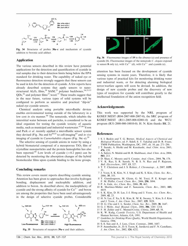
fluorescent sensors for cyanide (Fig. 34).64 In the absence of
cyanide, intramolecular charge transfer (ICT) in these systems
occurs efficiently and the fluorophores in the probes are
effectively quenched. On the other hand, the extent of ICT
from the amino moiety to the pyridinium nitrogen is reduced
in the presence of cyanide, which was attributed to enhanced
electron donation from the cyanide-complexed boronic acid to
the quaternary nitrogen. The decrease in ICT enhances the
intensity of blue-shifted fluorescence. These three water-
soluble fluorescent probes have been used to determine the
free cyanide concentration at concentrations up to physio-
logically lethal levels of 40.5 ppm.
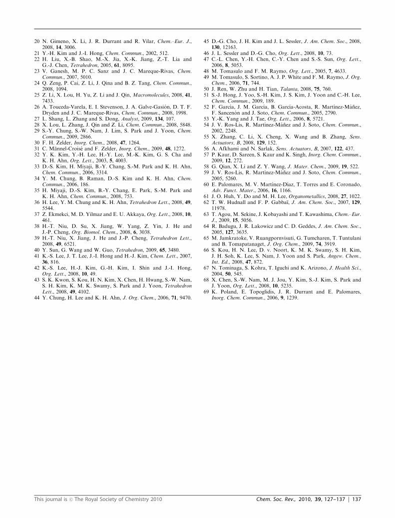
Recently, Tomapatanaget et al. reported acceptor–
donor–acceptor (A–D–A) systems (39a–c) composed of
naphthoquinone, imidazole and boronic acid moieties
(Fig. 35).65 The fluorescence band at 460 nm was switched
on upon the substitution of cyanide on the sensors in a
CTAB micelle. This was attributed to the ITC caused by
the poor acceptability on the boron center after cyanide
addition.
Fig. 29 (a) Structures of chromophores 32a and 32b. (b) Proposed
mechanism for the chemical reaction with cyanide.
Fig. 30 Structures of probes 33, 34a and 34b.
Fig. 31 Structures of receptor 35 and its adduct 35-I.
Fig. 32 Addition products of 36a and 36b with cyanide and fluoride,
respectively.
Fig. 33 Structures of receptor 37 and its adduct 37-I.
This journal is �c The Royal Society of Chemistry 2010 Chem. Soc. Rev., 2010, 39, 127–137 | 135
Application
The various sensors described in this review have potential
applications for the detection and quantification of cyanide in
real samples due to their detection limits being below the EPA
standard for drinking water. The capability of naked eye or
fluorescence detection strongly suggests that these sensors can
be used in kits for the detection of cyanide. A few reports have
already described systems that apply sensors to nano-
structured Al2O3 films,20 SAM,23 polymer backbones,24,25,52
QDs,26 and polymer films37/wool.57 These results suggest that
in the near future, various types of solid systems will be
configured to perform as sensitive and practical ‘‘dip-in’’
naked eye cyanide sensors.
Chemical analysis using portable microfluidic devices
enables environmental testing outside of the laboratory in a
low cost in situ manner.66 The nematode, which inhabits the
interstitial water between soil particles, is considered to be an
ideal organism for testing the cyanide toxicity of aquatic
media, such as municipal and industrial wastewater.67,68 Yoon
and Park et al. recently applied a microfluidic sensor system
they devised (Fig. 36a and b)29,43 to cell-imaging43 and in vivo
imaging of cyanide in Caenorhabditis elegans (Fig. 36c–e).29
The selective sensing of cyanide anions in water by using a
hybrid biomaterial composed of a mesoporous TiO2 film of
crystalline nanoparticles and the protein hemoglobin has also
been reported.69 Low levels of cyanide (o0.2 ppm) can be
detected by monitoring the absorption changes of the hybrid
biomolecular films upon cyanide binding to the heme groups.
Concluding remarks
This review covers recent reports describing cyanide sensing.
Attention has been given to approaches that involve hydrogen
bonding, displacement and nucleophilic addition, and
addition to boron. As described above, the nucleophilicity of
cyanide and the strong affinity of cyanide for Cu2+ and boron
are among the properties that have been used advantageously
in the design of selective cyanide probes. Considerable
attention has been focused on the development of cyanide
sensing systems in recent years. Therefore, it is likely that
various types of practical kits for monitoring drinking water
and industrial waste, or for detecting alarming biological
terror/warfare agents will soon be devised. In addition, the
design of new cyanide probes and the discovery of new
types of receptors for cyanide will contribute greatly to the
intellectual foundation of the anion recognition field.
Acknowledgements
This work was supported by the NRL program of
KOSEF/MEST (R04-2007-000-2007-0), the SRC program of
KOSEF/MEST (R11-2005-008-02001-0) and the WCU
program (R31-2008-000-10010-0). H. N. Kim thanks BK 21.
References
1 S. I. Baskin and T. G. Brewer, Medical Aspects of Chemical andBiological Warfare, ed. F. Sidell, E. T. Takafuji and D. R. Franz,TMM Publication, Washington, DC, 1997, ch. 10, pp. 271–286.
2 T. Suzuki, A. Hiolki and M. Kurahashi, Anal. Chim. Acta, 2003,476, 159.
3 A. Safavi, N. Maleki and H. R. Shahbaazi, Anal. Chim. Acta, 2004,503, 213.
4 D. Shan, C. Mousty and S. Cosnier, Anal. Chem., 2004, 76, 178.5 V. K. Rao, S. R. Suresh, N. B. S. N. Rao and P. Rajaram,Bull. Electrochem., 1997, 13, 327.
6 T. T. Christison and J. S. Rohrer, J. Chromatogr., A, 2007, 1155,31.
7 J. Yoon, S. K. Kim, N. J. Singh and K. S. Kim, Chem. Soc. Rev.,2006, 35, 355.
8 T. Gunnlaugsson, M. Glynn, G. M. Tocci, P. E. Kruger andF. M. Pfeffer, Coord. Chem. Rev., 2006, 250, 3094.
9 P. A. Gale, Acc. Chem. Res., 2006, 39, 465.10 R. Martınez-Manez and F. Sancanon, Chem. Rev., 2003, 103,
4419.11 S. K. Kim, D. H. Lee, J.-I. Hong and J. Yoon, Acc. Chem. Res.,
2009, 42, 23.12 H. N. Lee, Z. Xu, S. K. Kim, K. M. K. Swamy, Y. Kim, S.-J. Kim
and J. Yoon, J. Am. Chem. Soc., 2007, 129, 3828.13 D. G. Cho and J. L. Sessler, Chem. Soc. Rev., 2009, 38, 1647.14 G. J. Mohr, Anal. Bioanal. Chem., 2006, 386, 1201–1214.15 F. H. Zelder and C. Mannel-Croise, Chimia, 2009, 63, 58.16 K. W. Kulig, Cyanide Toxicity, U.S. Department of Health and
Human Services, Atlanta, GA, 1991.17 Guidelines for Drinking-Water Quality, World Health Organization,
Geneva, 1996.18 S.-S. Sun and A. J. Lees, Chem. Commun., 2000, 1687.19 P. Anzenbacher, Jr., D. S. Tyson, K. Jursıkova and F. N. Castellano,
J. Am. Chem. Soc., 2002, 124, 6232.
Fig. 34 Structures of probes 38a–c and mechanism of cyanide
addition to boronic acid adduct.
Fig. 35 Structures of receptors 39a–c and their adducts.
Fig. 36 Fluorescence images of 19 in the absence (a) and presence of
cyanide (b). Fluorescence images of the nematode C. elegans exposed
to sensor 8 only (c), with Cu2+ (d), with Cu2+ and cyanide (e).
136 | Chem. Soc. Rev., 2010, 39, 127–137 This journal is �c The Royal Society of Chemistry 2010
20 N. Gimeno, X. Li, J. R. Durrant and R. Vilar, Chem.–Eur. J.,2008, 14, 3006.
21 Y.-H. Kim and J.-I. Hong, Chem. Commun., 2002, 512.22 H. Liu, X.-B. Shao, M.-X. Jia, X.-K. Jiang, Z.-T. Lia and
G.-J. Chen, Tetrahedron, 2005, 61, 8095.23 V. Ganesh, M. P. C. Sanz and J. C. Mareque-Rivas, Chem.
Commun., 2007, 5010.24 Q. Zeng, P. Cai, Z. Li, J. Qina and B. Z. Tang, Chem. Commun.,
2008, 1094.25 Z. Li, X. Lou, H. Yu, Z. Li and J. Qin, Macromolecules, 2008, 41,
7433.26 A. Touceda-Varela, E. I. Stevenson, J. A. Galve-Gasion, D. T. F.
Dryden and J. C. Mareque-Rivas, Chem. Commun., 2008, 1998.27 L. Shang, L. Zhang and S. Dong, Analyst, 2009, 134, 107.28 X. Lou, L. Zhang, J. Qin and Z. Li, Chem. Commun., 2008, 5848.29 S.-Y. Chung, S.-W. Nam, J. Lim, S. Park and J. Yoon, Chem.
Commun., 2009, 2866.30 F. H. Zelder, Inorg. Chem., 2008, 47, 1264.31 C. Mannel-Croise and F. Zelder, Inorg. Chem., 2009, 48, 1272.32 Y. K. Kim, Y.-H. Lee, H.-Y. Lee, M.-K. Kim, G. S. Cha and
K. H. Ahn, Org. Lett., 2003, 5, 4003.33 D.-S. Kim, H. Miyaji, B.-Y. Chang, S.-M. Park and K. H. Ahn,
Chem. Commun., 2006, 3314.34 Y. M. Chung, B. Raman, D.-S. Kim and K. H. Ahn, Chem.
Commun., 2006, 186.35 H. Miyaji, D.-S. Kim, B.-Y. Chang, E. Park, S.-M. Park and
K. H. Ahn, Chem. Commun., 2008, 753.36 H. Lee, Y. M. Chung and K. H. Ahn, Tetrahedron Lett., 2008, 49,
5544.37 Z. Ekmekci, M. D. Yilmaz and E. U. Akkaya, Org. Lett., 2008, 10,
461.38 H.-T. Niu, D. Su, X. Jiang, W. Yang, Z. Yin, J. He and
J.-P. Cheng, Org. Biomol. Chem., 2008, 6, 3038.39 H.-T. Niu, X. Jiang, J. He and J.-P. Cheng, Tetrahedron Lett.,
2008, 49, 6521.40 Y. Sun, G. Wang and W. Guo, Tetrahedron, 2009, 65, 3480.41 K.-S. Lee, J. T. Lee, J.-I. Hong and H.-J. Kim, Chem. Lett., 2007,
36, 816.42 K.-S. Lee, H.-J. Kim, G.-H. Kim, I. Shin and J.-I. Hong,
Org. Lett., 2008, 10, 49.43 S. K. Kwon, S. Kou, H. N. Kim, X. Chen, H. Hwang, S.-W. Nam,
S. H. Kim, K. M. K. Swamy, S. Park and J. Yoon, TetrahedronLett., 2008, 49, 4102.
44 Y. Chung, H. Lee and K. H. Ahn, J. Org. Chem., 2006, 71, 9470.
45 D.-G. Cho, J. H. Kim and J. L. Sessler, J. Am. Chem. Soc., 2008,130, 12163.
46 J. L. Sessler and D.-G. Cho, Org. Lett., 2008, 10, 73.47 C.-L. Chen, Y.-H. Chen, C.-Y. Chen and S.-S. Sun, Org. Lett.,
2006, 8, 5053.48 M. Tomasulo and F. M. Raymo, Org. Lett., 2005, 7, 4633.49 M. Tomasulo, S. Sortino, A. J. P. White and F. M. Raymo, J. Org.
Chem., 2006, 71, 744.50 J. Ren, W. Zhu and H. Tian, Talanta, 2008, 75, 760.51 S.-J. Hong, J. Yoo, S.-H. Kim, J. S. Kim, J. Yoon and C.-H. Lee,
Chem. Commun., 2009, 189.52 F. Garcıa, J. M. Garcıa, B. Garcıa-Acosta, R. Martınez-Manez,
F. Sancenon and J. Soto, Chem. Commun., 2005, 2790.53 Y.-K. Yang and J. Tae, Org. Lett., 2006, 8, 5721.54 J. V. Ros-Lis, R. Martınez-Manez and J. Soto, Chem. Commun.,
2002, 2248.55 X. Zhang, C. Li, X. Cheng, X. Wang and B. Zhang, Sens.
Actuators, B, 2008, 129, 152.56 A. Afkhami and N. Sarlak, Sens. Actuators, B, 2007, 122, 437.57 P. Kaur, D. Sareen, S. Kaur and K. Singh, Inorg. Chem. Commun.,
2009, 12, 272.58 G. Qian, X. Li and Z. Y. Wang, J. Mater. Chem., 2009, 19, 522.59 J. V. Ros-Lis, R. Martınez-Manez and J. Soto, Chem. Commun.,
2005, 5260.60 E. Palomares, M. V. Martınez-Dıaz, T. Torres and E. Coronado,
Adv. Funct. Mater., 2006, 16, 1166.61 J. O. Huh, Y. Do and M. H. Lee, Organometallics, 2008, 27, 1022.62 T. W. Hudnall and F. P. Gabbaı, J. Am. Chem. Soc., 2007, 129,
11978.63 T. Agou, M. Sekine, J. Kobayashi and T. Kawashima, Chem.–Eur.
J., 2009, 15, 5056.64 R. Badugu, J. R. Lakowicz and C. D. Geddes, J. Am. Chem. Soc.,
2005, 127, 3635.65 M. Jamkratoke, V. Ruangpornvisuti, G. Tumcharen, T. Tuntulani
and B. Tomapatanaget, J. Org. Chem., 2009, 74, 3919.66 S. Kou, H. N. Lee, D. v. Noort, K. M. K. Swamy, S. H. Kim,
J. H. Soh, K. Lee, S. Nam, J. Yoon and S. Park, Angew. Chem.,Int. Ed., 2008, 47, 872.
67 N. Tominaga, S. Kohra, T. Iguchi and K. Arizono, J. Health Sci.,2004, 50, 545.
68 X. Chen, S.-W. Nam, M. J. Jou, Y. Kim, S.-J. Kim, S. Park andJ. Yoon, Org. Lett., 2008, 10, 5235.
69 K. Poland, E. Topoglidis, J. R. Durrant and E. Palomares,Inorg. Chem. Commun., 2006, 9, 1239.
This journal is �c The Royal Society of Chemistry 2010 Chem. Soc. Rev., 2010, 39, 127–137 | 137