Embed Size (px)
Citation preview

Sensors 2001, 1, 148-160
sensorsISSN 1424-8220© 2001 by MDPI
http://www.mdpi.net/sensors
Evaluation of Protein Adsorption on Chitosan Surfaces withReflectometry Interference Spectroscopy
Xiao Ying Lü∗∗, Yan Huang and Chao Qun Ma
Key Laboratory of Molecular and Biomolecular Electronics of the Ministry of Education, P. R. China.
Southeast University, 210096, Nanjing, China. Tel: (86)-25-3793430. Fax: (86)-25-7712719.
E-mail: [email protected]. Homepage: http://www.lmbe.seu.edu.cn
* Author to whom correspondence should be addressed.
Received: 4 September 2001 / Accepted: 25 September 2001 / Revised: 10 October 2001 /
Published: 12 October 2001
Abstract: Using a biomedical sensor setup RIfS we have investigated the kinetic behavior
of human albumin (Alb), human fibrinogen (Fib), and human immunoglobulin G (IgG)
adsorbed onto surfaces of chitosan. Polystyrene (PS) was used as the control material in this
study. The optical thickness of three kinds of proteins measured by RIfS was related to their
molecular dimensions and potential orientations on a film surface. According to the
operation principle of RIfS and the molecular dimensions of three kinds of proteins, the
adsorbed layers of proteins onto the surface of chitosan and PS was calculated by using a
newly introduced equation. The microstructure of the chitosan and polystyrene film and the
surfaces with adsorbed proteins were imaged by atomic force microscopy (AFM). With
AFM analyses the lateral distribution of the protein molecules on surfaces have been
recognized. The results show that the number of adsorbed layers of the three proteins on the
surface of chitosan are 0.635 for Alb, 0.158 for Fib and 0.0967 for IgG, and of polystyrene
are: 0.577 for IgG, 0.399 for Fib, 0.336 for Alb. This study confirmed that RIfS is a useful
tool for the analysis of plasma proteins adsorbed on a surface of biomaterials. Results show
that at first on the surface of chitosan film much more Alb than Fib was adsorbed which
demonstrated that chitosan has a antithrombus function. Secondly, on the surface of chitosan
film more Alb and less Fib were adsorbed than on the surface of PS film, which
demonstrated that chitosan has a better blood compatibility than polystyrene. Thirdly, the
calculated layer number of the three proteins indicated that on both chitosan and PS
substrates monolayer coatings form.
Key words: Biomedical sensor, RIfS, chitosan, Protein adsorption, Biomaterials

Sensors 2001, 1 149
1. Introduction
Optical interferomertry is well known for the measurement of physical parameters such as speed and
distance. Further, light interference was used for visual detection of antigen-antibody reactions [1].
Spectral interferometry has been considered as an interesting approach to detecting sensitively and
rapidly any changes of optical path length in thin polymer films caused by antigen–antibody interaction
in immunochemical sensing [2]. After the development of surface plasmon resonance (SPR) [3,4],
another successful optical approach, Reflectometry Interference Spectroscopy (RIfS), was introduced
as a highly sensitive and robust technique for direct label-free monitoring of the interaction of
biomolecules [5,6,7].
RIfS is based on the spectral distribution of white-light reflectance from transparent thin layers. A
distinct reflectance pattern with alternating maxima and minima results from the interference of beams
partially reflected from each interface of the interference layer. Binding detection with RIfS shows
little temperature dependence, allows avoidance of gold layers, and is promising for high throughput
screening. The simple test format and high sensitivity makes RIfS attractive for detecting and
characterizing the interaction of biomolecules. Biological material deposited at the surface during a
binding event increases the optical thickness of the interference layer, leading to a shift in the
interference pattern. This approach allows on-line monitoring of binding reactions with high resolution
[8]. The main advantages of the RIfS transducer are its ruggedness, the small active area, and the
simple construction [9].
A sensitive dynamic determination made on gaseous or liquid hydrocarbons as well as
immunoreactions with RIfS was reported in 1993 [6]. Then, an investigation of the specific binding of
low molecular weight ligands to immobilized receptors was carried out by using RIfS; two model
systems were studied in which the binding event was successfully detected [10]. Next, RIfS was
utilized for studying the hybridization of antisense with its complementary target sequences and
especially to investigate the interaction of terminal- and backbone-modified antisense ONs targeted to
multidrug resistance1b-mRNA [8]. Other applications with RIfS were in a dyeless optical detection of
ammonia in the gas phase, by using pH-responsive polymers. Also, clinical detection of HbsAGb was
accomplished [11, 12]. Recently, the application of RIfS for the evaluation of the biocompatibility of
biomaterials has been described [13].
Since the protein adsorption on the surface of biomaterials is an important factor for the adhesion of
platelet and biocompatibility, the characterization of adsorbed proteins on the surface of various
biomaterials, such as biopolymers, alloys, ceramics etc., has been extensively investigated by means of
different techniques [14-26].
Chitosan is a natural source polymer prepared by N-deacetylation of chitin, which is the main
structural component of crab and shrimp shells [27]. Intensive investigations have been carried out for
the structure, the chemistry, the biological properties, and the applications of chitosan [28].
Chitosan has been widely used as a new biomaterial since it is safe and has a good biocompatibility
with the human body [29-35]. Chitosan also has a haemostatic function [36] and may have an
antithrombogenic function. Some investigators reported the platelet adhesion on the surface of
modified chitosan [37,38]. However, the protein adsorption occurrs before the platelet adhesion and is
the first step of thrombosis. The aim of this study is to evaluate the protein adsorption behavior of

Sensors 2001, 1 150
human albumin (Alb), human fibrinogen (FIb), and human IgG on chitosan, by using RIfS. The
kinetics of the adsorption process is investigated. Polystyrene is used as the control material, since its
surface chemistry is well defined and it is frequently used as a model system in protein adsorption
studies [39].
2. Materials and Methods
2.1 RIfS Apparatus
The RIfS setup used has been developed in our laboratory [40]. The main elements of the setup
were a CCD spectrometer with a bifurcated optical fiber and an adsorption cell with a volume of about
30 µL. The bifurcated optical fiber was used to illuminate the transducer chip and to collect the light
reflected from the chip. The chip is a glass substrate on which a 20-nm Ta2O5 film was deposited in
order to enhance the interference efficiency. An original software was used to control the collection of
full spectra, to analyze the experimental data in real-time, and to draw the experimental curves on-line.
2.2 Preparation of Chitosan and PS films
Chitosan with a degree of deacetylation of 93.2% and of an average molecular weight of 750,000 Da
was purchased from Ocean Institute of Jiangsu, China. The material is powder form with 80-100nm
granularities and has a white color. The thin films of chitosan exposed to proteins were prepared
by the spin-coating method. At first, the chitosan was immersed in 0.5% acetic acid (Factory of
Chemical Reagents of Nanjing, China) with 10mg/mL. After the chitosan was dissolved completely,
the solution was filtered with a middle speed qualitative filter paper (Factory of Fuyang Special
Paper of Hangzhou, China). Then, 50-µL chitosan solution was pipetted out and dropped onto
the surface of the transducer chip (1.2cm*1.2cm ). A chitosan film was spin-coated on the
transducer chip using a spinning-machine at 1700 rpm. Typically, the film thickness was ~650 nm.
Polystyrene (Nunc Inc. Denmark) was dissolved in toluene ( Factory of Chemical Reagents of
Nanjing, China) at a concentration of 40mg/mL. The coating process of polystyrene film on the
transducer chip is same with the chitosan film. All materials and reagents as well as chemicals were of
analytical grade or better.
2.3. Proteins
Three kinds of proteins were used : human albumin (Alb), human fibrinogen (Fib), and human IgG
(Sigma, Dorset, UK). For each measurement a protein solution was prepared freshly in 10mM
phosphate buffer solution (PBS, pH=7.4) of a concentration of 0.1mg/mL. Deionized water was used to
prepare the aqueous solutions.
2.4 Protein Adsorption Measurement
The protein adsorption measurement has been performed as follows: the sensor chip coated with
chitosan/PS film was mounted into the adsorption cell carefully and fixed with two clips. Then, the

Sensors 2001, 1 151
peristaltic pump was started to drive PBS (pH=7.4) flow into the adsorption cell. The PBS cleaned the
surface of the chitosan/PS film, drove out air bubbles that may be present in the adsorption cell. Most
importantly, it produced a physiological pH-environment for the adsorption of proteins. After this
treatment, the protein solution flowed into the adsorption cell at a rate of 200 µL/min and the kinetic
adsorption process of proteins on the surface of the chitosan/PS film can be oberseved in situ on the
monitor of the computer. Until the kinitic adsorption curve of proteins became stable, PBS was
pumped into the adsorption cell again to wash the proteins which did not adsorb tightly onto the
material surface. The wash handing would get a response on the adsorption curve. The mark on the Y
axis of the Figs.1-4 give the thickness of the proteins adsorbted on the chitosan/PS film. For each kind
of proteins, at least three tests were performed (n=3).
2.5 Calculation of the Adsorbed Layer of Protein
Since the molecular dimensions of various proteins are different, a normalized treatment of the
experimental data was performed using a previously defined equation [13] in order to obtain the
effective adsorbed layer number Lp (Eq.1):
pp
p
xL
D=
(1)
where Dp is the molecular dimension of a single protein and px , the average thickness of each type
of proteins on the surface of the material intended to be probed with RIfS.
2.6 AFM Imaging
The microstructure of the chitosan and polystyrene films and the surfaces with adsorbed proteins
were imaged with a commercial atomic force microscope (AFM, Nanoscope IIIa, Digital Instrument,
Santababara, USA). The instrument was operated in Tapping Mode using silicon cantilevers oscillating
with the average amplitude of 100 nm and a resonance frequency between 200 and 450 kHz. The
scanning rate selected was 1 Hz. All images presented in this paper were obtained repeatedly and were
stable under the experimental conditions.
3. Results and Discussion
3.1 Kinetics of Protein Adsorption onto PS Films
In Fig.1 the kinetic adsorption curves of human Alb, IgG and Fib onto the surface of a PS film are
shown. The curves evidence that the adsorption behavior of the three proteins onto PS film are similar:
initially, the processes of all three proteins increase quickly in the initial stage and reach their
equilibrium values after about 500 s. Secondly, after washed with PBS the adsorption curves have
different tendencies: the curve of IgG falls about 0.2nm, which means some loosely adsorbed
molecules were removed from the PS film. Nevertheless, the curves of Alb and Fib show almost no
Sensors 2001, 1 152
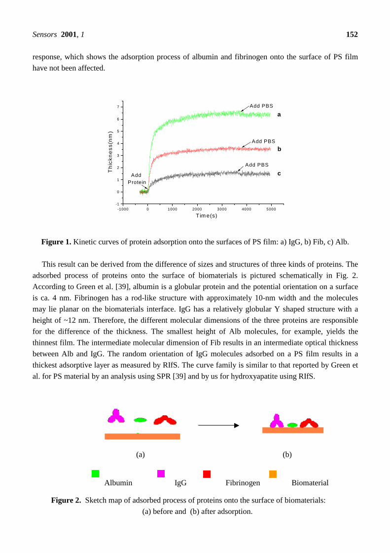
response, which shows the adsorption process of albumin and fibrinogen onto the surface of PS film
have not been affected.
-1000 0 1000 2000 3000 4000 5000-1
0
1
2
3
4
5
6
7
c
b
a
Add PBS
Add PBS
Add PBS
Add Protein
Th
ick
ne
ss
(nm
)
T im e(s)
Figure 1. Kinetic curves of protein adsorption onto the surfaces of PS film: a) IgG, b) Fib, c) Alb.
This result can be derived from the difference of sizes and structures of three kinds of proteins. The
adsorbed process of proteins onto the surface of biomaterials is pictured schematically in Fig. 2.
According to Green et al. [39], albumin is a globular protein and the potential orientation on a surface
is ca. 4 nm. Fibrinogen has a rod-like structure with approximately 10-nm width and the molecules
may lie planar on the biomaterials interface. IgG has a relatively globular Y shaped structure with a
height of ~12 nm. Therefore, the different molecular dimensions of the three proteins are responsible
for the difference of the thickness. The smallest height of Alb molecules, for example, yields the
thinnest film. The intermediate molecular dimension of Fib results in an intermediate optical thickness
between Alb and IgG. The random orientation of IgG molecules adsorbed on a PS film results in a
thickest adsorptive layer as measured by RIfS. The curve family is similar to that reported by Green et
al. for PS material by an analysis using SPR [39] and by us for hydroxyapatite using RIfS.
(a) (b)
Albumin IgG Fibrinogen Biomaterial
Figure 2. Sketch map of adsorbed process of proteins onto the surface of biomaterials:
(a) before and (b) after adsorption.

Sensors 2001, 1 153
3.2 Kinetics of Protein Adsorption onto Chitosan Films
The kinetic adsorption curves of human Alb, Fib, and IgG on the surface of a chitosan film are
shown in Fig. 3−5. According to these curves the adsorption behavior of three kinds of proteins onto
chitosan is dissimilar. For Alb, the adsorption processes amplifies slowly in the initial stage and
reaches its stable values after about 8000 s. This means that during this period the adsorption of
albumin on chitosan continued. Nevertheless, for Fib and IgG the adsorption processes is fast in the
initial stage and reaches its stable values after about 1500 s, which means that the adsorption process of
Fib and IgG on chitosan films reaches saturation fast. After washed with PBS, the curves of Alb, Fib,
and IgG fall apparently, which indicates more loosely adsorbed molecules were removed from the
chitosan film.
-2000 0 2000 4000 6000 8000 10000 12000 14000 16000 18000-0.5
0.0
0.5
1.0
1.5
2.0
2.5
3.0
3.5
Add PBS
Add Albumin
Th
ickn
ess
(nm
)
Time(s)
Figure 3. Kinetic curves of adsorbed Alb on the surface of chitosan film.
-2000 -1000 0 1000 2000 3000 4000 5000 6000-0.5
0.0
0.5
1.0
1.5
2.0
2.5
Add Fibrinogen
Add PBS
Thi
ckn
ess
(nm
)
Time(s)
Figure 4. Kinetic curves of adsorbed Fib on the surface of chitosan film.

Sensors 2001, 1 154
-1000 0 1000 2000 3000 4000 5000-0.5
0.0
0.5
1.0
1.5
2.0
2.5
Add IgG
Add P BS
Th
ick
ne
ss
(nm
)
T im e(s )
Figure 5. Kinetic curves of adsorbed IgG on the surfaces of chitosan film.
3.3 Comparing of the Kinetic Curves on Chitosan and PS
Fig. 6-8 compare the adsorption of the three kinds of proteins onto the surface of chitosan and PS
films. The curves reveal that on the surface of chitosan film more Alb was adsorbed than on the surface
of PS film, and conversely, on the surface of PS film more Fib and IgG were adsorbed than on the
surface of chitosan films.
-3000 0 3000 6000 9000 12000 15000
0.0
0.5
1.0
1.5
2.0
2.5
3.0
b
a
Add PBS
Add PBS
Add Albumin
Thi
ckne
ss(n
m)
Time(s)
Figure 6. Adsorbed Alb on the surfaces of chitosan and PS: a) chitosan, b) PS.

Sensors 2001, 1 155
-1000 0 1000 2000 3000 4000 5000 6000
0
1
2
3
4
Add Fibrinogen
Add PBS
Add PBS
b
a
Thi
ckne
ss(n
m)
Time(s)
Figure 7. Adsorbed Fib on the surfaces of chitosan and PS: a) PS, b) chitosan.
-1000 0 1000 2000 3000 4000 5000
0
1
2
3
4
5
6
7
b
a
Add PBS
Add PBS
Add IgG
Th
ick
ne
ss
(nm
)
T im e(s)
Figure 8. Adsorbed IgG on the surfaces of chitosan and PS: a) PS, b) chitosan.
3.4 Adsorbed Layers of Alb, Fib, and IgG on Chitosan and PS Films
As discussed above, each adsorption curve indicates the thickness of one particular protein adsorbed
onto the surface of one kind of material. Thus, the adsorptive thickness of one kind of proteins on films
of different materials can be compared directly according to the curve family as in Figs. 6-8. But with
the RIfS thickness a comparison of the adsorbed amount of different proteins on the same materials
cannot be carried out directly. Since the structure and size of three proteins are very different. After a
normalized treatment of the experimental data using the equation, the thickness of the adsorbed protein
layers was calculated, which indicates the effectively adsorbed amount of different proteins on one
particular material.

Sensors 2001, 1 156
RIfS thickness of three kinds of proteins on the PS surface decreases in the order (Tab.1): IgG
(6.35nm) > Fib (3.59nm) > Alb (1.34nm). The adsorbed layer number, calculated using the equation,
PS has same series: IgG (0.577) > Fib (0.399) > Alb. (0.336).
The thickness determined by RIfS for the three kinds of proteins on the chitosan surface decreases
in the order (Tab.2): Alb (2.54nm) > Fib (1.42nm) > IgG (1.06nm). By this order a special phenomena
for albumin adsorbed onto chitosan film is recognized. Despite the smallest structure the adsorbed RIfS
thickness of Alb is greatest among the three kinds of proteins. This result is very different from the
results of PS and of another studies [13, 39]. It indicates that chitosan adsorbed much more Alb than
Fib and IgG. In this case the adsorbed layer number of three kinds of proteins on chitosan decreases in
the same order: Alb (0.635) > Fib (0.158) > IgG (0.0967).
The calculated layer numbers of three kinds of proteins show that the total amount of each kind of
protein adsorbed on chitosan and PS occurs as monolayer adsorption.
According to previous studies, albumin has a thrombus-resistant ability, whereas fibrinogen
promotes platelet adhesion on a polymer surface [41]. The calculated number of adsorbed layers of the
three proteins on chitosan and PS evidences that the chitosan material shows a higher affinity to Alb
and lower affinity to Fib than PS. This means that chitosan has a better antithrombus function relative
to PS films.
Table 1. RIfS thickness and adsorbed layer number of three kinds of proteins on the PS film.
Thickness Proteinsx (n=3) SD
Adsorbed
Layer
Alb 1.34 0.0868 0.336
Fib 3.59 0.0586 0.399
IgG 6.35 0.0430 0.577
Table 2. RIfS thickness and adsorbed layer number of three kinds of proteins on the chitosan film.
Thickness Proteinsx (n=3) SD
Adsorbed
Layer
Alb 2.54 0.0193 0.635
Fib 1.42 0.0220 0.158
IgG. 1.06 0.0505 0.0967
3.5 AFM characteristic
AFM analysis is useful for probing the lateral distribution of the protein molecules with the
monolayer and confirmed indirectly the presence of the passively adsorbed protein on the surfaces of
biomaterials. The AFM image in Figs. 9(a) and figure10(a) are the microstructure of the chitosan and
PS surface, while in Figs. 9(b) and 10(b) the same surfaces with adsorbed protein albumin.
From the AFM image in Fig. 9(a), the pattern in the surface of chitosan material can be revealed.
The AFM images of albumin coated chitosan surface in figure9(b) reveal that the most area of the
surface of chitosan film was covered with the proteins.

Sensors 2001, 1 157
The AFM image of PS film in Fig. 10(a) termed as ultra-flat film [42]. The AFM image in Fig.
10(b) reveal that there is fewer albumin on PS film than on chitosan film in figure 9(b).
AFM analyses are consistent with the calculated adsorbed layer in Tab. 1. and Tab. 2.
(a) (b)
Figure 9. AFM image of microstructure of a chitosan surface (a) and with adsorbed albumin (b).
(a) (b)
Figure 10. AFM image of microstructure of a PS surface (a) and with adsorbed albumin (b).
4. Conclusions
This study confirmed that RIfS is a useful tool for analysis of plasma proteins adsorbed on a surface
of biomaterials. The number of adsorbed layers of three kinds of proteins on the surface of chitosan has
a series: Alb > Fib > IgG and PS has a series: IgG > Fib > Alb. Results reported in this paper show that
at first on the surface of chitosan film more Alb and fewer Fib adsorbs, which demonstrates that
chitosan has a antithrombus function and secondly, on the surface of chitosan film more Alb and less
Fib than on the surface of PS film, which demonstrated that chitosan has a better blood compatibility
than polystyrene. And thirdly, according to the calculated layer number of the three proteins all the
proteins on chitosan and on PS materials represents a monolayer adsorption.
Acknowledgements
We are thankful to the Ministry of Education of China for the support by the “Promotion Foundation
for Scholars from Abroad” (1998-2000).

Sensors 2001, 1 158
References
1. Kawaguchi, T.; Shiro, T.; Iwata, K. A highly sensitive device for visual detection of antigens and
antibodies by means of light interference. Sensors and Actuators B 1991, 3, 113-121.
2. Brecht, A.; Gauglitz, G.; Nahm, W. Interferometric measurement used in chemical and biochemical
sensors. Analysis 1992, 20, 135-140.
3. Myszka, D.G. Kinetic analysis of macromolecular interactions using surface plasmon resonance
biosensors. Current Opinion in Biotechnology 1997, 8, 50-57.
4. Green, R.J.; Frazier, R.A.; Shakesheff, K.M.; Davies, M.C.; Roberts, C.J.; Tendler, S.J.B. Surface
plasmon resonance analysis of dynamic biological interactions with biomaterials. Biomaterials
2000, 21, 1823-1835.
5. Brecht, A.; Ingenhoff, J.; Gauglitz, G. Direct monitoring of antigen-antibody interactions by
spectral interferometry. Sensors and Actuators B 1992, 6, 96-100.
6. Gauglitz, G.; Brecht, A.; Kraus, G.; Nahm, W. Chemical and biochemical sensors based on
interferometry at thin (multi-) layers. Sensors and Actuators B 1993, 11, 21-27.
7. Schmitt, H.M.; Brecht, A.; Piehler, J.; Gauglitz, G. An integrated system for optical biomolecular
interaction analysis. Biosensors & Bioelectronics 1997, 12(8), 809-816.
8. Sauer, M.; Brecht, A.; Charissé, K.; Maier, M.; Gerster, M.; Stemmler, I.; Gauglitz, G.; Bayer, E.
Interaction of Chemically Modified antisense oligonucleotides with sense DNA: a label-free
interaction study with reflectometric interference spectroscopy. Analytical Chemistry 1999, 71,
2850-2857.
9. Brecht, A.; Gauglitz, G. Optimised layer systems for immunosensors used on the RIFS transducer.
J Anal Chem. 1994, 349, 360-366.
10. Piehler, J.; Brecht, A.; Gauglitz, G.; Maul, C.; Grabley, S.; Zerlin, M. Specific binding of low
molecular weight ligands with direct optical detection. Biosensors & Bioelectronics 1997, 12(6),
531-538.
11. Rathgeb, F.; Gauglitz, G. Dyeless optical detection of ammonia in the gas phase using pH-
responsive polymers with reflectometric interference spectroscopy. Analytica Chimica Acta 1998,
372, 333-340.
12. Yu, F.; Yao, D.F.; Qian, W.P.; et al. Reflectometry. interference spectroscopy in detection of
hepatitis B surface antigen. Clin. Chem. 2000, 46(9), 1489-1490.
13. Lü, X.Y.; Huang, H.F.; Chen, D.M.; et al.. Real time in situ kinetic analysis of proteins adsorbed
onto the surface of hydroxyapatite film using RIfS. Proceedings of the first international conference
on biomaterials (China) July 24-26,2001, Beijing, China.
14. Sharma, C.P.; Sunny, M.C. Albumin adsorption on to aluminum oxide and polyurethane surfaces.
Biomaterials 1990, 11, 255-257.
15. Zhang, S.J.; Li, D.J.; Zhao, J.; Gu, H.Q. A study of blood protein adsorption on diamond-like
carbon. Chinese Conference on Biomaterials. Panyu. China 1999 Nov; 5-8.
16. Babensee, J.E.; Cornelius, R.M.; Brash, J.L.; Sefton, M.V. Immunoblot analysis of proteins
associated with HEMA-MMA microcapsules: Human serum proteins in vitro and rat proteins
following implantation. Biomaterials 1998, 19, 839-849.
Sensors 2001, 1 159
17. Kandori, K.; Fujiwara, A.; Mukai, M.; Yasukawa, A.; Ishikawa, T. Evaluation of the adsorption
affinity of proteins to calcium hydroxyapatites by desorption and pre-adsorption methods. Colloids
and Surfaces B: Biointerfaces 1998, 11, 313-320.
18. Klomp, A.J.A.; Engbers, G.H.M.; Mol, J.; Terlingen, J.G.A.; Feijen, J. Adsorption of proteins from
plasma at polyester non-wovens. Biomaterials 1999, 20, 1203-1211.
19. Chittur, K.K. FTIR/ATR for protein adsorption to biomaterial surfaces. Biomaterials 1998, 19,
357-369.
20. Zeng, H.; Chittur, K.K.; Lacefield, W.R. Analysis of bovine serum albumin adsorption on calcium
phosphate and titanium surfaces. Biomaterials 1999, 20, 377-384.
21. Kingshott, P.; Heather, A.W.; John, S.T.; Griesser, H.J. Direct detection of proteins adsorbed on
synthetic materials by matrix-assisted laser desorption ionization-mass spectrometry. Analytical
Biochemistry 1999, 273, 156-162.
22. Oleschuk, R.D.; McComb, M.E.; Chow, A.; Ens, W.; Standing, K.G.; Perresult, H.; Mariois, Y.;
King, M. Characterization of plasma proteins adsorbed onto biomaterials by MALDI-TOFMS.
Biomaterials 2000, 21, 1701-1710.
23. Ortega-Vinuesa, J.L.; Tengvall, P.; Wälivaara, B.; Lundström, I. Stagnant versus dynamic
conditions: a comparative adsorption study of blood proteins. Biomaterials 1998, 19, 251-262.
24. Zhang, M.; Desai, T.; Ferrari, M. Proteins and cells on PEG immobilized silicon surfaces.
Biomaterials 1998, 19, 953-960.
25. Elwing, H. Protein absorption and ellipsometry in biomaterial research. Biomaterials 1998, 19,
397-406.
26. Tengyall, P.; Lundström, I.; Liedberg, B. Protein adsorption studies on model organic surfaces: an
ellipsometric and infrared spectroscopic approach. Biomaterials 1998, 19, 407-422.
27. Hirano, S.; Zhang, M.; Nakagawa, M.; Miyata, T. Wet spun chitosan-collagen fibers, their chemical
N-modifications, and blood compatibility. Biomaterials 2000, 21, 997-1003.
28. Gu, H.Q.; Xu, G.F. Biomedical Materials. Publishing Company. Of Translate for Sci and Tech.
Tianjing, China. 1993, p316-324.
29. Zhang, J.X.; Tang, J.; Xu, B. Biocompatibility and safety evaluation of chitosan rod. J Biomed Eng.
(Chinese) 1996, 13(4), 293-297.
30. Jiang, X.S.; Wang, B.S.; Chen, C.; Li, X.G.; Sun, F.Y. The bioactivity and medical appliance of
chitin and its ramification. J Biomed Eng. (Chinese) 1996, 13(4), 353-356.
31. Andrew, C.A.W.; Khor, E.; Hastings, G.W. The influence of anionic chitin derivatives on calcium
phosphate crystallization. Biomaterials 1998, 19, 1309-1316.
32. Leroux, L.; Freche, Z., Frèche, M.; Lacout, J.L. Effects of various adjuvants (lactic acid, glycerol,
and chitosan) on the injectability of a calcium phosphate cement. Bone 1999 Supplement August;
25(2), 318-348.
33. Chenite, A.; Chaput, C.; Wang, D.; Combes, C.; Buschmann, M.D.; Hoemann, C.D.; Leroux, J.C.;
Atkinson, B.L.; Binette, F.; Selmani, A. Novel injectable neutral solutions of chitosan form
biodegradable gels in situ. Biomaterials 2000, 21, 2155-2161.

Sensors 2001, 1 160
34. Campos, A.M.D.; Sánchez, A.; Alonso, M.J. Chitosan nanoparticles: a new vehicle for the
improvement of the delivery of drugs to the ocular surface. Application to cyclosporin A.
International Journal of Pharmaceutics 2001, 224, 159-168.
35. Van der Lubben, I.M.; Verhoef, J.C.; Van Aelst, A.C.; Borchard, G.; Junginger, H.E. Chitosan
microparticles for oral vaccination: preparation, characterization and preliminary in vivo uptake
studies in murine Peyer’s patches. Biomaterials 2001, 22(7), 687-694.
36. Yang, J.; Tian, F.; Chen, S.Q. The mechanism of chitosan hemostasis and its application. Alien
Medical: Biomedical and engineering 2001, 24(2), 77-80.
37. Amiji, M.M. Platelet adhension and activation on an amphoteric chitosan derivative bearing
sulfonate groups. Colloids and Surfaces B:Biointerfaces 1998, 10, 263-271.
38. Lin, C.W.; Lin, J.C. Surface characterization and platelet compatibility evalution of surface-
sulfonated chitosan membrane. J Biomater Sci Polym Ed. 2001, 12(5), 543-557.
39. Green, R.J.; Davies, J.; Davies, M.C.; Robert, C.J.; Tendler, S.J.B. Surface plasmon resonance for
real time in situ analysis of protein adsorption to polymer surfaces. Biomaterials 1997, 18, 405-
413.
40. Yu, F. The establishment of reflectometry interference spectroscopy and its use in the token of
interface biomolecule. M.S.Thesis: Biomedical and Engineering Department of Southeast
University, Nanjing, China., July 2000.
41. Huang, S.L.; Chao, M.S.; Ruan, R.C.; Lai, J.Y. Microphase separated structure and protein
adsorption of polyurethanes with butadiene soft segment. European Polymer Journal 2000, 36,
285-294.
42. Qian, W.P.; Yao, D.F.; Yu, F.; Xu, B.; Zhou, R.; Bao, X.; Lu, Z.H. Immobilization of antibodies on
ultraflat polystyrene surfaces. AACC’s Oak Ridge Conference, Bosten, USA. 2000 May; 5-6.
Sample Availability: Available from the author.
© 2001 by MDPI (http://www.mdpi.net). Reproduction is permitted for noncommercial purposes.


![TRINIDAD AND TOBAGO GAZETTE 2001/2001 Gaz no 148.p… · 694 TRINIDAD AND TOBAGO GAZETTE [August 7, 2001] 1537—Continued APPOINTMENTS, ACTING APPOINTMENTS, ETC.—CONTINUED Appointment](https://img.dokumen.tips/doc/110x75/5e9201805d1ff9166c43de82/trinidad-and-tobago-20012001-gaz-no-148p-694-trinidad-and-tobago-gazette-august.jpg)