Embed Size (px)
Citation preview

Inserting membrane proteins in bacteria: characterizing the YidC and
Sec substrate determinants
Senior Honors Thesis
Presented in partial fulfillment of the requirements for graduation with distinction in
Biochemistry in the college of Arts and Sciences at The Ohio State University
By
Abdul Wasey
Spring 2013
Project Advisor: Dr. Ross E. Dalbey, Department of Chemistry
Project Mentors: Dr. Lu Zhu, Department of Chemistry
Raunak Soman, Department of Chemistry

Abstract
In this project, we investigated what determines whether a membrane protein inserts by the
YidC/Sec-independent pathway, the YidC-only pathway, or the YidC/Sec pathway. Specifically,
we wish to characterize the features of a membrane protein that determine path dependence. Our
studies find that a membrane protein with a highly hydrophobic transmembrane segment inserts
via the YidC/Sec-independent pathway. Addition of a negatively charged residue into the
translocated periplasmic region or the transmembrane segment seems to switch insertion
pathways to the YidC-only pathway, suggesting that charged residues in the translocated domain
or transmembrane region is a YidC substrate determinant. On the other hand, the addition of a
positively charged residue in the translocated domain or transmembrane of a membrane protein
switches its insertion to the YidC/Sec pathway. We tested our studies initially using a single
span membrane protein, and extended them to a protein that inserts with two hydrophobic
domains. Our current findings indicate that that the YidC and YidC-Sec pathway have opposite
charge requirements for membrane protein insertion.

Introduction
The assembly of proteins into biological membranes is an essential process for cell life. Once
inserted, membrane proteins perform a multitude of physiological functions such as metabolite
exchange and cell communication. Due to their vital roles in cell life, membrane proteins
comprise over 50% of all drug targets (Dalbey et al., 2007).
To study membrane insertion processes, many labs employ the use of simpler prokaryotic
systems to learn more about the complex membrane insertion events found in eukaryotic human
cells. This can be done because the basic machinery for translocating proteins into or across
membranes is remarkably conserved from Escherichia coli to humans (Samuelson et al., 2000).
In bacteria, most of the inner membrane proteins are membrane targeted cotranslationally by
SRP and inserted by the SecYEG machinery. Proteins in bacteria that exhibited no dependence
on SecYEG were thought to insert in the membrane without using protein translocase machinery.
Then, in 2000, the Dalbey lab discovered YidC (Samuelson et al., 2000), a membrane protein
insertion system that functions either with or without the Sec translocase (Dalbey et. al, 2007).
Such a finding laid the foundation for three pathways for membrane protein insertion: a
YidC/Sec pathway, a YidC only pathway, and a YidC/Sec-independent pathway (See Figure 1).
In E. coli some membrane proteins, such as KdpD, with a very hydrophobic transmembrane
(TM) segment insert independently of the Sec and YidC machinery. It is thought, however, their
insertion may yet be characterized by another mechanism involving an undiscovered protein
(Facey and Kuhn, 2003). The majority of proteins in E. coli, nevertheless, are inserted by the
YidC and Sec machinery, which work either independently or in concert to catalyze insertion.

In the YidC/Sec pathway, the Sec translocase is composed of the Sec translocation channel
SecYEG and accessory proteins SecA, SecDFYajC, and YidC (see Figure 2) (Driessen and
Nouwen, 2008). The SecA ATPase catalyses the translocation of hydrophilic domains by
moving the domains through the SecYEG channel (Papanikou et al., 2007). Sec DFYajC aids in
membrane protein insertion most likely by binding to the translocated domain of the inserting
protein as it emerges on the periplasmic side of the membrane (Nouwen and Driessen, 2002;
Tsukazaki et al.). In this way, SecDFYajC promotes the forward movement of the translocated
region of the membrane protein. Finally, the third accessory protein is YidC, which plays a
critical role for a subset of proteins. These include Subunit a of the F1Fo ATP synthase (Kol et
al., 2009) and NuoK (Price and Driessen, 2009). YidC also aids the Sec machinery in the
insertion and folding of the Sec dependent lac permease (Nagamori et al., 2004).
The second membrane insertion pathway is the YidC only pathway, where YidC inserts
membrane proteins independently from the Sec translocase. Although in some instances, a
portion of YidC is associated with the Sec translocase, much of it is free from the Sec machinery
since it is much more abundant than the SecYEG channel (Urbanus et al., 2002). YidC, acting
independently of the Sec translocase, inserts several crucial proteins, such as subunit c of the
F1F0 ATP synthase (van der Laan et al., 2004), M13 Procoat (Sumuelson et al., 2000), and pf3
coat protein (Chen et al., 2002).
Finally, the third pathway is the YidC/Sec-independent pathway, which is when hydrophobic
forces drive the insertion of the N-terminus of proteins such as leader peptidase into the
membrane without the use of the YidC/Sec machinery (Samuelseon et al., 2000). As mentioned
earlier, proteins that insert using the YidC machinery, such as Pf3 coat, can be made YidC
independent by increasing the hydrophobicity of the transmembrane segment (Ernst et al., 2011).

A major question in the membrane field is what determines whether the YidC/Sec, YidC only or
YidC/Sec independent pathway is used during membrane protein insertion. Does pathway
selection depend on the overall hydrophobicity of transmembrane regions of protein substrates?
Or is it related to the placement of charged residues in the transmembrane or translocated
domain? One hypothesis comes from Driessen, who reported that YidC in conjunction with the
Sec translocase was required for the insertion of subunit K of NADH:Ubiquinone
Oxidoreductase (NuoK) when negatively charged residues were present in the transmembrane
segments of NuoK (Price and Driessen, 2009). A second possibility is that YidC is needed for
inserting membrane proteins with transmembrane segments that have an unfavorable distribution
of positively charged residues around the TM segments (Gray et al. 2011). Finally, a third
hypothesis is YidC is needed for insertion of proteins with a weak hydrophobic TM segment,
which is based on the results that by increasing the hydrophobicity of the TM of YidC-dependent
Pf3 coat, the protein inserts independently of translocation machinery (Ernst et al., 2011).
Besides determining when YidC is required for insertion, it still also remains unclear when
SecYEG is needed for insertion.
In this honor’s thesis, we investigated the structural features of membrane proteins to begin
identifying translocase determinants. We made mutations in a single span model membrane
protein, followed by mutations in a protein that inserts with two hydrophobic domains, to
understand what determines pathway selection for insertion. In determining pathway selection
we could (1) identify YidC substrate determinants, (2) show that substrates with certain TM
features and translocated regions must be handled by the Sec translocase, in addition to YidC,
and (3) understand why the YidC/Oxa1/Alb3 family of proteins and the Sec translocase are
essential to all organisms and have been conserved throughout evolution. We found that

addition of a charged residue into the periplasmic tail or polar residues in the TM segment can
switch the insertion pathway from YidC/Sec-independent to the YidC or YidC/Sec dependent
pathway. We also found the Sec translocase is required in conjunction with YidC when a
positively charged residue is added to the TM or periplasmic domain.

Materials and Methods
Overview of Methodology
To change the structural features of membrane proteins, first site directed mutagenesis on the
appropriate model membrane protein using the Quick change method was performed. The
techniques in Maniatis et al. (Maniatis, 1982) were used for DNA manipulations in all
experiments. DNA sequencing of the entire gene confirmed all mutations. The four model
membrane proteins used in this study were the single span membrane proteins Pf3-LepTM, a-
LepTM, and Pf3-aTM, and the model protein that inserts with two hydrophobic domains
Procoat-Lep. The model proteins were cloned into the IPTG inducible pMS119 vector and
expressed in the YidC depletion JS7131 strain, or cloned into the pLZ1 vector and expressed in
the SecE depletion CM124 strain. After transforming plasmids into the appropriate strain,
Proteinase K mapping was performed.
First IPTG induction is used to express the model membrane protein. Then cells were radio-
labeled with [35
S]-methionine to detect the protein of interest. Cells were then converted into
spheroplasts and Proteinase K was added to cleave all periplasmically exposed protein domains.
A shift in the molecular weight of the model membrane protein upon adding Proteinase K
indicates the inner membrane protein inserted across the membrane. Then Immunoprecipitation
was performed to isolate the model membrane protein. Finally, the results were analyzed using
SDS Page and phophorimaging. (See Figure 3 for an overview of methodology)

Strains and growth conditions
The YidC depletion JS7131 strain was from our lab’s collection. The SecE depletion CM124
strain was from Beth Traxler and is described in (Traxler and Murphy, 1996). yidC and secE
genes in the JS7131 and CM124 strains are under the araBAD promoter.
JS7131 cells were cultured at 37°C for 3 h in LB media with 0.2% Arabinose (YidC expression
conditions) or 0.2% glucose (YidC depletion conditions) (Samuelson et al., 2000). The SecE
depletion strain CM124 was cultured in M9 media supplemented with 0.2% Arabinose with
0.4% glucose (SecE expression conditions) or 0.4% glucose (SecE depletion conditions) for 9 h
at 37°C (Traxler and Murphy, 1996). Prior to induction of the plasmid-encoded proteins in
JS7131 and CM124, the media was exchanged to M9 media (Miller, 1972) containing 0.5%
fructose and 50 µg/mL of each amino acid except Methionine, and shaken for 30 min at 37°C.
Protease-accessibility Assay
Expression of the constructs was induced by 1 mM IPTG for 3 min, and cells were labeled with
[35
S]-methionine for 1 min. Cells were then converted to spheroplasts (Delgado-Partin and
Dalbey, 1998). The [35
S]-labeled cells were then treated with Proteinase K (0.75 mg/mL) for 1
h. After inactivating the Proteinase K with 5 mM PMSF, the cells were precipitated with 20%
Tricholoracetic acid. The cell pellet was washed with ice cold acetone and solubilized with
SDS-Tris buffer (10 mM Tris-HCl, pH 8.0, 2% SDS). (See Figure 4 for a model of the Protease-
accessibility Assay)
Immunoprecipation
Samples in SDS-Tris buffer were diluted in Triton X-100 Tris buffer and immunoprecipitated
with antiserum to leader peptidase, which precipitates the Lep derivatives. All model membrane
proteins have a leader peptidase C-terminal domain that acts as a tag. The samples were also
immunoprecipitated with antiserum to OmpA (Outer Membrane Protein A), which serves as a
positive control for the spheroplasts formation. OmpA is not digested in intact E. coli but is
digested in spheroplasts.
Analysis of YidC or SecE dependence results
The samples were analyzed by SDS-PAGE and phosphorimaging. The radioactive dried SDS
PAGE gels were scanned using a Typhoon Trio Scanner. (See Figure 5 for SDS PAGE recipes).
Materials
Phenylmethlysulfonyl fluoride and lysozyme were from Sigma. Proteinase K was purchased
from Qiagen. IPTG was from Research Products International Corporation. Trans [35
S]-label, a
mixture of 85% [35
S]-methionine and 15% [35
S]-cysteine, 1000 Ci/mmol was from ICN.
Antiserum to leader peptidase (anti-Lep) and OmpA (anti-OmpA) were from our own laboratory
collection.

Results
The N-Tail and the TM segment determine the insertion pathway
To examine what features of a membrane protein determine the pathway used, we initially
studied three model membrane proteins that insert with a single hydrophobic domain. We then
later extended our studies to a fourth model membrane protein that inserts with two hydrophobic
domains. The model proteins differ only in the translocated N-tail region and/or TM segment.
The constructs are named with the first part representing the N-tail and the second part
representing the TM. Pf3-LepTM contains the Pf3coat periplasmic tail region attached to the
LepTM. a-LepTM contains the Foa (subunit a of the F1Fo ATP synthase) periplasmic tail region
fused to the N-terminus of Lep like the Pf3-LepTM construct. The third model protein Pf3-aTM
contains the Pf3 coat periplasmic tail region fused with the TM1 of Foa (See Figure 6 for the
three single hydrophobic domain model membrane proteins and Figure 7 for their amino acid
sequences). All of the membrane proteins that insert with a single hydrophobic domain contain
an Arginine introduced after TM2 to prevent the C-terminus from being translocated across the
membrane (Cao and Dalbey, 1994). The addition of the Arginine residue after TM2 of Lep
simplifies the Proteinase K assays by allowing only the translocation of the N-terminal tail to be
assayed since it is the only region translocated across the inner membrane.
The YidC dependence of the model membrane proteins was assayed using the methods described
above in Methodology. While Subunit a of F1Fo ATP synthase (Kol et al., 2009) and pf3 coat
protein (Samuelson et al., 2001) were known to be YidC dependent, Lep was found to be YidC
independent (Samuelson et al., 2000). When the Pf3 Coat N-tail was attached to Lep TM1, the
protein was found to be YidC independent. The challenge then became to investigate YidC

determinants. When the Pf3 coat N-tail was switched for the N-tail of Foa, a-LepTM was found
to be YidC dependent, indicating the N-tail of Foa was a YidC determinant. When the LepTM
of Pf3-LepTM was switched for TM1 of Foa, Pf3-aTM was found to be YidC dependent,
indicating the TM is also a YidC substrate determinant (See Figure 8 for a sample full length
SDS PAGE gel with an explanation of how to determine YidC dependence from the gel, and
Figure 9 for the results of the three model membrane proteins indicating N-tail and TM domains
as determinants for dictating insertion pathways). Here degradation of outer membrane protein
A (OmpA) was used as a positive control to indicate efficiency of spheroplasts formation, as it is
not digested in intact cells. OmpA is completely digested in all Proteinase K + studies showing
conversion to spheroplasts (Figure 9, right panels).
Addition of a charged residue in the N-tail determines YidC dependency
Comparing the N-tail of Foa to the N-tail of Pf3-LepTM (See Figures 6 and 7) indicates that
there are more negatively charged residues in the N-tail of Foa. To test whether negatively
charged residues determine YidC dependency, we tested Pf3-Lep V15D, which is when the Val
at residue 15 in the N-tail was mutated to negatively charged Asp. The JS7131 cells expressing
the Pf3-Lep V15D were grown under YidC expression and depletion conditions. As seen in
Figure 10, the PK digestion of the N-tail did not occur under YidC depletion conditions, but did
occur under YidC expression conditions. Additional studies also found that with a doubly
negatively charged mutant for Pf3-Lep TM V4E/V15D was also YidC dependent (Zhu et al.,
2013). We also tested if there was any difference in terms of YidC dependence if the charge was
negative or positive. Upon testing the mutant Pf3-LepTM V15R, it was found that YidC was
still needed for insertion (See Figure 10). Subsequent studies have shown that additional
positive charges block insertion due to acting as a barrier for translocation according to the

positive inside rule (Zhu et al., 2013). These results show that adding either a negatively or
positively charged residue to the N-tail can make the protein YidC dependent for insertion.
Decreased hydrophobicity in the TM segment indicates YidC dependency
We also tested charged residues located in the TM segment as a YidC substrate determinant
since YidC dependent Pf3-aTM has an unfavorable negatively charged residue within its TM
segment, while Pf3-LepTM does not. By adding a positively charged Arg in position 23 in place
of the Ala in the Pf3-LepTM transmembrane, the mutant was changed from YidC independent to
YidC dependent, as indicated by the shift in bands with Proteinase K digestion under YidC
expression conditions, but not under YidC depletion conditions in Figure 11. On the other hand,
by removing a negatively charged residue in the TM of Pf3-aTM D23A, where Ala replaced the
negatively charged Asp, the mutant was made YidC independent. Strikingly, upon substitution
of the charged residue in place of a polar residue as in Pf3-aTM D23N, the substrate remained
YidC dependent. Finally, the position of the charged residue in the TM did not seem to affect
YidC dependency, as can be seen from Pf3-aTM, D23A/F27R, where the positively charged
residue was moved further down the TM segment (Figure 11). The results show that not just
negative or positive charges in the TM dictate YidC dependency, but so do polar residues.
Additionally, where they are placed in the TM does not seem to change YidC dependency.
Subsequent positional scanning studies with Pf3-aTM, where charged or polar residues were
placed throughout the membrane, have confirmed this result (Zhu et. al, 2013). These results
show decreased hydrophobicity is a YidC substrate determinant.

The Sec translocase is required to translocate the N-tail or TM of a protein when a
positively charged residue is introduced, switching insertion to the YidC/Sec pathway.
All of the membrane protein mutants that were tested for YidC dependence, were subsequently
also tested for Sec dependence (Figure 12), using the CM124 SecE depletion strain as described
in the methodology. It has been well established that proteins requiring SecYEG machinery are
inhibited from being translocated across the membrane when SecE has been depleted (Traxler
and Murphy, 1996). SecE depletion leads to degradation of SecY. Aside from growth conditions,
the Proteinase K mapping assay is the same as with the JS7131 YidC depletion strain.
When a negative charge was introduced in the N-tail, as in Pf3-LepTM V15D, or when a positive
charge was removed from the N-Tail as in a-LepTM R24A, the mutant membrane proteins were
still found to be Sec independent (Figure 12). On the other hand, if a positive charge was
introduced in the N-tail, as in Pf3-LepTM V15R, it was found the membrane protein would
require the Sec translocase.
When a positive charge was introduced into the transmembrane region, as in Pf3-LepTM A23R,
it was found that the Sec translocase was also required in this case. The position of the positive
charge also did not seem to change the Sec dependence, as Pf3-aTM D23A/F27R still remained
Sec dependent. However, upon introducing a neutral slightly hydrophobic or even polar charged
residue in place of the negatively charged residue in the transmembrane in Pf3-LepTM D23A
and Pf3-LepTM D23N, the membrane protein still remained Sec independent (Figure 12). Such
results seem to indicate that the Sec translocase, in addition to YidC, is required to translocate an
N-tail or transmembrane region with a positive charge.
Extension of results to a model membrane protein that inserts with two hydrophobic
domains seems to confirm previous data
We next extended our studies to test whether a positive charge added in different places with a
protein that inserts with two hydrophobic domains, ProCoat-Lep, still leads to the Sec-dependent
membrane insertion pattern observed before. Figure 13 shows a model of how M13 ProCoat-
Lep inserts into the membrane and Figure 14 shows a sample SDS-Page gel and how to interpret
results. As shown in Figure 16, ProCoat-Lep WT is a YidC dependent, Sec-independent protein.
To test whether Sec dependence relied on a positive charge in the periplasmic domain, as it did
in the previous N-tail studies, we made mutations (highlighted in red in the amino acid sequence
of ProCoat-Lep in Figure 15) in the periplasmic loop region of the amino acid sequence
AEGDD. It was found that upon mutating AEGDD to ARGNN, which contains an additional
positive charge in the periplasmic domain, only a very slight Sec dependence was seen (See
Figure 17). However, in the mutants ANGRR (two positive charges) and ARGRR (three
positive charges), even greater Sec dependence was seen to the point where ARGRR was nearly
completely Sec dependent (Figure 17). As expected, all of the mutants were YidC dependent,
since the parent ProCoat-Lep is strictly dependent upon YidC for insertion.
Further studies will need to be completed to test whether the addition of a positive charge in the
transmembrane region of ProCoat-Lep dictates Sec dependence.

Discussion
This paper studied the insertion of four model membrane proteins, each of which had a
periplasmic N-tail and TM segment. We examined the structural features of the proteins to
determine whether they insert by the YidC/Sec-independent, YidC-only, or YidC/Sec-dependent
pathways. Mutations made within the model protein N-tail and TM regions helped us
understand what structural features determine pathway selection for insertion. Three key
observations from our studies can be made. One, a single charged residue in the N-tail region
can switch the insertion pathway for membrane proteins from YidC independent to YidC
dependent in E. coli. Two, a weakly hydrophobic TM segment is also a YidC substrate
determinant. And three, there are opposite charge requirements for the YidC and YidC/Sec
dependent pathways. A membrane protein with a negative charge in the N-tail or TM segment
seems to insert via the YidC pathway, whereas a protein with a positively charged residue inserts
by the YidC/Sec pathway.
Supporting the first observation, that negatively charges residues in periplasmic domains are a
YidC substrate determinant, is that many currently known YidC substrates contain negatively
charged residues in their periplasmic regions. For example, the YidC dependent substrates M13
ProCoat, Pf3 coat, subunit a and c (of F1Fo ATP synthase) all have negatively charged residues
in their periplasmic regions with few to no positively charged residues (Fillingame et al., 2000;
Price and Driessen, 2009). The YidC homolog in mitochondria, Oxa1, which aids in inserting
proteins into the mitochondrial inner membrane from the matrix, also aids in translocating a
large number of proteins with many negative charges (van der Laan et al., 2004). Our results,
however, also point out that the bacterial YidC is needed to insert proteins with a positive
charged residue in the translocated region.
The second observation, that a weakly hydrophobic TM segment is a YidC substrate
determinant, is also consistent with the known YidC-dependent substrate NuoK that has a
negatively charged residue in its TM segment (Price and Driessen, 2009). Our studies also show
that a weakly hydrophobic TM region is an important feature for determining YidC as a
requirement for insertion. Either a positive or negative charge introduced into a transmembrane
segment makes the protein strictly YidC dependent. This is different from the recent study
suggesting that YidC dependency for NuoK insertion was based only on the presence of two
negatively charged residues in the TM segment, and not on the presence of positively charged
residues (Price and Driessen, 2009).
Finally, our studies have shown the third observation, that there are opposite charge requirements
for the YidC and YidC/Sec dependent pathways. YidC seems to have limited translocation
capacity, since it seems to insert N-tail and TM regions with positively charge residues only in
concert with the Sec Translocase. A scenario that has been put forth is YidC binds to the
inserting membrane protein during membrane partitioning, and then the SecYEG channel binds
to the TM segment of the membrane protein to promote its translocation (Zhu et al., 2013). The
Sec translocase requirement seems to coincide with the Sec-dependent insertion we saw for the
a-LepTM, Pf3-LepTM, and ProCoat-Lep constructs which contain the added positively charged
residues in this project. We found that the Sec translocase was not needed for membrane
insertion for several negatively charged TM or N-tail mutants, but was needed when a positively
charged residue was introduced.
As for a final discussion point, a membrane protein with a highly hydrophobic TM segment has
the capacity to insert without the YidC or Sec translocase because the hydrophobic force of the
TM is sufficient to drive membrane translocation. With added negative or positive charges to the

N-tail, insertion requires YidC or YidC/Sec, as the hydrophobic force becomes insufficient to
translocate the polar N-tail region. Similarly, adding a polar or charged residue in the TM
prevents insertion spontaneously because the hydrophobic force of this region is reduced,
therefore requiring YidC or YidC/Sec for insertion.
Further studies will continue to shed light on the membrane protein insertion pathways that exist
in bacteria. Certainly additional mutants from the constructs in the project would help further
clarify this project’s results. Specifically, further TM mutants using the ProCoat-Lep model
protein would aid in understanding Sec translocation. Eventually, maybe even proteins that
seem to insert spontaneously into the membrane may be discovered to use an alternate membrane
protein insertion path. Only with continuous investigation will we know such findings.

References
Cao, G., and R.E. Dalbey. 1994. Translocation of N-terminal tails across the plasma membrane. Embo J.
13:4662-9.
Chen, M., J.C. Samuelson, F. Jiang, M. Muller, A. Kuhn, and R.E. Dalbey. 2002. Direct interaction of
YidC with the Sec-independent Pf3 coat protein during its membrane protein insertion. J. Biol.
Chem. 277:7670-5.
Dalbey, R., Koehler, C. and Fuyhikno, T. 2007. The enzymes; YidC: A membrane protein with multiple
functions in bacterial membrane biogenesis. Amsterdam. p. 93-105
Delgado-Partin, V.M., and R.E. Dalbey. 1998. The proton motive force, acting on acidic residues,
promotes translocation of amino-terminal domains of membrane proteins when the
hydrophobicity of the translocation signal is low. J. Biol. Chem. 273:9927-34.
Driessen, A.J., and N. Nouwen. 2008. Protein translocation across the bacterial cytoplasmic
membrane. Annu Rev Biochem. 77:643-67.
Ernst, S., A.K. Schonbauer, G. Bar, M. Borsch, and A. Kuhn. YidC-driven membrane insertion of single
fluorescent Pf3 coat proteins. J Mol Biol. 412:165-75.
Facey, S.J., and A. Kuhn. 2003. The sensor protein KdpD inserts into the Escherichia coli membrane
independent of the Sec translocase and YidC. Eur J Biochem. 270:1724-34.
Fillingame, R.H., W. Jiang, and O.Y. Dmitriev. 2000. Coupling H(+) transport to rotary catalysis in F-
type ATP synthases: structure and organization of the transmembrane rotary motor. J Exp
Biol. 203 Pt 1:9-17.
Kol, S., W. Majczak, R. Heerlien, J.P. van der Berg, N. Nouwen, and A.J. Driessen. 2009. Subunit a of
the F(1)F(0) ATP synthase requires YidC and SecYEG for membrane insertion. J Mol Biol.
390:893-901.
Maniatis, T., Fritsch, E. F., and Sambrook, J. 1982. Molecular Cloning: A Laboratory Manual. Cold
Spring Harbor Laboratory, Cold Spring Harbor, NY.
Miller, J.H. 1972. Experiments in Molecular Genetics. Cold Spring Harbor Laboratory, Cold Spring
Harbor, NY. pp. 431 pp.
Nagamori, S., I.N. Smirnova, and H.R. Kaback. 2004. Role of YidC in folding of polytopic membrane
proteins. J Cell Biol. 165:53-62.
Nouwen, N., and A.J. Driessen. 2002. SecDFyajC forms a heterotetrameric complex with YidC. Mol
Microbiol. 44:1397-405.

Papanikou, E., S. Karamanou, and A. Economou. 2007. Bacterial protein secretion through the
translocase nanomachine. Nat Rev Microbiol. 5:839-51.
Price, C.E., and A.J. Driessen. 2009. Conserved negative charges in the transmembrane segments of
subunit K of the NADH:ubiquinone oxidoreductase determine its dependence on YidC for
membrane insertion. J Biol Chem.
Samuelson, J., Chen, M., Jiang, F., Moller, I., Wiedmann, M., Kugn, A., Phillips and G., Dalbey, R. 2000.
YidC mediates membrane protein insertion in bacteria. Nature 406: 637-641.
Samuelson, J.C., F. Jiang, L. Yi, M. Chen, and J.W. de Gier. 2001. Function of YidC for the insertion of
M13 procoat protein in E. coli: translocation of mutants that show differences in their
membrane potential dependence and Sec requirement. J. Biol. Chem. 276:34847.
Traxler, B., and C. Murphy. 1996. Insertion of the polytopic membrane protein MalF is dependent on
the bacterial secretion machinery. J. Biol. Chem. 271:12394-400.
Tsukazaki, T., H. Mori, Y. Echizen, R. Ishitani, S. Fukai, T. Tanaka, A. Perederina, D.G. Vassylyev, T.
Kohno, A.D. Maturana, K. Ito, and O. Nureki. Structure and function
Urbanus, M.L., L. Frderberg, D. Drew, P. Bjork, and J.W.L. de Gier. 2002. Targeting, insertion, and
localization of Escherichia coli YidC. J. Biol. Chem. 277:12718.
van der Laan, M., P. Bechtluft, S. Kol, N. Nouwen, and A.J. Driessen. 2004. F1F0 ATP synthase subunit c
is a substrate of the novel YidC pathway for membrane protein biogenesis. J Cell Biol. 165:213-
22.
Zhu L., Wasey A., White S.H., Dalbey R.E. 2013. Charge-composition features of model single-span membrane proteins that determine selection of YidC and SecYEG translocase pathways in Escherichia coli J. Biol. Chem. 288: 7704-7716.

Figure 1: Membrane Insertion Pathways. Left: YidC acting in concert with the Sec translocase to insert
an inner membrane protein. Middle: YidC independently inserting a membrane protein. Right:
hydrophobic force of the TM segment is sufficient enough to drive insertion spontaneously.
Figure 2: Structure of the YidC Sec Translocase with its accessory proteins.

Figure 3: Overview of Methodology. A simplified step by step protocol of the methods used in
determining YidC and Sec dependency for model membrane proteins.
Analyze by SDS-page and phosphorimaging
Immunoprecipitate with Lep and OmpA antibody
Treat with Proteinase K and quench with PMSF
Convert cells into spheroplasts
IPTG induction and pulse label cells with 35S-Methionine
Resuspend cells in M9 medium
For JS7131: Culture cells for 3 h in LB with Ara (YidC expression condition) or Glc (YidC depletion condition). For CM124: culture in M9 with Ara and Glc (SecE expression) or Glc (SecE depletion
condition)
Transform plasmid into appropriate strain (JS7131 for YidC depletion condition or CM124 for Sec E depletion condition)
Perform site-directed mutagenesis and clone into appropriate plasmid

Figure 4: Proteinase K Accessibility Assay: Periplasmically exposed protein domains are cleaved by
proteinase K while cytoplasmic proteins and nontranslocated domains of membrane proteins remain
protected within spheroplasts. The difference in molecular weight after cleavage by Proteinase K can be
detected on an SDS PAGE gel.

Recipe for 15% SDS Page Gels
Reagents 15% SDS Running Gel: 15% SDS Stacking Gel
dH2O 1208 µL 819 µL
0.5 M Tris-HCl SDS, pH 6.8 0 µL 131 µL
1.5 M Tris-HCl SDS, pH 8.8 945 µL 0 µL
40% Acrylamide 1313 µL 337.5 µL
10% Ammonium Persulfate 35 µL 12.5 µL
10 TEMED (N, N, N’, N’ –
Tetramethyl ethylene diamine)
1.5 µL 1.25 µL
Figure 5: 15% SDS Gel Recipe. SDS Page gels were used to determine whether a shift in the molecular
weight of the model membrane protein occurred upon adding Proteinase K.
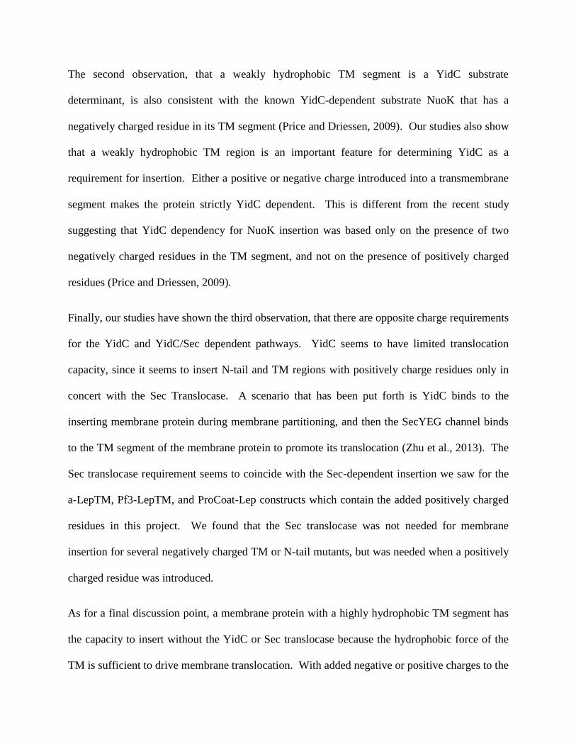
Figure 6: The topology of the three model membrane proteins Pf3-LepTM, a-LepTM, Pf3-aTM that
insert with a single hydrophobic domain. The four membrane proteins on the left (F1Fo ATPase, FoA,
Pf3 Coat, and Lep) indicate where the model membrane proteins were derived from. The model
membrane protein Pf3-LepTM has the N-tail of Pf3 Coat, and the TM1 of Lep. a-LepTM has the N-tail of
FoA and the TM1 of Lep. Pf3-aTM has the N-tail of Pf3 Coat and the TM1 of FoA.
Figure 7: Amino acid sequences of the three model membrane proteins that insert with a single
hydrophobic domain.

Figure 8: Sample full length SDS Page Gel. The model membrane protein here is Pf3-aTM with a Lep
tag and mutation D23N. A shift in the left two columns under Lep Ara (YidC expression) but a lack of a
shift under Lep Glc (YidC depletion) indicates that the protein is YidC dependent. Fragment indicates
Proteinase K had access to the inserted membrane protein. IPTG + induces expression of the protein of
interest Pf3-aTM (D23N), and IPTG – is a negative control; no major bands should be in those latter
columns. OmpA indicates spheroplast formation and if YidC depletion had a negative impact by
depleting the proton-motive-force too much. The precursor form of OmpA called Pro-OmpA would
accumulate on the gel if the pmf was over depleted, since OmpA is pmf-dependent for export across the
inner membrane.

Figure 9: The N tail and TM segment can function as a YidC substrate determinant. E. coli JS7131 cells
bearing the plasmids of the three different model membrane proteins were grown under YidC
expression or YidC depletion conditions, labeled, and analyzed for translocation of the N-tail using
Proteinase K accessibility assay described under methodology. The plasmids encoded Pf3-LepTM, a-
LepTM, and Pf3-aTM. Pf3-LepTM was found to be YidC independent, as indicated by the shift under
both YidC expression and depletion conditions. a-LepTM and Pf3-LepTM, on the other hand, were both
YidC dependent.
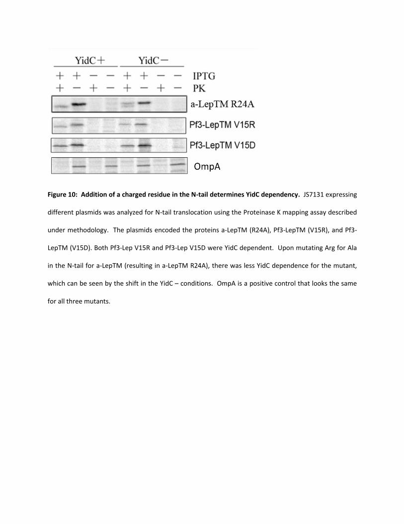
Figure 10: Addition of a charged residue in the N-tail determines YidC dependency. JS7131 expressing
different plasmids was analyzed for N-tail translocation using the Proteinase K mapping assay described
under methodology. The plasmids encoded the proteins a-LepTM (R24A), Pf3-LepTM (V15R), and Pf3-
LepTM (V15D). Both Pf3-Lep V15R and Pf3-Lep V15D were YidC dependent. Upon mutating Arg for Ala
in the N-tail for a-LepTM (resulting in a-LepTM R24A), there was less YidC dependence for the mutant,
which can be seen by the shift in the YidC – conditions. OmpA is a positive control that looks the same
for all three mutants.

Figure 11: Decreased hydrophobicity in the TM segment results in YidC dependency for insertion. E.
coli JS7131 cells expressing different plasmids were analyzed by Proteinase K mapping assay described
under methodology. The plasmids expressed the proteins Pf3-LepTM (A23R), Pf3-aTM (D23N), Pf3-aTM
(D23A, F27R) and Pf3-aTM (D23A). For Pf3-LepTM (A23R), Pf3-aTM (D23N), and Pf3-aTM (D23A, F27R),
YidC dependence can be observed. However, Pf3-aTM (D23A) is YidC independent, as seen by the shift
observed under both YidC expression and depletion conditions.
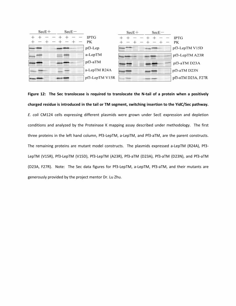
Figure 12: The Sec translocase is required to translocate the N-tail of a protein when a positively
charged residue is introduced in the tail or TM segment, switching insertion to the YidC/Sec pathway.
E. coli CM124 cells expressing different plasmids were grown under SecE expression and depletion
conditions and analyzed by the Proteinase K mapping assay described under methodology. The first
three proteins in the left hand column, Pf3-LepTM, a-LepTM, and Pf3-aTM, are the parent constructs.
The remaining proteins are mutant model constructs. The plasmids expressed a-LepTM (R24A), Pf3-
LepTM (V15R), Pf3-LepTM (V15D), Pf3-LepTM (A23R), Pf3-aTM (D23A), Pf3-aTM (D23N), and Pf3-aTM
(D23A, F27R). Note: The Sec data figures for Pf3-LepTM, a-LepTM, Pf3-aTM, and their mutants are
generously provided by the project mentor Dr. Lu Zhu.

Figure 13: Model of how ProCoat-Lep inserts into the membrane. Upon YidC-catalyzed insertion,
ProCoat-Lep gets cleaved by Leader Peptidase. This generates the mature coat protein called Coat-Lep,
which is seen on the very right hand side of the figure. Coat-Lep is then what Proteinase K digests if
ProCoat-Lep was inserted into the membrane.

Figure 14: A sample SDS PAGE Gel for ProCoat-Lep. The model membrane protein here is ProCoat-Lep,
wildtype. A shift in the left two columns under Lep Ara (YidC expression) but a lack of a shift under Lep
Glc (YidC depletion) indicates that the protein is YidC dependent. Fragment indicates Proteinase K had
access to the inserted PClep protein. (See Figure 13 for an explanation of how ProCoat-Lep inserts).
IPTG + induces expression of the protein of interest ProCoat-Lep and IPTG – is a negative control; no
major bands should be in those columns.

Figure 15: Amino acid sequence of ProCoat-Lep.
Figure 16: YidC and Sec dependence of ProCoat-Lep WT. Left: E. coli JS7131 cells expressing ProCoat-
Lep WT were analyzed by Proteinase K mapping assay described under methodology. ProCoat-Lep was
found to be YidC dependent. Right: E. coli CM124 cells expressing ProCoat-Lep WT were grown under
SecE expression and depletion conditions and analyzed by the Proteinase K mapping assay described
under methodology. ProCoat-Lep was found to be Sec independent. Note: Only IPTG+ conditions are
shown here.

Figure 17: Positive charges in the periplasmic loop of ProCoat-Lep seem to be consistent with the
YidC/Sec dependent pathway. Left: E. coli JS7131 cells expressing different plasmids were analyzed by
Proteinase K mapping assay described under methodology. The plasmids expressed the proteins
ProCoat-Lep (ARGNN), ProCoat-Lep (ANGRR), and ProCoat-Lep (ARGRR). Right: E. coli CM124 cells
expressing different plasmids were grown under SecE expression and depletion conditions and analyzed
by the Proteinase K mapping assay described under methodology. The plasmids expressed the proteins
ProCoat-Lep (ARGNN), ProCoat-Lep (ANGRR), and ProCoat-Lep (ARGRR).


![partial] fulfillment Town](https://img.dokumen.tips/doc/110x75/61cbd0e20c1fcf7e016f4ea2/partial-fulfillment-town.jpg)