Embed Size (px)
Citation preview
Molecular Human Reproduction Vol.8, No.9 pp. 805–810, 2002
Semenogelin I and II, the predominant human seminalplasma proteins, are also expressed in non-genital tissues
Åke Lundwall1,3, Anders Bjartell2, A.Yvonne Olsson1 and Johan Malm1
Departments of 1Laboratory Medicine and 2Urology, Lund University, University Hospital MAS, S-205 02 Malmo, Sweden
3To whom correspondence should be addressed. E-mail: [email protected]
Semenogelin I (SgI) and semenogelin II (SgII) are the dominating protein components of the coagulum formed by freshlyejaculated human semen. The primary source of these proteins is the seminal vesicles and, apart from a small production ofSgII in epididymis, they have not been detected in other tissues. In this report, we have re-examined the distribution of SgIand SgII transcripts and protein by RT–PCR and immunohistochemistry. Both SgI and SgII transcripts were demonstrated inseveral tissues, with the strongest signals coming from seminal vesicles, vas deferens, prostate, epididymis and trachea.Transcripts in the gastro-intestinal tract and skeletal muscle almost exclusively encoded SgI, whereas in kidney and testis, SgIItranscripts were predominant. By immunohistochemistry, the basal cell layer of the secretory epithelium in prostate, tracheaand bronchi was stained by antibodies recognizing both SgI and SgII. This is in contrast to the seminal vesicle and vas deferens,where the luminal cells were stained. The staining of skeletal muscle cells and a few scattered cells in the central nervous systemsuggests that semenogelin expression is not restricted to epithelial cells.
Key words: coagulation/semen/semenogelin/seminal plasma/transglutaminase
IntroductionSemenogelin I (SgI) and semenogelin II (SgII) are dominating proteinsof human seminal plasma that together with fibronectin give rise tothe gel-like coagulum of newly ejaculated semen (Lilja, 1985; Malmet al., 1996). They both originate from the glandular epithelium ofthe seminal vesicles, which secrete them at very high concentrations.At lower concentrations, SgII is also secreted by the epithelium ofthe epididymis (Bjartell et al., 1996).
SgI is a non-glycosylated protein of 439 aa residues with amolecular mass of 50 kDa (Lilja et al., 1989). The SgII molecule of63 kDa consists of 559 aa residues with a primary structure that is78% similar to that of SgI (Lilja and Lundwall, 1992). It has onepotential site for N-linked glycosylation and around half of themolecules in seminal plasma are glycosylated, yielding two molecularspecies with an apparent mass difference of 5 kDa (Lilja and Laurell,1985). The repeated structure with motifs abundant in Gln, Ser, Glyand Lys residues is characteristic to both SgI and SgII and mostconspicuous are highly conserved repeats of 60 aa residues in thecentral to C-terminal part of the molecules (Lilja and Lundwall,1992). In the SgI molecule, there are two such repeats and in theSgII molecule there are four. The two extra repeats in SgII accountfor the size difference between the two semenogelin molecules. Byway of the single Cys residue present in SgI and the two Cys residuesin SgII, the molecules are present as covalent homo- and hetero-multimers (Lilja and Laurell, 1985). In ejaculated sperm, the moleculesare also held together by non-covalent forces to yield a gel-likestructure that can be dissolved by 4 mol/l urea and high pH in thepresence of reducing agents (Malm et al., 1996). The gel structurealso dissolves spontaneously within minutes after ejaculation as theresult of proteolytic degradation of the semenogelin molecules by
© European Society of Human Reproduction and Embryology 805
prostate-specific antigen (PSA) (Lilja, 1985). Both SgI and SgII havebeen shown to function as excellent substrates of transglutaminaseand the repeated structure with an abundance of Gln and Lys isprobably a major explanation for this property (Peter et al., 1998).
Phylogenetic studies show that the genes encoding semenogelinsand related seminal vesicle-secreted proteins have undergone dramaticchanges during evolution. This is illustrated by the structural differ-ences between the semenogelins and the predominant proteinsexpressed by rodent seminal vesicles (Lundwall and Lazure, 1995).There is a very limited similarity in primary structure between thesemenogelins and the major proteins secreted by the rat and mouseseminal vesicles, even though they originate from homologous genes.This is because of the unusual evolution of otherwise ordinarilyconserved genes. The major part of the secreted protein is encodedby a single exon and the structural heterogeneity is caused by eithera differing selection of splice site or by an internal expansion of theexon during evolution (Lundwall and Lazure, 1995). The gene codingfor the major clot protein of guinea-pig semen is an interestingexample of the former (Hagstrom et al., 1996). The upstream promoterregion as well as the first exon and first intron is similar in sequenceto the two human semenogelin genes. Furthermore, the first intron ofthe guinea-pig gene also carries ~0.5 kb that are homologous to theprotein coding nucleotides in the beginning of the second exon ofthe semenogelin genes. It is therefore very likely that a relativelyrecent ancestor of the guinea-pig expressed a primitive semenogelin-like molecule that during evolution was omitted through the selectionof new splice sites. The high sequence similarity in the 5� end of thegenes, including the upstream promoter region, and a mutual highexpression level in seminal vesicles suggests that the guinea-pig genemight be regulated in a similar manner to the two semenogelin genes.Because the transcription of the guinea-pig gene has been reported
Å.Lunwall et al.
Table I. PCR primers. Nucleotides sequence of primers used for RT–PCRon transcripts of semenogelin I (SgI), semenogelin II (SgII) and adeninephosphoribosyltransferase (APRT).
Primer name Primer sequence Product size(bp)
5� SgI primer 5�-GCAGACACCAACATGGATCTCA-3� 1803� SgI primer 5�-CTGAGGTCAACTGACACCTTGA-3�5� SgII primer 5�-AGCATGAGGTTGCCCAAGATGA-3� 1303� SgII primer 5�-GAGGTCGGGTGACACCTTGC-3�5� APRT primer 5�-GACTACATCGCAGGCCTAGAC-3� 2573� APRT primer 5�-GCAGCGTTCATGGTTCCACCA-3�
to occur not only in seminal vesicles, but also in testis, kidney, liverand lung (Hagstrom et al., 1996), it is not unlikely that the humansemenogelin genes are expressed in more tissues than previouslyanticipated. Studies have indicated a role of semenogelin moleculesin relation to capacitation and motility of sperm (Robert and Gagnon,1996; de Lamirande et al., 2001). A non-genital expression of SgIand SgII would suggest that the molecule also has functions that areunrelated to sperm. To investigate this, semenogelin transcripts andimmunoreactivity were analysed in a collection of tissue samples.
Materials and methods
Tissue and semen samplesThe Helsinki Declaration regarding the use of human tissues was followed.For immunohistochemistry, archival tissues obtained at operations were used.All tissues were fixed within 30 min after removal in Bouin’s fixative (for4–18 h) or 4% buffered paraformaldehyde (overnight). All tissues werehistopathologically normal according to haematoxylin–eosin staining.
RNA samples for RT–PCR were isolated from tissue specimens homogen-ized in 4 mol/l guanidiniumthiocyanate as previously described (Chomczynskiand Sacchi, 1987). RNA preparations were made from single tissue specimens,except from epididymis and kidney where respectively five and four prepara-tions were made. Tissue samples from the urogenital tract and mammaryglands were from patients undergoing surgical treatment for neoplastic disease.The other specimens were taken at autopsy ~20 h post-mortem. RNA fromhuman lung, pancreas, salivary gland, skeletal muscle and trachea werepurchased from Clontech (Palo Alto, CA, USA). RNA samples were storedat –70°C.
Semen samples were collected from voluntary donors by masturbation.Liquefaction was achieved by incubation for 60–90 min at room temperature.Cells were removed by centrifugation at 800 g for 15 min. Unliquefiedsamples were collected in 40 mmol/l Tris–HCl, pH 9.7, 4 mol/l urea,25 mmol/l EDTA and 30 mmol/l dithiothreitol as previously described (Malmet al., 1996). All semen samples were stored at –20°C.
RT–PCROligo-dT-primed cDNA synthesis, using 3 µg of total RNA, was performedin a volume of 15 µl with the First-Strand cDNA synthesis kit (AmershamPharmacia Biotech, Uppsala, Sweden). The subsequent PCR was performedwith 2 µl cDNA, equivalent to ~0.4 µg of RNA, in a volume of 10 µl usingthe Advantage 2 PCR kit (Clontech) and 0.2 µmol/l gene-specific primers(Table I). The PCR was run in a DNA Engine, PTC-200 (MJ Research,Watertown, MA, USA) with a program that had an initial 1 min denaturationat 95°C followed by 35 cycles consisting of 30 s at 95°C and 1 min at 68°C.At the end of the program, there was also an extra 1 min incubation at 68°C.Reaction products were analysed by electrophoresis in 2.5% (w/v) agarosegels and visualized by exposure to UV light following staining with ethidiumbromide. Each sample was analysed at least three times for both SgI andSgII: once with primers for a single gene and twice with primers for both genes.
Immunological methodsAntiserum against SgII was raised in rabbits by standard procedures (Harlowand Lane, 1988) using protein purified from unliquefied semen (Malm et al.,
806
1996). Immunohistochemistry was performed using a detection kit, DakoChemMate™ Detection Kit peroxidase/carbazole, rabbit/mouse and a stainingmachine, Dako TechMate™ 500/1000 Instrument (Dako A/S, Denmark).Briefly, the sections were deparaffinized in xylene, rehydrated and treatedwith 0.3% H2O2 in methanol for 30 min at room temperature to quenchendogenous peroxidase activity. For antigen retrieval, tissue sections werefirst incubated with sodium citrate (10 mmol/l, pH 6.0) and boiled in amicrowave oven at 750 W for 2�3 min, and then digested with proteinase K(20 µg/ml in 20 mmol/l Tris–HCl, 2 mmol/l CaCl2, pH 7.5) for 25 min at37°C. The sections were incubated with the antiserum against SgII (diluted1:4000) for 60 min at room temperature, after which they were incubatedwith biotinylated secondary antibodies against rabbit IgG, which were includedin the ChemMate kit, for 60 min at room temperature. The immunoreactivitywas visualized using the manufacturer’s protocol for the peroxidase/AEC(3-amino-9-ethyl-carbazole) or DAB (3,3�diaminobenzidine tetrahydro-chloride) reagents in the ChemMate kit. The sections were counterstainedwith Mayer’s haematoxylin solution. As a negative control, adjacent tissuesections were processed by replacing the primary antibody with non-immunerabbit IgG diluted 1:8000 (Dako). At least three samples were analysed fromeach tissue.
Results
Detection of SgI and SgII transcriptsAn RT–PCR assay was developed for easy and swift detection ofsemenogelin transcripts in different tissues. PCR primers were selectedfrom cDNA sequences that showed low conservation between SgIand SgII. In order to reduce false signals by priming on genomicDNA present in small quantities in RNA preparations, the 3� primerswere designed to span the second intron of the genes. The transcriptof the housekeeping gene encoding adenine phosphoribosyltransferase(APRT) served as control of cDNA synthesis and PCR (Hidaka et al.,1987). Also with this transcript, false priming was avoided byselecting intron-spanning primers; in this case the 5� and 3� PCRprimers were designed to span introns 2 and 4 of the ARPT gene.The assay was optimized using RNA from seminal vesicles andepididymis, and distinct APRT, SgI and SgII signals were obtainedusing the protocol with 35 PCR cycles. By running the assay withsamples of cloned cDNA of known concentration, the detection limitwas estimated to be ~1 fg of transcript.
Figure 1 shows the PCR products run on agarose gel stainedwith ethidium bromide. The 257 bp APRT product is seen withsamples from all tissues as expected. Several samples also giverise to strong signals for both the 180 bp SgI product and the130 bp SgII product. In the gastro-intestinal tract there are mainlySgI transcripts, whereas in kidney, the SgII transcripts dominate.However, the relative abundance of SgI to SgII transcripts variedin different kidney samples and in some the SgI signal was asequally strong as the SgII signal. This did not depend on theanatomical location as the relative signal intensity was the samewhether samples were taken from the cortex or the medulla,although the intensity was consistently stronger in medulla samples.Several epididymis specimens were analysed and invariably bothSgI and SgII signals were found. However, as with the kidneysamples, the staining intensity of epididymis samples varied sothat the signal from some preparations yielded a stronger SgIIthan SgI signal and vice versa. The SgI signal in epididymis wasa surprise as results from previous investigations (Bjartell et al.,1996) suggested that only the SgII gene is transcribed in thistissue, with a restricted distribution to the caudal part. Becausecauda epididymis empties into vas deferens, there is a possibilitythat epididymal RNA preparations were contaminated with RNAfrom vas deferens. From the close relationship of the epitheliumof vas deferens and seminal vesicles it is reasonable to assume
Non-genital semenogelin expression
Figure 1. Expression of semenogelin (Sg)I and SgII mRNA. Transcripts were identified by RT–PCR and electrophoresis. The arrows to the right indicate thelocation of PCR products of adenine phosphoribosyltransferase (ARPT), SgI and SgII transcripts. The source of RNA is indicated below each lane.
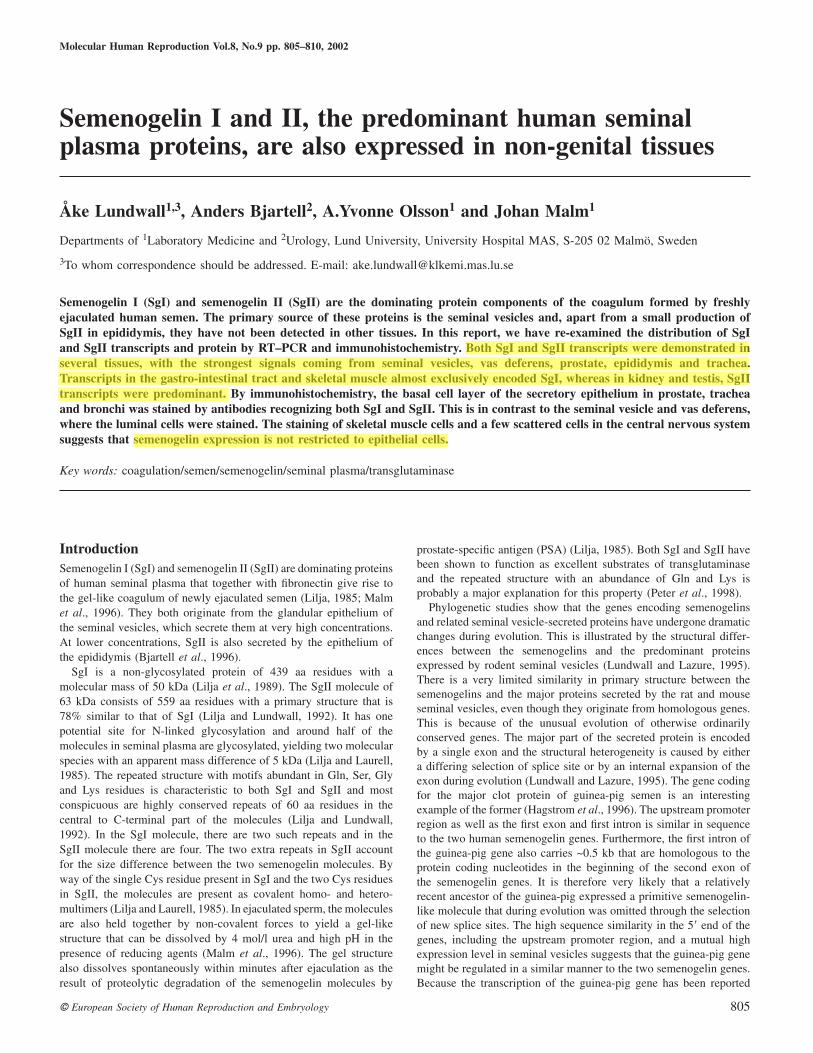
Figure 2. Transcripts in epididymis. Epididymis was dissected into itsconstituent parts from which RNA was extracted. Transcripts were amplifiedby RT–PCR and analysed by electrophoresis. Transcripts of adeninephosphoribosyltransferase (ARPT), semenogelin I (SgI) and semenogelin II(SgII) are indicated by the arrows, with the RNA source is indicated beloweach lane.
that vas deferens express SgI and SgII. Thus, the SgI signal inepididymis might originate from vas deferens contamination. Thiswas investigated by dissection of epididymis into its constituentparts which were subsequently analysed by RT–PCR. As can beseen in Figure 2, both vas deferens and cauda epididymis showstrong SgI and SgII signals, but weaker and very faint signals forboth transcripts are also seen in corpus and caput epididymisrespectively. As it is unlikely that the signals from corpus andcaput epididymis are the result of vas deferens contamination, boththe SgI and SgII genes are probably transcribed in epididymis.
Because of the strong signal from prostate RNA, transcriptswere also analysed in three prostate cancer cell lines. No, or veryfew, transcripts were detected in RNA from the hormone-insensitivecell line PC-3, but from the likewise hormone-insensitive DU 145cell line, a faint SgII signal was detected. In the hormone-sensitivecell line LNCaP both SgI and SgII transcripts were detected, butcompared with prostate tissue, the signal from cell lines was weak.This might indicate that the semenogelin genes are down-regulatedduring malignant transformation or that stromal cells instead ofepithelial cells are the main source of prostate SgI and SgII.
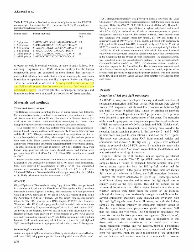
Immunohistochemical localization of semenogelinantigenicityThe specificity of the antiserum against SgII was analysed byWestern blotting (Figure 3). As can be seen, it could recognizeboth SgI and SgII as well as their degradation products. Theequally strong immunostaining of SgI and SgII suggests that the
807
Figure 3. Western blotting. Samples of 10 µl separated by SDS–PAGE andanalysed by Western blotting using an antiserum raised against humansemenogelin (Sg)II, but which also recognizes SgI. Samples are unliquefiedhuman seminal plasma diluted 1:70 (lane1), liquefied human seminalplasma diluted 1:10 (lane 2) and human blood plasma diluted 1:10(lane 3). Lane 4 show size markers stained by Coomassie Brilliant Blue,with sizes in kDa indicated to the right. The arrows indicate the location ofSgI and SgII.
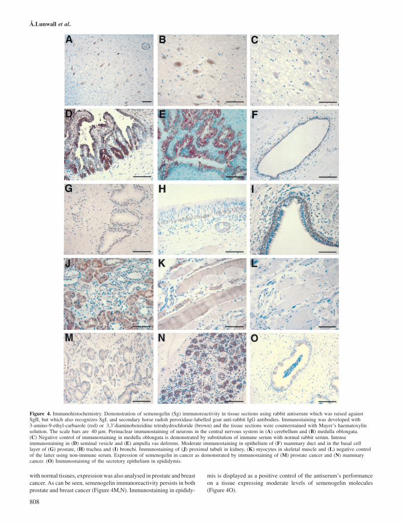
antiserum is unable to distinguish between SgI and SgII. Semenoge-lin immunoreactivity was thereafter analysed in tissue sectionsfrom a variety of organs. Tissue specimens from the gastro-intestinal tract, with moderate amounts of SgI transcripts accordingto RT–PCR, showed no specific semenogelin signal, as the weakstaining generated by immune sera did not significantly exceedbackground staining with non-immune sera. In contrast to this arethe discrete semenogelin signals seen in neural tissues, despitenegative RT–PCR results. The immunostaining appeared to bespecific for a discrete cell type that is present both in medullaoblongata and cerebellum (Figure 4A–C). The seminal vesiclesecretion contains very high concentrations of both SgI and SgII.In tissue sections of the seminal vesicles this appeared as anintense immunostaining of the secretory epithelium. An equallyintense staining was seen in vas deferens and ampulla vas,indicating high semenogelin concentration also in secretion fromthe epithelium of the spermatic ducts (Figure 4D,E). A muchweaker immunostaining was observed in breast tissue, prostate,trachea, bronchi, kidney and skeletal muscle (Figure 4F–L). In skeletalmuscle the immunostaining appeared to be specific for myocytes,whereas in breast tissue the epithelium lining the mammary ductswas stained and in the kidneys the proximal tubuli were stained. Theexperiments with prostate, trachea and bronchi were very interesting asthey all appeared to show immunostaining in the basal cell layer andnot in the luminal layer, as in the seminal vesicles. Because of the result
Å.Lunwall et al.
Figure 4. Immunohistochemistry. Demonstration of semenogelin (Sg) immunoreactivity in tissue sections using rabbit antiserum which was raised againstSgII, but which also recognizes SgI, and secondary horse radish peroxidase-labelled goat anti-rabbit IgG antibodies. Immunostaining was developed with3-amino-9-ethyl-carbazole (red) or 3,3�diaminobenzidine tetrahydrochloride (brown) and the tissue sections were counterstained with Mayer’s haematoxylinsolution. The scale bars are 40 µm. Perinuclear immunostaining of neurons in the central nervous system in (A) cerebellum and (B) medulla oblongata.(C) Negative control of immunostaining in medulla oblongata is demonstrated by substitution of immune serum with normal rabbit serum. Intenseimmunostaining in (D) seminal vesicle and (E) ampulla vas deferens. Moderate immunostaining in epithelium of (F) mammary duct and in the basal celllayer of (G) prostate, (H) trachea and (I) bronchi. Immunostaining of (J) proximal tubuli in kidney, (K) myocytes in skeletal muscle and (L) negative controlof the latter using non-immune serum. Expression of semenogelin in cancer as demonstrated by immunostaining of (M) prostate cancer and (N) mammarycancer. (O) Immunostaining of the secretory epithelium in epididymis.
with normal tissues, expression was also analysed in prostate and breastcancer. As can be seen, semenogelin immunoreactivity persists in bothprostate and breast cancer (Figure 4M,N). Immunostaining in epididy-
808
mis is displayed as a positive control of the antiserum’s performanceon a tissue expressing moderate levels of semenogelin molecules(Figure 4O).
Non-genital semenogelin expression
DiscussionIn this study, we showed that semenogelin transcripts and antigenicityare present in several tissues and not confined to the seminal vesiclesand epididymis as was previously assumed. Following ejaculation,SgI and SgII are degraded within minutes by PSA during the processknown as semen liquefaction (Lilja, 1985). Prolonged incubationleads to the formation of very small fragments and a progressiveloss of semenogelin immunoreactivity. Also, other proteases readilydegrade the semenogelin molecules and therefore it might be assumedthat their general structure renders them very sensitive to proteolyticdigestion (Deperthes et al., 1996). Because of this, proteolyticdegradation could be one explanation for the failure in previousreports to detect semenogelin antigenicity in tissues other than seminalvesicles and epididymis. At least in the seminal vesicles, the expressionlevels of the semenogelins are extremely high so even if most of thesemenogelin molecules are degraded, there will still be substantialquantities of antigen left over to be detected by the anti-semenogelinantibodies. The improved fixation procedure in combination withmicrowave radiation to expose antigen in this study has probablyalso increased the sensitivity.
It has previously been shown by in-situ hybridization that onlySgII is synthesized in the epididymis (Bjartell et al., 1996). This isin contrast to this report where both SgI and SgII transcripts weredetected. The discrepancy can probably be explained, at least partly,by sample heterogeneity. As the epididymal ratio of SgI to SgIItranscripts appears to vary significantly between preparations, theprevious investigation could have been conducted on samples withhigh levels of SgII transcripts and low levels of SgI transcripts,whereby the weak signal from the latter could have been ignored. Itis not clear what governs the variation in SgI to SgII transcript ratios,but it appears to predominantly reflect variations in SgI transcriptlevels. Thus, SgI transcript levels might be prone to fluctuations,something that could also be a factor behind the failure to detectsemenogelin immunoreactivity in the gastro-intestinal tract. Theexpression of the SgI gene could, for instance, have been differentin samples taken for RT–PCR compared to those taken for immuno-histochemistry. This can probably be addressed by simultaneousanalyses of transcripts and immunoreactivity in larger samples.Another indication that the SgI gene is differently regulated to theSgII gene was the discrepant expression in the hormone-sensitive cellline LNCaP and the hormone-insensitive cell line DU 145. Thetranscription of both the SgI gene and the SgII gene in LNCaP cellsversus almost sole transcription of the SgII gene in DU 145 cellssuggests that the SgI gene is more sensitive to hormone stimulation.
UniGene (http://www.ncbi.nlm.nih.gov/UniGene/) is a databasewhich contain clusters of expressed sequence tags (EST) that enablesidentification of tissues in which a specific gene is transcribed. InNovember 2001, the database contained 241 EST sequences encodingSgI and 84 EST sequences encoding SgII. Of the SgI transcripts, 226originated from prostate, 11 from skeletal muscle, two from breasttissue and two from kidney, and of the SgII transcripts, 78 originatedfrom prostate, six from skeletal muscle and one each from colon andpooled colon, kidney and stomach RNA. The EST sequences confirmthe here reported expression of both SgI and SgII in the prostate.The rest of the EST sequences also agree with our data, except forthose suggesting SgII transcription in skeletal muscle and colon. Thenumber of identified EST sequences in skeletal muscle exclude thepossibility that they represent cryptic transcripts. More likely theyrepresent transcripts from discrete subsets of cells that might differin number between different parts of the tissue. A similar reasoningmight also explain the negative RT–PCR in neural tissue in spiteof the immunostaining of certain cells in medulla oblongata andcerebellum.
809
SgI has been reported to be an inhibitor of sperm motility andcapacitation (Robert and Gagnon, 1996; de Lamirande et al., 2001).These activities have not been independently verified, so the biologicalrole of the semenogelins remains somewhat unclear. The here reportedfinding of both SgI and SgII transcripts and antigen in a variety oftissues suggest that the molecules have a function that is more farreaching than previously believed and not confined to a role connectedwith male fertility. In fact, it might very well be that the seminalvesicle synthesis should be regarded as a special case, where theprotein is over expressed so as to exert a specialized function in themale genital tract, while the function in other tissues is of moregeneral importance. In this context it is interesting to note that bothSgI and SgII were recently found in cell surface adhesion complexesof small cell lung carcinoma cell lines (Rodrigues et al., 2001). Themolecules could therefore very well turn out to be adhesion molecules.
The expression in lung cancers indicates that the semenogelinmolecules might serve as diagnostic and/or prognostic tumour markers(Rodrigues et al., 2001). In this report we also show semenogelinexpression in prostate and mammary cancer. Presently we are assessingthe utility of semenogelin immunostaining as a prognostic tool inthese cancers.
AcknowledgementsThe technical assistance of Margareta Persson, Elise Nilsson and BirgittaFrohm is acknowledged. This work was supported by grants from the SwedishResearch Council (project no. 8660) and the Swedish Cancer Society (projectno. 4564 and 4294).
ReferencesBjartell, A., Malm, J., Moller, C., Gunnarsson, M., Lundwall, Å. and Lilja,
H. (1996) Distribution and tissue expression of semenogelin I and II inman as demonstrated by in situ hybridization and immunocytochemistry.J. Androl., 17, 17–26.
Chomczynski, P. and Sacchi, N. (1987) Single-step method of RNA isolationby acid guanidinium thiocyanate-phenol-chloroform extraction. Anal.Biochem., 162, 156–159.
de Lamirande, E., Yoshida, K., Yoshiike, T.M., Iwamoto, T. and Gagnon, C.(2001) Semenogelin, the main protein of semen coagulum, inhibits humansperm capacitation by interfering with the superoxide anion generatedduring this process. J. Androl., 22, 672–679.
Deperthes, D., Frenette, G., Brillard-Bourdet, M., Bourgeois, L., Gauthier, F.,Tremblay, R.R. and Dube, J.Y. (1996) Potential involvement of kallikreinhK2 in the hydrolysis of the human seminal vesicle proteins after ejaculation.J. Androl., 17, 659–665.
Hagstrom, J.E., Fautsch, M.P., Perdok, M., Vrabel, A. and Wieben, E.D.(1996) Exons lost and found. Unusual evolution of a seminal vesicletransglutaminase substrate. J. Biol. Chem., 271, 21114–21119.
Harlow, E. and Lane, D. (eds) (1988) Antibodies. A Laboratory Manual. ColdSpring Harbor Laboratory Press, Cold Spring Harbor, New York, USA,p. 92.
Hidaka, Y., Tarle, S.A., O’Toole, T.E., Kelley, W.N. and Palella, T.D. (1987)Nucleotide sequence of the human APRT gene. Nucleic Acids Res., 15, 9086.
Lilja, H. (1985) A kallikrein-like serine protease in prostatic fluid cleaves thepredominant seminal vesicle protein. J. Clin. Invest., 76, 1899–1903.
Lilja, H. and Laurell, C.-B. (1985) The predominant protein in human seminalcoagulate. Scand. J. Clin. Lab. Invest., 45, 635–641.
Lilja, H. and Lundwall, A. (1992) Molecular cloning of epididymal andseminal vesicular transcripts encoding a semenogelin-related protein. Proc.Natl Acad. Sci. USA, 89, 4559–4563.
Lilja, H., Abrahamsson, P.A. and Lundwall, A. (1989) Semenogelin, thepredominant protein in human semen. Primary structure and identificationof closely related proteins in the male accessory sex glands and on thespermatozoa. J. Biol. Chem., 264, 1894–1900.
Lundwall, A. and Lazure, C. (1995) A novel gene family encoding proteinswith highly differing structure because of a rapidly evolving exon. FEBSLett., 374, 53–56.
Å.Lunwall et al.
Malm, J., Hellman, J., Magnusson, H., Laurell, C.B. and Lilja, H.(1996) Isolation and characterization of the major gel proteins in humansemen, semenogelin I and semenogelin II. Eur. J. Biochem., 238,48–53.
Peter, A., Lilja, H., Lundwall, A. and Malm, J. (1998) Semenogelin I andsemenogelin II, the major gel-forming proteins in human semen, aresubstrates for transglutaminase. Eur. J. Biochem., 252, 216–221.
810
Robert, M. and Gagnon, C. (1996) Purification and characterization of theactive precursor of a human sperm motility inhibitor secreted by the seminalvesicles: identity with semenogelin. Biol. Reprod., 55, 813–821.
Rodrigues, R.G., Panizo-Santos, A., Cashel, J.A., Krutzsch, H.C., Merino,M.J. and Roberts, D.D. (2001) Semenogelins are ectopically expressed insmall cell lung carcinoma. Clin. Cancer Res., 7, 854–860.
Submitted on January 10, 2002; accepted on May 20, 2002