Embed Size (px)
Citation preview

PAPER www.rsc.org/dalton | Dalton Transactions
Self-assembly of copper succinate nanoparticles to form anisotropicmesostructures
Aparna Ganguly,a,b Tokeer Ahmadb and Ashok K. Ganguli*a
Received 20th November 2008, Accepted 17th February 2009First published as an Advance Article on the web 16th March 2009DOI: 10.1039/b820778j
Uniform cylindrical rods of copper succinate dihydrate of several microns in length and 200 nm indiameter were obtained by the reverse micellar (microemulsion) method at room temperature usingCTAB as the surfactant. The rod-like structures are formed by an ordered assembly of sphericalparticles of 4–5 nm, which is facilitated by water molecules. The copper succinate particles, in theabsence of the microemulsion or surfactant, show only spherical geometry, while in the presence of thesurfactant, thicker rods (compared to as obtained by reverse micellar method) of varying length wereobtained. The formation of the rod-like structure is driven by the permanent dipole moment of thesuccinate ion, which leads to the oriented attachment of the nanoparticles in the presence of thesurfactant. A new phase (anhydrous copper succinate) is obtained upon heating the dihydrate at 75 ◦C,which shows branched and corrugated rods assembled from a random arrangement of nanoparticles.The water molecules appear to control the morphology of the rods giving smooth rods (orderedarrangement of nanoparticles) for the dihydrate while branched or disrupted rods with randomarrangement of nanoparticles are obtained for the anhydrous phase. The chain length of thedicarboxylic acid (ligand) appears to have a role in controlling the aspect ratio of these anisotropicmesostructures. The ability to generate suitable conditions for self assembly into orderednanostructures and to control the anisotropy would lead us towards a proper design of nanodevices.
Introduction
Synthesis of nanostructured compounds with controlled sizeand shape is a challenge and several methodologies have beendeveloped to realize them, such as chemical vapour deposition,1,2
ball milling,3,4 sol–gel5,6 and the microemulsion method usingreverse micelles.7–11 The microemulsion method leads to uniformand homogenous nanomaterials and has been well adapted forthe synthesis of metal and metal oxide nanoparticles.12,13 Recently,considerable interest has grown in the coordination chemistryof carboxylate ligands as their polymeric metal complexes areattractive for combining the properties of both organic andinorganic components. This has led to extensive studies on thesynthesis of nanostructured transition metal oxalates by reversemicellar method.14–18 These metal oxalates having metal ions in thedivalent state were found to crystallize with a rod-like morphology.The decomposition of these nanorods under specific conditionsyields pure phases of metal and metal oxide nanoparticles.
Here, we discuss our results on the effect of change of thecarboxylate ligand from the oxalate (used in previous studies) tothe succinate ion, on the morphology of the rods of these metalsuccinates. The bidentate succinate anion has two additional CH2
fragments compared to the oxalate anion and offers to createstructural diversity attributed to its various binding sites andthe conformational modes originating from its flexibility.19 The
aDepartment of Chemistry, Indian Institute of Technology, Hauz Khas, NewDelhi, 110016, India. E-mail: [email protected]; Fax: +91 112685 4715; Tel: +91 11 2659 1511bDepartment of Chemistry, Faculty of Natural Sciences, Jamia MilliaIslamia, New Delhi, 110025, India
objective of this investigation was to the study the role of the chainlength of the bidentate ligand on the aspect ratio of the succinaterods and also the texture of these nanorods. This study describesthe oriented self assembly of copper succinate nanoparticles toform nanorods and a plausible mechanism for it.
Experimental
Commercially available Cetyltetraammoniummethyl bromide(CTAB, Spectrochem, 99%), copper nitrate hydrate (BDH 97%),sodium succinate hexahydrate (CDH, 99%), 1-butanol (Qualigens,99.5%), isooctane (Spectrochem, 99%), chloroform (SRL 99.5%),ethanol (Merck, 99.9%) and methanol (Qualigens, 99%) were usedin the synthesis.
Microemulsions with CTAB as surfactant, 1-butanol as co-surfactant, isooctane as hydrocarbon phase and the aqueous phasewere prepared. The constituents of the microemulsion were takenin the following weight fractions: 16.76% of CTAB, 13.90% of n-butanol, 59.29% of isooctane and 10.05% of the aqueous phase.0.1 M metal nitrate (Cu 2+) solution was used as the aqueous phasein one microemulsion and 0.1 M sodium succinate hexahydratesolution in the second one. The reaction was accomplished bymixing the two microemulsions and stirred overnight at roomtemperature. The coloured (turquoise blue) precipitate formedwas separated from the apolar solvent and washed with 1 : 1mixture of CHCl3 and CH3OH. The precipitate was dried at roomtemperature and heated under specific conditions to obtain theoxide or the metal nanoparticles. Similar synthesis using onlythe surfactant (CTAB) in aqueous medium (absence of reversemicelles) and by co-precipitation method was carried out.
3536 | Dalton Trans., 2009, 3536–3541 This journal is © The Royal Society of Chemistry 2009
Dow
nloa
ded
by U
nive
rsity
of
Ten
ness
ee a
t Kno
xvill
e on
13
Mar
ch 2
013
Publ
ishe
d on
16
Mar
ch 2
009
on h
ttp://
pubs
.rsc
.org
| do
i:10.
1039
/B82
0778
JView Article Online / Journal Homepage / Table of Contents for this issue

XRD studies were carried out on a Bruker D-8 Advance X-raydiffractometer using Ni filtered Cu Ka radiation. Normal scanswere recorded with a 2q step of 0.02◦ and a residence time of1 s. With the help of X-ray line broadening studies, the crystallitesize could be obtained for the oxide particles. Thermogravimetric(TGA) and differential thermal analysis (DTA) were carried outon a Perkin-Elmer TGA–DTA instrument. Well ground and drysamples were loaded under flowing nitrogen with a heating rateof 5 ◦C min-1. SEM investigations were carried out on a ZEISSEVO 50 scanning electron microscope. HRTEM was recorded on aTechnai G2 20 (FEI) electron microscope operated at 200 kV. TEMspecimens were prepared by loading a drop of the ultrasonicallydispersed sample in ethanol on a carbon-coated copper grid anddried in air. Small angle X-ray scattering (SAXS) measurementwas carried out on a Bruker AXS Nanostar instrument. Zetapotential measurements were done on a Malvern Zetasizer, ZS90instrument. Magnetization studies of the metal succinate wasmeasured at temperatures ranging from 5–300 K, in appliedfields of up to 10 kOe with a quantumdesign physical propertiesmeasurement system.
Results and discussion
The powder X-ray diffraction pattern of copper succinate dihy-drate obtained using the microemulsion method could be indexedon the basis of a triclinic cell (Fig. 1a; JCPDS: 261767), whichmatches with the simulated pattern obtained from the single crystaldata20 (fig. 1b). Transmission electron microscopy showed theformation of rods with length greater than 1 mm and diameterof ~200 nm (Fig. 2a and b). The HRTEM image (Fig. 2c) showsthat the rods are made up of an ordered assembly of sphericalnanoparticles of size ~4–5 nm. To compare the synthesis of metalsuccinates in the absence of reverse micelles, a control experimentwas carried out where copper succinate dihydrate particles weresynthesized by a simple precipitation method. Spherical particlesof size ~200 nm were formed instead of rods as obtained usingreverse micelles (Fig. 3). This clearly indicates that the surfactantacts as a structure directing agent and helps in the formation ofthe rod-like structure. However, when the reaction was carriedout only in the presence of the surfactant (CTAB) and not themicroemulsion (reverse micelles), rods made up of small (4–5 nm)particles was again obtained (Fig. 4) but the diameter of thesenanorods were almost twice (500 nm) than the one obtained in thepresence of reverse micelles (200 nm). Apart from the diameter,the texture (smoothness) of the nanorod was affected by the use
Fig. 1 (a) PXRD pattern for copper succinate dihydrate synthesized usingreverse micellar (microemulsion) and (b) the simulated pattern from singlecrystal data.
of reverse micelles as in the absence of the microemulsion, butonly in the presence of surfactant, rods were obtained that werenot uniform. The aqueous reactor (core) in the reverse micelleswherein the metal ions and the dicarboxylate ions are allowedto react is small and the presence of organic phase outside thereactor restricts the growth leading to smaller diameter of the rods.While in the presence of only the surfactant (CTAB), not themicroemulsion, the aqueous media does not offer any restrictionon its growth and thus affects the aspect ratio of the metal
Fig. 2 (a) TEM micrographs for copper succinate dihydrate, (b) HRTEM shows the formation of rod from spherical particles and (c) HRTEMmicrographs at high magnification showing the lattice fringes.
This journal is © The Royal Society of Chemistry 2009 Dalton Trans., 2009, 3536–3541 | 3537
Dow
nloa
ded
by U
nive
rsity
of
Ten
ness
ee a
t Kno
xvill
e on
13
Mar
ch 2
013
Publ
ishe
d on
16
Mar
ch 2
009
on h
ttp://
pubs
.rsc
.org
| do
i:10.
1039
/B82
0778
J
View Article Online
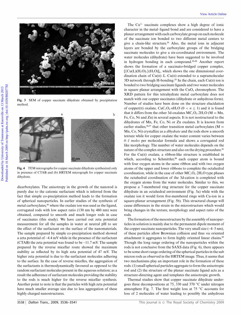
Fig. 3 SEM of copper succinate dihydrate obtained by precipitationmethod.
Fig. 4 TEM micrographs for copper succinate dihydrate synthesized onlyin presence of CTAB and (b) HRTEM micrograph for copper succinatedihydrate.
dicarboxylates. The anisotropy in the growth of the nanorod ispurely due to the cationic surfactant which is inferred from thefact that simple co-precipitation method leads to the formationof spherical nanoparticles. In earlier studies of the synthesis ofmetal carboxylates,16 where the oxalate ion was used as the ligand,corrugated rods with low aspect ratio (130 nm by 480 nm) wereobtained, compared to smooth and much longer rods in caseof succinates (this study). We have carried out zeta potentialmeasurement for all the samples in water at neutral pH to seethe effect of the surfactant on the surface of the nanomaterials.The sample prepared by simple co-precipitation method showeda zeta potential of ~4.4 mV while in the presence of the surfactant(CTAB) the zeta potential was found to be ~11.7 mV. The sampleprepared by the reverse micellar route showed the maximumstability as reflected by its high zeta potential of 47 mV. Thehigher zeta potential is due to the surfactant molecules adheringto the surface. In the case of reverse micelles, the aggregation ofthe surfactants is thermodynamically favoured as opposed to therandom surfactant molecules present in the aqueous solution; as aresult the adherence of surfactant molecules providing the stabilityto the rods is much higher for the reverse micellar synthesis.Another point to note is that the particles with high zeta potentialhave much smaller average size due to less aggregation of thesehighly charged nanostructures.
The Cu2+ succinate complexes show a high degree of ioniccharacter in the metal–ligand bond and are considered to have aplanar arrangement with each carboxylate group on each moleculeof the succinate ion bonded to two different metal centers togive a chain-like structure.21 Also, the metal ions in adjacentlayers are bonded by the carboxylate groups of the bridgingsuccinate molecules to give a six-coordinated environment. Thewater molecules (dihydrate) have been suggested to be involvedin hydrogen bonding in each compound.21,22 Another reportshows the formation of a succinato-bridged copper complex,[{Cu(L)(H2O)2}(H2O)]n, which shows the one dimensional coor-dination chain of Cu(II)–L–Cu(II) extended to a supramolecular3D network through H-bonding.23 In the chain, each Cu(II) ion isbonded to two bridging succinate ligands and two water moleculesin square planar arrangement with the CuO4 chromophore. TheXRD pattern for this tetrahydrate metal carboxylate does notmatch with our copper succinates (dihydrate or anhydrous form).Number of studies have been done on the structure elucidationof copper(II) oxalate, CuC2O4·nH2O (0 < n ≤ 1) and it is foundthat it differs from the other 3d-oxalates MC2O4·2H2O (M = Mn,Fe, Co, Ni and Zn) in several aspects. It is not isostructural to thedihydrates of Mn, Fe, Co, Ni or Zn oxalates. It is known fromearlier studies,16,17 that other transition metal carboxylates (M =Mn, Co, Ni) crystallize as a dihydrate and the rods show a smoothtexture while for copper oxalate the water content varies between0–1 moles per molecular formula and shows a corrugated rodlike morphology. The number of water molecules depends on thenature of the complex structure and also on the drying procedure.21
For the Cu(II) oxalate, a ribbon-like structure is established inwhich, according to Schmittler,24 each copper atom is boundwith four oxygen atoms in the same ribbon and with two oxygenatoms of the upper and lower ribbons to complete the octahedralcoordination, while in the case of other MC2O4·2H2O type phasesthe octahedral coordination of the 3d-cation is completed withthe oxygen atoms from the water molecules. Similar to this, wepropose a 7-membered ring structure for the copper succinatedihydrate in an octahedral environment (Fig. 5a) while with theoxalate ion it would form five-membered rings with Cu-ion in asquare-planar arrangement (Fig. 5b). This structural change willcause differences in the strain in the microstructure which wouldbring changes in the texture, morphology and aspect ratio of therods.
The formation of the mesostructure by the assembly of nanopar-ticles in solution is mainly due to the permanent dipole moment ofthe copper succinate nanoparticles. The very small size (~4–5 nm),of these particles allow Brownian collision and thus via orientedattachment it aggregates to form highly oriented linear chains.25
Though the long range ordering of the nanoparticles within therods is not conclusive from the SAXS data (Fig. 6), there appearsto be some short range ordering of the spherical particles in the submicron rods as observed in the HRTEM image. Thus, it seems thattwo mechanisms play an important role in the formation of theserods: (1) small spherical particles aggregate to form the anisotropicrod and (2) the structure of the planar succinate ligand acts as astructure-directing agent and templates the anisotropic growth.
Thermal studies show that copper succinate dihydrate under-goes three decompositions at 75, 330 and 370 ◦C under nitrogenatmosphere Fig. 7. The first weight loss at 75 ◦C accounts forloss of 2 molecules of water leading to possibly the anhydrous
3538 | Dalton Trans., 2009, 3536–3541 This journal is © The Royal Society of Chemistry 2009
Dow
nloa
ded
by U
nive
rsity
of
Ten
ness
ee a
t Kno
xvill
e on
13
Mar
ch 2
013
Publ
ishe
d on
16
Mar
ch 2
009
on h
ttp://
pubs
.rsc
.org
| do
i:10.
1039
/B82
0778
J
View Article Online

Fig. 5 Tentative structure of (a) copper succinate dihydrate and (b) copperoxalate monohydrate.
Fig. 6 SAXS pattern of copper succinate dihydrate.
copper succinate. The weight loss (subsequent two decompositionsteps) corresponds to the conversion of anhydrous succinate toelemental copper (at 390 ◦C). The decomposition of the metaldicarboxylate probably proceeds with stepwise cation reduction,from Cu2+ to Cu+ to Cu0. To investigate the structure of theintermediate phase obtained at 75 ◦C (as shown by TGA studies),the succinate precursor was heated in an oven at 75 ◦C for 6h.The X-ray pattern (Fig. 8a) of this sample does not match eitherwith the hydrated copper succinate (CuC4H4O4·2H2O) or withthat of the anhydrous copper succinate reported in JCPDS file:512293. Note that the X-ray powder pattern of the anhydroussuccinate reported in JCPDS files does not appear to be a carefulstudy since no description of cell parameters or the crystal
Fig. 7 Thermogravimetric analysis (TGA) and differential thermalanalysis (DTA) plots for the nanorods of copper succinate dihydrate.
Fig. 8 (a) PXRD pattern of copper succinate dihydrate heated at 75 ◦Cfor 6 h and (b) TGA of copper succinate dihydrate heated at 75 ◦C.
system is mentioned. It may be noted that the reflections forthe anhydrous succinate appear to have shifted non-uniformlyto higher d-values compared to the reflections observed for thedihydrate. The TGA (Fig 8b) for this anhydrous sample showsno water loss, which affirms our designation of this compoundto be an anhydrous form of copper succinate. The final weightloss for the anhydrous copper succinate to form the final productcorresponds to the formation of elemental Cu. TEM studies
This journal is © The Royal Society of Chemistry 2009 Dalton Trans., 2009, 3536–3541 | 3539
Dow
nloa
ded
by U
nive
rsity
of
Ten
ness
ee a
t Kno
xvill
e on
13
Mar
ch 2
013
Publ
ishe
d on
16
Mar
ch 2
009
on h
ttp://
pubs
.rsc
.org
| do
i:10.
1039
/B82
0778
J
View Article Online

Fig. 9 (a and b) TEM micrographs of copper succinate heated at 75 ◦C for 6 h and (c) HRTEM images for the copper succinate heated at 75 ◦C.
carried out on the 75 ◦C heated succinate powders show poorlycrystalline rods with branching (Fig 9a and b). HRTEM of theabove rods show assemblage of spherical particles to formthe rod-like structures (Fig 9c).Compared to our observation inthe dihydrate where we had smooth ordered superstructure, herein the anhydrous succinate we find that the rods show significantbranching. These studies show that the presence of water moleculesin the nanostructure plays an important role in the formation ofthe rod-like shape. At 75 ◦C, when the water molecules are lost(as evident from the TGA), we obtain corrugated and branchedrods as seen by TEM. It seems that the loss of water moleculesdisrupts the smooth morphology of the dihydrate rods and theplanar arrangement of the succinate ligand bonded to the copperion along with the water molecules.
On heating copper succinate dihydrate under flowing nitrogenat 800 ◦C, pure elemental copper is obtained (Fig 10a), which showspherical particles of various sizes (~100–300 nm) (Fig 10b).
Temperature dependent magnetic studies were carried out onthe copper succinate precursor. The nanorods of copper succinatedihydrate show a diamagnetic behaviour as is expected for copperII
dicarboxylates involving two Cu(II) ions in close proximity.20,22,26
Summary
Sub-micron rods of copper succinate dihydrate having a verysmooth texture have been obtained using the reverse micellar route.The length of the bidentate ligand modifies the ring size of thechelate formed and this seems to affect the morphology and aspectratio of the succinate nanorods as compared to the oxalate rodsstudied earlier. The importance of surfactant in the synthesis wasevident since simple co-precipitation yielded spherical particleswhile both the reverse micellar and surfactant-mediated routes ledto rod-shaped nanostructures. The loss of water at 75 ◦C disruptsthe ordered array of nanoparticles. Branched rods assembled from5 nm sized particles without any particular order is obtained. Innitrogen atmosphere, the metal succinate rods decompose to givepure Cu nanoparticles.
Acknowledgements
The authors thank CSIR and DST, Govt. of India for financialsupport. AG thanks UGC for a fellowship. We thank Prof.C. N. R. Rao, JNCASR for the SAXS measurement. All theexperiments (except magnetic measurements) have been carriedout in the facilities of IIT Delhi.
Fig. 10 (a) PXRD pattern of elemental copper obtained from thesuccinate precursor by heating it at 800 ◦C under flowing nitrogen and(b) TEM micrographs of nanosized copper particles.
References
1 N. Zink, J. Pansiot, J. Kieffer, H. A. Therese, M. Panthofer, F. Rocker,U. Kolb and W. Tremel, Chem. Mater., 2007, 19, 6391.
3540 | Dalton Trans., 2009, 3536–3541 This journal is © The Royal Society of Chemistry 2009
Dow
nloa
ded
by U
nive
rsity
of
Ten
ness
ee a
t Kno
xvill
e on
13
Mar
ch 2
013
Publ
ishe
d on
16
Mar
ch 2
009
on h
ttp://
pubs
.rsc
.org
| do
i:10.
1039
/B82
0778
J
View Article Online

2 D. Barreca, E. Comini, A. P. Ferrucci, A. Gasparotto, C. Maccato, C.Maragno, G. Sberveglieri and E. Tondello, Chem. Mater., 2007, 19,5642.
3 P. Balaz, E. Boldizarova, E. Godocıkovz and J. Briancin, Mater. Lett.,2003, 57, 1585.
4 H. W. Sheng, K. Lu and E. Ma, Acta Mater., 1998, 46, 5195.5 S. Zhou, L. Shi, J. Zhao, L. He and H. Yang, S. Zhang Phys. Rev. B,
2007, 76, 172407/1.6 A. Thurber, K. M. Reddy, V. Shutthanandan, M. H. Engelhard, C.
Wang, J. Hays and A. Punnoose, Phys. Rev. B, 2007, 76, 165206/1.7 M. Fernandez, B. Carolina, J. C. Hanson, X. Wang and J. A. Rodriguez,
J. Am. Chem. Soc., 2007, 129, 13604.8 A. Taleb, C. Petit and M. P. Pileni, Chem. Mater., 1997, 9, 950.9 K. Kandori, K. Konno and A. Kitahara, J. Colloid Inter. Sci., 1988,
122, 78.10 C. Beck, W. Hartl and R. Hempelmann, J. Mater. Res., 1998, 13, 3174.11 Applied Surface and Colloid Chemistry, ed. K. Holmberg, John Wiley
and Sons Ltd., vol. 1–2, 2001.12 A. Ledo, F. Martınez, M. A. L. Quintela and J. Rivas, Physica B, 2007,
398, 273.13 A. B. Chin and I. Yaacob, J. Mater. Process. Tech., 2007, 191, 235.
14 A. K. Ganguli and T. Ahmad, J. Nanosci. Nanotech., 2007, 7, 2029.15 T. Ahmad, S. Vaidya, N. Sarkar, S. Ghosh and A. K. Ganguli,
Nanotechnology, 2006, 17, 1236.16 T. Ahmad, R. Chopra, K. V. Ramanujachary, S. E. Lofland and A. K.
Ganguli, J. Nanosci. Nanotech., 2005, 5, 1840.17 T. Ahmad, K. V. Ramanujachary, S. E. Lofland and A. K. Ganguli,
J. Mater. Chem., 2004, 14, 3406.18 S. Vaidya, T. Ahmad, S. Agarwal and A. K. Ganguli, J. Am. Ceram.
Soc., 2007, 90, 863.19 D. Ghoshal, T. K. Maji, G. Mostafa, S. Sain, T. H. Lu, J. Ribas, E.
Zangrando and N. R. Chaudhuri, Dalton Trans., 2004, 1687.20 B. H. O. Connor and E. N. Maslen, Acta Crystallogr., 1966, 20, 824.21 K. M. A. Salaam, K. H. Halwani and S. A. Fakiha, Thermochim. Acta,
1992, 204, 311.22 R. K. Rastsvetaeva, D. Yu Pushcharovsky, N. G. Furmanova and H.
Sharp, Z. Kristallogr., 1996, 211, 808.23 D. Ghoshal, A. K. Ghosh, G. Mostafa, J. Ribas and N. R. Chaudhuri,
Inorg. Chim. Acta, 2007, 360, 1771.24 H. Schmitler, Crystal Res. Tech., 1985, 19, 1225.25 M. Ethayaraja and R. Bandopadhay, Langmuir, 2007, 23, 6418.26 P. G. Jasien and S. K. Dhar, J. Inorg. Nucl. Chem., 1980, 42, 924.
This journal is © The Royal Society of Chemistry 2009 Dalton Trans., 2009, 3536–3541 | 3541
Dow
nloa
ded
by U
nive
rsity
of
Ten
ness
ee a
t Kno
xvill
e on
13
Mar
ch 2
013
Publ
ishe
d on
16
Mar
ch 2
009
on h
ttp://
pubs
.rsc
.org
| do
i:10.
1039
/B82
0778
J
View Article Online