Embed Size (px)
Citation preview
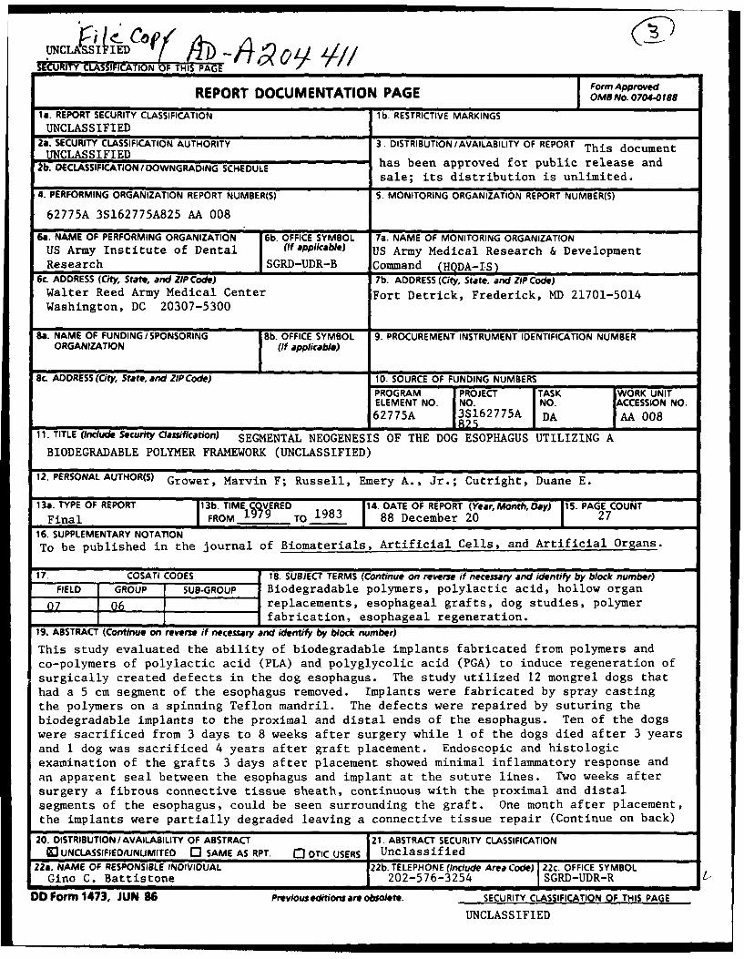
UNCLA~iIED(sI U IY LA SIFICATIO -O T I P GE 4
Form ApprovedREPORT DOCUMENTATION PAGE OMBNO. 0704-0188
la. REPORT SECURITY CLASSIFICATION lb. RESTRICTIVE MARKINGSUNCLASSIFIED
2a. SECURITY CLASSIFICATION AUTHORITY 3. DISTRIBUTION /AVAILABILITY OF REPORT This documentUNCLASSIFIEDDCLASSIFID N has been approved for public release andsale; its distribution is unlimited.
4. PERFORMING ORGANIZATION REPORT NUMBER(S) S. MONITORING ORGANIZATION REPORT NUMBER(S)
62775A 3S162775A825 AA 008
6a. NAME OF PERFORMING ORGANIZATION 6b. OFFICE SYMBOL 7a. NAME OF MONITORING ORGANIZATIONUS Army Institute of Dental (If applicable) US Army Medical Research & DevelopmentResearch I SGRD-UDR-B Command (HQDA-IS)
6c. ADDRESS (City, State, and ZIP Code) 7b. ADDRESS (City, State. and ZIP Code)Walter Reed Army Medical Center Fort Detrick, Frederick, MD 21701-5014Washington, DC 20307-5300
8a. NAME OF FUNDING/ SPONSORING Bb. OFFICE SYMBOL 9. PROCUREMENT INSTRUMENT IDENTIFICATION NUMBERORGANIZATION (If applicable)
Sc. ADDRESS (City, State, and ZIP Code) 10. SOURCE OF FUNDING NUMBERSPROGRAM PROJECT TASK WORK UNIT
62775 I 3f D A 0ELEMENT NO. NO. NO. CCESSION NO.
162775A 3S162775A DA AA 008
11. TITLE (Inlude Securiy Classification) SEGMENTAL NEOGENESIS OF THE DOG ESOPHAGUS UTILIZING A
BIODEGRADABLE POLYMER FRAMEWORK (UNCLASSIFIED)
12. PERSONAL AUTHOR(S) Grower, Marvin F; Russell, Emery A., Jr.; Cutright, Duane E.
13a. TYPE OF REPORT 13b. TIME COVERED 14. DATE OF REPORT (Year, Month, Day) 15. PAGE COUNTFinal FROM 1979 TO 1983 88 December 20 27
16. SUPPLEMENTARY NOTATIONTo be published in the journal of Biomaterials, Artificial Cells, and Artificial Organs.
17. COSATI CODES 18. SUBJECT TERMS (Continue on reverse if necessary and identify by block number)FIELD GROUP SUB-GROUP Biodegradable polymers, polylactic acid, hollow organ
07 06 replacements, esophageal grafts, dog studies, polymerfabrication, esophageal regeneration.
19. ABSTRACT (Continue on reverse if necessary and identify by block number)
This study evaluated the ability of biodegradable implants fabricated from polymers andco-polymers of polylactic acid (PLA) and polyglycolic acid (PGA) to induce regeneration ofsurgically created defects in the dog esophagus. The study utilized 12 mongrel dogs thathad a 5 cm segment of the esophagus removed. Implants were fabricated by spray castingthe polymers on a spinning Teflon mandril. The defects were repaired by suturing thebiodegradable implants to the proximal and distal ends of the esophagus. Ten of the dogswere sacrificed from 3 days to 8 weeks after surgery while 1 of the dogs died after 3 yearsand 1 dog was sacrificed 4 years after graft placement. Endoscopic and histologicexamination of the grafts 3 days after placement showed minimal inflammatory response andan apparent seal between the esophagus and implant at the suture lines. Two weeks aftersurgery a fibrous connective tissue sheath, continuous with the proximal and distalsegments of the esophagus, could be seen surrounding the graft. One month after placement,the implants were partially degraded leaving a connective tissue repair (Continue on back)
20. DISTRIBUTION/AVAILABILITY OF ABSTRACT 21. ABSTRACT SECURITY CLASSIFICATIONKI UNCLASSIFIED/UNLIMITED -- SAME AS RPT, [3 DTIC USERS Unclassified
22a. NAME OF RESPONSIBLE INDIVIDUAL 22b. TELEPHONE (include Area Code) 22c. OFFICE SYMBOLGino C. Battistone 202-576-3254 SGRD-UDR-R
DID Form 1473, JUN 86 Previous editions are obsolete. SECURITY CLASSIFICATION OF THIS PAGE
UNCLASSIFIED

19. ABSTRACT (Continued)
continuous with the proximal and distal ends of the esophagus. The repair area waslined with epithelium and enabled the dogs to drink freely and eat semisolid foods.In conclusion, it has been shown that it is possible to fabricate a biodegradableimplant which can stimulate regeneration of a hollow organ and which is compatiblewith long term survival. (UNCLASSIFIED)
. D~~TIC ,' l[
~C
SEGMENTAL NEOGENESIS OF THEDOG; ESOPHAGUS UTILIZING A :'t,,r
BIODEGRADABLE POLYMER FRAMEWORK.
Marvin F. Grower, D.D.S., Ph.D.Emery A. Russell, Jr., D.D.S., M.S.
Duane E. Cutright, D.D.S., M.S., Ph.D.
U. S. Army Institute of Dental Research, Division of Chemistry,Fort Meade, Maryland 20755
ABSTRACT
his study evaluated the ability of biodegradable implantsfabricated from polymers and co-polymers of polylactic acid (PLA)and polyglycolic acid (PGA) to induce regeneration of surgicallycreated defects in the dog esophagus. The study utilized 12 mongreldogs that had a 5 cm segment of the esophagus removed. Implantswere fabricated by spray casting the polymers on a spinning Teflonmandril. The defects were repaired by suturing the biodegradableimplants to the proximal and distal ends of the esophagus. Ten ofthe dogs were sacrificed from 3 days to 8 weeks after surgery while1 of the dogs died after 3 years and I dog was sacrificed 4 yearsafter graft placement. Endoscopic and histologic examination ofthe grafts 3 days after placement showed minimal inflammatoryresponse and an apparent seal between the esophagus and implant atthe suture lines. Two weeks after surgery a fibrous connectivetissue sheath, continuous with'the proximal and distal segments ofthe esophagus, could be seen surrounding the graft. One month afterplacement, the implants were partially degraded leaving aconnective tissue repair continuous with the proximal and distalends of the esophagus. The repair area was lined with epitheliumand enabled the dogs to drink freely and eat semisolid foods. Inconclusion, it has been shown that it is possible to fabricate abiodegradable implant which can stimulate regeneration of a holloworgan and which is compatible with long term survival. LuLt IOrd
A v I I I Ik3

I t* I I ~ I i i •
INTRODUCTION
The successful repair or replacement of hollow organs presents
a continuing challenge to the surgical profession. The repair of
such organs necessitated by atresia or traumatic avulsion such as
experienced in penetrating wounds requires immediate and complex
treatment. Often times multiple operations with secondary proce-_
dures at distant sites to bring autolgous tissue to the primary
site have been required to successfully replace an avulsed segment
of such organs.
The current therapy for repair and replacement of the diseased
or avulsed esophagus is by the use of autografts of viscus such as
the stomach [1], the colon [2], jejunal loops [3], isolated jejunal
segments [4], or split-thickness skin grafts [5]. None of these
procedures produce totally satisfactory results and complications
of reconstructive eophageal surgery may include [6]: necrosis of
the graft; infection; inadequate blood supply; difficulties in
suture retention; leakage at the anastomatic sites; stenosis of the
anastomosis between the esophagus and the graft; gastric stasis;
reflux; and eating disorders.
This study is a report on the use of biodegradable polymers to
construct esophageal implants which stimulated functional repair of
avulsive defects caused by excising a segment of the dog esophagus.
The polymeric materials selected to construct the biodegradable
esophageal implants used in this study were polylactic and polygly-
colic acid. These biodegradable polymers are aiiphatic polyesters
formed from the alpha-hydroxy-carbolic a:ids lactic acid and glyco-
lic acid. Studies on the fate and metabolism of these materials
have shown that they are eliminated primarily via respiration with
less than 10% eliminated via urine and feces and with trace amounts
in the tissue [7,8]. Surgical investigations utilizing various
forms of the polymers and copolymers as bone fixation devices
reported their disappearance via a vasofibroblastic and phagocytic
response accompanied with occasional giant cells [9,10,11]. These

same authors found no evidence of toxicity, antigenicity or
carcinogenicity and found the disappearance was accompanied by
restoration of normal morphology in bone and marrow and accompanied
by fibrous connective tissue and collagen in soft tissue sites.
The polymer polylactic acid was successfully used'as a mandi-
bular fixation device [11,121 and was used to repair blow out
fractures of the orbit [13]. Peripheral nerve cuffs constructed of
a copolymer of polylactic and polyglycolic acid prevented extrane-
ous connective tissue proliferation into the anastomotic site (141
and gave similar results when used as a tendon gliding device in
severely traumatized hands in primates [15].
Resorption rates of the PLA and PGA polymers and copolymers
have been determined to be between 4 weeks and 16 months [8,9,161.
These times can be varied according to the size, shape, method of
fibrication and the composition polymer or copolymer. In solid
form pure PGA or pure PLA have disappearance times of several
months duration while 50/50 ratios have disappearance times
measured in weeks. When these copolymers are sprayed into fibers
of 10-25 micron diameters disappearance rates can be reduced to 14
days or less.
On a conceptual basis, the use of a biodegradable polymer to
fabricate a successful hollow organ graft holds promise in that, if
successful, it would obviate the need for multiple operations and
the concern for the vascular integrity of the graft. In addition,
such structures would be readily available for immediate repair of
the defect and could be custom made, depending on the defect to be
repaired.
MATERIALS AND METHODS
The biodegradable polyier grafts were fabricated by dipping
and spraying polymers and copolyipers of polylactic and polyglycolic
acid dissolved in solvent onto a spinning Teflon mandril. The
materials used were as follows: (1) 1,1,1,3,3,3-Hexafluoro-2-
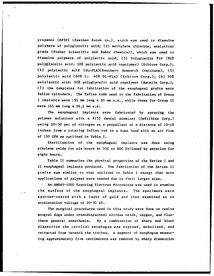
propanol (HFIP) (Eastman Kodak Co.), wiiich was used to dissolve
polymers of polyglycolic acid; (2) methylene chloride, analytical
grade (Fisher Scientific and Baker Chemical), which was used to
dissolve polymers of polylactic acid; (3) Polyglactin 910 (90%
polyglycolic acid: 10% polylactic acid copolymer) (Ethicon Corp.);
(4) polylactic acid (DL-PLA)(Southern Research Institute); (5)
polylactic acid (60% L: 40% DL-PLA) (Ethicon Corp.); (6) 50%
polylactic acid: 50% polyglycolic acid copolymer (Batelle Corp.);
(7) the templates for fabrication of the esophageal grafts were
Teflon cylinders. The Teflon rods used in the fabrication of Group
I implants were 150 mm long x 20 mm o.d., while those for Group II
were 145 mm long x 26.2 mm o.d.
The esophageal implants were fabricated by spraying the
polymer solutions with a #152 dental atomizer (DeVilbiss Corp.)
using 20-30 psi of nitrogen as a prdpellant at a distance of 10-18
inches from a rotating Teflon rod in a fume hood with an air flow
of 150 CFM as outlined in Table I.
Sterilization of the esophageal implants was done using
ethylene oxide for six hours at 55C to 60C followed by aeration for
eight hours.
Table II summaries the physical properties of the Series I and
II esophageal implants produced. The fabrication of the Series II
grafts was similar to that outlined in Table I except that more
applications of polymer were needed due to their larger size.
An.AMRAY-1000 Scanning Electron Microscope was used to examine
the surface of the esophageal implants. The specimens were
sputter-coated with a layer of gold and then examined at an
acceleration voltage of 20-30 kV.
The surgical procedures used in this study were done on twelve
mongrel dogs under oroendotracheal nitrous oxide, oxygen, and fluo-
thane general anesthesia. By a combination of sharp and blunt
dissection the cervical esophagus was exposed, mobolized, and
retracted from beneath the trachea. A segment of esophagus measur-
ing approximately five centimeters was removed by sharp dissection
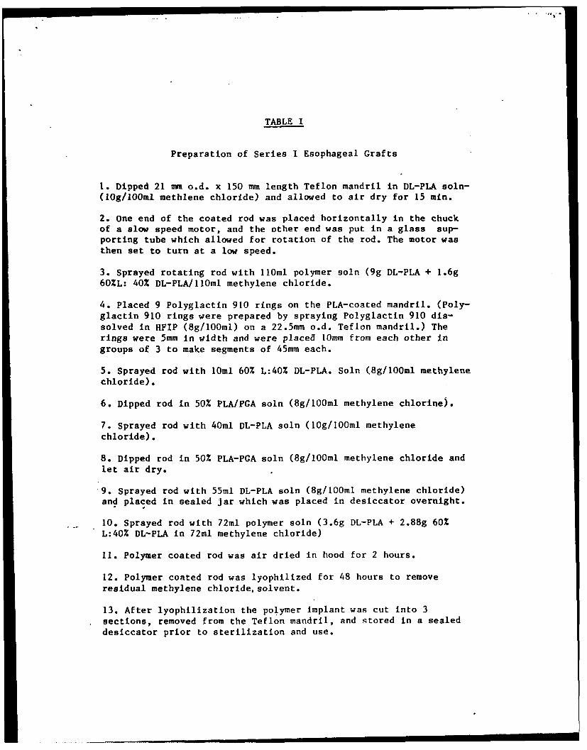
TABLE I
Preparation of Series I Esophageal Grafts
1. Dipped 21 mm o.d. x 150 mm length Teflon mandril in DL-PLA soln-(IOg/lOOml methlene chloride) and allowed to air dry for 15 min.
2. One end of the coated rod was placed horizontally in the chuckof a slow speed motor, and the other end was put in a glass sup-porting tube which allowed for rotation of the rod. The motor wasthen set to turn at a low speed.
3. Sprayed rotating rod with 1lOml polymer soln (9g DL-PLA + 1.6g60%L: 40% DL-PLA/IIOml methylene chloride.
4. Placed 9 Polyglactin 910 rings on the PLA-coated mandril. (Poly-glactin 910 rings were prepared by spraying Polyglactin 910 dis-
solved in HFIP (8g/1O0ml) on a 22.5mm o.d. Teflon mandril.) Therings were 5mm in width and were placed 10mm from each other ingroups of 3 to make segments of 45mm each.
5. Sprayed rod with 10ml 60% L:40% DL-PLA. Soln (8g/lOOml methylenechloride).
6. Dipped rod in 50% PLA/PGA soln (8g/10Oml methylene chlorine).
7. Sprayed rod with 40ml DL-PLA soln (10g/100ml methylenechloride).
8. Dipped rod in 50% PLA-PGA soln (8g/100ml methylene chloride andlet air dry.
9. Sprayed rod with 55m1 DL-PLA soln (8g/bOOml methylene chloride)and placed in sealed jar which was placed in desiccator overnight.
10. Sprayed rod with 72m1 polymer soln (3.6g DL-PLA + 2.88g 60%
L:40% DL-PLA in 72mi methylene chloride)
11. Polymer coated rod was air dried in hood for 2 hours.
12. Polymer coated rod was lyophilized for 48 hours to removeresidual methylene chloride, solvent.
13. After lyophilization the polymer implant was cut into 3sections, removed from the Teflon mandril, and stored in a sealeddesiccator prior to sterilization and use.
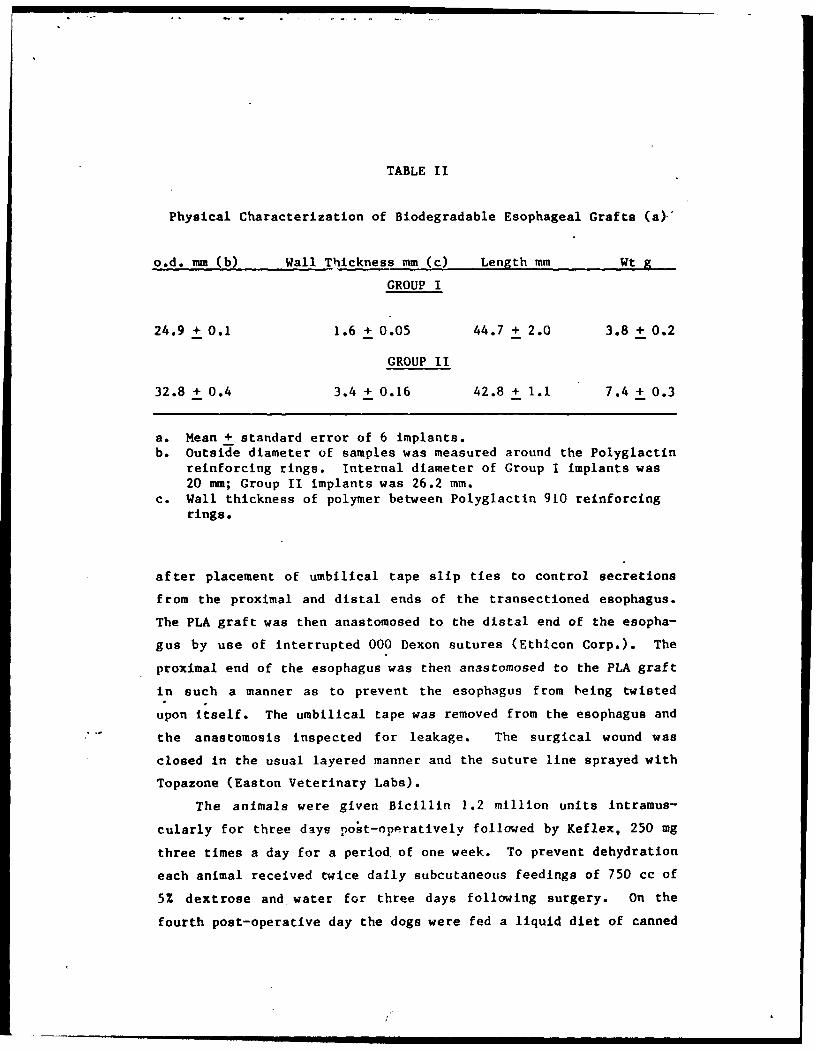
TABLE II
Physical Characterization of Biodegradable Esophageal Grafts (a)-
o.d. mm (b) Wall Thickness mm (c) Length mm Wt g
GROUP I
24.9 + 0.1 1.6 + 0.05 44.7 + 2.0 3.8 + 0.2
GROUP II
32.8 + 0.4 3.4 + 0.16 42.8 + 1.1 7.4 + 0.3
a. Mean + standard error of 6 implants.b. Outside diameter of samples was measured around the Polyglactin
reinforcing rings. Internal diameter of Group I implants was20 mm; Group II implants was 26.2 mm.
c. Wall thickness of polymer between Polyglactin 910 reinforcingrings.
after placement of umbilical tape slip ties to control secretions
from the proximal and distal ends of the transectioned esophagus.
The PLA graft was then anastomosed to the distal end of the esopha-
gus by use of interrupted 000 Dexon sutures (Ethicon Corp.). The
proximal end of the esophagus was then anastomosed to the PLA graft
in such a manner as to prevent the esophagus from being twisted
upon itself. The umbilical tape was removed from the esophagus and
the anastomosis inspected for leakage. The surgical wound was
closed in the usual layered manner and the suture line sprayed with
Topazone (Easton Veterinary Labs).
The animals were given Bicillin 1.2 million units intramus-
cularly for three days po~t-oppratively followed by Keflex, 250 mg
three times a day for a period of one week. To prevent dehydration
each animal received twice daily subcutaneous feedings of 750 cc of
5% dextrose and water for three days following surgery. On the
fourth post-operative day the dogs were fed a liquid diet of canned
dog food emulsified in water and fortified with fat (Pig Kalorie
Supplement, Haver-Lockhart Labs).
One dog was sacrificed at, 3 and 9 days post surgery and two
dogs at 14, 21, 30 and 56 days with an overdose of barbiturates.
Two of the animals were retained for long-term study.
The dog with a series I graft survived for 3 years and the
other animal implanted with a series II graft was sacrificed 4
years after graft placement.
At sacrifice the graft sites were immediately removed in a
cervical block to include the surrounding tissue and at least 2 cm
of normal esophagus at either end of the graft. Excess tissue was
trimmed off the specimens and they were placed in buffered 10%
formalin. After fixation, the graft sites were grossed serially
into 5 to 8 mm transverse segments and photographed. Tissue
sections were then prepared at 6 microns thickness and stained with
hematoxylin and eosin for histologic analysis.
RESULTS
Implant Construction
Spraying of the polymer solutions produced fibers of 3-25
microns in diameter and 2-10 cm in length which oriented themselves
in a circular manner as they attached to each other around the
rotating Teflon mandril. The spraying of methylene chloride
solutions of the amorphous structured DL-PLA produced shorter and
finer fibers of polymer while the presence of the more crystalline
L-PLA in the solutions produced longer and larger diameter fibers.
An example of the polymeric grafts, which were produced by
utilizing the procedures odtlined in Table I, is shown in Figure 1.
The fabricated grafts were rigid and showed very little tendency to
flex. The inner surface of the graft was smooth due to its being
composed of a solid film of PLA produced when the Teflon template
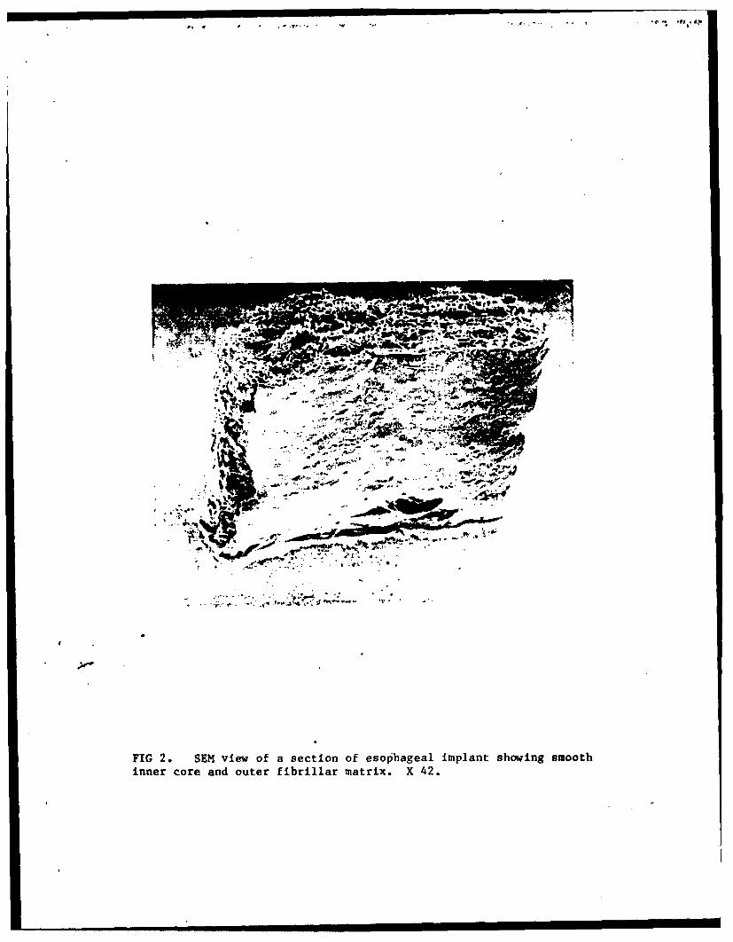
was dipped into the PLA solution. Figure 2 shows the scanning
I,

electron microscopic view of the smooth inner lumen as well as the
outer portion of the graft. The inner portions of the graft was
more solid in nature than the peripheria due to filling up of
inter-fibrillar networks by the 50Z PLA/PGA dips used in fabrica-
tion of the core portion of the implant. This solid core tended to
provide a certain rigidity to the implant. The outer half of the
implant was composed of a circular network of PLA fibers 3-25
microns in diameter which could be observed as a series of lamina-
tions around the inner core as seen in Figures 1 and 2.
The fibrillar outer coating of the graft allowed for the rapid
infiltration of fibrin and fibrovascular tissue into the implant
which resulted in a water-tight seal. These implants exhibited
initial resistance to flex and collapse; however, the consistency
of the wall material was flexible enough to allow the needle from a
3-0 Dexon suture (Ethicon Corp.) to be placed completely through
its wall. The sutures placed through the graft wall were retained
in position and the wall of the graft did not show any tendency to
tear after placement of the sutures. Comparison of the physical
properties of Group I implants and Group I implants (Table Ii)
shows that the Group 1I implants had an inner-diameter 6.2 mm
greater than Group I and a wall thickness at least two times as
thick. These larger implants were constructed to provide more
resistance to lumen collapse which was noted in some two-week
specimens, and to produce a larger diameter esophageal replacement
which would be more resiqtant to esophageal stricture during the
repair phase of healing.
Ethylene oxide sterilization of the implants caused an average
4% decrease in length and a 6% decrease in diameter from the dimen-
sions listed in Table II. This dimensional shrinkage was also
accompanied by a slight increase in flex resistance.
Clinical Findings
The post-operative periods progressed uneventfully. There was
very little swelling at the surgical site. Endoscopic examination
of the grafts in situ at three days and eight days showed an unob-
structured esophageal graft which was in continuity with the rest
of the esophagus. Oral administration of slurried canned dog food
was thus begun on day four after initial surgery. By the fourth
week after surgery, the dogs showed some difficulty in oral feeding
and endoscopic examination showed contraction of the repair tissues
present in the graft site. Dilation with a series of metal bougies
starting at a French #29 (9.5 mm) and ending at a French #45 (14.8
mm) was begun at this time and the graft sites were dilated biweek-
ly until two weeks prior to sacrifice in the two 56-day dogs.
The dog which survived 3 years after graft placement had
received a series I implant. It was dilated biweekly until six
months post surgery. Some esophageal constriction was seen at this
time, but was readily relieved by bougienage therapy and a size #45
French dilator freely passed the length of the esophagus after
dilation. This dog was then dilated monthly for the next three
months and then one more time two months later. At this time
bougienage therapy was terminated. At 18 months post surgery,
barium X-rays taken of the esophagus showed that an area of
stricture was present in the esophagus in the area of the implant.
However the dog ate well and showed an increase in body weight from
45 pounds prior to surgery to 48 pounds at that time. Three years
after tife initial surgery, the dog died. The necropsy report stated
that the cause of death may have been gastric torsion.
The second long-term survival animal had been implanted with a
series II graft. This dog was not dilated until 40 days after
surgery and then was dilated monthly for two months up to a size
#45 French dilator, after ldhich time it had received no further
dilations although the endoscope used to examine the esophagus had
a diameter equivalent to a #35 French dilator. Monthly endoscopic
examinations done over the next 9 months showed that no esophageal
constrictions were present and that the epithelium of the graft
area was continuous with that of the original esophagus.
At 18 months post surgery, endoscopy was again done. The endoscope
passed easily through the esophagus; the lining of the lumen
appeared to be completely epithelialized and only a slight area of
constriction was noted. The dog ate well and after an initial
weight loss post surgery, the dog eventually regained his preopera-
tive weight of 40 pounds.
The animal was sacrificed 4 years after placement of the
implant due to transfer of the principal investigators.
Gross and Histologic Findings
The gross morphology of the esophageal grafts after three days
of implantation is shown in Figure 3. Gross examination of the
implant site did not show any evidence of leakage around the
anastomosis between the polymer tube and the esophageal tissue.
All the original sutures were present and they showed only minimal
inflammatory reaction around them. The implant did not show any
evidence of collapse or loss of structural integrity at this time.
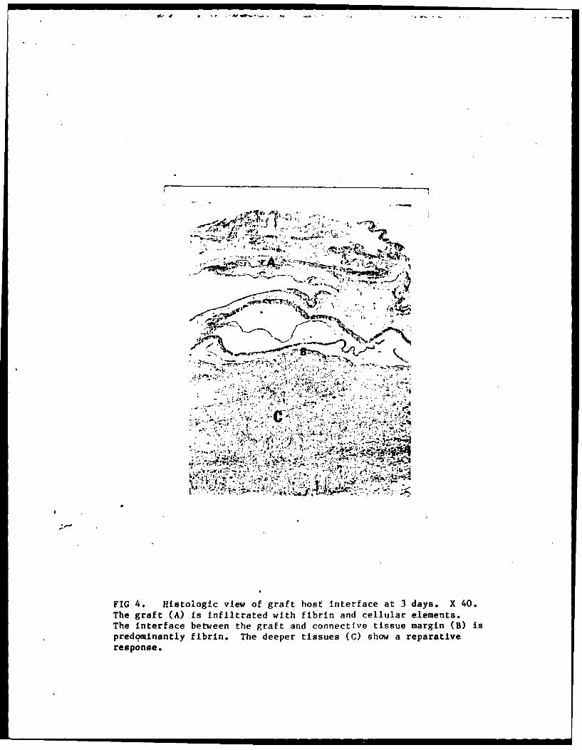
Histologic examination of the original connective tissue wall
of the esophagus and the graft itself as seen in Figure 4 showed a
layer of fibroblastic tissue demonstrating hemorrhage with early
vascular proliferation. At the interface between the graft and the
connective tissue there was evidence of platelet and fibrin accumu-
lation within the interstices of the graft, but little evidence of
organization. The inflammatory response to the graft and sutures
was minimal at this time. Although there was no proliferation of
the early fibroblastic tissue into the interstices of the graft, at
3 days there was a definite increase in mitotic activity in the
surrounding connective tisshes. This was evidenced by an increased
number of fibroblasts and small vascular channels proliferating
around the edges of the graft.
The gross section of the 9-day surgery sample shown in Figure
5 demonstrates that the intertace between the connective tissue and
the graft showed a maturation of the fibrous connective tissue and

ingrowth of the vasofibroblastic tissue into the interstices of the
graft material. The thickness of the connective tissue wall,.
measured from the outer surface of the tracheal cartilage rings,
was 2 mm. The trachael wall did not show any evidence of erosion
due to presence of the graft. Figure 5 also shows that the thinner
walled implants of Group I (Table I) exhibited a tendency to close
down; however, a sufficient opening for maintenance of the nutri-
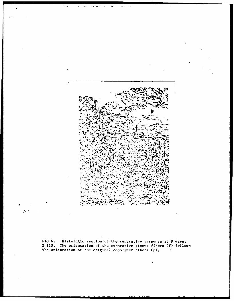
tional requirements of the dog was still present. Histologically,
there could be seen a beginning orientation of the connective
tissue fibers in a circular manner around the graft site (Figure
6). There was very little tendency to form giant cells surrounding
the polymer fibers. The interface between the graft and the
original connective tissue showed an ingrowth of 1.5 mm of fibrous
connective tissue into the interstices of the graft with minimal
inflammation present. At the margins, or at the interface between
the graft and the esophageal epithelium, there was a proliferation
of new epithelium at least 3 mm down over the graft site.
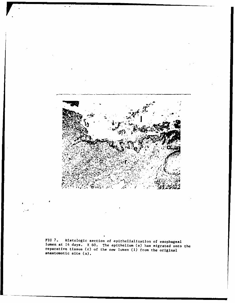
Examination of the 14-day samples revealed a vasofibrobastic
connective tissue wall of approximately 3 mm in thickness which
surrounded the esophageal implant. In certain areas of the
connective tissue graft interface there was multinucleated giant
cell formation where the polymer had been incorporated into the
tissue and this extended back about 1.5 mm from the apparent edge
of 'the graft itself, indicating that the vasofibroblastic ingrowth
had penetrated at least 1.5 to 2 mm into the graft. In addition,
there was minimal evidence of collagen organization into both
circular and longitudinal bands in the implant area as well as
epithelial migration down the edges of the draft itself as seen In
Figure 7.
Gross examination of the 307day samples, as seen in Figure 8,
showed a vasofibroblastic wall of about 5 mm (measured from the
outer tracheal ring) which had formed both as a result of tissue
growth around the implant and growth into the implant itself of
about 2.5 mm. Most of the inner core of the implant had become
-. .I "

delaminated from the outer sections which had induced the peri-
pherial fibroblastic response and, or which, has become invaded by
repair tissue. Histologically, the advancing front of granulation
tissue into the graft was characterized by occasional multinuclea-
ted giant cells which were phagocytosing the polymer after it had
been hydrolyzed. The collagen fibers present in the new esopha-
geal wall showed a variable orientation and there was variable
epithelial migration up to 5 mm from the anastomosis along the
inner aspect of the graft site.
By eight weeks the graft resorption and elimination was almost
complete and a collagenous tube with a wall thickness up to 5 mm
measured from the tracheal rings was present (Figure 9). In
addition, it was evident that the esophageal implants had not
affected or caused any erosion of the trachea. The lumen of the
esophagus was open although some contraction of the repair tissue
was evident as indicated by the narrowing of the esophageal lumen.
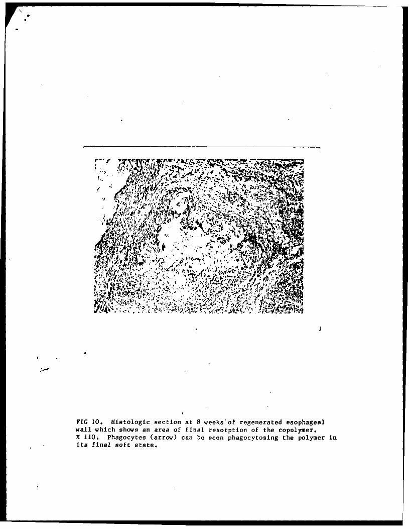
There was no macroscopic evidence of graft material. Microscopic
examination, however, showed some evidence of residual polymer
between the collagen fibers as seen in Figure 10. The Dexon
sutures on the other hand appeared to have become completely
degraded. Only minimal inflammation and slight edema were present
at the graft site and the collagen showed a variable orientation
with a small amount of circular orientation. There were islands
and sheets of epithelium growing over the lumenal surface of the
repair tissue in the area of the original graft, although some
areas were still not completely epithelialized at eight weeks.
Necropsy of the animal, which died after 3 years of graft
placement (series I), showed that the area of the esophagus
anterior to the graft site'showed some dilation while the esophagus
distal to the graft site (gastric end) was normal in appearance.
The graft site itself showed constriction and shrinkage to about
1.5 cm from its original 4 cm length.
Histologic analysis was not done due to the advanced
postmortem degeneration of the tissues. Gross and histologic
analysis of the 4 year dog was not done due to transfer of the
principal investigators.
DISCUSSION
This study demonstrated the feasibility of using a biodegrad-
able hollow organ implant fabricated from polylactic acid and poly-
glycolic acid polymers to serve as a template for regeneration of
an excised segment of the dog esophagus. The inner core provided
for rigidity of the implant as well as a seal which prevented
egress of stomach fluids into the surrounding tissues. The outer
covering of PLA fibers provided for strength and allowed for a
vasofibroblastic ingrowth into the graft as well as promoting
growth of a collagenous sheath of tissue around the peripheria of
the graft. The fabrication of 100% PLA into 3-25 micron fibers in
the outer covering, as was done in this study, allowed degradation
of some polymer in as little as 14 to 21 days by hydrolysis of
tissue fluids and cellular action; although some polymer incorpora-
ted into the repair tissue appeared to be retained for up to 8
weeks (Figure 10). This resorption time is less then the time
required for solid PLA plates.as plates are partially resorbed in
six weeks and total resorption may taks six months [11,121.
The decrease in size and increased resistence to flex which
occurred on ethylene oxide sterilization may have been due to an
increase in crystallinity of the polymer resulting from the heat
used in the sterilization process as suggested by a recent study by
Gindoe and Gupta [171.
The laminar construction of the grafts apparently allowed for
partial removal of the graft by a delamination of the inner core at
3-4 weeks post-surgery. This delamination appeared to be an
essential requirement for the success of these grafts since the
Polyglactin supporting rings in the implants only provided limited
resistence to graft collapse as was seen in some early specimens
(Figure 6). The use of more rigid cast or machine formed solid
rings of PLA or PGA as an inner support would probably prevent such
graft collapse while allowing for their degradation in stitu or in
the stomach if delamination of the graft occurred.
The fibrillar portions of the polymer implants in contact with
the cut ends of the esophagus as well as the connective tissue in
the implant site promoted a favorable healing reaction as evidenced
by the complete repair of the graft site. This repair was by a
collagenous sheath connecting the two ends of the esophagus and was
achieved as early as two weeks. Histologically, the final repair
seen at 8 weeks was by a dense hyalinized type of connective tissue
in sheath form. The connective tissue sheath allowed the animal to
eat freely with minimal discomfort until the fourth week after
surgery, when some contraction of the-new segments was noted. This
constriction was readily relieved by dilation of the esophagus with
metal bougies. Similar problems may also be seen with autogenous
viscus replacements 1181 and the course of bougienage therapy used
on the long term surviving dogs is similar to the therapeutic
regimens used on human patients with constrictive esophagitis [191.
The ability of the dog receiving a series I implant (OD -
24.9mm) to survive 25 months without bougienage therapy, and that
of the dog receiving a series.II implant (OD - 32.8mm) to survive
for at least 36 months without additional dilation therapy, sug-
gests that a functional maturation of the connective tissue at the
repair site eventually occurred. Nevertheless, the constriction
noted at 18 months in the dog receiving the series I graft as well
as the shrinkage and contraction noted 18 months later at necropsy
suggests that further dilation therapy may have reduced this
shrinkage. However, the main criteria for stimulating and main-
taining a physiologic repair may be the initial size of the polymer
graft. The dog which received the larger series II graft did not
appear to need additional dilation therapy after the initial series
in order to maintain its nutritional intake up to its sacrifice 4
years after graft placement.

These results compare favorably with results achieved using
viscus grafts in which patients report difficulties in eating and
slow weight gain for 12 to 18 months or longer [18).
The esophageal contraction and shrinkage noted on the necropsy
of the dog receiving the series I graft may or may not have been a
factor in the death of the dog due to possible gastric torsion,
since gastric torsion is a condition of unknown pathogenesis which
can occur in caged animals.
In conclusion, it has been shown in this study that: (1) It
is possible to construct a completely biodegradable off-the-
shelf graft which can replace lost segments of hollow organs.
(2) Regeneration of the hollow organ occurred by a new tube of
connective tissue lined by epithelium utilizing the technique of
neogenesis within a biodegradable polymer-copolymer framework. (3)
The replacement supported the dogs' nutritional intake require-
ments. (4) The replacement showed adequate strength and allowed for
maintenance of esophageal diameter by bougienage therapy. (5) It
did not appear to exhibit problems such as the need for multiple
operations, leakage at anastomosis sites, lack of blood supply, and
postoperative infections seen with other therapeutic procedures in
current use. (6) Grafts made of spun biodegradable PLA and PGA
co-polymers meet the general requirements for an effective and
easy-to-use replacement and should be studied further.
"In conducting research described in this report, the investigatorsadhered to the 'Guide for the Care and Use of Laboratory Animals'as promulgated by the Committee on the Revision of the Guide forLaboratory Animal Facilities and Care of the Institute of Labora-tory Animal Resources National Research Council."
"Commercial materials and equipment are identified in this reportto specify the investigative procedure. Such identification doesnot imply recommendation or endorsement, or that the materials andequipment are necessarily the best available for the purpose."
"The views of the authors do not purport to reflect the views ofthe Department of the Army or the Department of Defense."
REFERENCES
1. D. Gavriliu, "The Long-Term Clinical State After Resection withJejunal Replacement" in Surgery of the Oesophagus, (R.A. Smithand R.E. Smith, eds.) Vol. 29, Butterworth, London, 1975, p.33,
2. R. Belsey, Reconstruction of the esophagus with left colon.J. Thorac. Cardiovasc. Surg., 49, 33 (1965).
3. R.H.F. Brain, Steatorrhea in oesophagastric surgical practice.Proc. R. Soc. Med., 46, 438 (1953).
4. B. Seidenberg, S.S. Rosenak, E.S. Hurwitt, and M.L. Som,Immediate reconstruction of the cervical esophagus by arevascularized isolated jejunal segment. Ann. Surg., 149, 162,(1959).
5. M.T. Edgerton, One stage reconstruction of cervical esophagusor trachea. Surgery, 31, 239, (1952).
6. A.J. Gunning, and R. Marshall, The Oesophagus. Part II:Replacement of the oesophagus. Clin. Gastroenterol, 8, 293(1979).
7. R.K. Kulkarni, K.C. Pani, C. Neuman, and F. Leonard. Polylac-tic acid for surgical Implants. Arch. Surg., 93, 839, (1966).
8. J.M. Brady, D.E. Cutright, R.A. Miller, E.E. Hunsuck, and G.C.Battistone. Resorption rate, route of elimination and ultra-structure of the implant site of polylactic acid in the abdomi-nal wall of the rat. J. Biomed. Mater. Res., 7, 155, (1973).
9. R.A. Miller, J.M. Brady, and D.E. Cutright, Degradation ratesof oral resorbable implants (polylactates and polyglycolates).Rate of modification with cianges in PLA/PGA copolymer ratios.J. Biomed. Mater. Res., 11, 711, (1977).
10@ D.E. Cutright and E.E. Hunsuck, Tissue reaction to the biode-gradable polylactic acid suture. Oral Surg., 31(1), 134,(1971).
11. D.E. Cutright, E.E. Hunsuck, and J.D. Beasley III, Fracturereduction using a biodegradable material, polylactic acid. J.Oral Surg., 29, 393, (1971).
12. L. Getter, D.E. Cutright, S.N. Bhaskar, and J.K. Augsburg, Abiodegradable intraosseous appliance in the treatment ofmandibular fractures. J. Oral Surg., 30. 344, (1972).
13. D.E. Cutright and E.E. Hunsuck, The repair of fractures of theorbital floor using biodegradable polylactic acid. Oral Surg.,33(1), 28, 34, (1972).
14. R.L. Reid and D.E. Cutright, The utilization of biodegradablenerve cuffs as an adjunct in peripheral nerve anastomosis. InProgress.
15. D.E. Cutright and R.L. Reid, A biodegradable blocking agent toimprove tendon gliding - A primate study, The Hand, 7, 228,(1975).
16. D.E. Cutright, B. Perez, J.D. Beasley III, W.J. Larson, andW.R. Posey, Degradation rates of polymers and copolymers ofpolylactic and polyglycolic acids. Oral Surg., 38(1), 142,(1972).
17. R.M. Gindoe and R.K. Gupta, In vitro chemical degradation ofpoly(glycolic acid) pellets and fibers. J. App. Polymer Sci.33, 2411, (1987).
18. A.J. Gunning and R. Marshall. The oesophagus. Part II:Replacement of the oesophagus. Clini. Gastroenterol. 8, 292,(1979).
19. C.A. Flood, Bougienage therapy for constrictive esophagitis.Gastrointest. Endosc. 25, 130, (1979).

FIG 1. End view of a Group II biodegradable polymer esophagealgraft. X 1.5.
-L 7U t I
* * W 1
;; -4
FIG 2. SEll view of a section of esophageal implant showing smoothinner core and outer fibrillar matrix. X 42.

FIG 3. Three-day gross specimen of Ifhe anterior anastoinosis of
the esophagus with the polymer graft (longitudinal section). (a)
anastomosis site; (e) anterior esophagus; (g) main body of im-
plant. The adherence of tissue to graft and fibrin penetration
into graft is shown by the arrow.
--
FIG .Hitoloic vew f graft host inefc at3 as.X 0
The raf (A)is nfilratd wih fbrinandcelllarelemnts
The interfaceI. bewe th graf andconetie isse arin(Bi
predminntl firin.Thedeeer isse C hwarprtv
response1
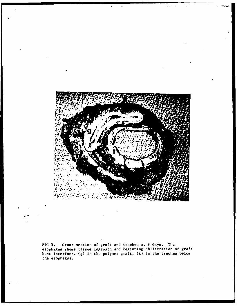
FIG 5. Gross section of graft and trfachea at 9 days. Theesophagus shows tissue ingrowth and beginning obliteration of grafthost interface. (g) Is the polymer graft; (t) is the trachea belowthe esophagus.
-- &
'AV1q .'"V
- 'V
FIG~ ~ ~ 6.* Hitloi seto fterprtv-epnea as
X 110 Theorienatio of te rearatie tisue fbers(f folwthe orenato ofte'rgna on'1or b -s()
1 tt
koaA 'ZI
4I~ ~-A' .d'
FIG ~ 7. Hsolgcseto o pthlaizto o sphgalumen at 14dy. X6. Teeihlu-e a irtdot h
reparative tisu (r ftenwlmn(1 rmteoiia
anastomoi sie a)
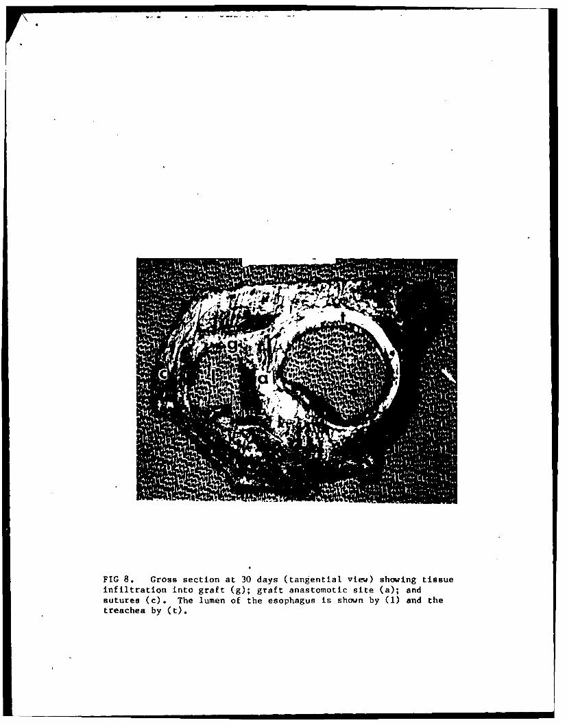
FIG 8. Gross section at 30 days (tangential view) showing tissueinfiltration into graft (g); graft anastomotic site (a); andsutures (c). The lumen of the esophagus is shown by (1) and thetreachea by (t).

M4 v
J V
dlrd
FIG 9. Gross section of 8-week esophageal implant (cross sec-
tion): Cr) tegenerated esophageal wall; (1) lumen of esophagus;
Wt trachea.
01
* ~ *V0
'16.
*~~ 1*41 *
II
FIG 10. Histologic section at 8 weeks'of regenerated esophagealvail which shows an area of final resorption of the copolymer.X 110. Phagocytes (arrow) can be seen phagocytosing the polymer inits final soft state.