Embed Size (px)
Citation preview

SECTION II
METHODOLOGICAL PROCEDURES
77

Chapter 1 - MINIMAL REQUIREMENTS FORIDENTIFICATION
The correct identification of microorganisms depends on the availa-bility of pure cultures. A mycobaterial culture contaminated by other micro-organisms will lead to false positive or negative results in the identificationtests.
The minimal requirements for identification (230) are:
• A pure culture• A culture with at least 20 colonies; in the case of cultures with fewer
colonies it is advisable to make a subculture, so that the amount ofbacteria is enough for the performance of all the tests.
The first step for identification is, thus, to make a smear of the culture,stained by the acid fast stain (either Ziehl-Neelsen or Kinyoun methods).
The objectives of the smear are:
• to confirm that the culture is composed of acid-fast bacilli (AFB)• to confirm that the culture is not contaminated with other bacterial
species• to verify the presence of cord formation, which suggests that the
culture could be of M. tuberculosis
The second step is the observation of the culture aspect.The objectives of this observation are:
• to confirm the purity of the culture by verifying the absence ofcontamination
• to evaluate the colony aspect: pigmentation and morphology.
1.1. Bacterial suspension preparation
• small screw-capped bottle• glass beads, 3 mm in diameter• distilled water or phosphate buffer pH 7.4
Place 10 glass beads in a bottle containing 1 ml of distilled water orphosphate buffer. Sterilize by autoclaving.
Suspension preparation: use a sterile swab to transfer certain amountof bacterial growth from the primary culture into the suspension bottle (a 10µl loop can also be used instead of a swab but the swab gives a more homo-
Practical handbook for the phenotypic and genotypic identification of mycobacteria
79

geneous suspension). Make a homogeneous and heavy suspension with thehelp of the swab and the glass beads. Let stand for 5 minutes to allow thelarger residual clumps of bacteria and aerosol generated to settle.
The suspension can be made with phosphate buffer or distilled water.In the case of mycobacteria the use of buffer is not important due to theparticularity of the mycobacteria wall. The use of distilled water is more ad-visable because it is simpler and there is less risk of contamination than usingthe buffer.
This bacterial suspension is used to inoculate test media and biochem-ical tests in liquid media. For media inoculation, see below. For biochemicaltests performed in liquid media, the inoculum will vary according to the test,and will be explained in the corresponding test description.
1.2. Inoculation of the test media
Using a pipette or a bacteriological loop (sterile), inoculate 10 µl ofthe suspension in test media. The inoculum should not be spread over thesurface of the medium but streaked down the middle to facilitate the exam-ination of growth, as the border between growth and medium is more easilyobserved.
1.3. Preservation of strains
There are several ways of preserving mycobacterial strains:• a simple method of storing cultures grown on egg media, is to
keep them in a freezer at –20oC . The viability of bacteria is main-tained for more than one year
• to save space in the freezer, it is recommended to store the bacteriasuspended in a small tube with 10% glycerol, 1% glutamate or10% glucose in a freezer at –20oC. The viability is maintained for atleast one year.
• Cultures in a freezer at –20oC, using 20% skim milk as a medium.The viability is maintained for more than two years.
• We recommend the glass embroidery beads method.
1.3.1. Conservation of mycobacteria using glass embroidery beads at–70°C
• Wash the glass beads with water and detergent.• Dry the glass beads at 45 °C
SECTION II - METHODOLOGICAL PROCEDURES
80

• Place 6-10 glass beads in small plastic flasks or microcentrifuge vi-als; sterilize by autoclaving.
• Put 0,5 ml of Sauton media with 10% glycerol in the microcentri-fuge vial; be careful to avoid contamination.
• Identify the microcentrifuge vial with labels on the side and on thecap of the vial.
• Place a loopful of bacterial growth (it is important that the bacteriais in an exponential growth phase) and stir the medium with theloop to dissolve the clumps.
• Let the microcentrifuge vial settle for 15 minutes.• Pipette out and discard all medium• Place the vials in boxes and make a map of each box, with the
numbers of the strains for quick localization.• Freeze at -70°C. (viability is maintained for up to 10 years)
1.3.2. A modification of the method for conservation of mycobacteria inskim milk at -70oC (adding 10% glycerol)
• Use fresh cultures in Lowenstein-Jensen (2-3 weeks)• Prepare skim milk (Difco ref 232100) according to manufacturer
instructions and add 10% glycerol• Aliquot 3 ml in 13x100 tubes and autoclave for 10 min,121oC• Use an sterile swab to make a heavy bacterial suspension• Homogenize and aliquot 1 ml in 1.8 ml sterile cryovials• Keep at -70oC (viability is maintained for up to 10 years)
1.4. Staining procedures
1.4.1. Ziehl-Neelsen
MaterialsBasic fuchsinEthanol 95%Phenol crystalsHydrochloric acid, concentratedMethylene blue chlorideDistilled water
Preparation- Fuchsin: dissolve 3 g of basic fuchsin in 100 ml of 95% ethanol.- Phenol: dissolve 5 g of phenol crystals in 100 ml of water (place it in a
water-bath until it dissolves completely)
Practical handbook for the phenotypic and genotypic identification of mycobacteria
81

- Carbol fuchsin: mix 10 ml of fuchsin solution with 90 ml of phenol solu-tion. Filter the carbol fuchsin prior to use.
- Acid alcohol: carefully add 30 ml of concentrated hydrochloric acid to 970ml of 95% ethanol; mix gently.
- Methylene blue – dissolve 3 g of methylene blue chloride in 1000 ml ofdistilled water.
Procedure- Prepare the smear and allow it to dry at room temperature- Heat fix the smear passing the slide through a Bunsen burner flame as for
other bacteriological smears- Place the slides on a staining rack- Cover the entire surface of each slide with carbol fuchsin- Using a Bunsen burner or cotton and alcohol flame, gently heat the slides
until vapour rises.DO NOT ALLOW THEM TO BOIL OR DRY.- Allow the stain to remain on the slides for 5 minutes. Maintain heat
throughout this period in a way that vapour rises 3 times during the 5minutes.
- Gently wash the stain from the slide.- Cover the slides with acid alcohol; leave it on the slides for 3 minutes.- Rinse the slides again.- Counter stain with methylene blue for 1 minute, and rinse again.
1.4.2. Kinyoun (100)
MaterialBasic fuchsinEthyl alcohol (95%)Destilled waterAcid alcoholMethylene bluePhenol
Preparation• Kinyoun carbolfuchsin:
- Basic fuchsin 4 g- Ethyl alcohol 20.0 ml
(dissolve and add slowly while shaking)- Distilled water 100.0 ml- Liquefied phenol (melted crystals) 8 g
SECTION II - METHODOLOGICAL PROCEDURES
82

• Acid-alcohol- Ethyl alcohol (95%) 97.0 ml- Concentrated hydrochloric acid 3.0 ml
• Counter stain:- Methylene blue chloride 0.3 g- Distilled water 100.0 ml
Procedure- Heat-fix smears on slide warmer at 65oC to 75oC for 2 hours or overnight.
An alternative is to use a Bunsen burner passing the slide through theflame without overheating.
- Cover smear with a 2x3 cm piece of filter paper (to hold the stain on theslide and to filter out any undissolved crystals fo dye)
- Flood the paper strip with Kinyoun carbol fuchsin- Stain for 5 minutes (no heat is necessary)- Remove the paper strip with forceps- Rinse off stain from the slide with tap water- Decolorize with acid-alcohol for 2 minutes- Rinse with water and drain- Repeat decolorization for 1-2 minutes only if the smear remains red and
rinse with water- Flood slide for 3-4 minutes with methylene blue- Rinse off with water- Air dry (do not blot) and observe under the microscope
1.4.3. Auramine-rhodamine (57)
MaterialAuramine ORhodamineGlycerolPhenolDistilled waterHydrochloric acidEthyl alcohol (70%)Potassium permanganate (KMnO4)
PreparationAuramine-rhodamineSolution 1: Dissolve 1.5 g of auramine O and 0.75 g of rhodamine B in 75 ml
Practical handbook for the phenotypic and genotypic identification of mycobacteria
83
of glycerolSolution 2: Mix 10 ml of phenol with 50 ml of distilled water
Working solution: Combine solutions 1 and 2. Use magnetic stirring deviceand mix for 24 hours. Filter stain through glass wool and place it in a darkbottle at 4oC up to 3 months.
Decolorizing agent- 0.5% acid-alcohol: Add 0.5 ml of concentrated hydro-chloric acid to 100 ml of 70% ethanol. Label and store at room temperatureup to 3 months.
Counterstain - Potassium permanganate: Dissolve 0.5 g of potassium per-manganate in 100 ml of distilled water. Label and store at room temperaturefor up to 3 months.
Procedure- Flood the slide with fluorochrome stain- Allow the smear to stain for 15 minutes (be sure the stain remains covering
the smear, do not heat, do not use filter paper)- Rinse the slide with water and drain excess water from the slide- Flood the slide with 0.5% acid alcohol- Allow the smear to decolorize for 2 minutes- Rinse the slide with water, drain excess of water- Flood the slide with counterstain- Allow the smear to air dry (do not blot)- Examine the smear with fluorescence microscope as soon as possible using
40X objective (to confirm morphology an objective of 100X may be used)- This smears can be stained again with Ziehl Neelsen or Kinyoun after im-
mersion oil is removed with xylene to confirm positive smears and mor-phology
SECTION II - METHODOLOGICAL PROCEDURES
84

1.5. Culture media preparation
1.5.1. Sauton Medium with 10% Glycerol
This medium is used for conservation of mycobacteria at –20°C or–70°C.
MaterialL-asparagine 4 gMagnesium sulfate (MgSO4.7H2O) 0.5 gDipotassium phosphate (K2H2PO4) 0.5 gCitric acid 2 gFerric ammonium citrate 0.05 gGlycerol 100 mlDistilled water 1000 mlFinal pH 7,2
PreparationDissolve the salts in boiling water. To adjust pH, add NaOH 4% very care-fully. Filtrate through filter paper. Autoclave at 121oC for 15 minutes.
1.5.2. Lowenstein-Jensen Medium
Lowenstein-Jensen (LJ) is the most common medium used for myco-bacterial culture. LJ medium containing glycerol favours M. tuberculosisgrowth, while LJ medium with pyruvate instead of glycerol enhances M. bo-vis growth.
MaterialMineral salt solutionPotassium dihydrogen phosphate anhydrous (KH2PO4) 2.4 gMagnesium sulphate (MgSO4.7H2O) 0.24 gMagnesium citrate 0.6 gAsparagine 3.6 gGlycerol 12 mlDistilled water 600 mlDissolve the ingredients in order in the distilled water by heating. Autoclaveat 121oC for 30 minutes. Cool at room temperature. This solution can bekeeps indefinitely and may be stored in suitable amounts in the refrigerator.
Practical handbook for the phenotypic and genotypic identification of mycobacteria
85
Malachite green solution 2%Malachite green dye 2.0 gSterile distilled water 100 ml
Using aseptic techniques dissolve the dye in sterile distilled water by placingthe solution in the incubator for 1-2 hours. This solution may precipitate orchange to a less-deeply coloured solution. In either case discard and preparea fresh solution.
Homogenised whole eggsFresh hens’ eggs, not more than seven days old, are cleaned by scrubbingthoroughly with a hand brush in warm water and plain alkaline soap. Let theeggs soak for 30 minutes in the soap solution. Rinse eggs thoroughly inrunning water and soak them in 70% ethanol for 15 minutes. Before han-dling the clean dry eggs scrub the hands and wash them. Crack the eggswith a sterile knife into a sterile flask and beat them with a sterile egg whiskor in a sterile blender.
PreparationPut the ingredients aseptically in a large sterile flask and mix wellMineral salt solution 600 mlMalachite green solution 20 mlHomogenized eggs (20-25 eggs, depending on size) 1000 mlThe complete egg medium is distributed in 6-8 ml volumes in sterile 14 mlor 28 ml bottles or in 20 ml volumes in 20 x 50mm screw-capped test tubes,and the caps are tightly closed. Inspissate the medium within 15 minutes ofdistribution to prevent sedimentation of the heavier ingredients.
Coagulation of mediumBefore loading, heat the inspissator to 80oC. Place the bottles in a slantedposition in the inspissator and coagulate the medium for 45 minutes at 80oCto 85oC (since the medium has been prepared with sterile precautions thisheating is to solidify the medium, not to sterilize).The quality of egg media deteriorates when coagulation is done at too hightemperatures or for too long. Discolouration of the coagulated medium maybe due to excessive temperature. The appearance of little holes or bubbleson the surface of the medium also indicates faulty coagulation procedures.Poor quality media should be discarded.
SECTION II - METHODOLOGICAL PROCEDURES
86

Sterility checking and storageAfter coagulation, the whole batch or a representative sample of culturetubes should be incubated at 35oC to 37oC for 24 hours for sterility control.The medium should be dated and stored in the refrigerator or at room tem-perature, with caps tightly closed. The medium can be kept up to threemonths if it does not show drying aspect.
1.5.3. Lowenstein-Jensen Medium with pyruvate
This medium is useful for M.bovis cultivation.
Follow the same preparation of LJ medium, substituting glycerol for 8.0 gsodium pyruvate in the mineral solution preparation.
1.5.4. Ogawa Medium
This medium is cheaper than LJ because it is made without asparag-ine.
MaterialMineral salt solutionPotassium dihydrogen phosphate anhydrous (KH2PO4) 3.0 gSodium glutamate 3.0 gGlycerol 18 mlDistilled water 300 mlDissolve the ingredients in distilled water by heating. Autoclave at 121oC for30 minutes. Cool at room temperature. This solution keeps indefinitely andmay be stored in suitable amounts in the refrigerator.
PreparationMineral salt solution 300 mlHomogenized eggs (12-16 eggs, depending on size) 600 mlMalachite green solution 18 mlThe final pH of the medium is 6.8.The medium is mixed well and distributed in 6-8 ml volumes in sterile 14 mlor 28 ml bottles or in 20 ml volumes in 20 x 50mm screw-capped test tubes,and the caps are tightly closed. For coagulation and sterility control see LJmedium preparation.
Practical handbook for the phenotypic and genotypic identification of mycobacteria
87

1.5.5. Acid-Buffered Ogawa Medium
MaterialMineral salt solutionPotassium dihydrogen phosphate anhydrous (KH2PO4) 9.0 gSodium glutamate 3.0 gGlycerol 18 mlDistilled water 300 mlDissolve the ingredients in distilled water by heating. Autoclave at 121oC for30 minutes. Cool at room temperature. This solution keeps indefinitely andmay be stored in suitable amounts in the refrigerator.
PreparationMineral salt solution 300 mlHomogenized eggs (12-16 eggs, depending on size) 600 mlMalachite green solution 2% 18 ml
The final pH of the medium is 6.2.
The medium is mixed well and distributed in 6-8 ml volumes in sterile 14 mlor 28 ml bottles or in 20 ml volumes in 20 x 50mm screw-capped test tubes,and the caps are tightly closed. For coagulation and sterility control see LJmedium preparation.
1.5.6. Middlebrook media
Media prepared from dehydrated material should be prepared in accord-ance with directions given by manufacturer.
Middlebrook 7H9 Broth
Middlebrook and Cohn 7H10 Agar Base
Middlebrook and Cohn 7H11.Add the following antimicrobial agents per litre of 7H11 agar:- Carbenicillin 50 µg- Polymyxin B 200.000 U- Amphotericin B10 µg- Trimethoprim lactate 20 µg
SECTION II - METHODOLOGICAL PROCEDURES
88

Oleic acid – Albumin – Dextrose – Catalase (OADC)
- Dissolve 50 g bovine albumin fraction V in 900 ml freshly prepared saline(0.85% NaCl)
- Add 30 ml sodium oleate prepared as follow:0.05N NaOH 30.0 mlOleic acid 0.6 mlWarm to 56°C and swirl gently to dissolve
- Adjust to pH 7.0 with 4% NaOH- Heat in water bath at 56°C for 1 hour- Add 40 ml of sterile 50% solution of dextrose prepared as follows:
To 30 ml of boiling distilled water, add 25 g of dextrose (glucose).Stir to dissolve.Add distilled water to complete 50 ml total volumeAutoclave at 121°C for 10 minutes
This constitutes the sterile OAD solution
- Prepare sterile catalase as follows: add 0.02 ml of catalase (technical grade)to 20 ml of 0.85% saline (this contains 1000 µg/ml) and sterilize by mem-brane filtration through a 0.2 µm membrane.
- Add 2 ml of the sterile catalase to each 100 ml of OAD solution.- Sterilize the complete OADC solution by filtration through a 0.2 µm mem-
brane. To facilitate this, use a pre-filter with a 0.45 µm membrane whilethe OAD solution is still warm.
- Dispense 20 ml volumes in sterile screw cap tubes- Incubate at 37°C for 24 hours before use to check for sterility- Store at 4°C in air-tight containers. DO NOT FREEZE.
1.5.7. Dubos broth
It is a commercial ready-to-use base to which sterile albumin or serumis added, according to manufacturer’s recommendations.
Practical handbook for the phenotypic and genotypic identification of mycobacteria
89


Chapter 2 - PHENOTYPIC IDENTIFICATION
The Runyon classification of non-tuberculous mycobacteria can be aguideline when choosing what are the appropriate identification tests to beperformed. It is based on the rate of growth, production of pigment in thedark or only after exposure to light. On the basis of this the non-tuberculousmycobacteria are divided into four groups:
• Group I: slow growers – photochromogen: actively growing culturesdevelop yellow pigment on exposure to light but fail to producepigment in the dark. Cultures require 2-6 weeks of incubation be-fore visible growth appears (example: M. kansasii, M. marinum)
• Group II: slow growers – scotochromogen: pigment is produced inthe light or dark. Cultures require 2-6 weeks of incubation beforevisible growth appears. (M. scrofulaceum, M. gordonae, M. szulgai)
• Group III: slow growers – nonchromogen: this group contains bothpotential pathogenic and non-pathogenic species. Most are non-pigmented and extremely slow growers (M. avium-intracellulare, M.xenopi, M. terrae)
• Group IV: rapid growers – characterized by their ability to grow rap-idly, in 2 to 7 days. They may be pigmented or non-pigmented.Colonies are generally smooth but rough variants may occur (M.fortuitum complex, M. peregrinum, M. abscessus, M. chelonae)
Tests for identification of mycobacteria are depicted in ANNEX 2.Slow and rapid growers need different biochemical tests for identification.Flowcharts illustrated in ANNEX 1 suggest different approaches that can beused for identification.
Clinical laboratories should be able to differentiate M. tuberculosisfrom NTM and the most common mycobacteria usually present in clinicalspecimens, i.e, MAC, M. kansasii, M. gordonae and rapidly growing myco-bacteria.
A minimum set of phenotypic tests is necessary to identify these my-cobacteria:
1. pigment2. growth at 25 oC3. growth at 45 oC4. niacin production5. nitrate reduction6. growth in the presence of PNB
Practical handbook for the phenotypic and genotypic identification of mycobacteria
91

7. picric acid8. peptone agar9. arylsulfatase 3 and 14 days10. Tween hydrolysis at 7 and 14 days11. urease
Reference laboratories should be able to identify most mycobacterialspecies. For this purpose the following additional tests are recommended:
12. NaCl 5%13. β-galactosidase14. tellurite reduction15. citrate16. mannitol17. inositol18. hydroxylamine19. isoniazid20. acid phosphatase21. iron uptake22. pyrazinamidase23. streptomycin24. oxigen preference
2.1. Procedures
Test procedures described here are based on descriptions from se-lected references (36, 48, 100, 105, 106, 122, 132, 158, 230, 258)
It is very important to carry out all tests with active cultures of twoweeks for the rapid growers and of 3-4 weeks for the slow growers. Culturesolder than 5 weeks are not appropriate. It is essential always to makepositive and negative controls for all tests. Whenever a specific negative con-trol is mentioned, pure substrate can be used as negative control for testsbased on substrates.
Bacterial suspensions: In a centrifuge tube put 3 ml of distilled water.With a swab pick up a good amount of bacterial growth from the LJ mediaand mix with water, making a heavy suspension. Dispense one drop of thissuspension on LJ tubes to check for pigment production, growth at 25°C,37°C and 45°C, and to check for growth with drugs. Especially for identifica-tion of mycobacteria from skin lesions, incubate all tests at 25°C. Always inoc-ulate one tube of 7H10 or LJ to serve as control of growth for all tests.
SECTION II - METHODOLOGICAL PROCEDURES
92

2.1.1. Pigment production
ProcedureInoculate 3 LJ tubes with the bacterial suspensionTube 1 - incubate at 37°CTube 2 - incubate at 37°C wrapped in black paper or aluminum foilTube 3 - incubate at 25°C wrapped in black paper or aluminum foilAs an alternative to obtain darkness, tubes 2 and 3 could be put inside a box.
ReadingCompare growth in tube 1 with growth in tubes 2 and 3:a - If colonies in tube 1 and 2 are pigmented = scotochromogenicb - If colonies in tube 1 are not pigmented, uncover tubes 2 and 3, and placethem under a 60 W lamp or a bulb of white light at a distance of 20-25 cm,for a period of 2 to 3 hours. Observe pigment production after incubation at37°C for 24, 48 and 72 hours.
InterpretationGrowth of non-pigmeted colonies in tubes 1, 2 and 3: non-chromogenicGrowth of pigmented colonies in tubes 1 and 2: scotochromogenicGrowth of pigmented colonies in tube 2 after exposure to light: photochro-mogenicGrowth of pigmented colonies in tube 3 after exposure to light: photochro-mogenic at 25°C (M. szulgai is scotochromogenic at 37°C and photochro-mogenic at 25°C)
A. Martin
Figure 2 - The three central tubes show pigment production; the tubes atthe left and right are non-pigmented. Some colonies are rough (first threetubes) and others are smooth (last two tubes)
Practical handbook for the phenotypic and genotypic identification of mycobacteria
93

A. Martin
Figure 3 - M. gordonae: scotochromogen. The culture on the left was incu-bated in the dark while the culture on the right was incubated under light.Both showed pigment.
A. Martin
Figure 4 - M. kansasii: photochromogen. The culture on the left was incu-bated in the dark and did not produce pigment. The culture on the rightproduced pigment after exposure to light
SECTION II - METHODOLOGICAL PROCEDURES
94

A. Martin
Figure 5 - M. fortuitum: non-chromogen. Pigment is not produced in cul-tures after being incubated in the dark or under light
2.1.2. Rate of growth
ProcedureInoculate 1 LJ tube with a MacFarland 1 suspension and incubate at 37°C
ReadingRead in the 7th day - if it is negative incubate 7 more days. If there is nogrowth after 14 days, incubate for another week.
InterpretationIf growth appears in less than 7 days = rapid growerIf growth appears after 7 days = slow grower
AlternativeUse 1 peptone agar tube and 1 picric acid agar tube.Prepare the peptone agar according to manufacturer’s recommendation.Dispense 4 ml in screw cap tubes (16x150). Autoclave for 15 minutes andcoagulate with inclination.Prepare one batch of Sauton or Middlebrook 7H10-OADC agar with picricacid at 0.2% dissolved in water. Dispense and coagulate: same as peptoneagar.
Interpretation- If growth appears in both media, peptone agar and picric acid = rapid
grower- If there is no growth in peptone agar and picric acid = slow grower
Practical handbook for the phenotypic and genotypic identification of mycobacteria
95

2.1.3. Growth in the presence of TCH
Procedure:Prepare 5 µg/ml Thiophene carboxylic acid hydrazide (TCH) (weight 50 mgof TCH in 5 ml sterile distilled water). Transfer 1 ml of this solution to a tubewith 9,0 ml of distilled water. Add 1 ml to 100 ml of LJ medium. Distribute intubes and coagulate with inclination, at 80°C for 45 minutes.Positive control: M. tuberculosis
ReadingObserve after 14 days. If there is no growth after 14 days, incubate for an-other week.
InterpretationGrowth in both media (with and without TCH) = resistant to TCHGrowth only in the medium without TCH = sensitive to TCH
AlternativeQuadrant Petri dishes: prepare 2 quadrants with Middlebrook 7H10-OADCmedium with TCH and two quadrants with medium without TCH. Onequadrant with TCH and one without should be inoculated with the test my-cobacterium. The same must be done with the positive control.
2.1.4. Growth in the presence of 5% NaCl
ProcedurePrepare one batch of LJ medium, add NaCl to final concentration of 5%,making sure of mixing well.Positive control: M. fortuitum
ReadingObserve after 14 days. If there is no growth after 14 days, incubate for an-other week.
InterpretationCompare growth in both tubes (test and control).If test tube shows at least 50% of the growth observed in the control tube:PositiveIf test tube shows less than 50% of the growth observed in the control tube:Negative
SECTION II - METHODOLOGICAL PROCEDURES
96
2.1.5. Growth in the presence of PNB
ProcedurePrepare Middlebrook 7H10-OADC or LJ, adding PNB at 0.5 mg/mL beforeautoclaving or coagulating; dispense in Petri dishes or screw capped tubes.To prepare PNB, dissolve 1.25g in NaOH 1N with agitation and warming,complete to a volume of 10 mL of distilled water, aliquot and keep at -20oCfor up to 3 months (stock solution concentration is 125 mg/mL, one mL isadded to 250 mL of culture media).Positive control: M. fortuitum
ReadingObserve after 14 days. If there is no growth after 14 days, incubate for an-other week.
InterpretationGrowth in the presence of PNB: Non tuberculous mycobacterium
2.1.6. Growth in the presence of SM
ProcedurePrepare 2 µg/ml Streptomycin (SM) (weight 2 mg in 2 ml sterile distilledwater). Transfer 1 ml of this solution to a tube with 9,0 ml of distilled water.Add 1 ml of this solution to 100 ml of LJ.Positive control: M. avium
ReadingObserve after 14 days. If there is no growth after 14 days, incubate for an-other week.
InterpretationGrowth = resistant to SMNo growth = sensitive to SM
2.1.7. Growth in the presence of INH
ProcedurePrepare 10 µg/ml INH (dilute 100 mg of isoniazid in 10 ml of sterile distilledwater). Dilute 1 ml of this solution in 9 ml of sterile distilled water. Add 1 mlof this solution to 100 ml of LJ.Positive control: M. avium
Practical handbook for the phenotypic and genotypic identification of mycobacteria
97

ReadingObserve after 14 days. If there is no growth after 14 days, incubate for an-other week.
InterpretationGrowth = resistant to INHNo growth = sensitive to INH
2.1.8. Growth in the presence of HA
ProcedurePrepare 500 µg/ml Hydroxylamine (HA) (dilute 0.5 g of hydroxylamine in10 ml of sterile distilled water). Add 1 ml of this solution to 100 ml of LJ.Positive control: M. fortuitum
ReadingObserve after 14 days. If there is no growth after 14 days, incubate for an-other week.
InterpretationGrowth = resistant to HANo growth = sensitive to HA
2.1.9. Semi-quantitative catalase
ProcedurePrepare LJ and dispense in 5 ml tubes of 20x150 (long tubes), coagulate invertical position. Inoculate one drop of bacterial suspension.Positive control: M. fortuitum
ReadingAfter two weeks of incubation add 1 ml of a fresh solution of Tween-perox-ide and wait for 5 minutes.Tween-peroxide solution:1 – H2O2 30% (commercial 110 vol)2 – Tween 80 10% (10 ml Tween + 90 ml distilled water), autoclave3 – mix equal parts of the reagent 1 and 2 immediately before use
InterpretationAfter 5 minutes, measure with a ruler the height of the produced column of foam> 45 mm: positive for semiquantitative catalase.< 45 mm: negative for semiquantitative catalase
SECTION II - METHODOLOGICAL PROCEDURES
98

E. Roxo
Figure 6 – Semi-quantitative catalase test. The column of foam is above 45mm yielding a positive semi-quantitative catalase test.
2.1.10. Nitrate Reduction test
MaterialSubstrate: Sodium nitrate 22 mM, pH 7NaNO3 0.85 gKH3PO4 1.17 gNa2HPO4.7H2O 3.36 gDistilled water 1000 mlThe final pH is 7Dispense in several bottles, autoclave 15 minutes at 121oC and keep at 4oC
Reagent A: slowly add 50 ml of HCl to 50 ml of water (Attention: never addwater to acid).Reagent B: sulphanilamide (0.2 g in 100 ml of water)Reagent C: N-naphthylethylene-diamide (0.1 g in 100ml of water).Keep reagents A, B, and C in dark flasks under refrigeration. Discard if colorchanges, or if precipitation is observed.
Practical handbook for the phenotypic and genotypic identification of mycobacteria
99

Procedure:Dispense 2 ml of the substrate in screw cap tubes (13x100) and inoculatewith several colonies of mycobacteria. Incubate at 35 - 37°C for 2 hours.After 2 hours add:1 drop of reagent A2 drops of reagent B2 drops of reagent CPositive control: M. fortuitum
ReadingObserve immediate appearance of clear pink to purple color.Color observation:Faint pink +/-Clear pink 1+Deep pink 2+Red 3+Deep red 4+Purple red 5+
Interpretation3+ to 5+: positiveno color formation, 1+ to 2+: negativeNOTE: Confirm true negatives by adding a small amount of zinc powder toall the negatives; if there is NaNO3 in the suspension, when adding the Zinc,it will immediately turn to pink and it will a be true negative, if there is nocolor formation after addition of Zinc this means that the reaction occurredand went further than nitrite reduction and it is a positive reaction.
2.1.11. Acid phosphatase
MaterialSubstrate: 0.5 M phenolphthalein diphosphate
Solution A - 0.2 M acetic acid
Store at 4°C for no longer than 1 year.
Solution B - 0.2 M sodium acetate
Autoclave, store at 4°C for no longer than 1 year.
SECTION II - METHODOLOGICAL PROCEDURES
100

Mix 14.5 ml solution A + 85.5 ml solution BHeat for 30 minCool down at room temperature and add 100 mg phenolphthalein diphos-phate for 100 ml solution.Store at 4°C for no longer than 6 weeks.
Na2CO3 10%:Na2CO3 20 gDistilled water 200 ml
Autoclave, store at 4°C for no longer than 1 year.
ProcedureUse a fresh culture on LJ. Rinse the tube with approximately 1.5 ml sterilewater and transfer the obtained bacterial suspension to another tube. Fillone tube with sterile water as control. Add 0.5 ml phenolphthalein diphos-phate. Incubate the tubes for 2h at 37°C. Add 0.5 ml Na2CO3 (10%). Checkthe colour.Positive control: M. fortuitum
ReadingObserve immediate appearance of the red color.
InterpretationRed: positiveColourless, faint pink: negative
A. Martin
Figure 7 - Acid phosphatase test. The tube on the left shows a negativeresult; the other three tubes show positive results.
Practical handbook for the phenotypic and genotypic identification of mycobacteria
101

2.1.12. Urease
MaterialSubstrate: urea-indole solutionL-triptophan 3 gKH2PO4 1 gK2HPO4 1 gNaCl 5 galcohol 95o 10 ml0.2% phenol-red solution 12.5 mldistilled water 900 mlDissolve by heating gently without boiling. After cooling down, adjust pH to7.0. Filter with paper and autoclave for 15 minutes at 121oC. After coolingdown, add 100 ml of sterile 20% urea solution (20g urea in 100 ml distilledwater, sterilize by filtration through a 0.22 µ filter). Dispense 1.5 ml in sterile13X100 mm screw cap tubes. Keep in refrigerator in the dark.
ProcedureInoculate substrate in tubes with one loopful of bacteriaPositive control: M. fortuitum
ReadingObserve color after 2 and 18 hours.
Interpretationchange of color from red to pink: positiveno change of color: negative
A. Martin
Figure 8 - Urease test. The tube on the left is the negative control, the centertube is a positive control and the tube at the right is a positive test.
SECTION II - METHODOLOGICAL PROCEDURES
102

Alternative:Disks of Urea (BBL TAXO Cod 231737)
ProcedurePlace 1 disk in a screw cap tube (13x100) plus 0.5 ml of sterile distilledwater, add several colonies of an active culture. Incubate at 35 - 37°C
ReadingMake readings after one hour and daily for 3 serial days.
Interpretationcolor of the medium changes to purple or dark pink: positiveno change of color: negative
2.1.13. Pyrazinamidase 6 days (PZAse)
MaterialMedium: Dissolve 6.5 g of Dubos broth base in 1000 ml of distilled water;add 0.1 g of pyrazinamide, 2.0 g pyruvic acid sodium salt and 15 g of agar.Heat to dissolve the agar and dispense in 5 ml amounts of 16x125 mmscrew cap tubes. Autoclave for 15 minutes at 121oC. Allow the medium tosolidify in an upright position. Store in the refrigerator for several months.
Reagent: 1% ferrous ammonium sulphatePrepare the solution just before use: 0.1 g of ferrous ammonium sulphate in10 ml of distilled water.
ProcedureInoculate the surface of the medium with a heavy bacterial growth (take itwith a 10 µl bacterial loop) from a new culture (2 to 3 weeks old). Theinoculum should be heavy; insufficient bacterial growth may cause a falsenegative result. Incubate at 37° C.Control + : M. tuberculosis H37RaControl - : M. avium complex, M. bovis BCG
ReadingAfter 6 days, add 1 ml of 1% ferrous ammonium sulphate solution to eachtube; refrigerate for 3 hours.
Practical handbook for the phenotypic and genotypic identification of mycobacteria
103

Interpretationformation of a pink band in the subsurface of the agar that diffuses into themedium: positive, sensitive to PZAno pink band in agar: negative, resistant to PZA
2.1.14. Arylsulfatase 3 days (quick and slow growers) and 14 days (slowgrowers)
MaterialReagent: 0.08 M phenolphthalein disulfateDissolve 2.6 g of phenolphthalein disulfate, tripotassium salt, in 50 ml ofdistilled water. Sterilize by membrane filtration, 0.22 µm pore size and storein the refrigerator.
Substrate: liquid medium Dubos broth (or Middlebrook 7H9)Prepare two bottles of media with180 ml of Dubos broth for the 3 days test180 ml of Dubos broth for the 14 days testSterilize by autoclaving for 15 minutes at 121oC.After cooling at room temperature, aseptically add 20 ml of commercialBacto Dubos Medium Albumin (or ADC) to each bottle.
For the 3-day test medium, aseptically add 2.5 ml of 0.08 M phenolphthaleindisulfate.For the 14-day test medium, aseptically add 7.5 ml of 0.08 M phenol-phthalein disulfate.
Dispense 2 ml of each substrate, aseptically, into sterile 12x120 screw captubes, and store in the refrigerator. Mark the tubes with color codes or num-bers 3 and 14, to identify the 3- and 14-day substrates.
Developer: 2 N sodium carbonateDissolve 10.6 g of anhydrous Na2CO3 in 100 ml of distilled water. Sterilizeby 0.22 µm pore membrane filtration. Keep at room temperature.
ProcedureFor each strain, inoculate 2 drops of bacterial suspension in one tube of3-day and one of 14-day sustrate. Incubate at 37oC.Positive control: M. fortuitum
SECTION II - METHODOLOGICAL PROCEDURES
104

ReadingAfter 3 days of incubation add 4 drops of 2N sodium carbonate to the 3-daysubstrate tube.After 14 days of incubation add 4 drops of 2N sodium carbonate to the14-day substrate tube.
Interpretationred or pink color: positive. Positive result may be compared with a set ofcolor standards and the intensity of the color recorded in number of +.no color change: negative
2.1.15. β-Galactosidase
MaterialMedium: Modified Dubos Broth MediumKH2PO4 1.0 gNa2SO4 2.48 gMgSO4.7H2O 0.6 gC6H5Na3O7.2H2O 1.5 gC4H8N2O3.H2O 2.0 g10% Tween 80 5.0 mlDissolve in 100 ml of distilled water and complete to 1000 ml of distilled water.Autoclave.
SubstrateDissolve 0.1 g of 2-nitrophenyl-β-d-galactopyranoside in a small volume ofModified Dubos Medium, filter and complete to 100ml. Add 7.5 ml of OADCenrichment. Distribute 2 ml in screw cap tubes (16x160). Keep in the refrig-erator.
ProcedureInoculate 1 tube of substrate with a heavy inoculum (from solid or liquidculture). Incubate at 35-37°C for 7 days.Positive control: M. chelonae
ReadingAt day 7 just check the color
Practical handbook for the phenotypic and genotypic identification of mycobacteria
105

Interpretationbright yellow color: positiveno color: negative
2.1.16. Niacin production
MaterialsNiacin strips (BBL-Difco)
ProcedureUse a 4-5 week old culture having at least 50 to 100 colonies on solid eggmedium.Add 1.5 ml of sterile distilled water to the culture. Using a pipette or a loop,gently scrape off surface growth and stab the medium to permit extractionof the niacin. Put the tubes in a horizontal position so that the liquid coversthe surface of the medium. Allow to remain in this position for 30 minutes.Carefully remove approximately 0.6 ml of the liquid with a pipette andtransfer to a sterile 13x100 mm screw cap tube.Place one niacin strip with the arrow downward into each tube (positivecontrol, negative control and tests tubes), and immediately seal the tubes.Leave at room temperature and occasionally agitate the tube.Positive control: M. tuberculosis H37Ra
ReadingAfter 15 minutes observe the color of the liquid in the bottom of the tubeagainst a white background.
Interpretationyellow color: positive. Color only on the strip is not considered positive, thismay be due to oxidation of chemicals, especially at the top of the strip.no color change: negative.
SECTION II - METHODOLOGICAL PROCEDURES
106

A. Martin
Figure 9 - Niacin test. The tubes on the left show a negative and positiveresult using the niacin chemical test. The tubes on the right show negativeand positive results using the niacin test strips.
2.1.17. Iron uptake
ProcedureAdd 5 g of ferric ammonium citrate on 200 ml of LJ medium. Distribute inscrew cap tubes 16x150 mm; coagulate at 80°C for 45 minutes.
Inoculate with one drop of the bacterial suspension. Incubate at 37°C fortwo weeks or until the culture grows.Positive control: M. fortuitumNegative control: M. chelonae
ReadingObserve after 14 days.
InterpretationColonies of brown color or brown orange: Positive for iron uptakeColonies of the same color as colonies in LJ: Negative for iron uptake
2.1.18. Tween 80 hydrolysis
Procedurephosphate buffer 67 mM pH 7.0Solution A: Dissolve 9.47 g of anhydrous Na2HPO4 in 1000 ml distilled water.Solution B: Dissolve 9.07 g of KH2PO4 in 1000 ml distilled water.
Practical handbook for the phenotypic and genotypic identification of mycobacteria
107

To prepare 100 ml of phosphate buffer mix 61.1 ml of solution A with 38.9of solution B. Check the pH of final solution.
Add the following, in order, to 100 ml of phosphate buffer: 0.5 ml of Tween80 and 2 ml of a 0.1% aqueous solution of neutral red. Dispense 2 ml into13x100 mm screw cap tubes. Autoclave for 15 minutes at 121oC. The sub-strate should be amber after autoclaving. Store in the dark in the refrigeratorfor no more than two weeks.
Inoculate the tubes with the organisms from a culture 2-3 weeks old. Makea heavy inoculum picking up the colonies with a swab. Incubate at 37oC fortwo weeks.Positive control: M. kansasii
ReadingObserve after 5 and 10 days.
Interpretationchange of color from amber to pink or red: positiveno change of color: negativeNOTE: it is necessary that the medium changes color. If colonies are red, butthe medium remains amber, the test is reported as negative.
A. Martin
Figure 10 - Tween hydrolysis test. The tube on the left shows a negativeresult while the tube on the right is a positive test.
SECTION II - METHODOLOGICAL PROCEDURES
108

2.1.19. Tellurite reduction
MaterialReagents:Potassium tellurite solution 0.2% (Potassium tellurite 0.1 g in 50 ml distilledwater)Dispense 2 ml in small tubes or vials and autoclave. Store at 4°C.
ProcedurePrepare 7H9 medium and supplement it with 0.5 ml of Tween 80 for each1000 ml of medium (do not use glycerol), autoclave and then supplementwith ADC. Dispense 2.5 ml in screw cap tubes. Inoculate 1 tube with onedrop of bacterial suspension. Incubate at 37°C for 7 days (shake to stimulategrowth). After day 7 add 2 drops of tellurite solution. Re-incubate at 37°Cfor 3 more days (do not shake the tubes during this re-incubation)Positive control: MACNegative control: M. terrae complex
ReadingObserve after 3 days the formation of a black metallic precipitate. If it isnegative, incubate for 6 more days and read again.
InterpretationFormation of black metallic precipitate: positiveNo formation of precipitate black: negative. Some species produce a lightbrown or gray precipitate that must be considered as negative.
E. Roxo
Figure 11 - Tellurite test. The tube on the left shows a negative result. Thetube on the right shows a black metallic precipitate indicating a positive test.
Practical handbook for the phenotypic and genotypic identification of mycobacteria
109

2.1.20. Utilization of carbon sources
MaterialsReagents(NH4)2SO4 2.4 gKH2PO4 0.5 gMgSO4.7H2O 0.5 gAgar 2% 20 gDistilled water 950 mlSodium citrate 5.6 gMannitol 5 gInositol 5 g
ProcedureBasal medium
Dissolve in distilled water all reagents except citrate, inositol or mannitolAdjust the basal medium pH at 7.0Autoclave for 20 minutes at 121°C
Citrate mediumAllow cooling at 56oC in a water bathDissolve 5.6 g of sodium citrate in 50 ml in distilled waterSterilize using membrane filtrationAseptically add to the basal mediumAliquot 8 ml per tubeAllow solidifying in a slant position
Mannitol mediumAdjust the basal medium pH at 7.2 before autoclavationAllow cooling at 56oC in a water bathDissolve 5.0 g of mannitol in 50 ml in distilled waterSterilize using membrane filtrationAseptically add to the basal mediumAliquot 8 ml per tubeAllow solidifying in a slant position
Inositol mediumAdjust the basal medium pH at 7.2 before autoclavationAllow cooling at 56oC in a water bathDissolve 5.0 g of inositol in 50 ml in distilled water
SECTION II - METHODOLOGICAL PROCEDURES
110

Sterilize using membrane filtrationAseptically add to the basal mediumAliquot 8 ml per tubeAllow solidifying in a slant position
ProcedureUsing a 7-day-old 7H9-ADC broth or a suspension from LJ culture, makeserial tenfold dilutions in sterile saline until no turbidity is visible. Use this lastsuspension to inoculate 0.1 ml onto each of the carbon souce media andone control slant. Incubate all slants at 37oCPositive control: M. smegmatis for the three media
ReadingObserve growth after 14 days
Interpretationgrowth on the test medium (citrate, inositol or mannitol): positiveno growth on test media: negative
2.1.21. Oxygen preference
ProcedureAdd pure agar to make a 0.1% semisolid Middlebrook 7H9 medium. Dis-pense in 10 ml amounts in screw-capped bottles. Pipette 0.2 ml of bacterialsuspension about 1 cm below the medium surface and mix carefully, avoid-ing bubbles and aeration.
ReadingObserve after 14 days. If there is no growth after 14 days, incubate for an-other week.InterpretationGrowth at or near the surface = aerobicGrowth as a band 10-20 mm below the surface (sometimes extending up-wards) = microaerophilic.
Practical handbook for the phenotypic and genotypic identification of mycobacteria
111


Chapter 3 - MOLECULAR IDENTIFICATION
3.1. Equipment
Thermal CyclerMicrocentrifugeWater baths or dry baths at 37°C, 65°C, and 80°CVortex mixerSubmarine gel electrophoresis system and power supplyGel documentation (Polaroid camera + UV transiluminator or Gel Documen-tation System)Micropipettes of different volumesTips and microcentrifuge tubes (autoclaved)
3.2. DNA extraction
VERY IMPORTANT!It is very important to use separate rooms for DNA extraction, DNA amplifi-cation and detection. It is especially important also to have separate individ-ual micropipettes, tubes, etc for each of these steps. Tips and microcentri-fuge tubes need to be sterilized by autoclave before use.Contamination of reagents and DNA with amplification products from pre-vious reactions can be a problem and has to be strictly avoided.Commercial tests have mechanisms to overcome contamination with previ-ous amplicons, but special care must be taken when in-house PCR protocolsare used. Commercial tests have protocols for DNA extraction that havebeen standardized for these kits.
3.2.1. Solutions
1x TE buffer(10 mM Tris/HCl, pH 8.0, 1 mM EDTA)1.211 g Tris2 ml 0.5 M EDTA.Adjust pH to 8.0 with concentrated HClAdd distilled water to a final volume of 1 L.Autoclave.Store at room temperature for no longer than one year.
Practical handbook for the phenotypic and genotypic identification of mycobacteria
113

Lysozyme solution10 mg lysozyme/ml distilled water.Store in small aliquots at -20°C for no longer than one year.
Proteinase K10 mg proteinase K/ml distilled water.Store in small aliquots at -20°C for no longer than one year.
10% SDS10 g SDS/100 ml distilled water.Dissolve by heating at 65°C. Do not autoclave.Store at room temperature for no longer than 1 month.
CTAB/NaCl solutionDissolve 4.1 g NaCl in 80 ml distilled water. While stirring, add 10 g CTAB(N-cetyl- N,N,N,-trimethyl ammonium bromide). If necessary, heat the so-lution to 65°C. Adjust the volume to 100 ml with distilled water.Store at room temperature for no longer than 6 months.
24:1 chloroform/isoamyl alcoholMix 24 volumes of chloroform with 1 volume of isoamyl alcohol.Store at room temperature for no longer than one year.
25:24:1 phenol/chloroform/isoamyl alcoholMix 25 volumes of phenol with 24 volumes of chloroform and 1 volume ofisoamyl alcohol.Store at room temperature for no longer than one year.
70 % EthanolMix 7 volumes absolute ethanol with 3 volumes distilled water.Store at room temperature for no longer than one year.
3.2.2. DNA extraction from clinical samples
1. collect samples in 1.5mL microcentrifuge tubes2. centrifuge at 10,000 x g for 5 min3. discard supernatant and wash the pellet twice with TE buffer, each time
centrifuging at 10,000 x g for 5 min4. discard supernatant without disturbing the pellet
SECTION II - METHODOLOGICAL PROCEDURES
114

5. resuspend in 75 µL of TE 1X and 25 µL of lysozyme solution (final con-centration of 2.5mg/mL) and incubate for 30 min at 37°C
6. add 3 µL of proteinase K (final concentration of 150 µg/mL), plus 20 µLof 10% SDS (final concentration of 1%) and complete with TE to a finalvolume of 200µL
7. incubate at 65°C with occasional agitation8. extract DNA with 300 µL of phenol/chloroform/isoamyl alcohol
(25:24:1), centrifuge at 14,000 x g for 5 min, and transfer the aqueousphase to a clean microcentrifuge tube
9. add 30 µL of sodium acetate 3M pH 4.810. precipitate DNA with 1 vol isopropanol (300 µL), agitate manually, and
centrifuge at 14,000 x g for 15 min.11. discard supernatant and add cold ethanol 70% (300 µL)12. centrifuge at 14,000 x g for 5 min13. discard the supernatant and let dry at room temperature14. resuspend in 10 µL TE15. use 2 µL for PCR
3.2.3. DNA extraction from pure cultures
Be sure to work with pure cultures before starting (see chapter 1)!
Protocol 1 (complete) (234)1. Resuspend several loopful of bacteria in 400 µl of TE 1X2. Inactivate the bacteria at 80°C for 20 min3. Cool at room temperature and add 50 mL of lysozyme solution4. Vortex and incubate at 37°C for at least 1 h under agitation or for 12 h
standstill.5. Add 70 mL of SDS 10% and 5 mL of proteinase K6. Vortex and incubate at 65°C for 10min7. Add 100 mL of NaCl 5M and 100 mL of CTAB/NaCl solution8. Vortex until the liquid content becomes white (″milky″) and incubate
for 10 min at 65°C9. Add 750 mL of chloroform/isoamylic alcohol (24:1), vortex for 10 sec
and centrifuge at room temperature for 5 min at 14,000 x g.10. Transfer supernatant to a clean microcentrifuge tube and add 0.6 vol-
umes (z450 mL) of isopropanol11. Incubate the mixture at –20°C for 30 min and centrifuge for 15 min at
14,000 x g.
Practical handbook for the phenotypic and genotypic identification of mycobacteria
115
12. Discard the supernatant, wash the pellet with 1 mL of ethanol 70% andcentrifuge for 5 min at 14,000 x g
13. Add to precipitate the DNA 20-30 mL of TE and conserve at 4°C forimmediate use or at –20°C for future use.
Protocol 2 (simple)1. Transfer a loopful of culture in solid medium to a microcentrifuge tube
and add 100 µL of distilled water. If liquid cultures are used, transfer 1mL to a microcentrifuge tube, centrifuge at 14,000 x g for 5 min andresuspend in 100 µL of distilled water.
2. Inactivate the bacteria at 80°C for 20 min3. Centrifuge at 14,000 x g for 5 min and wash the pellet with saline by
centrifugation4. Resuspend bacteria in 0.2 – 1 mL (depending on bacterial mass) of TET
buffer (Triton X-100 1% in TE)5. Boil once for 10 minutes and freeze at –20°C overnight.6. Alternatively, perform 3 freeze-and-boil cycles of 10 minutes each7. Store at –20°C until use8. Thaw and centrifuge samples briefly and use 5-10 mL of supernatant for
amplification
Protocol 3 (simple) (260)1. Follow steps 1 and 2 of the Protocol 22. Let it cool to room temperature, add 100 µL of chloroform, and vortex
briefly (10 sec)3. Incubate for 20 min at 80°C.4. Incubate for 20 min at -20°C5. Thaw and centrifuge for 3 min at 14,000 x g.6. Collect the aqueous phase supernatant in a clean microcentrifuge tube7. Store at –20°C until use
3.3. Molecular identification of M. tuberculosis complex
Prepare 48 µL of the reaction mix and add 2 µL of DNA.
SECTION II - METHODOLOGICAL PROCEDURES
116
3.3.1. Protocol for amplification of the 123 bp fragment from IS6110
PRIMERS IS1: 5’ CCTGCGAGCGTAGGCGTCGGIS2: 5’ CTCGTCCAGCGCCGCTTCGG
REACTIONMIX
5 µL of 10 x Taq polymerase buffer
1.5 µL of 50 mM MgCl25 µL of 1 mM dNTP1 µL of each primer at 25 pmoles/µL0.2 µL Taq DNA Polymerase 5 U/µLadd water and DNA up to 50 µL
Final concentrationKCl 50 mM, Tris-HCl10 mM pH81.5 mM100 mM0.5 mM1 U
AMPLIFI-CATION
1 cycle 950C 5 min940C 2 min
40 cycles H600C 2 min720C 2 min
1 cycle 720C 7 minPRODUCT 123 bp
3.3.2. Protocol for amplification of the 245 bp fragment from IS6110
PRIMERS INS1: 5’ CGTGAGGGCATCGAGGTGGCINS2: 5’ GCGTAGGCGTCGGTGACAAA
REACTIONMIX
5 µL of 10 x Taq polymerase buffer
1.5 µL of 50 mM MgCl25 µL of 1 mM dNTP1 µL of each primer at 25 pmoles/µL0.2 µL Taq DNA Polymerase 5 U/µLadd water and DNA up to 50 µL
Final concentrationKCl 50 mM, Tris-HCl10 mM pH81.5 mM100 mM0.5 mM1 U
AMPLIFI-CATION
1 cycle 950C 5 min940C 2 min
40 cycles H600C 2 min720C 2 min
1 cycle 720C 7 minPRODUCT 245 bp
Practical handbook for the phenotypic and genotypic identification of mycobacteria
117

3.3.3. Protocol for amplification of the mtp40 fragment
PRIMERS PT1: 5’ CAACGCGCCGTCGGTGGPT2: 5’ CCCCCCACGGCACCGC
REACTIONMIX
5 µL of 10 x Taq polymerase buffer
2.5 µL of 50 mM MgCl210 µL of 1 mM dNTP1 µL of each primer at 20 pmoles/µL0.2 µL Taq DNA Polymerase 5 U/µLadd water and DNA up to 50 µL
Final concentrationKCl 50 mM, Tris-HCl10 mM pH82.5 mM200 mM0.4 mM1 U
AMPLIFI-CATION
1 cycle 950C 10 min35 cycles H 940C 1 min
740C 2min1 cycle 720C 7 min
PRODUCT 396 bp
3.3.4. Protocol for gyrB-RFLP
PCR
PRIMERS Mtubf: 5’TCGGACGCGTATGCGATATCMtubr: 5’ ACATACAGTTCGGACTTGCG
REACTIONMIX
5 µL of 10 x Taq polymerase buffer
1.5 µL of 50 mM MgCl25 µL of 1 mM dNTPs5 µL of each primer at 20 pmoles/µL0.25 µL Taq DNA Polymerase at 5 U/µLadd water and DNA up to 50 µL
Final concentrationKCl 50 mM, Tris-HCl10 mM pH81.5 mM100 mM2 mM1.25 U
AMPLIFI-CATION
1 cycle 950C 5 min940C 1 min
35 cycles H 650C 1 min720C 1.5min
1 cycle 720C 7 minPRODUCT 1020 bp
Visualize 10 µL of the reaction in 0.8 - 1% agarose gel to verify amplification.
SECTION II - METHODOLOGICAL PROCEDURES
118

Enzymatic digestionUse 5µL of the PCR product separately for digestion with RsaI and TaqI.
Reaction condition5 µL of PCR product1µL buffer specific for each enzyme1µL enzyme3 µL watertotal volume = 10µL
Incubate digestions at 37°C for 1 h.
Agarose gel
5 x TBE(Tris 445 mM, boric acid 445 mM, EDTA 10 mM, pH 8.2)54 g Tris27.5 g boric acid3.72 g EDTA or 20 mL 0.5 M EDTA pH 8.0.do not adjust pHAdd distilled water to a final volume of 1 LAutoclaveStore at room temperature for no longer than one year.
Prepare 2% agarose gel in 0.5 x TBE. Ethidium bromide 0.5 µg/mL finalconcentration can be added directly to the gel or the gel can be stained afterelectrophoresis. Subject digestion products to electrophoresis at 5V/cm (dis-tance between electrodes). In 2 slots at both extreme of sthe gel run 50bpladder for calculation of band sizes.
M 1 2 3 1 2 3 M
Rsa I Taq I S. Leao
Figure 12 - 2% agarose gel showing gyrB-RFLP patterns of M. tuberculosiscomplex members. M = 50bp ladder, 1 = M. africanum I, 2 = M. bovis / M.bovis BCG, 3 = M. tuberculosis / M. africanum II .
Practical handbook for the phenotypic and genotypic identification of mycobacteria
119

Rsa Ib c
Taq Ib c
660-385 440-160-130-105-100-80 M. microti
560-385-100 H 440-270-130-100-80
440-160-130-105-100-80
M. tuberculosis / M. africanum II / M. canettii
M. africanum I / M. pinnipedii
480-385-100 440-160-130-105-100-80 M. bovis / M. bovis BCG
Figure 13 – Algorithm of gyrB-RFLP patterns.
3.4. Molecular identification of NTM
3.4.1. PRA (PCR-Restriction Enzyme Analysis)
PCR
PRIMERS Tb11: 5’ ACCAACGATGGTGTGTCCATTb12: 5’ CTTGTCGAACCGCATACCCT
REACTIONMIX
5 µL of 10 x Taq polymerase buffer
1.5 µL of 50 mM MgCl210 µL of 1 mM dNTPs5 µL of glycerol1 µL of each primer at 25 pmoles/µL0.2 µL Taq DNA Polymerase 5 U/µLadd water and DNA up to 50 µL
Final concentrationKCl 50 mM, Tris-HCl10 mM pH81.5 mM200 mM10%0.5 mM1 U
AMPLIFI-CATION
1 cycle 950C 5 min940C 1 min
45 cycles 650C 1 minH720C 1 min
1 cycle 720C 7 minPRODUCT 441 bp
Visualize 10 µL of the reaction in 1% agarose gel to verify amplification.
Enzymatic digestionUse 10-15µL of the PCR product separately for digestion with BstE II and Hae III.
Reaction conditions15 µL of PCR product2.5µL buffer specific for each enzyme1µL enzyme6.5 µL watertotal volume = 25µL
SECTION II - METHODOLOGICAL PROCEDURES
120
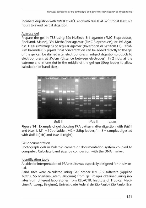
Incubate digestion with BstE II at 60°C and with Hae III at 37°C for at least 2-3hours to avoid partial digestion.
Agarose gelPrepare the gel in TBE using 3% NuSieve 3:1 agarose (FMC Bioproducts,Rockland, Maine), 3% MethaPhor agarose (FMC Bioproducts), or 4% Agar-ose 1000 (Invitrogen) or regular agarose (Invitrogen or SeaKem LE). Ethid-ium bromide 0.5 µg/mL final concentration can be added directly to the gelor the gel can be stained after electrophoresis. Subject digestion products toelectrophoresis at 5V/cm (distance between electrodes). In 2 slots at theextreme and in one slot in the middle of the gel run 50bp ladder to allowcalculation of band sizes.
S. LeaoBstE II Hae IIIFigure 14 - Example of gel showing PRA patterns after digestion with BstE IIand Hae III. M1 = 50bp ladder, M2 = 25bp ladder, 1 – 8 = samples digestedwith BstE II (left) and Hae III (right).
Gel documentationPhotograph gels in Polaroid camera or documentation system coupled tocomputer. Calculate band sizes by comparison with the DNA marker.
Identification tableA table for interpretation of PRA results was especially designed for this Man-ual.Band sizes were calculated using GelCompar II v. 2.5 software (AppliedMaths, St- Martens-Latem, Belgium) from gel images obtained using iso-lates from different laboratories from RELACTB: Institute of Tropical Medi-cine (Antwerp, Belgium), Universidade Federal de Sao Paulo (Sao Paulo, Bra-
Practical handbook for the phenotypic and genotypic identification of mycobacteria
121

zil), Instituto Adolfo Lutz (Sao Paulo, Brazil), Fundacao Oswaldo Cruz (Rio deJaneiro, Brazil), Instituto Malbran (Buenos Aires, Argentina), Pasteur Institute(Guadelupe), and Instituto Pedro Kourı (Havana, Cuba).Consensus BstEII and HaeIII band sizes were obtained by comparison of sizescalculated from gel images (empirical data) with patterns described by Tel-enti et al. (226), Devallois et al. (53), Brunello et al. (26), da Silva Rocha et al.(46), and available at PRASITE (http://app.chuv.ch/prasite) (published data).PRA patterns from frequently encountered and clinically important spe-cies were selected. For interpretation of patterns not included in thishandbook the reader is referred to published tables and to patterns availableon the Internet (PRASITE http://app.chuv.ch/prasite, PRAONLINEhttp://www.ioc.fiocruz.br/praonline).
SECTION II - METHODOLOGICAL PROCEDURES
122
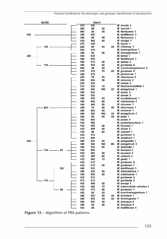
Figure 15 – Algorithm of PRA patterns.
Practical handbook for the phenotypic and genotypic identification of mycobacteria
123
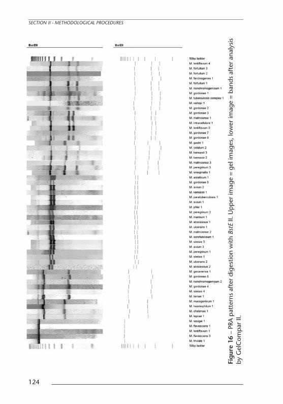
Fig
ure
16–
PRA
pat
tern
saf
ter
dige
stio
nw
ithBs
tEII.
Up
per
imag
e=
geli
mag
es,l
ower
imag
e=
band
saf
ter
anal
ysis
byG
elC
omp
arII.
SECTION II - METHODOLOGICAL PROCEDURES
124

Fig
ure
17–
PRA
pat
tern
saf
ter
dige
stio
nw
ithH
aeIII
.Up
per
imag
e=
geli
mag
es,l
ower
imag
e=
band
saf
ter
anal
ysis
byG
elC
omp
arII.
Practical handbook for the phenotypic and genotypic identification of mycobacteria
125