Embed Size (px)
Citation preview

Section VI. Drugs Used to Treat Disease of the Blood, Inflammation, & Gout Chapter 33. Agents Used in Anemias; Hematopoietic Growth Factors Katzung PHARMACOLOGY, 9e > Section VI. Drugs Used to Treat Disease of the Blood, Inflammation, & Gout > Chapter 33. Agents Used in Anemias; Hematopoietic Growth Factors >
Agents Used in Anemias; Hematopoietic Growth Factors: Introduction
Hematopoiesis, the production from undifferentiated stem cells of circulating erythrocytes, platelets, and leukocytes, is a remarkable process that produces over 200 billion new cells per day in the normal person and even greater numbers of blood cells in people with conditions that cause loss or destruction of blood cells. The hematopoietic machinery resides primarily in the bone marrow in adults and requires a constant supply of three essential nutrients—iron, vitamin B12, and folic acid—as well as the presence of hematopoietic growth factors, proteins that regulate the proliferation and differentiation of hematopoietic cells. Inadequate supplies of either the essential nutrients or the growth factors result in deficiency of functional blood cells. Anemia, a deficiency in oxygen-carrying erythrocytes, is the most common and easily treated of these conditions, but thrombocytopenia and neutropenia are not rare and in some forms are amenable to drug therapy. In this chapter, we first consider treatment of anemia due to deficiency of iron, vitamin B12, or folic acid and then turn to the medical use of hematopoietic growth factors to combat anemia, thrombocytopenia, and neutropenia. Katzung PHARMACOLOGY, 9e > Section VI. Drugs Used to Treat Disease of the Blood, Inflammation, & Gout > Chapter 33. Agents Used in Anemias; Hematopoietic Growth Factors >
Agents Used in Anemias
Iron
Basic Pharmacology
Iron deficiency is the most common cause of chronic anemia—anemia that develops over time. Like other forms of chronic anemia, iron deficiency anemia leads to pallor, fatigue, dizziness, exertional dyspnea, and other generalized symptoms of tissue ischemia. The cardiovascular adaptations to chronic anemia—tachycardia, increased cardiac output, vasodilation—can worsen the condition of patients with underlying cardiovascular disease.
Iron forms the nucleus of the iron-porphyrin heme ring, which together with globin chains forms hemoglobin. Hemoglobin reversibly binds oxygen and provides the critical mechanism for oxygen delivery from the lungs to other tissues. In the absence of adequate iron, small erythrocytes with insufficient hemoglobin are formed, giving rise to microcytic hypochromic anemia.
Pharmacokinetics
The body has an elaborate system for maintaining the supply of the iron required for hematopoiesis.

It involves specialized transport and storage proteins whose concentrations are regulated by the body's demand for hemoglobin synthesis and adequate iron stores (Table 33–1). The vast majority of the iron used to support hematopoiesis is reclaimed from catalysis of the hemoglobin in old erythrocytes. Normally, only a small amount of iron is lost from the body each day, so dietary requirements are small and easily fulfilled by the iron available in a wide variety of foods. However, in special populations with either increased iron requirements (eg, growing children, pregnant women) or increased losses of iron (eg, menstruating women), iron requirements can exceed normal dietary supplies and iron deficiency can develop.
Table 33–1. Iron Distribution in Normal Adults.1,2
Iron Content (mg) Men Women Hemoglobin 3050 1700 Myoglobin 430 300 Enzymes 10 8 Transport (transferrin) 8 6 Storage (ferritin and other forms) 750 300 Total 4248 2314
1Adapted, with permission, from Brown EB: Iron deficiency anemia. In: Cecil Textbook of Medicine, 16th ed. Wyngaarden JB, Smith LH (editors). Saunders, 1982.
2Values are based on data from various sources and assume that "normal" men weigh 80 kg and have a hemoglobin of 16 g/dL and that "normal" women weigh 55 kg and have a hemoglobin of 14 g/dL.
Absorption
Iron is normally absorbed in the duodenum and proximal jejunum, though the more distal small intestine can absorb iron if necessary. The average diet in the USA contains 10–15 mg of elemental iron daily. A normal individual without iron deficiency absorbs 5–10% of this iron, or about 0.5–1 mg daily. Iron absorption increases in response to low iron stores or increased iron requirements. Total iron absorption increases to 1–2 mg/d in normal menstruating women and may be as high as 3–4 mg/d in pregnant women. Infants and adolescents also have increased iron requirements during rapid growth periods.
Iron is available in a wide variety of foods but is especially abundant in meat. The iron in meat protein can be efficiently absorbed, since heme iron in meat hemoglobin and myoglobin can be absorbed intact without first having to be broken down into elemental iron. Iron in other foods, especially vegetables and grains, is often tightly bound to phytates or other complexing agents and may be much less available for absorption. Nonheme iron in foods and iron in inorganic iron salts and complexes must be reduced to ferrous (Fe2+) iron before it can be absorbed by the intestinal mucosal cells. Such absorption is decreased by the presence of chelators or complexing agents in the intestinal lumen and is increased in the presence of hydrochloric acid and vitamin C.

Iron crosses the intestinal mucosal cell by active transport. The rate of iron uptake is regulated by mucosal cell iron stores such that more iron is transported when stores are low. Together with iron split from absorbed heme, the newly absorbed iron can be made available for immediate transport from the mucosal cell to the plasma via transferrin or can be stored in the mucosal cell as ferritin, a water-soluble complex consisting of a core crystal of ferric hydroxide covered by a shell of a specialized storage protein called apoferritin. In general, when total body iron stores are high and iron requirements by the body are low, newly absorbed iron is diverted into ferritin in the intestinal mucosal cells rather than being transported to other sites. When iron stores are low or iron requirements are high, however, newly absorbed iron is immediately transported from the mucosal cells to the bone marrow for the production of hemoglobin.
Transport
Iron is transported in the plasma bound to transferrin, a -globulin that specifically binds ferric iron. The transferrin-ferric iron complex enters maturing erythroid cells by a specific receptor mechanism. Transferrin receptors—integral membrane glycoproteins present in large numbers on proliferating erythroid cells—bind the transferrin-iron complex and internalize the iron, releasing it within the cell. The transferrin and transferrin receptor are then recycled, providing an efficient mechanism for incorporating iron into hemoglobin in developing red blood cells.
Increased erythropoiesis is associated with an increase in the number of transferrin receptors on developing erythroid cells. Iron store depletion and iron deficiency anemia are associated with an increased concentration of serum transferrin.
Storage
Iron binds avidly to a protein, apoferritin, and forms the complex ferritin. Iron is stored, primarily as ferritin, in intestinal mucosal cells and in macrophages in the liver, spleen, and bone. Apoferritin synthesis is regulated by the levels of free iron. When these levels are low, apoferritin synthesis is inhibited and the balance of iron binding shifts toward transferrin. When free iron levels are high, more apoferritin is produced in an effort to safely sequester more iron and protect organs from the toxic effects of excess free iron.
Ferritin is also detectable in plasma. Since the ferritin present in plasma is in equilibrium with storage ferritin in reticuloendothelial tissues, the plasma (or serum) ferritin level can be used to estimate total body iron stores.
Elimination
There is no mechanism for excretion of iron. Small amounts are lost by exfoliation of intestinal mucosal cells into the stool, and trace amounts are excreted in bile, urine, and sweat. These losses account for no more than 1 mg of iron per day. Because the body's ability to increase excretion of iron is so limited, regulation of iron balance must be achieved by changing intestinal absorption and storage of iron, in response to the body's needs.
Clinical Pharmacology
Indications for the Use of Iron
The only clinical indication for the use of iron preparations is the treatment or prevention of iron deficiency anemia. Iron deficiency is commonly seen in populations with increased iron

requirements. These include infants, especially premature infants; children during rapid growth periods; and pregnant and lactating women. Iron deficiency also occurs frequently after gastrectomy and in patients with severe small bowel disease that results in generalized malabsorption. Iron deficiency in these gastrointestinal conditions is due to inadequate iron absorption.
The most common cause of iron deficiency in adults is blood loss. Menstruating women lose about 30 mg of iron with each menstrual period; women with heavy menstrual bleeding may lose much more. Thus, many premenopausal women have low iron stores or even iron deficiency. In men and postmenopausal women, the most common site of blood loss is the gastrointestinal tract. Patients with unexplained iron deficiency anemia should be evaluated for occult gastrointestinal bleeding.
As iron deficiency develops, storage iron decreases and then disappears; next, serum ferritin decreases; and then serum iron decreases and iron-binding capacity increases, resulting in a decrease in iron-binding (transferrin) saturation. Thereafter, anemia begins to develop. Red cell indices (mean corpuscular volume [MCV]: normal = 80–100 fL; mean corpuscular hemoglobin concentration [MCHC]: normal = 32–36 g/dL) are usually low normal when iron deficiency anemia is mild, but cells become progressively more microcytic (low MCV) and hypochromic (low MCHC) as anemia becomes more severe. By the time iron deficiency is diagnosed, serum iron is usually less than 40 g/dL; total iron-binding capacity (TIBC) is greater than 400 g/dL; iron-binding saturation is less than 10%; and serum ferritin is less than 10 g/L. These laboratory measurements can be used to confirm a diagnosis of iron deficiency anemia in patients who present with signs and symptoms of microcytic anemia.
Treatment
The treatment of iron deficiency anemia consists of administration of oral or parenteral iron preparations. Oral iron corrects the anemia just as rapidly and completely as parenteral iron in most cases if iron absorption from the gastrointestinal tract is normal.
Oral Iron Therapy
A wide variety of oral iron preparations are available. Since ferrous iron is most efficiently absorbed, only ferrous salts should be used. Ferrous sulfate, ferrous gluconate, and ferrous fumarate are all effective and inexpensive and are recommended for the treatment of most patients.
Different iron salts provide different amounts of elemental iron, as shown in Table 33–2. In an iron-deficient individual, about 50–100 mg of iron can be incorporated into hemoglobin daily, and about 25% of oral iron given as ferrous salt can be absorbed. Therefore, 200–400 mg of elemental iron should be given daily to correct iron deficiency most rapidly. Patients unable to tolerate such large doses of iron can be given lower daily doses of iron, which results in slower but still complete correction of iron deficiency. Treatment with oral iron should be continued for 3–6 months. This will correct the anemia and replenish iron stores.
Table 33–2. Some Commonly Used Oral Iron Preparations.
Preparation Tablet
Size Elemental Iron per Tablet
Usual Adult Dosage (Tablets per Day)
Ferrous sulfate, hydrated
325 mg 65 mg 3–4

Ferrous sulfate, desiccated
200 mg 65 mg 3–4
Ferrous gluconate 325 mg 36 mg 3–4 Ferrous fumarate 200 mg 66 mg 3–4 Ferrous fumarate 325 mg 106 mg 2–3
Common adverse effects of oral iron therapy include nausea, epigastric discomfort, abdominal cramps, constipation, and diarrhea. These effects are usually dose-related and can often be overcome by lowering the daily dose of iron or by taking the tablets immediately after or with meals. Some patients have less severe gastrointestinal adverse effects with one iron salt than another and benefit from changing preparations. Patients taking oral iron develop black stools; this itself has no clinical significance but may obscure the diagnosis of continued gastrointestinal blood loss.
Parenteral Iron Therapy
Parenteral therapy should be reserved for patients with documented iron deficiency unable to tolerate or absorb oral iron and patients with extensive chronic blood loss who cannot be maintained with oral iron alone. This includes patients with various postgastrectomy conditions and previous small bowel resection, inflammatory bowel disease involving the proximal small bowel, and malabsorption syndromes.
Iron dextran is a stable complex of ferric hydroxide and low-molecular-weight dextran containing 50 mg of elemental iron per milliliter of solution. Iron-sucrose complex and iron sodium gluconate complex are newer, alternative preparations. These agents can be given either by deep intramuscular injection or by intravenous infusion. Adverse effects of parenteral iron therapy include local pain and tissue staining (brown discoloration of the tissues overlying the injection site), headache, light-headedness, fever, arthralgias, nausea and vomiting, back pain, flushing, urticaria, bronchospasm, and, rarely, anaphylaxis and death.
Most adults with iron deficiency anemia require 1–2 g of replacement iron, or 20–40 mL of iron dextran. Most physicians prefer to give the entire dose in a single intravenous infusion in several hundred milliliters of normal saline over 1–2 hours. Intravenous administration eliminates the local pain and tissue staining that often occur with the intramuscular route and allows delivery of the entire dose of iron necessary to correct the iron deficiency at one time. There is no clear evidence that any of the adverse effects, including anaphylaxis, are more likely to occur with intravenous than with intramuscular administration.
Owing to the risk of a hypersensitivity reaction, a small test dose of iron dextran should always be given before full intramuscular or intravenous doses are given. Patients with a strong history of allergy and patients who have previously received parenteral iron are more likely to have hypersensitivity reactions following treatment with parenteral iron dextran.
Clinical Toxicity
Acute Iron Toxicity
Acute iron toxicity is seen almost exclusively in young children who have ingested a number of iron tablets. Although adults are able to tolerate large doses of oral iron without serious consequences, as

few as ten tablets of any of the commonly available oral iron preparations can be lethal in young children. Patients taking oral iron preparations should be instructed to store tablets in child-proof containers out of the reach of children.
Large amounts of oral iron cause necrotizing gastroenteritis, with vomiting, abdominal pain, and bloody diarrhea followed by shock, lethargy, and dyspnea. Subsequently, improvement is often noted, but this may be followed by severe metabolic acidosis, coma, and death. Urgent treatment of acute iron toxicity is necessary, especially in young children. Activated charcoal, a highly effective adsorbent for most toxins, does not bind iron and thus is ineffective. Whole bowel irrigation (see Chapter 59: Management of the Poisoned Patient) should be performed to flush out unabsorbed pills. Deferoxamine, a potent iron-chelating compound, can be given systemically to bind iron that has already been absorbed and to promote its excretion in urine and feces. Appropriate supportive therapy for gastrointestinal bleeding, metabolic acidosis, and shock must also be provided.
Chronic Iron Toxicity
Chronic iron toxicity (iron overload), also known as hemochromatosis, results when excess iron is deposited in the heart, liver, pancreas, and other organs. It can lead to organ failure and death. It most commonly occurs in patients with inherited hemochromatosis, a disorder characterized by excessive iron absorption, and in patients who receive many red cell transfusions over a long period of time.
Chronic iron overload in the absence of anemia is most efficiently treated by intermittent phlebotomy. One unit of blood can be removed every week or so until all of the excess iron is removed. Iron chelation therapy using parenteral deferoxamine is much less efficient as well as more complicated, expensive, and hazardous, but it can be useful for severe iron overload that cannot be managed by phlebotomy.
VItamin B12
Vitamin B12 serves as a cofactor for several essential biochemical reactions in humans. Deficiency of vitamin B12 leads to anemia, gastrointestinal symptoms, and neurologic abnormalities. While deficiency of vitamin B12 due to an inadequate supply in the diet is unusual, deficiency of B12 in adults—especially older adults—due to abnormal absorption of dietary vitamin B12 is a relatively common and easily treated disorder.
Chemistry
Vitamin B12 consists of a porphyrin-like ring with a central cobalt atom attached to a nucleotide. Various organic groups may be covalently bound to the cobalt atom, forming different cobalamins. Deoxyadenosylcobalamin and methylcobalamin are the active forms of the vitamin in humans. Cyanocobalamin and hydroxocobalamin (both available for therapeutic use) and other cobalamins found in food sources are converted to the above active forms. The ultimate source of vitamin B12 is from microbial synthesis; the vitamin is not synthesized by animals or plants. The chief dietary source of vitamin B12 is microbially derived vitamin B12 in meat (especially liver), eggs, and dairy products. Vitamin B12 is sometimes called extrinsic factor to differentiate it from intrinsic factor, a protein normally secreted by the stomach.
Pharmacokinetics
The average diet in the USA contains 5–30 g of vitamin B12 daily, 1–5 g of which is usually

absorbed. The vitamin is avidly stored, primarily in the liver, with an average adult having a total vitamin B12 storage pool of 3000–5000 g. Only trace amounts of vitamin B12 are normally lost in urine and stool. Since the normal daily requirements of vitamin B12 are only about 2 g, it would take about 5 years for all of the stored vitamin B12 to be exhausted and for megaloblastic anemia to develop if B12 absorption stopped. Vitamin B12 in physiologic amounts is absorbed only after it complexes with intrinsic factor, a glycoprotein secreted by the parietal cells of the gastric mucosa. Intrinsic factor combines with the vitamin B12 that is liberated from dietary sources in the stomach and duodenum, and the intrinsic factor-vitamin B12 complex is subsequently absorbed in the distal ileum by a highly specific receptor-mediated transport system. Vitamin B12 deficiency in humans most often results from malabsorption of vitamin B12, due either to lack of intrinsic factor or to loss or malfunction of the specific absorptive mechanism in the distal ileum. Nutritional deficiency is rare but may be seen in strict vegetarians after many years without meat, eggs, or dairy products.
Once absorbed, vitamin B12 is transported to the various cells of the body bound to a plasma glycoprotein, transcobalamin II. Excess vitamin B12 is transported to the liver for storage. Significant amounts of vitamin B12 are excreted in the urine only when very large amounts are given parenterally, overcoming the binding capacities of the transcobalamins (50–100 g).
Pharmacodynamics
Two essential enzymatic reactions in humans require vitamin B12 (Figure 33–1). In one, methylcobalamin serves as an intermediate in the transfer of a methyl group from N�5-methyltetrahydrofolate to methionine (Figure 33–1 A; Figure 33–2, reaction 1). In the absence of vitamin B12, conversion of the major dietary and storage folate, N5-methyltetrahydrofolate, to tetrahydrofolate, the precursor of folate cofactors, cannot occur. As a result, a deficiency of folate cofactors necessary for several biochemical reactions involving the transfer of one-carbon groups develops. In particular, the depletion of tetrahydrofolate prevents synthesis of adequate supplies of the deoxythymidylate (dTMP) and purines required for DNA synthesis in rapidly dividing cells as shown in Figure 33–3, reaction 2. The accumulation of folate as N5-methyltetrahydrofolate and the associated depletion of tetrahydrofolate cofactors in vitamin B12 deficiency have been referred to as the "methylfolate trap." This is the biochemical step whereby vitamin B12 and folic acid metabolism are linked and explains why the megaloblastic anemia of vitamin B12 deficiency can be partially corrected by ingestion of relatively large amounts of folic acid. Folic acid can be reduced to dihydrofolate by the enzyme dihydrofolate reductase (Figure 33–2, reaction 3) and thus serve as a source of the tetrahydrofolate required for synthesis of the purines and dTMP that are needed for DNA synthesis.
Figure 33–1.

Enzymatic reactions that use vitamin B12. See text for details.
Figure 33–2.

Enzymatic reactions that use folates. Section 1 shows the vitamin B12-dependent reaction that allows most dietary folates to enter the tetrahydrofolate cofactor pool and becomes the "folate trap" in vitamin B12 deficiency. Section 2 shows the dTMP cycle. Section 3 shows the pathway by which folate enters the tetrahydrofolate cofactor pool. Double arrows indicate pathways with more than one intermediate step.
The other enzymatic reaction that requires vitamin B12 is isomerization of methylmalonyl-CoA to succinyl-CoA by the enzyme methylmalonyl-CoA mutase (Figure 33–1 B). In vitamin B12 deficiency, this conversion cannot take place, and the substrate, methylmalonyl-CoA, accumulates. In the past, it was thought that abnormal accumulation of methylmalonyl-CoA causes the neurologic manifestations of vitamin B12 deficiency. However, newer evidence instead implicates the disruption of the methionine synthesis pathway as the cause of neurologic problems. Whatever the biochemical explanation for neurologic damage, the important point is that administration of folic acid in the setting of vitamin B12 deficiency will not prevent neurologic manifestations even though it will largely correct the anemia caused by the vitamin B12 deficiency.
Clinical Pharmacology
Vitamin B12 is used to treat or prevent deficiency. There is no evidence that vitamin B12 injections have any benefit in persons who do not have vitamin B12 deficiency. The most characteristic clinical manifestation of vitamin B12 deficiency is megaloblastic anemia. The typical clinical findings in

megaloblastic anemia are macrocytic anemia (MCV usually > 120 fL), often with associated mild or moderate leukopenia or thrombocytopenia (or both), and a characteristic hypercellular bone marrow with megaloblastic maturation of erythroid and other precursor cells. Vitamin B12 deficiency also causes a neurologic syndrome that usually begins with paresthesias and weakness in peripheral nerves and progresses to spasticity, ataxia, and other central nervous system dysfunctions. A characteristic pathologic feature of the neurologic syndrome is degeneration of myelin sheaths followed by disruption of axons in the dorsal and lateral horns of the spinal cord and in peripheral nerves. Correction of vitamin B12 deficiency arrests the progression of neurologic disease, but it may not fully reverse neurologic symptoms that have been present for several months. Although most patients with neurologic abnormalities caused by vitamin B12 deficiency have full-blown megaloblastic anemias when first seen, occasional patients have few if any hematologic abnormalities.
Once a diagnosis of megaloblastic anemia is made, it must be determined whether vitamin B12 or folic acid deficiency is the cause. (Other causes of megaloblastic anemia are very rare.) This can usually be accomplished by measuring serum levels of the vitamins. The Schilling test, which measures absorption and urinary excretion of radioactively labeled vitamin B12, can be used to further define the mechanism of vitamin B12 malabsorption when this is found to be the cause of the megaloblastic anemia.
The most common causes of vitamin B12 deficiency are pernicious anemia, partial or total gastrectomy, and diseases that affect the distal ileum, such as malabsorption syndromes, inflammatory bowel disease, or small bowel resection.
Pernicious anemia results from defective secretion of intrinsic factor by the gastric mucosal cells. Patients with pernicious anemia have gastric atrophy and fail to secrete intrinsic factor (as well as hydrochloric acid). The Schilling test shows diminished absorption of radioactively labeled vitamin B12, which is corrected when hog intrinsic factor is administered with radioactive B12, since the vitamin can then be normally absorbed.
Vitamin B12 deficiency also occurs when the region of the distal ileum that absorbs the vitamin B12-intrinsic factor complex is damaged, as when the ileum is involved with inflammatory bowel disease, or when the ileum is surgically resected. In these situations, radioactively labeled vitamin B12 is not absorbed in the Schilling test, even when intrinsic factor is added. Other rare causes of vitamin B12 deficiency include bacterial overgrowth of the small bowel, chronic pancreatitis, and thyroid disease. Rare cases of vitamin B12 deficiency in children have been found to be secondary to congenital deficiency of intrinsic factor and congenital selective vitamin B12 malabsorption due to defects of the receptor sites in the distal ileum.
Since almost all cases of vitamin B12 deficiency are caused by malabsorption of the vitamin, parenteral injections of vitamin B12 are required for therapy. For patients with potentially reversible diseases, the underlying disease should be treated after initial treatment with parenteral vitamin B12. Most patients, however, do not have curable deficiency syndromes and require lifelong treatment with vitamin B12 injections.
Vitamin B12 for parenteral injection is available as cyanocobalamin or hydroxocobalamin. Hydroxocobalamin is preferred because it is more highly protein-bound and therefore remains longer in the circulation. Initial therapy should consist of 100–1000 g of vitamin B12 intramuscularly daily or every other day for 1–2 weeks to replenish body stores. Maintenance therapy consists of 100–1000 g intramuscularly once a month for life. If neurologic abnormalities are present, maintenance therapy injections should be given every 1–2 weeks for 6 months before

switching to monthly injections. Oral vitamin B12-intrinsic factor mixtures and liver extracts should not be used to treat vitamin B12 deficiency; however, oral doses of 1000 g of vitamin B12 daily are usually sufficient to treat patients with pernicious anemia who refuse or cannot tolerate the injections.
Folic Acid
Reduced forms of folic acid are required for essential biochemical reactions that provide precursors for the synthesis of amino acids, purines, and DNA. Folate deficiency is not uncommon, even though the deficiency is easily corrected by administration of folic acid. The consequences of folate deficiency go beyond the problem of anemia because folate deficiency is implicated as a cause of congenital malformations in newborns and may play a role in vascular disease (see Folic Acid Supplementation: A Public Health Dilemma).
Chemistry
Folic acid (pteroylglutamic acid) is a compound composed of a heterocycle, p-aminobenzoic acid, and glutamic acid (Figure 33–3). Various numbers of glutamic acid moieties may be attached to the pteroyl portion of the molecule, resulting in monoglutamates, triglutamates, or polyglutamates. Folic acid can undergo reduction, catalyzed by the enzyme dihydrofolate reductase ("folate reductase"), to give dihydrofolic acid (Figure 33–2, reaction 3). Tetrahydrofolate can subsequently be transformed to folate cofactors possessing one-carbon units attached to the 5-nitrogen, to the 10-nitrogen, or to both positions (Figure 33–2). The folate cofactors are interconvertible by various enzymatic reactions and serve the important biochemical function of donating one-carbon units at various levels of oxidation. In most of these, tetrahydrofolate is regenerated and becomes available for reutilization.
Figure 33–3.
The structure and numbering of atoms of folic acid. (Reproduced, with permission, from Murray RK et al: Harper's Biochemistry, 24th ed. McGraw-Hill, 1996)
Pharmacokinetics
The average diet in the USA contains 500–700 g of folates daily, 50–200 g of which is usually

absorbed, depending on metabolic requirements (pregnant women may absorb as much as 300–400 g of folic acid daily). Various forms of folic acid are present in a wide variety of plant and animal
tissues; the richest sources are yeast, liver, kidney, and green vegetables. Normally, 5–20 mg of folates are stored in the liver and other tissues. Folates are excreted in the urine and stool and are also destroyed by catabolism, so serum levels fall within a few days when intake is diminished. Since body stores of folates are relatively low and daily requirements high, folic acid deficiency and megaloblastic anemia can develop within 1–6 months after the intake of folic acid stops, depending on the patient's nutritional status and the rate of folate utilization.
Unaltered folic acid is readily and completely absorbed in the proximal jejunum. Dietary folates, however, consist primarily of polyglutamate forms of N�5-methyltetrahydrofolate. Before absorption, all but one of the glutamyl residues of the polyglutamates must be hydrolyzed by the enzyme -1-glutamyl transferase ("conjugase") within the brush border of the intestinal mucosa. The monoglutamate N�5-methyltetrahydrofolate is subsequently transported into the bloodstream by both active and passive transport and is then widely distributed throughout the body. Inside cells, N�5-methyltetrahydrofolate is converted to tetrahydrofolate by the demethylation reaction that requires vitamin B12 (Figure 33–2, reaction 1).
Pharmacodynamics
Tetrahydrofolate cofactors participate in one-carbon transfer reactions. As described above in the section on vitamin B12, one of these essential reactions produces the dTMP needed for DNA synthesis. In this reaction, the enzyme thymidylate synthase catalyzes the transfer of the one-carbon unit of N�5,N�10-methylenetetrahydrofolate to deoxyuridine monophosphate (dUMP) to form dTMP (Figure 33–2, reaction 2). Unlike all of the other enzymatic reactions that utilize folate cofactors, in this reaction the cofactor is oxidized to dihydrofolate, and for each mole of dTMP produced, one mole of tetrahydrofolate is consumed. In rapidly proliferating tissues, considerable amounts of tetrahydrofolate can be consumed in this reaction, and continued DNA synthesis requires continued regeneration of tetrahydrofolate by reduction of dihydrofolate, catalyzed by the enzyme dihydrofolate reductase. The tetrahydrofolate thus produced can then reform the cofactor N�5,N�10-methylenetetrahydrofolate by the action of serine transhydroxy- methylase and thus allow for the continued synthesis of dTMP. The combined catalytic activities of dTMP synthase, dihydrofolate reductase, and serine transhydroxymethylase are often referred to as the dTMP synthesis cycle. Enzymes in the dTMP cycle are the targets of two anticancer drugs; methotrexate inhibits dihydrofolate reductase, and a metabolite of 5-fluorouracil inhibits thymidylate synthase (see Chapter 55: Cancer Chemotherapy).
Cofactors of tetrahydrofolate participate in several other essential reactions. As described above, N�5-methy- lenetetrahydrofolate is required for the vitamin B12-dependent reaction that generates methionine from homocysteine (Figure 33–1 A; Figure 33–2, reaction 1). In addition, tetrahydrofolate cofactors donate one-carbon units during the de novo synthesis of essential purines. In these reactions, tetrahydrofolate is regenerated and can reenter the tetrahydrofolate cofactor pool.
Clinical Pharmacology
Folate deficiency results in a megaloblastic anemia that is microscopically indistinguishable from the anemia caused by vitamin B12 deficiency (see above). However, folate deficiency does not cause the characteristic neurologic syndrome seen in vitamin B12 deficiency. In patients with megaloblastic anemia, folate status is assessed with assays for serum folate or for red blood cell folate. Red blood cell folate levels are often of greater diagnostic value than serum levels, since serum folate levels tend to be quite labile and do not necessarily reflect tissue levels.

Folic acid deficiency, unlike vitamin B12 deficiency, is often caused by inadequate dietary intake of folates. Alcoholics and patients with liver disease develop folic acid deficiency because of poor diet and diminished hepatic storage of folates. There is also evidence that alcohol and liver disease interfere with absorption and metabolism of folates. Pregnant women and patients with hemolytic anemia have increased folate requirements and may become folic acid-deficient, especially if their diets are marginal. Evidence implicates maternal folic acid deficiency in the occurrence of fetal neural tube defects, eg, spina bifida. (See Folic Acid Supplementation: A Public Health Dilemma.) Patients with malabsorption syndromes also frequently develop folic acid deficiency. Folic acid deficiency is occasionally associated with cancer, leukemia, myeloproliferative disorders, certain chronic skin disorders, and other chronic debilitating diseases. Patients who require renal dialysis also develop folic acid deficiency, because folates are removed from the plasma each time the patient is dialyzed.
Folic acid deficiency can be caused by drugs that interfere with folate absorption or metabolism. Phenytoin, some other anticonvulsants, oral contraceptives, and isoniazid can cause folic acid deficiency by interfering with folic acid absorption. Other drugs such as methotrexate and, to a lesser extent, trimethoprim and pyrimethamine, inhibit dihydrofolate reductase and may result in a deficiency of folate cofactors and ultimately in megaloblastic anemia.
Parenteral administration of folic acid is rarely necessary, since oral folic acid is well absorbed even in patients with malabsorption syndromes. A dose of 1 mg of folic acid orally daily is sufficient to reverse megaloblastic anemia, restore normal serum folate levels, and replenish body stores of folates in almost all patients. Therapy should be continued until the underlying cause of the deficiency is removed or corrected. Therapy may be required indefinitely for patients with malabsorption or dietary inadequacy. Folic acid supplementation to prevent folic acid deficiency should be considered in high-risk patients, including pregnant women, alcoholics, and patients with hemolytic anemia, liver disease, certain skin diseases, and patients on renal dialysis. Katzung PHARMACOLOGY, 9e > Section VI. Drugs Used to Treat Disease of the Blood, Inflammation, & Gout > Chapter 33. Agents Used in Anemias; Hematopoietic Growth Factors >
Folic Acid Supplementation: A Public Health Dilemma
By January 1998, all products made from enriched grains in the USA were required to be supplemented with folic acid. This FDA ruling was issued to reduce the incidence of congenital neural tube defects. Scientific studies show a strong correlation between maternal folic acid deficiency and the incidence of neural tube defects such as spinal bifida and anencephaly. The FDA requirement for folic acid supplementation is a public health measure aimed at the significant number of women in the USA who do not receive prenatal care and are not aware of the importance of adequate folic acid ingestion for preventing birth defects in their babies. Pregnant women have increased requirements for folic acid; at least 400 g/d is recommended. It is estimated that the level of folic acid fortification now required in enriched grain products provides an additional 80–100 g of folic acid per day to the diet of women of childbearing age and 70–120 g/d to the diet of middle-aged and older adults.
There may be an added benefit for adults. N�5-methyltetrahydrofolate is required for the conversion of homocysteine to methionine (Figure 33–1; Figure 33–2, reaction 1). Impaired synthesis of N�5-methyltetrahydrofolate results in elevated serum concentrations of homocysteine. Data from several sources suggest a positive correlation between elevated serum homocysteine and occlusive vascular diseases such as ischemic heart disease and stroke. Clinical data suggest that the

folate supplementation program has improved the folate status and reduced the prevalence of hyperhomocysteinemia in a population of middle-aged and older adults who did not use vitamin supplements. It is possible, though as yet unproved, that the increased ingestion of folic acid will also reduce the risk of vascular disease in this population.
While these two potential benefits of supplemental folic acid are compelling, the decision to require folic acid in grains was—and still is—controversial. As described in the text, ingestion of folic acid can partially or totally correct the anemia caused by vitamin B12 deficiency. However, folic acid supplementation will not prevent the potentially irreversible neurologic damage caused by vitamin B12 deficiency. People with pernicious anemia and other forms of vitamin B12 deficiency are usually identified because of signs and symptoms of anemia, which tend to occur before neurologic symptoms. The opponents of folic acid supplementation are concerned that increased folic acid intake in the general population will mask vitamin B12 deficiency and increase the prevalence of neurologic disease in our elderly population. To put this in perspective, approximately 4000 pregnancies, including 2500 live births, in the USA each year are affected by neural tube defects. In contrast, it is estimated that over 10% of the elderly population in the USA, or several million people, are at risk of the neuropsychiatric complications of vitamin B12 deficiency (Rothenberg, 1999). In acknowledgment of this controversy, the FDA kept its requirements for folic acid supplementation at a somewhat low level. They also recommend that all adults should keep their ingestion of folic acid below 1 mg/d. Katzung PHARMACOLOGY, 9e > Section VI. Drugs Used to Treat Disease of the Blood, Inflammation, & Gout > Chapter 33. Agents Used in Anemias; Hematopoietic Growth Factors >
Hematopoietic Growth Factors
The hematopoietic growth factors are glycoprotein hormones that regulate the proliferation and differentiation of hematopoietic progenitor cells in the bone marrow. The first growth factors to be identified were called colony-stimulating factors because they could stimulate the growth of colonies of various bone marrow progenitor cells in vitro. In the past decade, many of these growth factors have been purified and cloned, and their effects on hematopoiesis have been extensively studied. Quantities of these growth factors sufficient for clinical use are produced by recombinant DNA technology.
Of the known hematopoietic growth factors, erythropoietin (epoetin alfa), granulocyte colony-stimulating factor (G-CSF), granulocyte-macrophage colony-stimulating factor (GM-CSF), and interleukin 11 are currently in clinical use. Thrombopoietin is undergoing clinical trials and will probably become available soon. Other potentially useful hematopoietic factors are still in development.
The hematopoietic growth factors have complex effects on the function of a wide variety of cell types, including nonhematologic cells. Their utility in other areas of medicine, particularly as potential anticancer and anti-inflammatory drugs, is being investigated.
Erythropoietin
Chemistry & Pharmacokinetics
Erythropoietin, a 34-39 kDA glycoprotein, was the first human hematopoietic growth factor to be isolated. It was originally purified from the urine of patients with severe anemia. Recombinant human erythropoietin (rHuEpo, epoetin alfa) is produced in a mammalian cell expression system

using recombinant DNA technology. After intravenous administration, erythropoietin has a serum half-life of 4–13 hours in patients with chronic renal failure. It is not cleared by dialysis. It is measured in international units (IU). Darbopoetin alfa is a glycosylated form of erythropoietin and differs from it functionally only in having a twofold to threefold longer half-life.
Pharmacodynamics
Erythropoietin stimulates erythroid proliferation and differentiation by interacting with specific erythropoietin receptors on red cell progenitors. It also induces release of reticulocytes from the bone marrow. Endogenous erythropoietin is produced by the kidney in response to tissue hypoxia. When anemia occurs, more erythropoietin is produced by the kidney, signaling the bone marrow to produce more red blood cells. This results in correction of the anemia provided that bone marrow response is not impaired by red cell nutritional deficiency (especially iron deficiency), primary bone marrow disorders (see below), or bone marrow suppression from drugs or chronic diseases.
Normally there is an inverse relationship between the hematocrit or hemoglobin level and the serum erythropoietin level. Nonanemic individuals have serum erythropoietin levels of less than 20 IU/L. As the hematocrit and hemoglobin levels fall and anemia becomes more severe, the serum erythropoietin level rises exponentially. Patients with moderately severe anemias usually have erythropoietin levels in the 100–500 IU/L range, and patients with severe anemias may have levels of thousands of IU/L. The most important exception to this inverse relationship is in the anemia of chronic renal failure. In patients with renal disease, erythropoietin levels are usually low because the kidneys cannot produce the growth factor. These patients are the most likely to respond to treatment with exogenous erythropoietin. In most primary bone marrow disorders (aplastic anemia, leukemias, myeloproliferative and myelodysplastic disorders, etc) and most nutritional and secondary anemias, endogenous erythropoietin levels are high, so there is less likelihood of a response to exogenous erythropoietin (but see below).
Clinical Pharmacology
The availability of erythropoietin has had a significant positive impact for patients with chronic renal failure. Erythropoietin consistently improves the hematocrit and hemoglobin level and usually eliminates the need for transfusions in these patients. An increase in reticulocyte count is usually observed in about 10 days and an increase in hematocrit and hemoglobin levels in 2–6 weeks. Most patients can maintain a hematocrit of about 35% with erythropoietin doses of 50–150 IU/kg intravenously or subcutaneously three times a week. Failure to respond to erythropoietin is most commonly due to concurrent iron deficiency, which can be corrected by giving oral iron. Folate supplementation may also be necessary in some patients.
In selected patients, erythropoietin may also be useful for the treatment of anemia due to primary bone marrow disorders and secondary anemias. This includes patients with aplastic anemia and other bone marrow failure states, myeloproliferative and myelodysplastic disorders, multiple myeloma and perhaps other chronic bone marrow malignancies, and the anemias associated with chronic inflammation, AIDS, and cancer. Patients with these disorders who have disproportionately low serum erythropoietin levels for their degree of anemia are most likely to respond to treatment with this growth factor. Patients with endogenous erythropoietin levels of less than 100 IU/L have the best chance of response, though patients with erythropoietin levels between 100 and 500 IU/L respond occasionally. These patients generally require higher erythropoietin doses (150–300 IU/kg three times a week) to achieve a response, and responses are often incomplete.
Erythropoietin has been used successfully to offset the anemia produced by zidovudine treatment in

patients with HIV infection and in the treatment of the anemia of prematurity. It can also be used to accelerate erythropoiesis after phlebotomies, when blood is being collected for autologous transfusion for elective surgery, or for treatment of iron overload (hemochromatosis).
Erythropoietin is one of the drugs banned by the International Olympic Committee. The use of erythropoietin by athletes is based on their hope that increased red blood cell concentration will increase oxygen delivery and improve performance.
Toxicity
The most common adverse effects of erythropoietin are associated with a rapid increase in hematocrit and hemoglobin and include hypertension and thrombotic complications. These difficulties can be minimized by raising the hematocrit and hemoglobin slowly and by adequately monitoring and treating hypertension. Allergic reactions have been infrequent and mild.
Myeloid Growth Factors
Chemistry & Pharmacokinetics
G-CSF and GM-CSF, the two myeloid growth factors currently available for clinical use, were originally purified from cultured human cell lines. Recombinant human G-CSF (rHuG-CSF; filgrastim) is produced in a bacterial expression system using recombinant DNA technology. It is a nonglycosylated peptide of 175 amino acids, with a molecular weight of 18 kDa. Recombinant human GM-CSF (rHuGM-CSF; sargramostim) is produced in a yeast expression system using recombinant DNA technology. It is a partially glycosylated peptide of 127 amino acids, with three molecular species with molecular weights of 15,500, 15,800, and 19,500. These preparations have serum half-lives of 2–7 hours after intravenous or subcutaneous administration. Pegfilgrastim, a covalent conjugation product of filgrastim and a form of polyethylene glycol, has a much longer serum half-life than recombinant G-CSF, and so it can be injected once per myelosuppressive chemotherapy cycle instead of daily for several days.
Pharmacodynamics
The myeloid growth factors stimulate proliferation and differentiation by interacting with specific receptors found on various myeloid progenitor cells. These receptors are members of the superfamily of receptors that transduce signals by association with cytoplasmic tyrosine kinases in the JAK/STAT pathway (see Chapter 2: Drug Receptors & Pharmacodynamics). G-CSF stimulates proliferation and differentiation of progenitors already committed to the neutrophil lineage. It also activates the phagocytic activity of mature neutrophils and prolongs their survival in the circulation. G-CSF also has a remarkable ability to mobilize hematopoietic stem cells, ie, to increase their concentration in peripheral blood. This biologic effect underlies a major advance in transplantation—the use of peripheral blood stem cells (PBSCs) instead of bone marrow stem cells for autologous and allogeneic hematopoietic stem cell transplantation (see below).
GM-CSF has broader biologic actions than G-CSF. It is a multipotential hematopoietic growth factor that stimulates proliferation and differentiation of early and late granulocytic progenitor cells as well as erythroid and megakaryocyte progenitors. Like G-CSF, GM-CSF also stimulates the function of mature neutrophils. GM-CSF acts together with interleukin-2 to stimulate T cell proliferation and appears to be a locally active factor at the site of inflammation. GM-CSF mobilizes peripheral blood stem cells, but it is significantly less efficacious than G-CSF in this

regard.
Clinical Pharmacology
Neutropenia, a common adverse effect of the cytotoxic drugs used to treat cancer, puts patients at high risk of serious infection. Unlike the treatment of anemia and thrombocytopenia, transfusion of neutropenic patients with granulocytes collected from donors is performed rarely and with limited success. The introduction of G-CSF in 1991 represented a milestone in the treatment of chemotherapy-induced neutropenia. This growth factor dramatically accelerates the rate of neutrophil recovery after dose-intensive myelosuppressive chemotherapy (Figure 33–4). It reduces the duration of neutropenia and usually raises the nadir, the lowest neutrophil count seen following a cycle of chemotherapy.
Figure 33–4.
Effects of G-CSF (color) or placebo (black line) on absolute neutrophil count (ANC) after cytotoxic chemotherapy for lung cancer. Doses of chemotherapeutic drugs were administered on days 1 and 3. G-CSF or placebo injections were started on day 4 and continued daily through day 12 or 16. The first peak in ANC reflects the recruitment of mature cells by G-CSF. The second peak reflects a marked increase in new neutrophil production by the bone marrow under stimulation by G-CSF. Treated patients in this study had fewer days of neutropenia, days of antibiotic treatment, and days of hospitalization. They also had a lower incidence of infections. (Normal ANC is 2.2–8.6 x 10⁹/L.) (Modified and reproduced, with permission, from Crawford et al: Reduction by granulocyte colony-stimulating factor of fever and neutropenia induced by chemotherapy in patients with small-cell lung cancer. N Engl J Med 1991;325:164.)
While the ability of G-CSF to increase neutrophil counts after myelosuppressive chemotherapy is nearly universal, its impact upon clinical outcomes is more variable. Some clinical trials have shown that G-CSF reduces episodes of febrile neutropenia, requirements for broad-spectrum antibiotics, and days of hospitalization; however, other trials failed to find these favorable outcomes. To date, no clinical trial has shown improved survival in cancer patients treated with G-CSF. Clinical guidelines for the use of G-CSF after cytotoxic chemotherapy have been published (Ozer et al, 2001). These guidelines recommend reserving G-CSF for patients with a prior episode of febrile neutropenia after cytotoxic chemotherapy, patients receiving dose-intensive chemotherapy, patients at high risk of febrile neutropenia, and patients who are unlikely to survive an episode of febrile neutropenia. Pegfilgrastim is an alternative to G-CSF for prevention of chemotherapy-induced febrile neutropenia. Pegfilgrastim can be administered less frequently, and it may shorten the period of severe neutropenia slightly more than G-CSF.

Like G-CSF and pegfilgrastim, GM-CSF also reduces the duration of neutropenia after cytotoxic chemotherapy. It has been more difficult to show that GM-CSF reduces the incidence of febrile neutropenia, probably because GM-CSF itself can induce fever. In the treatment of chemotherapy-induced neutropenia, G-CSF, 5 g/kg/d, or GM-CSF, 250 g/m2/d, is usually started within 24–72 hours after completing chemotherapy and is continued until the absolute neutrophil count is > 10,000 cells/ L. Pegfilgrastim is given as a single dose instead of as daily injections.
The utility and safety of the myeloid growth factors in the postchemotherapy supportive care of patients with acute myeloid leukemia (AML) has been the subject of a number of clinical trials. Since leukemic cells arise from progenitors whose proliferation and differentiation are normally regulated by hematopoietic growth factors, including GM-CSF and G-CSF, there was concern that myeloid growth factors could stimulate leukemic cell growth and increase the rate of relapse. The results of randomized clinical trials suggest that both G-CSF and GM-CSF are safe following induction and consolidation treatment of myeloid and lymphoblastic leukemia. There has been no evidence that these growth factors reduce the rate of remission or increase relapse rate. On the contrary, the growth factors accelerate neutrophil recovery and reduce infection rates and days of hospitalization. Both G-CSF and GM-CSF have FDA approval for treatment of patients with AML.
G-CSF and GM-CSF have also been shown to be effective in treating the neutropenia associated with congenital neutropenia, cyclic neutropenia, myelodysplasia, and aplastic anemia. Many patients with these disorders respond with a prompt and sometimes dramatic increase in neutrophil count. In some cases this results in a decrease in the frequency of infections. Since G-CSF and GM-CSF do not stimulate the formation of erythrocytes or platelets, they are sometimes used in combination with other growth factors for treatment of pancytopenia.
The myeloid growth factors play an important role in autologous stem cell transplantation for patients undergoing high-dose chemotherapy. High-dose chemotherapy with autologous stem cell support is increasingly being used to treat patients with tumors that are resistant to standard doses of chemotherapeutic drugs. The high-dose regimens produce extreme myelosuppression; the myelosuppression is then counteracted by reinfusion of the patient's own hematopoietic stem cells (which are collected prior to chemotherapy). The administration of G-CSF or GM-CSF early after autologous stem cell transplantation has been shown to reduce the time to engraftment and to recovery from neutropenia in patients receiving stem cells obtained either from bone marrow or from peripheral blood. These effects are seen in patients being treated for lymphoma or for solid tumors. G-CSF and GM-CSF are also used to support patients who have received allogeneic bone marrow transplantation for treatment of hematologic malignancies or bone marrow failure states. In this setting, the growth factors speed the recovery from neutropenia without increasing the incidence of acute graft-versus-host disease.
Probably the most important role of the myeloid growth factors in transplantation is for mobilization of peripheral blood stem cells (PBSCs). Stem cells collected from peripheral blood have nearly replaced bone marrow as the hematopoietic preparation used for autologous transplantation. The cells can be collected in an outpatient setting with a procedure that avoids much of the risk and discomfort of bone marrow collection, including the need for general anesthesia. In addition, there is evidence that PBSC transplantation results in more rapid engraftment of all hematopoietic cell lineages and in reduced rates of graft failure or delayed platelet recovery. The use of PBSCs for allogeneic transplantation is also being investigated. In allogeneic transplantation, donors are treated with G-CSF in order to mobilize their PBSCs prior to leukapheresis, the procedure that separates the fraction containing stem cells from the other components in blood.

G-CSF is the cytokine most commonly used for PBSC mobilization because of its increased efficacy and reduced toxicity compared with GM-CSF. To mobilize stem cells, patients or donors are given 5–10 g/kg/d subcutaneously for 4 days. On the fifth day, they undergo leukapheresis. The success of PBSC transplantation depends upon transfusion of adequate numbers of stem cells. CD34, an antigen present on early progenitor cells and absent from later, committed, cells, is used as a marker for the requisite stem cells. The goal is to reinfuse at least 5 x 10⁶ CD34 cells/kg; this number of CD34 cells usually results in prompt and durable engraftment of all cell lineages. It can take several separate leukaphereses to collect enough CD34 cells, especially from older patients and patients who have been exposed to radiotherapy or chemotherapy.
Toxicity
Although the two growth factors have similar effects on neutrophil counts, G-CSF is used more frequently because it is better tolerated. G-CSF can cause bone pain, which clears when the drug is discontinued. GM-CSF can cause more severe side effects, particularly at higher doses. These include fevers, malaise, arthralgias, myalgias, and a capillary leak syndrome characterized by peripheral edema and pleural or pericardial effusions. Allergic reactions may occur but are infrequent. Splenic rupture is a rare but serious complication of the use of G-CSF for PBSC.
Megakaryocyte Growth Factors
Chemistry & Pharmacokinetics
Interleukin-11 (IL-11) is a 65–85 kDa protein produced by fibroblasts and stromal cells in the bone marrow. Oprelvekin, the recombinant form of interleukin-11 approved for clinical use, is produced by expression in E coli. The half-life of IL-11 is 7–8 hours when the drug is injected subcutaneously.
Thrombopoietin, a 65–85 kDa glycosylated protein, is constitutively expressed by a variety of organs and cell types. Hepatocytes appear to be the major source of human thrombopoietin, and patients with cirrhosis and thrombocytopenia have low serum thrombopoietin levels. Recombinant thrombopoietin is produced by expression in human cells; the recombinant product contains two intramolecular disulfide bonds and a number of carbohydrate side chains.
Pharmacodynamics
Interleukin-11 acts through a specific cell surface cytokine receptor to stimulate the growth of multiple lymphoid and myeloid cells. It acts synergistically with other growth factors to stimulate the growth of primitive megakaryocytic progenitors and, most importantly, increases the number of peripheral platelets and neutrophils.
Acting through its own cytokine receptor, thrombopoietin also independently stimulates the growth of primitive megakaryocytic progenitors. In addition, it stimulates mature megakaryocytes and even activates mature platelets to respond to aggregation-inducing stimuli. The critical in vivo role of thrombopoietin has been demonstrated in genetically engineered knockout mice who lack either thrombopoietin or its receptor. These mice have marked thrombocytopenia but do not display anemia or leukopenia.
Clinical Pharmacology
Patients with thrombocytopenia have a high risk of hemorrhage. While platelet transfusion is

commonly used to treat thrombocytopenia, this procedure can cause adverse reactions in the recipient; furthermore, a significant number of patients fail to exhibit the expected increase in platelet count.
Interleukin-11 is the first growth factor to gain FDA approval for treatment of thrombocytopenia. It is approved for the secondary prevention of thrombocytopenia in patients receiving cytotoxic chemotherapy for treatment of nonmyeloid cancers. Clinical trials show that it reduces the number of platelet transfusions required by patients who experienced severe thrombocytopenia after a previous cycle of chemotherapy. Although IL-11 has broad stimulatory effects on hematopoietic cell lineages in vitro, it does not appear to have significant effects on the leukopenia or neutropenia caused by myelosuppressive chemotherapy. Interleukin-11 is given by subcutaneous injection at a dose of 50 g/kg/d. It is started 6–24 hours after completion of chemotherapy and continued for 14–21 days or until the platelet count passes the nadir and rises to > 50,000 cells/ L.
Recombinant thrombopoietin is still an investigational agent. The primary focus of current clinical trials is for the treatment of chemotherapy-induced thrombocytopenia and thrombocytopenia accompanying hematologic stem cell transplantation. Other trials are looking into the possibility of administering thrombopoietin to normal donors in order to increase the number of cells recovered by platelet apheresis. Approval of the latter application will require that thrombopoietin be shown to have an excellent short- and long-term safety profile.
Toxicity
The most common side effects of interleukin-11 are fatigue, headache, dizziness, and cardiovascular effects. The cardiovascular effects include anemia (due to hemodilution), dyspnea (due to fluid accumulation in the lungs), and transient atrial arrhythmias. Hypokalemia has also been seen in some patients. All of these adverse effects appear to be reversible. In the limited clinical trial data available thus far, recombinant thrombopoietin appears to be well tolerated. Katzung PHARMACOLOGY, 9e > Section VI. Drugs Used to Treat Disease of the Blood, Inflammation, & Gout > Chapter 33. Agents Used in Anemias; Hematopoietic Growth Factors >
Preparations Available
Darbopoetin alfa (Aranesp)
Parenteral: 25, 40, 60, 100, 150, 200, 300, 500 g/mL for IV or SC injection
Deferoxamine (Desferal)
Parenteral: 500 mg vials for IM, SC, or IV injection
Epoetin alfa (erythropoietin, Epo) (Epogen, Pro- crit)
Parenteral: 2000, 3000, 4000, 10,000, 20,000 IU/mL vials for IV or SC injection
Filgrastim (G-CSF) (Neupogen)
Parenteral: 300 g vials for IV or SC injection

Folic acid (folacin, pteroylglutamic acid) (generic)
Oral: 0.4, 0.8, 1 mg tablets
Parenteral: 5 mg/mL for injection
Iron (generic)
Oral: See Table 33–2.
Parenteral (Iron dextran) (InFeD, DexFerrum): 50 mg elemental iron/mL
Parenteral (Sodium ferric gluconate complex) (Ferrlecit): 12.5 mg elemental iron/mL
Parenteral (Iron sucrose)(Venofer): 20 mg elemental iron/mL
Oprelvekin (interleukin-11) (Neumega)
Parenteral: 5 mg vials for SC injection
Pegfilgrastim (Neulasta)
Parenteral: 10 mg/mL solution in single-dose syringe
Sargramostim (GM-CSF) (Leukine)
Parenteral: 250, 500 g vials for IV infusion
Vitamin B12 (generic cyanocobalamin or hydroxo- cobalamin)
Oral (cyanocobalamin): 100, 500, 1000 g tablets, 100, 250, 500 g lozenges
Nasal (Nascobal): 5000 g/mL (500 g/spray)
Parenteral (cyanocobalamin): 100, 1000 g/mL for IM or SC injection
Parenteral (hydroxocobalamin): 1000 g/mL for IM injection only Chapter 34. Drugs Used in Disorders of Coagulation Katzung PHARMACOLOGY, 9e > Section VI. Drugs Used to Treat Disease of the Blood, Inflammation, & Gout > Chapter 34. Drugs Used in Disorders of Coagulation >
Drugs Used in Disorders of Coagulation: Introduction
Excessive bleeding and thrombosis may represent altered states of hemostasis. Impaired hemostasis results in spontaneous bleeding; stimulated hemostasis results in thrombus formation. The drugs used to arrest abnormal bleeding and to inhibit thrombosis are the subjects of this chapter.
Mechanisms of Blood Coagulation

Thrombogenesis
Hemostasis is the spontaneous arrest of bleeding from a damaged blood vessel. The normal vascular endothelial cell is not thrombogenic, and circulating blood platelets and clotting factors do not normally adhere to it to an appreciable extent. The immediate hemostatic response of a damaged vessel is vasospasm. Within seconds, platelets stick to the exposed collagen of the damaged endothelium (platelet adhesion) and to each other (platelet aggregation). Platelets then lose their individual membranes and form a gelatinous mass during viscous metamorphosis. This platelet plug quickly arrests bleeding but must be reinforced by fibrin for long-term effectiveness. Fibrin reinforcement results from local stimuli to blood coagulation: the exposed collagen of damaged vessels and the membranes and released contents of platelets (Figure 34–1). The local production of thrombin not only releases platelet adenosine diphosphate (ADP), a powerful inducer of platelet aggregation, but also stimulates the synthesis of prostaglandins from the arachidonic acid of platelet membranes. These powerful substances are composed of two groups of eicosanoids (Chapter 18: The Eicosanoids: Prostaglandins, Thromboxanes, Leukotrienes, & Related Compounds) that have opposite effects on thrombogenesis. Thromboxane A2 (TXA2) is synthesized within platelets and induces thrombogenesis and vasoconstriction. Prostacyclin (PGI2) is synthesized within vessel walls and inhibits thrombogenesis. Serotonin (5-HT) is also released from the platelets, stimulating further aggregation and vasoconstriction.
Figure 34–1.
Thrombus formation at the site of the damaged vascular wall (EC, endothelial cell) and the role of platelets and clotting factors. Platelet membrane receptors include the glycoprotein (GP) Ia receptor, binding to collagen (C); GP Ib receptor binding, von Willebrand factor (vWF), and GP

IIb/IIIa, which binds fibrinogen and other macromolecules. Antiplatelet prostacyclin (PGI2) is released from the endothelium. Aggregating substances released from the degranulating platelet include ADP, TXA2, and 5-HT. Production of factor Xa is detailed in Figure 34–2. (Redrawn and reproduced, with permission, from Simoons ML, Decker JW: New directions in anticoagulant and antiplatelet treatment. [Editorial.] Br Heart J 1995;74:337.)
The platelet is central to normal hemostasis and to all thromboembolic disease. A white thrombus forms initially in high-pressure arteries by adherence of circulating platelets to areas of abnormal endothelium as described above. The growing thrombus of aggregated platelets reduces arterial flow. This localized stasis triggers fibrin formation, and a red thrombus forms around the nidal white thrombus.
A red thrombus can form around a white thrombus as mentioned above or de novo in low-pressure veins, initially by adherence of platelets (as in arteries) but followed promptly by the process of blood coagulation so that the bulk of the thrombus forms a long tail consisting of a fibrin network in which red cells are enmeshed. These tails become detached easily and travel as emboli to the pulmonary arteries. Such emboli often arise from a deep venous thrombosis (DVT)—a thrombus in the veins of the legs or pelvis. Although all thrombi are mixed, the platelet nidus dominates the arterial thrombus and the fibrin tail the venous thrombus. Arterial thrombi cause serious disease by producing local occlusive ischemia; venous thrombi, by giving rise to distant embolization.
Blood Coagulation
Blood coagulates by the transformation of soluble fibrinogen into insoluble fibrin. Several circulating proteins interact in a cascading series of limited proteolytic reactions. At each step, a clotting factor zymogen (eg, factor VII) undergoes limited proteolysis and becomes an active protease (eg, factor VIIa). Thus, each protease factor activates the next clotting factor until finally a solid fibrin clot is formed. Fibrinogen (factor I), the soluble precursor of fibrin, is the substrate for the enzyme thrombin (factor IIa). This protease is formed during coagulation by activation of its zymogen, prothrombin (factor II). Prothrombin is bound by calcium to a platelet phospholipid (PL) surface, where activated factor X (Xa), in the presence of factor Va, converts it into circulating thrombin. Several of the blood clotting factors are targets for drug therapy (Table 34–1).
Table 34–1. Blood Clotting Factors and Drugs That Affect Them.1
Component or Factor
Common Synonym Target for the Action of:
I Fibrinogen II Prothrombin Heparin (IIa); warfarin
(synthesis) III Tissue thromboplastin IV Calcium V Proaccelerin VII Proconvertin Warfarin (synthesis) VIII Antihemophilic factor (AHF) IX Christmas factor, plasma thromboplastin Warfarin (synthesis)

component (PTC) X Stuart-Prower factor Heparin (Xa); warfarin
(synthesis) XI Plasma thromboplastin antecedent (PTA) XII Hageman factor XIII Fibrin-stabilizing factor Proteins C and S Warfarin (synthesis) Plasminogen Thrombolytic enzymes,
aminocaproic acid
1See Figure 34–2 and text for additional details.
The main initiator of blood coagulation is the tissue factor (TF)/factor VIIa pathway. The exposure of TF on damaged endothelium binds and activates circulating factor VII (Figure 34–2). This complex, in turn, activates factors X and IX, with the eventual generation of thrombin. Thrombin, in turn, activates upstream proteins, primarily factors V, VIII, and XI, resulting in further thrombin generation. Additionally, thrombin is a potent activator of platelets, converts fibrinogen to fibrin, and activates factor XIII, resulting in an insoluble, cross-linked fibrin molecule.
Figure 34–2.
A model of blood coagulation. With tissue factor (TF), factor VII forms an activated complex (VIIa-TF) that catalyzes the activation of factor IX to factor IXa. Activated factor XIa also

catalyzes this reaction. Tissue factor pathway inhibitor (TFPI) inhibits the catalytic action of the VIIa-TF complex. The cascade proceeds as shown, resulting ultimately in the conversion of fibrinogen to fibrin, an essential component of a functional clot. The two major anticoagulant drugs, heparin and warfarin (an oral anticoagulant), have very different actions. Heparin, acting in the blood, directly activates anticlotting factors, specifically antithrombin, which inactivates the factors enclosed in rectangles. Warfarin, acting in the liver, inhibits the synthesis of the factors enclosed in circles. Proteins C and S exert anticlotting effects by inactivating activated factors Va and VIIIa.
The TF/factor VII/factor X process is inhibited and regulated by tissue factor pathway inhibitor (TFPI). Oral anticoagulant drugs inhibit the hepatic synthesis of several clotting factors. Heparin inhibits the activity of several of these activated clotting factors by enhancing the anticoagulant activity of antithrombin, which inactivates the serine proteases IIa, IXa, Xa, XIa, and XIIa. The endogenous anticoagulants protein C and protein S diminish amplification in the blood clotting cascade by proteolysis of factors Va and VIIIa.
Regulation of Coagulation & Fibrinolysis
Blood coagulation and thrombus formation must be confined to the smallest possible area to achieve local hemostasis in response to bleeding from trauma or surgery without causing disseminated coagulation or impaired blood flow. Two major systems regulate and delineate these processes: fibrin inhibition and fibrinolysis.
Plasma contains protease inhibitors that rapidly inactivate the coagulation proteins as they escape from the site of vessel injury. The most important proteins of this system are 1-antiprotease, 2-macroglobulin, 2-antiplasmin, and antithrombin. If this system is overwhelmed, generalized intravascular clotting may occur. This process is called disseminated intravascular coagulation (DIC) and may follow massive tissue injury, cell lysis in malignant neoplastic disease, obstetric emergencies such as abruptio placentae, or bacterial sepsis.
The central process of fibrinolysis is conversion of inactive plasminogen to the proteolytic enzyme plasmin. Injured cells release activators of plasminogen. Plasmin remodels the thrombus and limits the extension of thrombosis by proteolytic digestion of fibrin.
Regulation of the fibrinolytic system is useful in therapeutics. Increased fibrinolysis is effective therapy for thrombotic disease. Tissue plasminogen activator (t-PA), urokinase, and streptokinase all activate the fibrinolytic system (Figure 34–3). Conversely, decreased fibrinolysis protects clots from lysis and reduces the bleeding of hemostatic failure. Aminocaproic acid is a clinically useful inhibitor of fibrinolysis. Heparin and the oral anticoagulant drugs do not affect the fibrin-olytic mechanism.
Figure 34–3.

Schematic representation of the fibrin-olytic system. Plasmin is the active fibrinolytic enzyme. Several clinically useful activators are shown on the left in bold. Anistreplase is a combination of streptokinase and the proactivator plasminogen. Aminocaproic acid (right) inhibits the activation of plasminogen to plasmin and is useful in some bleeding disorders. Katzung PHARMACOLOGY, 9e > Section VI. Drugs Used to Treat Disease of the Blood, Inflammation, & Gout > Chapter 34. Drugs Used in Disorders of Coagulation >
Basic Pharmacology of the Anticoagulant Drugs
Indirect Thrombin Inhibitors
The indirect thrombin inhibitors are so named because their antithrombotic effect is exerted by their interaction with antithrombin. Unfractionated heparin (UFH), low-molecular-weight heparin (LMWH), and the synthetic pentasaccharide fondaparinux bind to antithrombin and enhance its inactivation of factor Xa. UFH and to a lesser extent LMWH also enhance antithrombin's inactivation of thrombin (IIa).
Heparin
Chemistry & Mechanism of Action
Heparin is a heterogeneous mixture of sulfated mucopolysaccharides. It binds to endothelial cell surfaces and a variety of plasma proteins. As noted above, its biologic activity is dependent upon the plasma protease inhibitor antithrombin. Antithrombin inhibits clotting factor proteases, especially thrombin (IIa), IXa, and Xa, by forming equimolar stable complexes with them. In the absence of heparin, these reactions are slow; in the presence of heparin, they are accelerated 1000-fold. Only about a third of the molecules in commercial heparin preparations have an accelerating effect because the remainder lack the unique pentasaccharide sequence needed for high-affinity binding to antithrombin (Figure 34–4). The active heparin molecules bind tightly to antithrombin
and cause a conformational change in this inhibitor. The conformational change of antithrombin exposes its active site for more rapid interaction with the proteases (the activated clotting factors). Heparin catalyzes the antithrombin-protease reaction without being consumed. Once the antithrombin-protease complex is formed, heparin is released intact for renewed binding to more antithrombin.
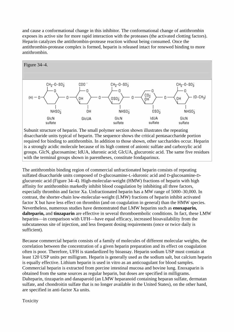
Figure 34–4.
Subunit structure of heparin. The small polymer section shown illustrates the repeating disaccharide units typical of heparin. The sequence shows the critical pentasaccharide portion required for binding to antithrombin. In addition to those shown, other saccharides occur. Heparin is a strongly acidic molecule because of its high content of anionic sulfate and carboxylic acid groups. GlcN, glucosamine; IdUA, iduronic acid; GlcUA, glucuronic acid. The same five residues with the terminal groups shown in parentheses, constitute fondaparinux.
The antithrombin binding region of commercial unfractionated heparin consists of repeating sulfated disaccharide units composed of D-glucosamine-L-iduronic acid and D-glucosamine-D-glucuronic acid (Figure 34–4). High-molecular-weight (HMW) fractions of heparin with high affinity for antithrombin markedly inhibit blood coagulation by inhibiting all three factors, especially thrombin and factor Xa. Unfractionated heparin has a MW range of 5000–30,000. In contrast, the shorter-chain low-molecular-weight (LMW) fractions of heparin inhibit activated factor X but have less effect on thrombin (and on coagulation in general) than the HMW species. Nevertheless, numerous studies have demonstrated that LMW heparins such as enoxaparin, dalteparin, and tinzaparin are effective in several thromboembolic conditions. In fact, these LMW heparins—in comparison with UFH—have equal efficacy, increased bioavailability from the subcutaneous site of injection, and less frequent dosing requirements (once or twice daily is sufficient).
Because commercial heparin consists of a family of molecules of different molecular weights, the correlation between the concentration of a given heparin preparation and its effect on coagulation often is poor. Therefore, UFH is standardized by bioassay. Heparin sodium USP must contain at least 120 USP units per milligram. Heparin is generally used as the sodium salt, but calcium heparin is equally effective. Lithium heparin is used in vitro as an anticoagulant for blood samples. Commercial heparin is extracted from porcine intestinal mucosa and bovine lung. Enoxaparin is obtained from the same sources as regular heparin, but doses are specified in milligrams. Dalteparin, tinzaparin and danaparoid (an LMW heparanoid containing heparan sulfate, dermatan sulfate, and chondroitin sulfate that is no longer available in the United States), on the other hand, are specified in anti-factor Xa units.
Toxicity

The major adverse effect of heparin is bleeding. This risk can be decreased by scrupulous patient selection, careful control of dosage, and close monitoring of the activated partial thromboplastin time (aPTT) in those patients receiving unfractionated heparin. Levels for UFH may also be determined by protamine titration (therapeutic levels 0.2–0.4 unit/mL) or anti-Xa units (therapeutic levels 0.3–0.7 unit/mL). Weight-based dosing of the LMW heparins results in predictable pharmacokinetics and plasma levels in patients with normal renal function. Therefore, LMW heparin levels are not generally measured except in the setting of renal insufficiency, obesity, and pregnancy. LMW heparin levels are determined by anti-Xa units. Peak therapeutic levels are 0.5–1 unit/mL for twice daily dosing, determined 4 hours after administration, and approximately 1.5 units/mL for once daily dosing. Elderly women and patients with renal failure are more prone to hemorrhage. Heparin is of animal origin and should be used cautiously in patients with allergy. Increased loss of hair and reversible alopecia have been reported. Long-term heparin therapy is associated with osteoporosis and spontaneous fractures. Heparin accelerates the clearing of postprandial lipemia by causing the release of lipoprotein lipase from tissues, and long-term use is associated with mineralocorticoid deficiency.
Heparin causes transient thrombocytopenia in 25% or more of patients and severe thrombocytopenia in 5%. Mild platelet reduction within the first 5 days of therapy may result from heparin-induced aggregation that is postulated to be benign and transient in character. A smaller subset of patients may develop an antibody-mediated thrombocytopenia that is associated with paradoxical thrombosis. In these instances, the heparin-induced antibody is directed against the heparin-platelet factor 4 complex. These antigen-antibody complexes bind to Fc receptors on adjacent platelets, causing aggregation and thromboembolism. The following points should be considered in all patients receiving heparin: Platelet counts should be performed frequently; thrombocytopenia should be considered to be heparin-induced; any new thrombus can be the result of heparin; and thromboembolic disease thought to be heparin-induced should be treated by discontinuance of heparin and administration of an alternative drug, such as a direct thrombin inhibitor (see below). Administration of warfarin alone is contraindicated since it may exacerbate the prothrombotic state associated with heparin-induced thrombocytopenia.
Contraindications
Heparin is contraindicated in patients who are hypersensitive to the drug, are actively bleeding, or have hemophilia, significant thrombocytopenia, purpura, severe hypertension, intracranial hemorrhage, infective endocarditis, active tuberculosis, ulcerative lesions of the gastrointestinal tract, threatened abortion, visceral carcinoma, or advanced hepatic or renal disease. Heparin should be avoided in those patients who have recently had surgery of the brain, spinal cord, or eye and in patients who are undergoing lumbar puncture or regional anesthetic block. Despite the apparent lack of placental transfer, heparin should be used in pregnant women only when clearly indicated.
Administration & Dosage
The indications for the use of heparin are described in the section on clinical pharmacology. A plasma concentration of heparin of 0.2–0.4 unit/mL (by protamine titration) or 0.3–0.7 unit/mL (anti-Xa units) usually prevents pulmonary emboli in patients with established venous thrombosis. This concentration of heparin will prolong the activated partial thromboplastin time (aPTT) to 2–2.5 times that of the control value. This degree of anticoagulant effect should be maintained throughout the course of continuous intravenous heparin therapy. When intermittent heparin administration is used, the aPTT should be measured 6 hours after the administered dose to maintain prolongation of the aPTT to 2–2.5 times that of the control value.

Continuous intravenous administration of heparin is accomplished via an infusion pump. After an initial bolus injection of 80-100 units/kg, a continuous infusion of about 15-22 units/kg/h is required to maintain the aPTT at 2–2.5 times control. Patients with acute pulmonary emboli often require larger doses than these during the first few days because of binding to a variety of acute phase proteins, such as factor VIII and von Willebrand factor, and increased heparin clearance. Subcutaneous administration of heparin, as in low-dose prophylaxis, is achieved with 5000 units every 8–12 hours. Because of the danger of hematoma formation at the injection site, heparin must never be administered intramuscularly.
Prophylactic enoxaparin is given subcutaneously in a dosage of 30 mg twice daily or 40 mg once daily. Full-dose enoxaparin therapy is 1 mg/kg subcutaneously every 12 hours. This corresponds to a therapeutic anti-factor Xa level of 0.5–1 unit/mL. Selected patients may be treated with enoxaparin 1.5 mg/kg once a day, with a target anti-Xa level of 1.5 units/mL. The prophylactic dose of dalteparin is 5000 units subcutaneously once a day; therapeutic dosing is 200 units/kg once a day for venous disease or 120 units/kg every 12 hours for acute coronary syndrome. The use of LMW heparins is discouraged or contraindicated in patients with renal insufficiency or body weight greater than 150 kg.
The synthetic pentasaccharide molecule fondaparinux (Figure 34–4) avidly binds antithrombin with high specific activity, resulting in efficient inactivation of factor Xa. Fondaparinux has a long half-life of 15 hours, allowing for once-daily dosing by subcutaneous administration. A series of phase 3 studies in orthopedic patients comparing fondaparinux 2.5 mg subcutaneously daily beginning 6 hours postoperatively versus enoxaparin either 40 mg daily or 30 mg twice daily found fondaparinux to be superior in preventing development of asymptomatic deep venous thrombosis. A phase 3 study evaluating the use of fondaparinux coupled with warfarin compared with LMWH plus warfarin in the treatment of acute venous thromboembolic disease found these two treatment approaches equivalent. Increased bleeding with fondaparinux is seen in patients administered the drug sooner than 6 hours postoperatively, in those who weigh less than 50 kg, and in those with renal insufficiency.
Reversal of Heparin Action
Excessive anticoagulant action of heparin is treated by discontinuance of the drug. If bleeding occurs, administration of a specific antagonist such as protamine sulfate is indicated. Protamine is a highly basic peptide that combines with heparin as an ion pair to form a stable complex devoid of anticoagulant activity. For every 100 units of heparin remaining in the patient, administer 1 mg of protamine sulfate intravenously; the rate of infusion should not exceed 50 mg in any 10-minute period. Excess protamine must be avoided; it also has an anticoagulant effect. Neutralization of LMW heparin by protamine is incomplete. Limited experience suggests that 1 mg of protamine sulfate may be used to partially neutralize 1 mg of enoxaparin. Protamine will not reverse the activity of fondaparinux. Excess danaparoid can be removed by plasmapheresis.
Direct Thrombin Inhibitors
The direct thrombin inhibitors (DTI) are a relatively new class of agents that exert their anticoagulant effect by directly binding to the active site of thrombin, thereby inhibiting thrombin's downstream effects. The DTIs bind thrombin without additional binding proteins, such as antithrombin, and they do not bind to other plasma proteins, such as platelet factor 4. Hirudin and bivalirudin are bivalent DTIs in that they bind at both the catalytic or active site of thrombin as well as at a substrate recognition site. Argatroban and melagatran are small molecules that bind only at the thrombin active site.
Hirudin
For a number of years, surgeons have used medicinal leeches (Hirudo medicinalis) to prevent thrombosis in the fine vessels of reattached digits. Hirudin is a specific, irreversible thrombin inhibitor from the leech that is now available in recombinant form as lepirudin. Its action is independent of antithrombin, which means it can reach and inactivate fibrin-bound thrombin in thrombi. Lepirudin has little effect on platelets or the bleeding time. Like heparin, it must be administered parenterally and is monitored by the aPTT. Lepirudin is FDA-approved for use in patients with thrombosis related to heparin-induced thrombocytopenia. This drug has a short half-life, but it accumulates in renal insufficiency and no antidote exists. Up to 40% of patients on long-term infusions develop an antibody directed against the thrombin-lepirudin complex. These antigen-antibody complexes are not cleared by the kidney and may result in an enhanced anticoagulant effect.
Bivalirudin, another bivalent inhibitor of thrombin, is administered intravenously, with a rapid onset and offset of action. The drug has a short half-life with clearance that is 20% renal and the remainder metabolic. Bivalirudin inhibits platelet activation and been FDA-approved for use in percutaneous coronary angioplasty.
Argatroban is a small molecule thrombin inhibitor that is FDA approved for use in patients with heparin-induced thrombocytopenia (HIT) with or without thrombosis and coronary angioplasty in patients with HIT. It, too, has a short half-life, is given by continuous intravenous infusion, and monitoring is done by aPTT. Its clearance is not affected by renal disease but is dependent on liver function. The drug requires dose reduction in patients with liver disease. Patients on argatroban will demonstrate elevated INRs because of test interference, rendering the transition to warfarin difficult.
Melagatran is the fourth parenteral drug in this class. It and its oral prodrug, ximelagatran, are under intensive study. Attractive features of ximelagatran include predictable pharmacokinetics and bioavailability—allowing for fixed dosing and predictable anticoagulant response; no need for routine coagulation monitoring; lack of interaction with P450-interacting drugs; rapid onset and offset of action—allowing for immediate anticoagulation and thus no need for overlap with additional anticoagulant drugs. A published phase 3 trial in patients status post major orthopedic surgery found ximelagatran equivalent to warfarin in preventing postoperative DVT. Clinical trials in patients with acute DVT and chronic atrial fibrillation are on-going.
Warfarin & the Coumarin Anticoagulants
Chemistry & Pharmacokinetics
The clinical use of the coumarin anticoagulants can be traced to the discovery of an anticoagulant substance formed in spoiled sweet clover silage. It produced a deficiency of plasma prothrombin and consequent hemorrhagic disease in cattle. The toxic agent was identified as bishydroxycoumarin and synthesized as dicumarol. This drug and its congeners, most notably warfarin (Figure 34–5), are widely used as rodenticides in addition to their application as antithrombotic agents in humans. Warfarin is the most reliable member of this group, and the other coumarin anticoagulants are almost never used in the USA.
Figure 34–5.

Structural formulas of several oral anticoagulant drugs and of vitamin K. The carbon atom of warfarin shown at the asterisk is an asymmetric center.
Warfarin is generally administered as the sodium salt and has 100% bioavailability. Over 99% of racemic warfarin is bound to plasma albumin, which may contribute to its small volume of distribution (the albumin space), its long half-life in plasma (36 hours), and the lack of urinary excretion of unchanged drug. Warfarin used clinically is a racemic mixture composed of equal amounts of two enantiomorphs. The levorotatory S-warfarin is four times more potent than the dextrorotatory R-warfarin. This observation is useful in understanding the stereoselective nature of several drug interactions involving warfarin.
Mechanism of Action
Coumarin anticoagulants block the -carboxylation of several glutamate residues in prothrombin and factors VII, IX, and X as well as the endogenous anticoagulant proteins C and S (Figure 34–2). The blockade results in incomplete molecules that are biologically inactive in coagulation. This protein carboxylation is physiologically coupled with the oxidative deactivation of vitamin K. The anticoagulant prevents reductive metabolism of the inactive vitamin K epoxide back to its active hydroquinone form (Figure 34–6). Mutational change of the responsible enzyme, vitamin K epoxidereductase, can give rise to genetic resistance to warfarin in humans and especially in rats.
Figure 34–6.
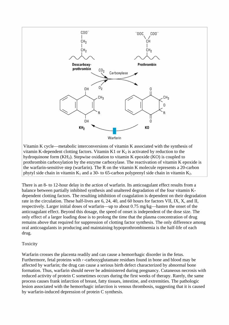
Vitamin K cycle—metabolic interconversions of vitamin K associated with the synthesis of vitamin K-dependent clotting factors. Vitamin K1 or K2 is activated by reduction to the hydroquinone form (KH2). Stepwise oxidation to vitamin K epoxide (KO) is coupled to prothrombin carboxylation by the enzyme carboxylase. The reactivation of vitamin K epoxide is the warfarin-sensitive step (warfarin). The R on the vitamin K molecule represents a 20-carbon phytyl side chain in vitamin K1 and a 30- to 65-carbon polyprenyl side chain in vitamin K2.
There is an 8- to 12-hour delay in the action of warfarin. Its anticoagulant effect results from a balance between partially inhibited synthesis and unaltered degradation of the four vitamin K-dependent clotting factors. The resulting inhibition of coagulation is dependent on their degradation rate in the circulation. These half-lives are 6, 24, 40, and 60 hours for factors VII, IX, X, and II, respectively. Larger initial doses of warfarin—up to about 0.75 mg/kg—hasten the onset of the anticoagulant effect. Beyond this dosage, the speed of onset is independent of the dose size. The only effect of a larger loading dose is to prolong the time that the plasma concentration of drug remains above that required for suppression of clotting factor synthesis. The only difference among oral anticoagulants in producing and maintaining hypoprothrombinemia is the half-life of each drug.
Toxicity
Warfarin crosses the placenta readily and can cause a hemorrhagic disorder in the fetus. Furthermore, fetal proteins with -carboxyglutamate residues found in bone and blood may be affected by warfarin; the drug can cause a serious birth defect characterized by abnormal bone formation. Thus, warfarin should never be administered during pregnancy. Cutaneous necrosis with reduced activity of protein C sometimes occurs during the first weeks of therapy. Rarely, the same process causes frank infarction of breast, fatty tissues, intestine, and extremities. The pathologic lesion associated with the hemorrhagic infarction is venous thrombosis, suggesting that it is caused by warfarin-induced depression of protein C synthesis.

Administration & Dosage
Treatment with warfarin should be initiated with standard doses of 5–10 mg rather than the large loading doses formerly used. The initial adjustment of the prothrombin time takes about 1 week, which usually results in a maintenance dose of 5–7 mg/d. The prothrombin time should be increased to a level representing a reduction of prothrombin activity to 25% of normal and maintained there for long-term therapy. When the activity is less than 20%, the warfarin dosage should be reduced or omitted until the activity rises above 20%.
The therapeutic range for oral anticoagulant therapy is now defined in terms of an international normalized ratio (INR). The INR is the prothrombin time ratio (test/control) obtained if the more sensitive international reference thromboplastin made from human brain is used rather than the less sensitive rabbit brain thromboplastin used in North America. Randomized prospective studies with the INR system have resulted in use of lower doses of anticoagulant drug and less bleeding, yet the efficacy is equal to that of the regimens with higher doses. These less intensive and lower dose regimens are efficacious for many therapeutic indications. Analysis of several clinical trials in patients with artificial heart valves has led to the recommendation that dosage be adjusted to achieve an INR of 2.5–3.5.
Drug Interactions
The oral anticoagulants often interact with other drugs and with disease states. These interactions can be broadly divided into pharmacokinetic and pharmacodynamic effects (Table 34–2). Pharmacokinetic mechanisms for drug interaction with oral anticoagulants are mainly enzyme induction, enzyme inhibition, and reduced plasma protein binding. Pharmacodynamic mechanisms for interactions with warfarin are synergism (impaired hemostasis, reduced clotting factor synthesis, as in hepatic disease), competitive antagonism (vitamin K), and an altered physiologic control loop for vitamin K (hereditary resistance to oral anticoagulants).
Table 34–2. Pharmacokinetic and Pharmacodynamic Drug and Body Interactions with Oral Anticoagulants.
Increased Prothrombin Time Decreased Prothrombin Time Pharmacokinetic Pharmacodynamic Pharmacokinetic Pharmacodynamic Amiodarone Drugs Barbiturates Drugs Cimetidine Aspirin (high doses) Cholestyramine Diuretics Disulfiram Cephalosporins, third-
generation Rifampin Vitamin K
Metronidazole1
Heparin Body factors
Fluconazole1
Hereditary resistance
Phenylbutazone1
Body factors Hypothyroidism
Sulfinpyrazone1
Hepatic disease

Trimethoprim-sulfamethoxazole
Hyperthyroidism
1Stereoselectively inhibits the oxidative metabolism of the (S)-warfarin enantiomorph of racemic warfarin.
The most serious interactions with warfarin are those that increase the anticoagulant effect and the risk of bleeding. The most dangerous of these interactions are the pharmacokinetic interactions withthe pyrazolones phenylbutazone and sulfinpyrazone. These drugs not only augment the hypoprothrombinemia but also inhibit platelet function and may induce peptic ulcer disease (see Chapter 36: Nonsteroidal Anti-Inflammatory Drugs, Disease-Modifying Antirheumatic Drugs, Nonopioid Analgesics, & Drugs Used in Gout). The mechanisms for their hypoprothrombinemic interaction are a stereoselective inhibition of oxidative metabolic transformation of S-warfarin (the more potent isomer) and displacement of albumin-bound warfarin, increasing the free fraction. For this and other reasons, neither phenylbutazone nor sulfinpyrazone is in common use in the USA. Metronidazole, fluconazole, and trimethoprim-sulfamethoxazole also stereoselectively inhibit the metabolic transformation of S-warfarin, whereas amiodarone, disulfiram, and cimetidine inhibit metabolism of both enantiomorphs of warfarin. Aspirin, hepatic disease, and hyperthyroidism augment warfarin pharmacodynamically—aspirin by its effect on platelet function and the latter two by increasing the turnover rate of clotting factors. The third-generation cephalosporins eliminate the bacteria in the intestinal tract that produce vitamin K and, like warfarin, also directly inhibit vitamin K epoxide reductase. Heparin directly prolongs the prothrombin time by inhibiting the activity of several clotting factors.
Barbiturates and rifampin cause a marked decrease of the anticoagulant effect by induction of the hepatic enzymes that transform racemic warfarin. Cholestyramine binds warfarin in the intestine and reduces its absorption and bioavailability.
Pharmacodynamic reductions of anticoagulant effect occur with vitamin K (increased synthesis of clotting factors), the diuretics chlorthalidone and spironolactone (clotting factor concentration), hereditary resistance (mutation of vitamin K reactivation cycle molecules), and hypothyroidism (decreased turnover rate of clotting factors).
Drugs with no significant effect on anticoagulant therapy include ethanol, phenothiazines, benzodiazepines, acetaminophen, opioids, indomethacin, and most antibiotics.
Reversal of Action
Excessive anticoagulant effect and bleeding from warfarin can be reversed by stopping the drug and administering vitamin K1 (phytonadione), fresh-frozen plasma, prothrombin complex concentrates (PCC) such as Bebulin and Proplex T, and recombinant factor VIIa (rFVIIa). The disappearance of excessive effect is not correlated with plasma warfarin concentrations but rather with reestablishment of normal activity of the clotting factors. A modest excess of anticoagulant effect without bleeding may require no more than cessation of the drug. The warfarin effect can be rapidly reversed in the setting of severe bleeding with the administration of a prothrombin complex or recombinant factor VIIa coupled with intravenous vitamin K.
Analogs & Variants
Vitamin K antagonists other than warfarin are seldom used, because they have less favorable

pharmacologic properties or greater toxicity. Dicumarol is incompletely absorbed and frequently causes gastrointestinal symptoms. Phenprocoumon has a long half-life of 6 days—a disadvantage should toxicity occur. The indanedione group, which includes phenindione and diphenadione, has potentially serious adverse effects in the kidney and liver and is of little clinical use. Katzung PHARMACOLOGY, 9e > Section VI. Drugs Used to Treat Disease of the Blood, Inflammation, & Gout > Chapter 34. Drugs Used in Disorders of Coagulation >
Basic Pharmacology of the Fibrinolytic Drugs
Fibrinolytic drugs rapidly lyse thrombi by catalyzing the formation of the serine protease plasmin from its precursor zymogen, plasminogen (Figure 34–3). These drugs (streptokinase, alteplase, anistreplase, tissue plasminogen activator, reteplase, tenecteplase, and urokinase) create a generalized lytic state when administered intravenously. Thus, both protective hemostatic thrombi and target thromboemboli are broken down. The section Thrombolytic Drugs for Acute Myocardial Infarction describes the use of these drugs in one major application.
Pharmacology
Streptokinase is a protein (but not an enzyme in itself) synthesized by streptococci that combines with the proactivator plasminogen. This enzymatic complex catalyzes the conversion of inactive plasminogen to active plasmin. Urokinase is a human enzyme synthesized by the kidney that directly converts plasminogen to active plasmin. Plasmin itself cannot be used because naturally occurring inhibitors in plasma prevent its effects. However, the absence of inhibitors for urokinase and the streptokinase-proactivator complex permit their use clinically. Plasmin formed inside a thrombus by these activators is protected from plasma antiplasmins, which allows it to lyse the thrombus from within.
Anistreplase (anisoylated plasminogen streptokinase activator complex; APSAC) consists of a complex of purified human plasminogen and bacterial streptokinase that has been acylated to protect the enzyme's active site. When administered, the acyl group spontaneously hydrolyzes, freeing the activated streptokinase-proactivator complex. This product (recently discontinued in the USA) allows for rapid intravenous injection, greater clot selectivity (ie, more activity on plasminogen associated with clots than on free plasminogen in the blood), and more thrombolytic activity.
Plasminogen can also be activated endogenously by tissue plasminogen activators (t-PA). These activators preferentially activate plasminogen that is bound to fibrin, which (in theory) confines fibrinolysis to the formed thrombus and avoids systemic activation. Human t-PA is manufactured as alteplase by means of recombinant DNA technology.
Reteplase is another recombinant human t-PA from which several amino acid sequences have been deleted. Reteplase is less expensive to produce than t-PA. Because it lacks the major fibrin-binding domain, reteplase is less fibrin-specific than t-PA. Tenecteplase is a mutant form of t-PA that has a longer half-life, and it can be given as an intravenous bolus. Tenecteplase is slightly more fibrin-specific than t-PA.
Indications & Dosage
Use of fibrinolytic drugs by the intravenous route is indicated in cases of multiple pulmonary emboli that are not massive enough to require surgical management. Intravenous fibrinolytic drugs

are also indicated in cases of central deep venous thrombosis such as the superior vena caval syndrome and ascending thrombophlebitis of the iliofemoral vein. They have also been used intra-arterially, especially for peripheral vascular disease.
Thrombolytic therapy in the management of acute myocardial infarction requires careful patient selection, the use of a specific thrombolytic agent, and the benefit of adjuvant therapy. Considerable controversy surrounds the question of greater safety or efficacy of t-PA compared with the other thrombolytic agents (see Thrombolytic Drugs for Acute Myocardial Infarction).
Streptokinase is administered by intravenous infusion of a loading dose of 250,000 units, followed by 100,000 units/h for 24–72 hours. Patients with antistreptococcal antibodies can develop fever, allergic reactions, and therapeutic resistance. Urokinase requires a loading dose of 300,000 units given over 10 minutes and a maintenance dose of 300,000 units/h for 12 hours. Alteplase (t-PA) is given by intravenous infusion of 60 mg over the first hour and then 40 mg at a rate of 20 mg/h. Reteplase is given as two intravenous bolus injections of 10 units each, separated by 30 minutes. Tenecteplase is given as a single intravenous bolus of 0.5 mg/kg. Anistreplase is given as a single intravenous injection of 30 units over 3–5 minutes. A single course of fibrinolytic drugs is expensive: hundreds of dollars for streptokinase and thousands for urokinase and t-PA.
Recombinant tissue plasminogen activator has increasingly been used for patients presenting with acute stroke symptoms. A recent outcomes study demonstrated an advantage with respect to neurologic disability at 1 year in those patients with acute ischemic stroke who received intravenous t-PA within 3 hours after onset of symptoms. Katzung PHARMACOLOGY, 9e > Section VI. Drugs Used to Treat Disease of the Blood, Inflammation, & Gout > Chapter 34. Drugs Used in Disorders of Coagulation >
Thrombolytic Drugs for Acute Myocardial Infarction
Background: The paradigm shift in 1980 on the causation of acute myocardial infarction to acute coronary occlusion by a thrombus created the rationale for thrombolytic therapy of this common lethal disease. At that time—and for the first time—intravenous thrombolytic therapy for acute myocardial infarction in the European Cooperative Study Group trial was found to reduce mortality significantly. Later studies, with thousands of patients in each trial, provided enough statistical power for the 20% reduction in mortality to be considered statistically significant. Although the standard of care in areas with adequate facilities and experience in percutaneous coronary intervention (PCI) now favors catheterization and placement of a stent, thrombolytic therapy is still very important where PCI is not readily available.
Patients: The selection of patients for thrombolytic therapy is critical. The diagnosis of acute myocardial infarction is made clinically and is confirmed by electrocardiography. Patients with ST segment elevation and bundle branch block on electrocardiography do best; those with ST segment depression or a normal ECG do less well; and those with non-Q-wave acute myocardial infarction may even be harmed. All trials to date show the greatest benefit for thrombolytic therapy when it is given early, within 6 hours after symptomatic onset of acute myocardial infarction.
Clinical trials: One of the trials (ISIS-3) showed that streptokinase plus aspirin performed as well as recombinant tissue-type plasminogen activator (rt-PA) or complex formulations of streptokinase such as anistreplase (APSAC). The GUSTO trial showed a small advantage for the much more expensive t-PA over streptokinase, but with a significantly higher risk of hemorrhagic stroke. Nine clinical trials—each containing over 1000 patients with suspected acute myocardial infarction—
reported 11.6% mortality at 35 days in the control group and 9.5% in the treatment group, an 18% reduction in mortality.
Adjunctive drugs: The best results occur with thrombolytic drugs supplemented by other drugs. -Blocker drugs reduced myocardial ischemia and infarct size, prevented arrhythmias, decreased reinfarction, and improved survival in ISIS-1 and GISSI-1. Aspirin alone and with streptokinase reduced mortality in ISIS-2. Nitroglycerin, given early in acute myocardial infarction for pain relief, had no beneficial effect on mortality in GISSI-3 and ISIS-4. Oral anticoagulants are best used in patients with depressed left ventricular function or systemic embolization. Heparin was a necessary adjunct for t-PA in GUSTO but was less useful with streptokinase plus aspirin because of the increased risk of bleeding. ACE inhibitors reduced infarct expansion and arrhythmias and improved survival in GISSI-3. Direct thrombin inhibitors like hirudin and bivalirudin are undergoing clinical trials to better determine their efficacy in conjunction with thrombolytic therapy (TIMI-9B, GUSTO-2A, OASIS, HERO).
Summary: Thrombolytic drugs reduce the mortality of acute myocardial infarction. The early and appropriate use of any thrombolytic drug probably transcends individual advantages. Adjunctive drugs like aspirin, -blockers, and ACE inhibitors reduce mortality even further. The principles of management are outlined in part 7 of the American Heart Association Guidelines, 2000. Katzung PHARMACOLOGY, 9e > Section VI. Drugs Used to Treat Disease of the Blood, Inflammation, & Gout > Chapter 34. Drugs Used in Disorders of Coagulation >
Basic Pharmacology of Antiplatelet Agents
Platelet function is regulated by three categories of substances. The first group consists of agents generated outside the platelet that interact with platelet membrane receptors, eg, catecholamines, collagen, thrombin, and prostacyclin. The second category contains agents generated within the platelet that interact with membrane receptors, eg, ADP, prostaglandin D2, prostaglandin E2, and serotonin. The third group comprises agents generated within the platelet that act within the platelet, eg, prostaglandin endoperoxides and thromboxane A2, the cyclic nucleotides cAMP and cGMP, and calcium ion. From this list of agents, several targets for platelet inhibitory drugs have been identified (Figure 34–1): inhibition of prostaglandin metabolism (aspirin), inhibition of ADP-induced platelet aggregation (clopidogrel, ticlopidine), and blockade of GP IIb/IIIa receptors on platelets (abciximab, tirofiban, and eptifibatide). Dipyridamole and cilostazol are additional antiplatelet drugs.
Aspirin
The prostaglandin thromboxane A2 is an arachidonate product that causes platelets to change shape, to release their granules, and to aggregate (see Chapter 18: The Eicosanoids: Prostaglandins, Thromboxanes, Leukotrienes, & Related Compounds). Drugs that antagonize this pathway interfere with platelet aggregation in vitro and prolong the bleeding time in vivo. Aspirin is the prototype of this class of drugs. Drugs that modulate the intraplatelet concentration of cAMP do not prolong the bleeding time. However, dietary therapy can be useful in the prophylaxis of thrombosis. Ingestion of the unsaturated fatty acid eicosapentaenoic acid, which is high in cold water fish, generates prostaglandin I3, an effective antiaggregating substance like prostacyclin, and thromboxane A3, which is much less active than TXA2 (Chapter 18: The Eicosanoids: Prostaglandins, Thromboxanes, Leukotrienes, & Related Compounds).
As described in Chapter 18: The Eicosanoids: Prostaglandins, Thromboxanes, Leukotrienes, &

Related Compounds, aspirin inhibits the synthesis of thromboxane A2 by irreversible acetylation of the enzyme cyclooxygenase. Other salicylates and nonsteroidal anti-inflammatory drugs also inhibit cyclooxygenase but have a shorter duration of inhibitory action because they cannot acetylate cyclooxygenase, ie, their action is reversible.
The FDA has approved the use of 325 mg/d for primary prophylaxis of myocardial infarction but urges caution in this use of aspirin by the general population except when prescribed as an adjunct to risk factor management by smoking cessation and lowering of blood cholesterol and blood pressure. Meta-analysis of many published trials of aspirin and other antiplatelet agents confirms the value of this intervention in the secondary prevention of vascular events among patients with a history of vascular events.
Clopidogrel & Ticlopidine
Clopidogrel and ticlopidine reduce platelet aggregation by inhibiting the ADP pathway of platelets. These drugs are thienopyridine derivatives that achieve their antiplatelet effects by irreversibly blocking the ADP receptor on platelets. Unlike aspirin, these drugs have no effect on prostaglandin metabolism. Randomized clinical trials with both drugs report efficacy in the prevention of vascular events among patients with transient ischemic attacks, completed strokes, and unstable angina pectoris. Use of clopidogrel or ticlopidine to prevent thrombosis is now considered standard practice in patients undergoing placement of a coronary stent.
Adverse effects of ticlopidine include nausea, dyspepsia, and diarrhea in up to 20% of patients, hemorrhage in 5%, and, most seriously, leukopenia in 1%. The leukopenia is detected by regular monitoring of the white blood cell count during the first 3 months of treatment. Development of thrombotic thrombocytopenic purpura (TTP) has also been associated with the ingestion of ticlopidine. The dosage of ticlopidine is 250 mg twice daily. It is particularly useful in patients who cannot tolerate aspirin. Doses of ticlopidine less than 500 mg/d may be efficacious with fewer adverse effects.
Clopidogrel has fewer adverse effects than ticlopidine and is rarely associated with neutropenia. Thrombotic thrombocytopenic purpura associated with clopidogrel has recently been reported. Because of its superior side effect profile and dosing requirements, clopidogrel is preferred over ticlopidine. The antithrombotic effects of clopidogrel are dose-dependent; within 5 hours after an oral loading dose of 300 mg, 80% of platelet activity will be inhibited. The maintenance dose of clopidogrel is 75 mg/d, which achieves maximum platelet inhibition. The duration of the antiplatelet effect is 7–10 days.
Blockade of Platelet Gp IIb/IIIa Receptors
The glycoprotein IIb/IIIa inhibitors are used in patients with acute coronary syndromes. These drugs target the platelet IIb/IIIa receptor complex (Figure 34–1). The IIb/IIIa complex functions as a receptor mainly for fibrinogen and vitronectin but also for fibronectin and von Willebrand factor. Activation of this receptor complex is the "final common pathway" for platelet aggregation. There are approximately 50,000 copies of this complex on the platelet surface. Persons lacking this receptor have a bleeding disorder called Glanzmann's thrombasthenia.
Abciximab, a humanized monoclonal antibody directed against the IIb/IIIa complex including the vibronectin receptor, was the first agent approved in this class of drugs. It has been approved for use in percutaneous coronary intervention and in acute coronary syndromes. Eptifibatide is an analog of the sequence at the extreme carboxyl terminal of the delta chain of fi-brinogen, which mediates

the binding of fibrinogen to the receptor. Tirofiban is a smaller molecule with similar properties. Eptifibatide and tirofiban inhibit ligand binding to the IIb/IIIa receptor by their occupancy of the receptor but do not block the vibronectin receptor.
The three agents described above are administered parenterally. Oral formulations of IIb/IIIa antagonists have been developed and are in various stages of development. Thus far, however, lack of efficacy and significant thrombocytopenia have prevented progress with the oral analogs.
Additional Antiplatelet-Directed Drugs
Dipyridamole is a vasodilator that inhibits platelet function by inhibiting adenosine uptake and cyclic GMP phosphodiesterase activity. Dipyridamole by itself has little or no beneficial effect. Therefore, therapeutic use of this agent is primarily in combination with aspirin to prevent cerebrovascular ischemia. It may also be used in combination with warfarin for primary prophylaxis of thromboemboli in patients with prosthetic heart valves. A combination of dipyridamole complexed with 25 mg of aspirin is now available for secondary prophylaxis of cerebrovascular disease.
Cilostazol is a newer phosphodiesterase inhibitor that promotes vasodilation and inhibition of platelet aggregation. Cilostazol is used primarily to treat intermittent claudication. Katzung PHARMACOLOGY, 9e > Section VI. Drugs Used to Treat Disease of the Blood, Inflammation, & Gout > Chapter 34. Drugs Used in Disorders of Coagulation >
Clinical Pharmacology of Drugs Used to Prevent Clotting
Venous Thrombosis
Risk Factors
Inherited Disorders
The inherited disorders characterized by an tendency to form thrombi (thrombophilia) derive from either quantitative or qualitative abnormalities of the natural anticoagulant system. Deficiencies in the natural anticoagulants antithrombin, protein C, and protein S account for approximately 15% of selected patients with juvenile or recurrent thrombosis and 5–10% of unselected cases of acute venous thrombosis. Additional causes of thrombophilia include the factor V Leiden mutation, hyperhomocystinemia, and the prothrombin 20210 mutation that together account for the greater number of hypercoagulable patients.
Acquired Disease
The increased risk of thromboembolism associated with arrhythmia, primarily atrial fibrillation, and the placement of mechanical heart valves has long been recognized. Similarly, prolonged bed rest, high-risk surgical procedures, and the presence of cancer are clearly associated with an increased incidence of deep venous thrombosis and embolism.
Antithrombotic Management

Prevention
Primary prevention of venous thrombosis reduces the incidence of and mortality rate from pulmonary emboli. Heparin and warfarin may be used to prevent venous thrombosis. Subcutaneous administration of low-dose unfractionated heparin, low-molecular-weight heparin, or fondaparinux provides effective prophylaxis. Warfarin is also effective but requires laboratory monitoring of the prothrombin time.
Treatment of Established Disease
Treatment for established venous thrombosis is initiated with unfractionated or low-molecular-weight heparin for the first 5–7 days, with an overlap with warfarin. Once therapeutic effects of warfarin have been established, therapy with warfarin is continued for a minimum of 3–6 months. Patients with recurrent disease or identifiable, nonreversible risk factors may be treated indefinitely. Small thrombi confined to the calf veins may be managed without anticoagulants if there is documentation over time that the thrombus is not extending.
Warfarin readily crosses the placenta. It can cause hemorrhage at any time during pregnancy as well as developmental defects when administered during the first trimester. Therefore, venous thromboembolic disease in pregnant women is generally treated with heparin, best administered by subcutaneous injection.
Arterial Thrombosis
Activation of platelets is considered an essential process for arterial thrombosis. Thus, treatment with platelet-inhibiting drugs such as aspirin and ticlopidine or clopidogrel is indicated in patients with transient ischemic attacks and strokes or unstable angina and acute myocardial infarction. In angina and infarction, these drugs are often used in conjunction with -blockers, calcium channel blockers, and fibrinolytic drugs. Katzung PHARMACOLOGY, 9e > Section VI. Drugs Used to Treat Disease of the Blood, Inflammation, & Gout > Chapter 34. Drugs Used in Disorders of Coagulation >
Drugs Used in Bleeding Disorders
VItamin K
Vitamin K confers biologic activity upon prothrombin and factors VII, IX, and X by participating in their postribosomal modification. Vitamin K is a fat-soluble substance found primarily in leafy green vegetables. The dietary requirement is low, because the vitamin is additionally synthesized by bacteria that colonize the human intestine. Two natural forms exist: vitamins K1 and K2. Vitamin K1 (phytonadione; Figure 34–5) is found in food. Vitamin K2 (menaquinone) is found in human tissues and is synthesized by intestinal bacteria.
Vitamins K1 and K2 require bile salts for absorption from the intestinal tract. Vitamin K1 is available clinically in 5 mg tablets and 50 mg ampules. Onset of effect is delayed for 6 hours but the effect is complete by 24 hours when treating depression of prothrombin activity by excess warfarin or vitamin K deficiency. Intravenous administration of vitamin K1 should be slow, because rapid infusion can produce dyspnea, chest and back pain, and even death. Vitamin K repletion is best achieved with intravenous or oral administration, because its bioavailability after subcutaneous administration is erratic. Vitamin K1 is currently administered to all newborns to prevent the

hemorrhagic disease of vitamin K deficiency, which is especially common in premature infants. The water-soluble salt of vitamin K3 (menadione) should never be used in therapeutics. It is particularly ineffective in the treatment of warfarin overdosage. Vitamin K deficiency frequently occurs in hospitalized patients in intensive care units because of poor diet, parenteral nutrition, recent surgery, multiple antibiotic therapy, and uremia. Severe hepatic failure results in diminished protein synthesis and a hemorrhagic diathesis that is unresponsive to vitamin K.
Plasma Fractions
Sources & Preparations
Deficiencies in plasma coagulation factors can cause bleeding (Table 34–3). Spontaneous bleeding occurs when factor activity is less than 5–10% of normal. Factor VIII deficiency (classic hemophilia, or hemophilia A) and factor IX deficiency (Christmas disease, or hemophilia B) account for most of the heritable coagulation defects. Concentrated plasma fractions are available for the treatment of these deficiencies. Administration of plasma-derived, heat- or detergent-treated factor concentrates and recombinant factor concentrates are the standard treatments for bleeding associated with hemophilia. Lyophilized factor VIII concentrates are prepared from large pools of plasma. Transmission of viral diseases such as hepatitis B and C and AIDS is reduced or eliminated by pasteurization and by extraction of plasma with solvents and detergents. The best use of these therapeutic materials requires diagnostic specificity of the deficient factor and quantitation of its activity in plasma. Intermediate purity factor VIII concentrates (as opposed to recombinant or high-purity concentrates) contain significant amounts of von Willebrand factor. Humate-P is a factor VIII concentrate that is approved by the FDA for the treatment of bleeding associated with von Willebrand disease.
Table 34–3. Therapeutic Products for the Treatment of Coagulation Disorders.
Factor Deficiency State Hemostatic
Levels Half-Life of Infused Factor
Replacement Source
I Hypofibrinogenemia 1 g/dL 4 days Cryoprecipitate
FFP II Prothrombin
deficiency 30–40% 3 days Prothrombin complex
concentrates (intermediate purity factor IX concentrates)
V Factor V deficiency 20% 1 day FFP VII Factor VII
deficiency 30% 4-6 hours FFP
Prothrombin complex concentrates (intermediate purity factor IX concentrates)
rFVIIa
VIII Hemophilia A 30–50%
100% for major bleeding or trauma
12 hours Recombinant factor VIII products
Plasma-derived high purity concentrates
1Cryoprecipitate
Some patients with mild deficiency will respond to DDAVP
IX Hemophilia B
Christmas disease
30–50%
100% for major bleeding or trauma
24 hours Recombinant F IX products
Plasma-derived high purity concentrates
X Stuart-Prower defect 25% 36 hours FFP
Prothrombin complex concentrates
XI Hemophilia C 30–50% 3 days FFP XII Hageman defect Not required Treatment not necessary Von Willebrand
Von Willebrand disease
30% Approximately 10 hours
Intermediate purity factor VIII concentrates that contain von Willebrand factor
Some patients respond to DDAVP
1Cryoprecipitate XIII Factor XIII
deficiency 5% 6 days FFP
Cryoprecipitate
FFP, fresh frozen plasma.
Antithrombin and activated protein C concentrates are available for the appropriate indications that include thrombosis in the setting of antithrombin deficiency and sepsis respectively.
1Cryoprecipate should be used to treat bleeding in the setting of factor VIII deficiency and von Willebrand disease only in an emergency in which pathogen-inactivated products are not available.
Clinical Uses
An uncomplicated hemorrhage into a joint should be treated with sufficient factor VIII or factor IX replacement to maintain a level of at least 30–50% of the normal concentration for 24 hours. Soft

tissue hematomas require a minimum of 100% activity for 7 days. Hematuria requires at least 10% activity for 3 days. Surgery and major trauma require a minimum of 100% activity for 10 days. The initial loading dose for factor VIII is 50 units/kg of body weight to achieve 100% activity of factor VIII from a baseline of 1%, assuming a normal hemoglobin. Each unit of factor VIII per kilogram of body weight raises its activity in plasma 2%. Replacement should be administered every 12 hours. Factor IX therapy requires twice the dose of factor VIII, but with an administration of about every 24 hours because of its longer half-life. Recombinant factor IX has only 80% recovery compared to plasma-derived factor IX products. Therefore, dosing with recombinant factor IX requires 120% of the dose used with the plasma-derived product.
Desmopressin acetate (arginine vasopressin) increases the factor VIII activity of patients with mild hemophilia A or von Willebrand disease. It can be used in preparation for minor surgery such as tooth extraction without any requirement for infusion of clotting factors if the patient has a documented adequate response. High-dose intranasal desmopressin (see Chapter 17: Vasoactive Peptides) is available and has been shown to be efficacious and well tolerated by patients.
Freeze-dried concentrates of plasma containing prothrombin, factors IX and X, and varied amounts of factor VII (Proplex, etc) are commercially available for treating deficiencies of these factors (Table 34–3). Each unit of factor IX per kilogram of body weight raises its activity in plasma 1.5%. Heparin is often added to inhibit coagulation factors activated by the manufacturing process. However, addition of heparin does not eliminate all thromboembolic events.
Some preparations of factor IX concentrate contain activated clotting factors, which has led to their use in treating patients with inhibitors or antibodies to factor VIII or factor IX. Two products are available expressly for this purpose: Autoplex (with factor VIII correctional activity) and Feiba (with factor VIII inhibitor bypassing activity). These products are not uniformly successful in arresting hemorrhage, and the factor IX inhibitor titers often rise after treatment with them. Acquired inhibitors of coagulation factors may also be treated with porcine factor VIII (for factor VIII inhibitors) and recombinant activated factor VII. Recombinant activated factor VII (NovoSeven) is being increasingly used to treat coagulopathy associated with liver disease and major blood loss in trauma and surgery. These recombinant and plasma-derived factor concentrates are very expensive, and the indications for them are very precise. Therefore, close consultation with a hematologist knowledgeable in this area is essential.
Cryoprecipitate is a plasma protein fraction obtainable from whole blood. It is used to treat deficiencies or qualitative abnormalities of fibrinogen, such as that which occurs with disseminated intravascular coagulation and liver disease. A single unit of cryoprecipitate contains 300 mg of fibrinogen.
Cryoprecipitate may also be used for patients with factor VIII deficiency and von Willebrand disease if desmopressin is not indicated and a pathogen-inactivated recombinant or plasma-derived product is not available. The concentration of factor VIII and von Willebrand factor in cryoprecipitate is not as great as that found in the concentrated plasma fractions. Moreover, cryoprecipitate is not treated in any manner to decrease the risk of viral exposure. For infusion, the frozen cryoprecipitate unit is thawed and dissolved in a small volume of sterile citrate-saline solution and pooled with other units. Rh-negative women with potential for childbearing should receive only Rh-negative cryoprecipitate because of possible contamination of the product with Rh-positive blood cells.
Fibrinolytic Inhibitors: Aminocaproic Acid

Aminocaproic acid (EACA), which is chemically similar to the amino acid lysine, is a synthetic inhibitor of fibrinolysis. It competitively inhibits plasminogen activation (Figure 34–3). It is rapidly absorbed orally and is cleared from the body by the kidney. The usual oral dosage of EACA is 6 g four times a day. When the drug is administered intravenously, a 5 g loading dose should be infused over 30 minutes to avoid hypotension. Tranexamic acid is an analog of aminocaproic acid and has the same properties. It is administered orally with a 15 mg/kg loading dose followed by 30 mg/kg every 6 hours, but the drug is not currently available in the United States.
Clinical uses of aminocaproic acid are as adjunctive therapy in hemophilia, as therapy for bleeding from fibrinolytic therapy, and as prophylaxis for rebleeding from intracranial aneurysms. Treatment success has also been reported in patients with postsurgical gastrointestinal bleeding and postprostatectomy bleeding and bladder hemorrhage secondary to radiation- and drug-induced cystitis. Adverse effects of the drug include intravascular thrombosis from inhibition of plasminogen activator, hypotension, myopathy, abdominal discomfort, diarrhea, and nasal stuffiness. The drug should not be used in patients with disseminated intravascular coagulation or genitourinary bleeding of the upper tract, eg, kidney and ureters, because of the potential for excessive clotting.
Serine Protease Inhibitors: Aprotinin
Aprotinin is a serine protease inhibitor ("serpin") that inhibits fibrinolysis by free plasmin and may have other antihemorrhagic effects as well. It also inhibits the plasmin-streptokinase complex in patients who have received that thrombolytic agent. Aprotinin will reduce bleeding—by as much as 50%—from many types of surgery, especially that involving extracorporeal circulation for open heart procedures and liver transplantation. It is currently approved for use in patients undergoing coronary artery bypass grafting who are at high risk of excessive blood loss. In placebo-controlled trials, adverse effects of aprotinin were little different from those reported in patients in the placebo group. In larger studies, a possible association with anaphylaxis has been reported in < 0.5% of cases. Therefore, a small test dose is recommended before the full therapeutic dose is given. Katzung PHARMACOLOGY, 9e > Section VI. Drugs Used to Treat Disease of the Blood, Inflammation, & Gout > Chapter 34. Drugs Used in Disorders of Coagulation >
Preparations Available
Abciximab (ReoPro)
Parenteral: 2 mg/mL for IV injection
Alteplase recombinant [t-PA] (Activase*)
Parenteral: 50, 100 mg lyophilized powder to reconstitute for IV injection
Aminocaproic acid (generic, Amicar)
Oral: 500 mg tablets; 250 mg/mL syrup
Parenteral: 250 mg/mL for IV injection
Anisindione (Miradon)

Oral: 50 mg tablets
Antihemophilic factor [factor VIII, AHF] (Alphanate, Bioclate*, Helixate*, Hemofil M, Koate-HP, Kogenate*, Monoclate, Recombinate,* others)* Recombinant product.
Parenteral: in vials
Anti-inhibitor coagulant complex (Autoplex T, Feiba VH Immuno)
Parenteral: in vials
Antithrombin III (Thrombate III)
Parenteral: 500, 1000 IU powder to reconstitute for IV injection
Aprotinin (Trasylol)
Parenteral: 10,000 units/mL in 100 and 200 mL vials
Argatroban
Parenteral: 100 mg/ml in 2.5 mL vials
Bivalirudin (Angiomax)
Parenteral: 250 mg per vial
Cilostazol (Pletal)
Oral: 50, 100 mg tablets
Clopidogrel (Plavix)
Oral: 75 mg tablets
Coagulation factor VIIa recombinant (Novo- Seven*)
Parenteral: 1.2, 4.8 mg powder/vial for IV injection
Dalteparin (Fragmin)
Parenteral: 2500, 5000, 10,000 anti-factor Xa units/0.2 mL for SC injection only
Danaparoid (Orgaran)
Parenteral: 750 anti-Xa units/vial
Dipyridamole (Persantine)
Oral: 25, 50, 75 mg tablets

Oral combination product (Aggrenox): 200 mg extended-release dipyridamole plus 25 mg aspirin
Enoxaparin (low-molecular-weight heparin, Lovenox)
Parenteral: pre-filled, multiple-dose syringes for SC injection only
Eptifibatide (Integrilin)
Parenteral: 0.75, 2 mg/mL for IV infusion
Factor VIIa: see Coagulation factor VIIa recombi- nant
Factor VIII: see Antihemophilic factor
Factor IX complex, human (AlphaNine SD, Bebulin VH, BeneFix*, Konyne 80, Mononine, Profilnine SD, Proplex T, Proplex SX-T)
Parenteral: in vials
Fondaparinux (Arixtra)
Parenteral: 2.5 mg in 0.5 mL single-dose prefilled syringes
Heparin sodium (generic, Liquaemin)
Parenteral: 1000, 2000, 2500, 5000, 10,000, 20,000, 40,000 units/mL for injection
Lepirudin (Refludan*)
Parenteral: 50 mg powder for IV injection
Phytonadione [K1] (generic, Mephyton, AquaMe- phyton)
Oral: 5 mg tablets
Parenteral: 2, 10 mg/mL aqueous colloidal solution or suspension for injection
Protamine (generic)
Parenteral: 10 mg/mL for injection
Reteplase (Retavase*)
Parenteral: 10.8 IU powder for injection
Streptokinase (Streptase)
Parenteral: 250,000, 750,000, 1,500,000 IU per vial powders to reconstitute for injection
Tenecteplase (TNKase)

Parenteral: 50 mg powder for injection
Ticlopidine (Ticlid)
Oral: 250 mg tablets
Tinzaparin (Innohep)
Parenteral: 20,000 anti-Xa units/mL for subcutaneous injection only
Tirofiban (Aggrastat)
Parenteral: 50, 250 g/mL for IV infusion
Tranexamic acid (Cyklokapron)
Oral: 500 mg tablets
Parenteral: 100 mg/mL for IV infusion
Urokinase (Abbokinase)
Parenteral: 250,000 IU per vial for systemic use
Warfarin (generic, Coumadin)
Oral: 1, 2, 2.5, 3, 4, 5, 6, 7.5, 10 mg tablets Chapter 35. Agents Used in Hyperlipidemia Katzung PHARMACOLOGY, 9e > Section VI. Drugs Used to Treat Disease of the Blood, Inflammation, & Gout > Chapter 35. Agents Used in Hyperlipidemia >
Acronyms
ACAT: Acyl-CoA:cholesterol acyltransferase Apo: Apolipoprotein CETP: Cholesteryl ester transfer protein HDL: High-density lipoproteins HMG-CoA: 3-Hydroxy-3-methylglutaryl-coenzyme A IDL: Intermediate-density lipoproteins LCAT: Lecithin:cholesterol acyltransferase LDL: Low-density lipoproteins Lp(a): Lipoprotein(a) LPL: Lipoprotein lipase PPAR- : Peroxisome proliferator-activated receptor-alphaVLDL: Very low density lipoproteins