Embed Size (px)
Citation preview

WIHS Manual of Operations (01/30/2018)
Section 44, Page 1 of 78
THE WOMEN’S INTERAGENCY HIV STUDY
SECTION 44: ECG PROTOCOL
A. BACKGROUND
The advent of potent antiretroviral therapy (ART) has transformed the care of HIV infection, with life expectancy for people living with HIV now approaching that of the general uninfected population.1 A counterweight to this success, however, is the finding that HIV-infected individuals receiving ART are susceptible to a range of chronic aging-related diseases.2 Notable among these is atherosclerotic cardiovascular disease (CVD), which is a major factor accounting for the rise in non-AIDS mortality among people with HIV.3 Indeed, studies have shown that HIV-infected people have a 1.5-fold risk of coronary heart disease (CHD) events as compared with their HIV-uninfected counterparts.4
Various factors have been cited to account for increased CVD risk. These include persistent immune activation and inflammation resulting from translocation of gut bacteria following obliteration of mucosal adaptive immunity during initial HIV infection or from persistence of HIV in tissue reservoirs; associated co-infections, such as HCV, and consequent hepatic fibrosis; a high burden of behavioral and traditional atherosclerosis risk factors among HIV-infected individuals; and off-target effects of ART on metabolism and mitochondrial function.4 The importance of proper control of HIV to reduce clinical CHD events has been documented in various cohorts composed predominantly of men.4 Additional evidence for this in women has come from studies applying carotid ultrasound to measure subclinical atherosclerotic disease in the Women’s Interagency HIV Study (WIHS) and the Multicenter AIDS Cohort Study (MACS), which have shown that low CD4+ T-cell count and/or increased immune activation and senescence are associated cross-sectionally with carotid atherosclerosis and carotid stiffness in HIV-infected versus HIV-uninfected individuals.5-7 More recently, a longitudinal evaluation in WIHS and MACS participants showed that HIV-infected individuals had a 1.6-fold greater risk of new carotid atheroma formation after adjusting for cardiometabolic factors as compared with HIV-uninfected individuals.8 A similar increase in risk of new carotid plaque formation was seen even among HIV-infected participants with persistent viral suppression relative to their HIV-uninfected counterparts.8 Notably, these heightened risks of carotid atheromatous disease were comparable in women and men, underscoring similar susceptibility in both sexes.
Apart from the impact on atherosclerosis, however, there is evidence that HIV and related factors have important effects on the myocardium.9 This is highlighted by the finding of marked prevalences of mild left ventricular systolic dysfunction and, especially, diastolic dysfunction by echocardiography in studies of HIV-infected individuals receiving ART.10 Such abnormalities may result from increased myocardial fibrosis or fat deposition as a result of HIV, other pathogens, associated risk factors, or ART.11 In accord with these findings, the risk of incident heart failure has been documented to be higher in HIV-infected than HIV-uninfected individuals.12 Furthermore, there are reports that HIV-infected people have an increased risk of atrial fibrillation,13 as well as a heightened risk of sudden cardiac death.14
In connection with dysrhythmic events in particular, mouse models have shown that HIV itself leads to prolongation of repolarization through a direct influence on K+ channels.15

WIHS Manual of Operations (01/30/2018)
Section 44, Page 2 of 78
Moreover, a variety of ART and other drugs frequently used in the context of HIV are well known to prolong the QT interval, thereby increasing the risk of torsade de pointes and sudden cardiac death.16 Such associations have been explored in several clinical reports, which have found HIV, and especially advanced or uncontrolled HIV infection, to be associated with QTc prolongation.16-19 Nevertheless, these reports have had limited sample size, have been uncontrolled or lacked well-matched HIV-uninfected control groups, and have had only limited characterization of HIV-related factors and therapy.
Additional information on dysrhythmia risk can be obtained from assessment of abnormal autonomic tone, as it is established that HIV’s neurotropism leads it to affect the autonomic nervous system.20 In fact, assessment of heart rate variability (HRV) through time-domain measures available on a 10-second ECG strip have been shown to be prognostic for mortality in non-HIV infected cohorts.21, 22 Analysis of such time-domain HRV measures has been applied at baseline to mostly ART-treated HIV-infected men participating in a large clinical trial, showing that non-boosted protease-inhibitor use was associated with greater HRV than non-nucleoside reverse transcriptase-inhibitor use.20
While there is considerable evidence of ischemic and non-ischemic myocardial disease in connection with HIV infection, there remain gaps in understanding the extent and especially the basis for the heightened risk of dysrhythmia in people living with HIV, particularly in women. Assessment of ECG abnormalities and autonomic dysfunction in the WIHS will provide important information on the scope of electrical cardiac derangements and their determinants in this understudied population.
B. REFERENCES
1. Wada N, Jacobson LP, Cohen M, French A, Phair J, Munoz A. Cause-specific life expectancies after 35 years of age for human immunodeficiency syndrome-infected and human immunodeficiency syndrome-negative individuals followed simultaneously in long-term cohort studies, 1984-2008. Am J Epidemiol 2013;177(2):116-25.
2. High KP, Brennan-Ing M, Clifford DB, Cohen MH, Currier J, Deeks SG, Deren S, Effros RB, Gebo K, Goronzy JJ, Justice AC, Landay A, Levin J, Miotti PG, Munk RJ, Nass H, Rinaldo CR, Jr., Shlipak MG, Tracy R, Valcour V, Vance DE, Walston JD, Volberding P, HIV OARWGo, Aging. HIV and aging: state of knowledge and areas of critical need for research. A report to the NIH Office of AIDS Research by the HIV and Aging Working Group. J Acquir Immune Defic Syndr 2012;60 Suppl 1:S1-18.
3. Ingle SM, May MT, Gill MJ, Mugavero MJ, Lewden C, Abgrall S, Fatkenheuer G, Reiss P, Saag MS, Manzardo C, Grabar S, Bruyand M, Moore D, Mocroft A, Sterling TR, D'Arminio Monforte A, Hernando V, Teira R, Guest J, Cavassini M, Crane HM, Sterne JA, Antiretroviral Therapy Cohort C. Impact of risk factors for specific causes of death in the first and subsequent years of antiretroviral therapy among HIV-infected patients. Clin Infect Dis 2014;59(2):287-97.
4. Kaplan RC, Hanna DB, Kizer JR. Recent Insights Into Cardiovascular Disease (CVD) Risk Among HIV-Infected Adults. Curr HIV/AIDS Rep 2016;13(1):44-52.
5. Kaplan RC, Kingsley LA, Gange SJ, Benning L, Jacobson LP, Lazar J, Anastos K, Tien PC, Sharrett AR, Hodis HN. Low CD4+ T-cell count as a major atherosclerosis risk factor in HIV-infected women and men. AIDS 2008;22(13):1615-24.

WIHS Manual of Operations (01/30/2018)
Section 44, Page 3 of 78
6. Seaberg EC, Benning L, Sharrett AR, Lazar JM, Hodis HN, Mack WJ, Siedner MJ, Phair JP, Kingsley LA, Kaplan RC. Association between human immunodeficiency virus infection and stiffness of the common carotid artery. Stroke 2010;41(10):2163-70.
7. Kaplan RC, Sinclair E, Landay AL, Lurain N, Sharrett AR, Gange SJ, Xue X, Hunt P, Karim R, Kern DM, Hodis HN, Deeks SG. T cell activation and senescence predict subclinical carotid artery disease in HIV-infected women. J Infect Dis 2011;203(4):452-63.
8. Hanna DB, Post WS, Deal JA, Hodis HN, Jacobson LP, Mack WJ, Anastos K, Gange SJ, Landay AL, Lazar JM, Palella FJ, Tien PC, Witt MD, Xue X, Young MA, Kaplan RC, Kingsley LA. HIV Infection Is Associated With Progression of Subclinical Carotid Atherosclerosis. Clin Infect Dis 2015;61(4):640-50.
9. Remick J, Georgiopoulou V, Marti C, Ofotokun I, Kalogeropoulos A, Lewis W, Butler J. Heart failure in patients with human immunodeficiency virus infection: epidemiology, pathophysiology, treatment, and future research. Circulation 2014;129(17):1781-9.
10. Cerrato E, D'Ascenzo F, Biondi-Zoccai G, Calcagno A, Frea S, Grosso Marra W, Castagno D, Omede P, Quadri G, Sciuto F, Presutti D, Frati G, Bonora S, Moretti C, Gaita F. Cardiac dysfunction in pauci symptomatic human immunodeficiency virus patients: a meta-analysis in the highly active antiretroviral therapy era. Eur Heart J 2013;34(19):1432-6.
11. Holloway CJ, Ntusi N, Suttie J, Mahmod M, Wainwright E, Clutton G, Hancock G, Beak P, Tajar A, Piechnik SK, Schneider JE, Angus B, Clarke K, Dorrell L, Neubauer S. Comprehensive cardiac magnetic resonance imaging and spectroscopy reveal a high burden of myocardial disease in HIV patients. Circulation 2013;128(8):814-22.
12. Butt AA, Chang CC, Kuller L, Goetz MB, Leaf D, Rimland D, Gibert CL, Oursler KK, Rodriguez-Barradas MC, Lim J, Kazis LE, Gottlieb S, Justice AC, Freiberg MS. Risk of heart failure with human immunodeficiency virus in the absence of prior diagnosis of coronary heart disease. Arch Intern Med 2011;171(8):737-43.
13. Hsu JC, Li Y, Marcus GM, Hsue PY, Scherzer R, Grunfeld C, Shlipak MG. Atrial fibrillation and atrial flutter in human immunodeficiency virus-infected persons: incidence, risk factors, and association with markers of HIV disease severity. J Am Coll Cardiol 2013;61(22):2288-95.
14. Tseng ZH, Secemsky EA, Dowdy D, Vittinghoff E, Moyers B, Wong JK, Havlir DV, Hsue PY. Sudden cardiac death in patients with human immunodeficiency virus infection. J Am Coll Cardiol 2012;59(21):1891-6.
15. Brouillette J, Grandy SA, Jolicoeur P, Fiset C. Cardiac repolarization is prolonged in CD4C/HIV transgenic mice. J Mol Cell Cardiol 2007;43(2):159-67.
16. Zipes DP, Wellens HJJ. Sudden Cardiac Death. Circulation 1998;98(21):2334-2351.
17. Sani MU, Okeahialam BN. QTc interval prolongation in patients with HIV and AIDS. J Natl Med Assoc 2005;97(12):1657-61.
18. Moss AJ. Measurement of the QT interval and the risk associated with QTc interval prolongation: A review. The American Journal of Cardiology 1993;72(6):B23-B25.
19. Qaqa AY, Shaaban H, DeBari VA, Phung S, Slim J, Costeas CA, Perez G, Shamoon FE. Viral load and CD4+ cell count as risk factors for prolonged QT interval in HIV-infected

WIHS Manual of Operations (01/30/2018)
Section 44, Page 4 of 78
subjects: a cohort nested case-control study in an outpatient population. Cardiology 2010;117(2):105-11.
20. Lebech A-M, Kristoffersen US, Mehlsen J, Wiinberg N, Petersen CL, Hesse B, Gerstoft J, Kjaer A. Autonomic dysfunction in HIV patients on antiretroviral therapy: studies of heart rate variability. Clinical Physiology and Functional Imaging 2007;27(6):363-367.
21. Kocheril AG, Bokhari SAJ, Batsford WP, Sinusas AJ. Long QTc and Torsades de Pointes in Human Immunodeficiency Virus Disease. Pacing and Clinical Electrophysiology 1997;20(11):2810-2816.
22. Correia D, Rodrigues De Resende LAP, Molina RJ, Ferreira BDC, Colombari F, Barbosa CJDG, Da Silva VJD, Prata A. Power Spectral Analysis of Heart Rate Variability in HIV-Infected and AIDS Patients. Pacing and Clinical Electrophysiology 2006;29(1):53-58.
C. SPECIFIC AIMS AND HYPOTHESES
Our overarching hypothesis is that HIV infection leads to abnormal cardiac autonomic function and results in more common ECG derangements in women, and that HIV-specific factors associated with poorer control or specific medications heighten such aberrations. We will address the following specific aims:
AIM 1. To compare measures of HRV (i.e., time-domain measures) and electrical abnormalities (e.g., atrial dysrhythmias, conduction block, prolonged QTc) obtained by 12-lead surface ECG in HIV-infected and HIV-uninfected women after adjustment for behavioral and clinical risk factors.
AIM 2. To evaluate the relationship of HIV-specific factors (CD4+ T-cell count, antecedent persistent viral suppression) and medications (protease inhibitors, specific nucleoside reverse transcriptase inhibitors, antibiotics, antihistamines) with HRV and ECG abnormalities in women with HIV infection.
D. RELEVANCE TO WIHS
The proposed study addresses high-priority HIV/AIDS research areas, namely, the comorbidity of cardiovascular disease associated with long-term HIV disease and antiretroviral therapy, and the problem of health disparities in this regard. There are clear synergies, notably with ongoing echocardiography and cardiac magnetic resonance imaging in the Bronx and Brooklyn field centers of the WIHS, and the extension of echocardiography to remaining field centers. Such different cardiac assessment modalities will allow parallel investigation of electrocardiographic and cardiac structural and functional features in the WIHS cohort. In addition, the ECG abnormalities may be linked to cardiovascular events in the future, allowing assessment of their implications for cardiovascular morbidity and mortality in the cohort.
E. OVERVIEW
The proposed study plans performance of 12-lead ECG in all active consenting participants of the WIHS (n=2355), allowing cross-sectional evaluation of the role of HIV and HIV-related factors as determinants of ECG and short-term HRV abnormalities in this cohort.

WIHS Manual of Operations (01/30/2018)
Section 44, Page 5 of 78
The major steps in the WIHS ECG Study research plan are as follows:
1) Development of protocols and procedures led by the PI, EPICARE ECG Reading Center (ERC), site PIs, and WDMAC
2) Submission of IRB modifications
3) Purchase of ECG machines
4) Training of site project directors and staff at a centralized reading session organized by EPICARE Reading Center
5) Site certification of ECG staff following successful transmission of 2 qualifying studies
6) Teleconference with WIHS Project Directors to review study procedures/alerts
7) Provision of continual feedback to ECG staff by local cardiologists and ERC
8) NHLBI staff will monitor project progress and research direction by attending:
a) Biannual WIHS EC in-person meetings
b) Monthly WIHS ECG Study Group conference calls, as necessary
c) Bi-weekly WIHS EC teleconference calls
d) Possible informal site visits to understand the site operations in conducting the WIHS ECG Study
e) Meeting with WIHS investigators at national conferences such as the annual American Heart Association Scientific Sessions and the Conference on Retroviruses and Opportunistic Infections
F. TIMELINE
The ECG study is planned for Visit 47 only (October 1, 2017 through March 31, 2018). The ECG should be performed at the core visit. In circumstances where it is impossible to do the ECG at the core visit, but the participant is willing to return for a separate visit, schedule the ECG within 4 weeks of the completed core visit.
If the site is not able to complete the ECG at the core visit or within 4 weeks of the core visit, the site should complete the ECG during the Visit 48 core visit.
G. ROLES OF ERC, WIHS SITES, WDMAC
1. EPICARE ECG READING CENTER (ERC)
Dr. Elsayed Soliman will lead the ECG Reading Center at Wake Forest (EPICARE). Dr. Soliman will assist with development of protocols and procedures at the sites; lead the centralized training session for site project directors and staff; oversee efforts to ensure successful transmission of ECG data to the ERC and the maintenance of ECG data quality; implement interpretation of ECGs using Minnesota coding and Nova coding, along with quantitative measures of ECG waveforms; conduct analyses of heart rate variability measures; and assure timely transmission of ECG data to WDMAC. EPICARE has extensive experience in the conduct and interpretation of ECGs in multi-center prospective cohort studies, and will lend its expertise to the successful execution of the proposed study.

WIHS Manual of Operations (01/30/2018)
Section 44, Page 6 of 78
WIHS SITES SITE ACTIVITIES: Procedures and protocols for the local performance and interpretation of ECGs across all WIHS sites will be developed by the WIHS ECG PI, Dr. Jorge Kizer, in collaboration with Dr. Soliman, the WIHS PIs, and WDMAC. The sites will submit IRB protocols for approval; purchase ECG machines; ensure training and certification of the site project director and ECG technician; recruit and enroll participants in the ECG study; perform the 12-lead ECGs; transmit results electronically to the ERC; ensure the timely interpretation of ECGs by the site’s primary or secondary cardiologist; and immediate review of ECGs by a site cardiologist, along with appropriate referrals.
12-LEAD ELECTROCARDIOGRAPHY: Standard 12-lead electrocardiography will be performed using GE MAC 3500 machines. Three successive ECGs will be obtained for each participant in order to allow calculation of short-term HRV measures. ECGs will be transmitted electronically to the ERC using virtual private networks or by analog phone line, as determined by the infrastructure of each site.
Transmission will be performed at least once or twice weekly as dictated by site workload demands. In the event of transmission difficulties, an SD card in the ECG machine will be used to backup the studies electronically. Hard copies of ECGs will be photocopied and kept as backups at each site. Machine ECGs will be deleted one day after confirmation of receipt by the ERC is obtained, to ensure that the electronic copies have been successfully stored on the ERC’s servers.
LOCAL ECG INTERPRETATION: Hard copies of the ECGs will be regularly provided to the site primary or secondary cardiologist for interpretation. If there is an abnormality on any of the three ECGs, or if a discrepancy is found, all three ECGs should be provided to the site cardiologist for interpretation. If all three ECGs are normal or consistent, the site should only provide the site cardiologist with the last ECG. Cardiologists will provide their final read of the ECG by annotating their interpretation on the ECG itself. The ECGs will be interpreted as normal, non-specific findings, or abnormal ECG. Letters will be generated for mailing to participants and, if appropriate consent is given, to their physicians, indicating the ECG findings. The plan is for ECG interpretations to be completed within 7 days of the ECG performance date.
NOTE: If there are budgetary concerns at the WIHS sites regarding the cost of mailing, the site can decide not to send a “normal ECG” letter to the participant after the cardiologist has interpreted the tracing. If the site chooses not to send “normal ECG” letters and the ECG printout indicates normal findings on the day of the ECG, the site can provide the ECG printout to the participant. The site should notate “preliminary findings” on top of the ECG printout and ensure the participant understands that the findings have not been confirmed by the cardiologist. The site should inform the participant that if they do not hear from the site within 2 weeks, the participant can assume the cardiologist confirmed the normal findings.
CRITICAL ALERTS: Critical findings will be identified based on the interpretation provided on top of the ECG tracings by the ECG machine’s automated algorithms. Refer to Appendix G for a list of critical and non-critical alert statements. Critical alerts will be identified by any result statement that includes any of the modifiers listed in Section 1 and/or any of the statements listed in Section 1. ECG technicians will be instructed to

WIHS Manual of Operations (01/30/2018)
Section 44, Page 7 of 78
immediately contact the site cardiologist when such automated interpretations appear on the ECG.
For sites with clinicians (non-cardiologists), staff may refer certain statements that may not qualify as alerts when known to be previously present and unchanged (e.g., atrial fibrillation, prolonged QT) for immediate pre-review by the clinician before deciding to contact a site cardiologist. The clinician can decide whether such statements merit immediate referral of the ECG to cardiology (with the understanding that such referral is encouraged for all but the most clear-cut situations).
The ECG will be faxed or hand-delivered to the cardiologist, who will provide a prompt over-read, elicit further history from the participant as appropriate, and make a triaging determination. Such determination will range from dialing 911 to escorting to the Emergency Department, to expedited or routine follow-up with a physician.
Note, if any of the non-critical statements listed in Section 2 appear with any of the modifiers listed in Section 1, they should be considered critical alerts and the site cardiologist should be notified immediately.
WIHS DATA MANAGEMENT AND ANALYSIS CENTER (WDMAC) WDMAC will assist with the development and dissemination of the ECG performance, transfer, and data management protocols, including tracking ECG completion and receipt of data, using an interactive data management system. All protocols will be placed on the WIHS study administration website. WDMAC will also develop the codebooks for the ECG data, and programs for quality assurance. WDMAC investigators will collaborate with site and reading center investigators in the analysis of the data and disseminating results according to concepts that are approved by the WIHS Executive Committee.
Figure 1: Participating Organizations Core Roles Overview
Partner Role in ECG
WIHS site staff 1. administer ECG 2. complete ECGNOTI final disposition form (Appendix A) 3. complete ECGAER adverse event reporting form is an adverse
event is observed or reported (Appendix C) 4. transmit ECG results electronically to EPICARE ECG Reading
Center (daily) 5. contact local reading physician immediately in the event of an
alert statement on the ECG; document Alert (use Appendix F as an example; complete ECGCA critical alert documentation form (Appendix B)
6. send ECG hard copy results to the local reading physician for reading (frequency to be determined by site, recommended weekly for non-alert ECG)
7. prepare and mail ECG result letters to the participant and/or their physician
Local reading physician 1. review ECG hard copy results (non-urgent scans) 2. review ECG urgent alerts and respond to sites before the
participant leaves the clinic

WIHS Manual of Operations (01/30/2018)
Section 44, Page 8 of 78
3. send results of review to site
WDMAC 1. review and run checks on the database (after first 2 weeks, monthly thereafter)
EPICARE ECG Reading Center
1. monitor data quality 2. interpret outcomes 3. send database and summary report to WDMAC (after first 2
weeks, monthly thereafter)
H. PRE-VISIT PREPARATION
1. PROGRAM ECG MACHINE FOR TRANSMISSION TO EPICARE
See Appendix C of the ECG Assessment Manual (MOP)
2. TRAINING OF STAFF IN ECG
Staff who did not participate in the Chicago training must be trained in person
o Include hands on practice including applying the leads for the ECG machine
Training videos are strongly recommended
o ECG: https://www.youtube.com/watch?v=SYAn8N7DZFk#action=share
3. CERTIFICATION OF STAFF PERFORMING ECG
Prior to administering the ECG on WIHS participants, each staff person must become certified by performing and transmitting 2 ECG sets (3 ECGs each) of sufficient quality.
o Staff may use the same person and the same leads for the 2 certification ECGs. However, all leads must be completely removed and reapplied between the two tests (allowing ~15 minutes between the two).
Staff should follow the instructions for data entry for the certification process (Table 3 in the ECG Assessment Manual). Site contacts will get the results of certification for their site.
4. EPICARE ACCOUNTS
WDMAC will request EPICARE accounts for each newly certified ECG technician.
Site must email WDMAC for additional staff accounts.
Contact WDMAC if no EPICARE account information is received within 3 days of request.
I. ECG
1. ECG ELIGIBILITY CRITERIA
Not recommended for participants with known adhesive allergies (latex-free).
8-hour fasting is recommended prior to the ECG, but not essential. Do not reschedule due to non-fasting.
2. MATERIALS AND EQUIPMENT
ECG machine

WIHS Manual of Operations (01/30/2018)
Section 44, Page 9 of 78
ECGNOTI Disposition Form (Appendix A)
ECGCA Critical Alert Documentation Form (Appendix B)
ECGAER Adverse Event Reporting Form (Appendix C)
GE MAC 3500 Electrocardiograph with its 12-lead acquisition module
Flexible measuring tape
Telephone jack cable
Scissors
Felt tip non-toxic washable markers
The CERC contact list (Appendix A, ECG Assessment Manual)
Reference guides for “Patient Data Entry” (Table 2, ECG Assessment Manual)
Reference guide for “Transmission of ECG” (Section 3.3.4, ECG Assessment Manual)
GE MAC 3500 operation manual
GE MAC 3500 ECG paper
Disposable silver chloride electrodes
Alcohol swabs and gauze pads
Cotton surgical tape
Examining table disposable paper
3. ECG TESTING
Site Procedure
Ensure comfortable temperature (not too cold) in room prior to ECG.
Perform ECG testing on participant as per detailed in the ECG Assessment Manual.
Output an ECG hard copy of the tracing for each of the three ECGs performed.
Evaluate all printouts for any ECG ALERTS as defined in Appendix G.
Storage and Transmitting of ECGs
Each center should be transmitting frequently and on a regular basis (e.g., at the end of each day) to avoid loss of electronic data. It can take about 5 minutes per ECG, or up to 15 minutes per participant. Overnight transfer is an option if you have a high volume to transfer.
Before transmitting new scans:
Per protocol, three good quality ECGs should be saved per participant. Check to ensure that there are not extra poor-quality ECGs (e.g., flat lines). If so, delete before transmitting.
Check to ensure that all WIHSIDs are valid.

WIHS Manual of Operations (01/30/2018)
Section 44, Page 10 of 78
After transmitting scans:
Check the EPICARE website Confirmation Tab to see if your transmission has gone through. Note that the site does not update in real-time and there may be up to a 70-minute lag during regular business hours.
DELETING ECG data from the GE MAC 3500 ECG
Allow one business day after your transmission is visible on the EPICARE website before deleting the ECG(s) from the GE MAC 3500. This will allow the EPICARE’s database server to be backed-up. At that point you may delete the records.
Back-up storage
If there’s an error that prevents transmission, switch to a back-up SD card after 30 participants have had ECGs or after 1 week (whichever is less). Mail the SD card to EPICARE.
The GE MAC 3500 system supports only SD cards formatted for the FAT or FAT16 file systems. Recommend 256 MB or 512 MB SD cards.
ECG ALERTS The ECG technician should refer to Appendix G to determine if any of the ECG statements on the ECG printout should be considered critical. If any critical alerts are identified, the ECG technician should contact the site cardiologist immediately and document on the ECGCA form.
4. PROCEDURE FOR ROUTINE NON-ALERT RESULTS
If NO ALERTS are seen on printout:
o Select the third ECG to provide to the site cardiologist for interpretation (This assumes that diagnostic statements for all three ECGs are uniform, and that the third ECG is the best quality one. Otherwise, send the one of highest quality, or all three ECGs, for cardiologist interpretation.)
o Photocopy tracing(s)
o Place tracing(s) in designated clinic location for later delivery to local reading physician
o File copies to keep in case of loss
Transmit electronic data to EPICARE Reading Center for each of the three ECGs performed
At regular intervals, site will:
Collect ECG hard copies
Send to local reading physician for reading
Record-keeping as per site (example: count, date sent, person sending)
Reading Physician Procedure (NOTE: Centers may tailor procedures)
Receive regular delivery of ECG hard copies (weekly, for example)

WIHS Manual of Operations (01/30/2018)
Section 44, Page 11 of 78
Handwrite any edits to the automated machine reading directly on the tracing (“Final Interpretation”)
The site may ask the reading physician to include coding to represent the type of result letter that should be produced
o (1) = normal (see NOTE on page 6)
o (2) = non-specific findings
o (3) = abnormality that needs further evaluation
Final ECG interpretation confirmed by physician signature directly on the tracing
o Suggest establishing deadlines for turnaround times by the clinicians – should aim to have a 7-day turnaround time for non-critical alerts
Return the printout with finalized/signed ECG interpretation to the site
Site Procedure
Receive delivery of ECG hard copies with reading physician signature
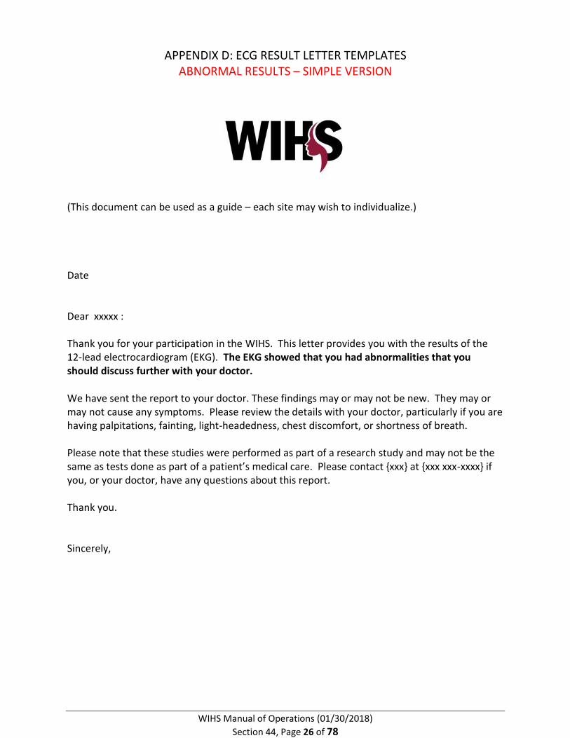
Use Final Interpretation to produce normal, abnormal or “non-specific findings” results letter (Appendix D)
o Suggest establishing deadlines, as above
o Site may select to include more specific findings in participant results letter by using terms written on ECG tracing along with the description of these terms (see Appendix E: Glossary of ECG results terms)
5. PROCEDURE FOR ALERT RESULTS
If an ALERT (as defined in Appendix G) is seen on printout:
o Stay calm and do not communicate distress to the participant. The ECG is highly sensitive and only in very rare cases will the participant require immediate medical attention.
o Immediately transmit all three ECG tracings to the reading physician while the participant is onsite. Options include:
Use phone to take pictures of tracings and printed diagnostic statement on top of the ECG printout; text or email photos
Fax hard copies
Hand carry tracings to physician
o Contact the reading physician (by text, email, page or phone call) and receive response
If no response, call/page backup reading physician
If no response, call/page site PI
o Document event and actions as per site protocol (i.e., save in Excel or Access) in an alert log (see an example in Appendix F). Save to your site

WIHS Manual of Operations (01/30/2018)
Section 44, Page 12 of 78
directory on the first Friday of each month with the date in the excel file, regardless if there were any alerts or not during the past month.
o Complete ECGCA Critical Alert Documentation Form (Appendix B)
Transmit electronic data to EPICARE Reading Center
Reading Physician Procedure (NOTE: Centers may tailor procedures)
Receive text, page or phone call from site
Review ECG tracing(s) with ALERT diagnostic statement
Communicate decision on how to proceed to the site
Possible decisions:
o No immediate follow-up needed
o Site to advise participant to follow-up with personal physician in XX days
o Site to advise participant to immediately go to hospital (ONLY if participant is in distress or recommended by reviewing physician)
o Site to call 911 (ONLY if recommended by reviewing physician or participant otherwise in distress and in need of medical attention; do NOT call if participant is asymptomatic)
Document the physician’s recommended follow-up as per institutional protocol
Handwrite any edits to the automated machine reading directly on the tracing (“Final Interpretation”)
The site may ask the reading physician to include coding to communicate the type of result letter to be produced
o (1) = normal interpretation produce normal result letter (see NOTE on page 6)
o (2) = non-specific findings produce non-specific result letter
o (3) = abnormality that needs further evaluation produce abnormal result letter
Final ECG interpretation confirmed by physician signature directly on the tracing
o Suggest establishing deadlines for turnaround times by the clinicians
Return the printout with finalized/signed ECG interpretation to the site
Site Procedure
Receive instructions from reading physician in response to ALERT Final interpretation
o If no immediate follow-up needed, reassure participant and proceed to produce letter (see NOTE on page 6)
o If reading physician advises follow-up with personal physician, inform participant and produce appropriate letter to physician
WIHS Manual of Operations (01/30/2018)
Section 44, Page 13 of 78
o If reading physician advises participant to immediately go to hospital, inform participant and arrange for transport to hospital; produce appropriate letter to physician [NOTE: This will be extremely unlikely]
o If reading physician advises site to call 911, then call 911 and inform participant; produce appropriate letter to physician [NOTE: This will be extremely unlikely]
Review Final Interpretation on tracing with reading physician signature
Produce normal, abnormal or “non-specific findings” results letter according to Final Interpretation on tracing (Appendix D)
o Suggest establishing deadlines
o Site may select to include more specific findings in participant results letter by using terms written on ECG tracing along with the description of these terms (see Appendix E: Glossary of ECG results terms)
Adverse Event
Definition: An “Adverse Event” is a significant medical event which is felt to be at least possibly related to the research study.
Fill out ECGAER Adverse Event Reporting Form (Appendix C) if the participant reports an adverse event while having an ECG performed.
Note: Mild, transient skin irritation from the lead placement without development of a frank rash or hives is not considered an adverse event.
A PDF of the form is available on the WIHS Admin website.
If the adverse event is not readily treatable with oral Benadryl or OTC hydrocortisone 1% cream, the participant should be seen by the site clinician, with appropriate referral made for care if needed.
Notify WDMAC of all Adverse Events.
6. DISPOSITION FORM – ECG SECTION
“ECG Disposition Form” (ECGNOTI, Appendix A)
Fill in this form following ECG testing of a participant
Indicate whether the ECG was successfully performed; and if not, what was the reason
indicate the participant’s preference for having ECG result letters sent to herself and/or her doctor
Collect the following additional data:
o Fasting status
o Electrode Location
o If alert conditions were noted (Y/N)

WIHS Manual of Operations (01/30/2018)
Section 44, Page 14 of 78
o If the participant appears to be intoxicated (note: this will be defined as readily noticeable intoxication of the participant by the clinic staff)
Enter into Apollo

WIHS Manual of Operations (01/30/2018)
Section 44, Page 15 of 78
APPENDIX A: WOMEN'S INTERAGENCY HIV STUDY FORM ECGNOTI: ECG FINAL DISPOSITION FORM
INSTRUCTIONS: THE PURPOSE OF THIS FORM IS TO TRACK IN THE DATA MANAGEMENT SYSTEM (APOLLO) EACH TIME A PARTICIPANT IS SCREENED AND/OR ENROLLED IN THE ECG PROTOCOL.
A1. PARTICIPANT ID |___|-|___|___|-|___|___|___|___|-|___| A2. FORM VERSION: 1 0 / 0 2 / 1 7 A3. FORM COMPLETED BY: |___|___|___| A4. SCREENING DATE: |___|___| / |___|___| / |___|___| M D Y
A5. WAS ECG SUCCESSFULLY COMPLETED?
YES ..............................1 (A6) NO ..............................2
a. PRIMARY REASON ECG NOT COMPLETED?
PARTICIPANT NOT INTERESTED/DID NOT CONSENT ...............................1 PARTICIPANT HAS NO TIME .....................................................................2 UNCOMFORTABLE/PAINFUL ....................................................................3 SAFETY CONCERN .....................................................................................4 HARDWARE MALFUNCTION ....................................................................5 LACK OF SUPPLIES ....................................................................................6 INSUFFICIENT TIME OR ROOM NOT AVAILABLE ......................................7 OTHER REASON ........................................................................................8
SPECIFY: _________________________________
b. WERE ANY ECG DATA COLLECTED (IF PARTIALLY COMPLETED)?
YES ..............................1 NO ..............................2 (END)
A6. DATE ECG COMPLETED: |___|___| / |___|___| / |___|___| M D Y A7. WIHS CORE VISIT NUMBER AT WHICH ECG IS COMPLETED: |___|___| A8. RESULTS LETTER WILL BE SENT TO: YES NO
a. DOCTOR............................................................................1 2
b. PARTICIPANT ....................................................................1 2

WIHS Manual of Operations (01/30/2018)
Section 44, Page 16 of 78
WIHSID
A9. HAS IT BEEN 8 OR MORE HOURS SINCE THE PARTICIPANT LAST ATE AND/OR DRANK
ANYTHING OTHER THAN WATER, INCLUDING CANDY AND CHEWING GUM?
YES ..............................1 NO ..............................2 A10. ELECTRODE LOCATION MEASUREMENTS (APPROXIMATED TO THE NEAREST 0.5”):
a. NV LINE: |___|___|.|___|
b. MID-CHEST: |___|___|.|___| A11. WERE ANY ALERT CONDITIONS NOTED?
YES ..............................1 NO ..............................2 A12. DOES THE PARTICIPANT APPEAR TO BE INTOXICATED?
YES ..............................1 NO ..............................2 A13. TECHNICIAN NAME: _____________________________________
A14. DATE ECG DATA UPLOADED TO EPICARE: |___|___| / |___|___| / |___|___| M D Y A15. TIME ECG DATA UPLOADED TO EPICARE: |___|___|:|___|___| AM ..................1 PM ..................2

WIHS Manual of Operations (01/30/2018)
Section 44, Page 17 of 78
APPENDIX B: WOMEN'S INTERAGENCY HIV STUDY FORM ECGCA: ECG CRITICAL ALERT DOCUMENTATION FORM
INSTRUCTIONS: THE PURPOSE OF THIS FORM IS TO TRACK THE OCCURRENCE OF CRITICAL ALERTS FOR PARTICIPANTS ENROLLED IN THE ECG PROTOCOL.
A1. PARTICIPANT ID |___|-|___|___|-|___|___|___|___|-|___| A2. FORM VERSION: 0 1 / 3 0 / 1 8 A3. FORM COMPLETED BY: |___|___|___| A4. ECG DATE: |___|___| / |___|___| / |___|___| M D Y
A5. NATURE OF CRITICAL ALERT: YES NO HEART RATE < 40 BPM…………………………………………………………………………….. 1 2 HEART RATE > 120 BPM…………………………………………………………………………… 1 2 VENTRICULAR TACHYCARDIA……………………………………………………………………. 1 2 ATRIAL FIBRILLATION OR FLUTTER……………………………………………………………. 1 2 COMPLETE ATRIOVENTRICULAR BLOCK/THIRD DEGREE HEART BLOCK/COMPLETE HEART BLOCK…………………………………………. 1 2 WOLFF-PARKINSON-WHITE SYNDROME…………………………………………………… 1 2 ACUTE MYOCARDIAL ISCHEMIA……………………………………………………………….. 1 2 ACUTE MYOCARDIAL INFARCTION/INJURY………………………………………………. 1 2 PROLONGED CORRECTED QT INTERVAL (QTc > 500 MS)………………………….. 1 2 OTHER ALERT 1………………………………………………………………………………………… 1 2
SPECIFY OTHER ALERT 1: _________________________________
OTHER ALERT 2………………………………………………………………………………………… 1 2
SPECIFY OTHER ALERT 2: _________________________________
OTHER ALERT 3………………………………………………………………………………………… 1 2
SPECIFY OTHER ALERT 3: _________________________________
OTHER ALERT 4………………………………………………………………………………………… 1 2
SPECIFY OTHER ALERT 4: _________________________________
OTHER ALERT 5………………………………………………………………………………………… 1 2
SPECIFY OTHER ALERT 5: _________________________________
A6. PHYSICIAN ALERTED: ____________________________________________ A7. PHYSICIAN DECISION: ____________________________________________ A8. PARTICIPANT ACTIONS: ___________________________________________

WIHS Manual of Operations (01/30/2018)
Section 44, Page 18 of 78
WIHSID
A9. OTHER COMMENTS: ______________________________________________ _______________________________________________________________ _______________________________________________________________
PROMPT: THIS FORM WILL NOT BE DATA ENTERED.

WIHS Manual of Operations (01/30/2018)
Section 44, Page 19 of 78
APPENDIX C: WOMEN'S INTERAGENCY HIV STUDY FORM ECGAER: ADVERSE EVENT REPORTING FORM
INSTRUCTIONS: THIS FORM SHOULD BE FILLED OUT BY THE PERSON WHO IS NOTIFIED OF OR OBSERVES AN ADVERSE EVENT THAT IS FELT TO BE AT LEAST POSSIBLY RELATED TO THE RESEARCH STUDY. IF THE ADVERSE EVENT IS NOT READILY TREATABLE WITH ORAL BENADRYL OR OVER-THE-COUNTER TOPICAL 1% HYDROCORTISONE CREAM, THE PARTICIPANT SHOULD BE SEEN BY THE SITE CLINICIAN, WITH APPROPRIATE REFERRAL MADE FOR CARE IF NEEDED. WDMAC SHOULD BE NOTIFIED OF ALL ADVERSE EVENTS.
A1. PARTICIPANT ID: |___|-|___|___|-|___|___|___|___|-|___| A2. FORM VERSION: 1 0 / 0 1 / 1 7a A3. FORM COMPLETED BY: |___|___|___| A4. DATE FORM COMPLETED: |___|___| / |___|___| / |___|___| M D Y
A5. DATE OF ADVERSE EVENT: |___|___| / |___|___| / |___|___| M D Y
A6. DATE OF DISCOVERY OF |___|___| / |___|___| / |___|___| ADVERSE EVENT M D Y
A7. DETAILED DESCRIPTION OF ADVERSE EVENT (E.G., SKIN REACTION):
_____________________________________________________________________
_____________________________________________________________________
_____________________________________________________________________
_____________________________________________________________________
_____________________________________________________________________
_____________________________________________________________________
_____________________________________________________________________
_____________________________________________________________________
_____________________________________________________________________
_____________________________________________________________________
A8. WHAT IS THE CURRENT STATUS OF THE PARTICIPANT?
_____________________________________________________________________
_____________________________________________________________________
_____________________________________________________________________

WIHS Manual of Operations (01/30/2018)
Section 44, Page 20 of 78
WIHSID
A9. DID THE EVENT RESOLVE?
YES ..............................1 NO ..............................2 (A10) A. IF YES, DATE OF RESOLUTION: |___|___| / |___|___| / |___|___| M D Y
B. HOW DID IT RESOLVE (E.G., TREATED, RESOLVED ON ITS OWN, ETC.)?:
_____________________________________________________________________
_____________________________________________________________________
_____________________________________________________________________
_____________________________________________________________________
A10. WAS THIS EVENT ANTICIPATED (LISTED IN THE CONSENT FORM RISK SECTION)?
YES ..............................1 NO ..............................2 (A11)
A. IF ANTICIPATED, WAS THE EVENT ANY OF THE FOLLOWING?
RASH ...........................1 HIVES ..........................2
A11. IS IT MORE LIKELY THAN NOT THAT THIS PROBLEM/EVENT WAS RELATED TO RESEARCH
PROCEDURES OR INTERVENTIONS?
YES ..............................1 NO ..............................2 (A12)
A. IF YES, CHOOSE THE STRENGTH OF ATTRIBUTION TO THE STUDY.
NOT RELATED .............1 PROBABLY NOT
RELATED .....................2 POSSIBLY RELATED .....3
PROBABLY RELATED ...4 DEFINITELY RELATED ..5

WIHS Manual of Operations (01/30/2018)
Section 44, Page 21 of 78
WIHSID
A12. ALL UNANTICIPATED ADVERSE EVENTS SHOULD BE REPORTED TO YOUR LOCAL IRB. WAS
THIS EVENT REPORTED TO YOUR LOCAL IRB?
YES ..............................1 NO ..............................2
PROMPT: IF THE UNANTICIPATED ADVERSE EVENT HAS NOT YET BEEN REPORTED TO YOUR LOCAL IRB, NOTIFY WDMAC WHEN IT HAS BEEN REPORTED.
PROMPT: THIS FORM WILL NOT BE DATA ENTERED.

WIHS Manual of Operations (01/30/2018)
Section 44, Page 22 of 78
APPENDIX D: ECG RESULT LETTER TEMPLATES NORMAL RESULTS – DETAILED VERSION
(This document can be used as a guide – each site may wish to individualize.) Date Dear xxxxx : Thank you for your participation in the Women’s Interagency HIV Study (WIHS). This letter provides you with the results of your 12-lead resting electrocardiogram (EKG). We did not detect any concerning abnormalities on your EKG. {We have sent the detailed report to your doctor. You can review the details with your doctor [ONLY IF REQUESTED BY PARTICIPANT]} It is recommended that everyone strive to lead a heart-healthy lifestyle and reduce any risk factors for heart disease, such as high cholesterol, high blood pressure, diabetes, cigarette smoking, or others. Please note that these studies were performed as part of a research study and may not be the same as tests done as part of a patient’s medical care. Please contact the { SHARE } office at {xxx xxx-xxxx} if you, or your doctor, have any questions about this report. Thank you. Sincerely,

WIHS Manual of Operations (01/30/2018)
Section 44, Page 23 of 78
APPENDIX D: ECG RESULT LETTER TEMPLATES
NORMAL RESULTS – SIMPLE VERSION
(This document can be used as a guide – each site may wish to individualize.) Date Dear xxxxx : Thank you for your participation in the WIHS. This letter provides you with the results of your 12-lead electrocardiogram (EKG). Your EKG was normal. {We have sent the detailed report to your doctor. You can review the details with your doctor [ONLY IF REQUESTED BY PARTICIPANT]} Please note that these studies were performed as part of a research study and may not be the same as tests done as part of a patient’s medical care. Please contact the { SHARE } office at {xxx xxx-xxxx} if you, or your doctor, have any questions about this report. Thank you. Sincerely,

WIHS Manual of Operations (01/30/2018)
Section 44, Page 24 of 78
APPENDIX D: ECG RESULT LETTER TEMPLATES
NON-SPECIFIC RESULTS – SIMPLE VERSION
(This document can be used as a guide – each site may wish to individualize.)
Date Dear xxxxx : Thank you for your participation in the WIHS study. This letter provides you with the results of your 12-lead electrocardiogram (EKG). Your EKG showed results that need further interpretation. We have sent the report to your doctor. These findings may or may not be new. They may or may not cause any symptoms. Please review the details with your doctor, particularly if you are having palpitations, fainting, light-headedness, chest discomfort, or shortness of breath. Please note that these studies were performed as part of a research study and may not be the same as tests done as part of a patient’s medical care. Please contact {xxx} at {xxx xxx-xxxx} if you, or your doctor, have any questions about this report. Thank you. Sincerely,

WIHS Manual of Operations (01/30/2018)
Section 44, Page 25 of 78
APPENDIX D: ECG RESULT LETTER TEMPLATES
ABNORMAL RESULTS – DETAILED VERSION
(This document can be used as a guide – each site may wish to individualize.) Date Dear xxxxx : Thank you for your participation in the Women’s Interagency HIV Study (WIHS). This letter provides you with the results of your 12-lead resting electrocardiogram (EKG). The EKG showed that you had: {Insert the appropriate option from below: 1) Extra heart beats from the upper chambers of your heart for 3 or more consecutive beats 2) Extra heart beats from the lower chambers of your heart for 3 or more consecutive beats 3) Abnormal heart conduction (right bundle branch block, left bundle branch block) 4) Heart beats from a pacemaker 5) Slow heart conduction 6) Non-specific abnormalities (use this for nonspecific ST and T wave changes, first degree
AVB, LVH, left axis deviation). }
{We have sent the report to your doctor. These findings may or may not be new. They may or may not cause any symptoms. Please review the details with your doctor, particularly if you are having palpitations, fainting, light-headedness, chest discomfort, or shortness of breath. [ONLY IF REQUESTED BY PARTICIPANT]} It is recommended that everyone strive to lead a heart-healthy lifestyle and reduce any risk factors for heart disease, such as high cholesterol, high blood pressure, diabetes, cigarette smoking, or others. Please note that these studies were performed as part of a research study and may not be the same as tests done as part of a patient’s medical care. Please contact {LOCAL CARDIOLOGIST READER} at {xxx xxx-xxxx} if you, or your doctor, have any questions about this report. Thank you. Sincerely,
WIHS Manual of Operations (01/30/2018)
Section 44, Page 26 of 78
APPENDIX D: ECG RESULT LETTER TEMPLATES ABNORMAL RESULTS – SIMPLE VERSION
(This document can be used as a guide – each site may wish to individualize.) Date Dear xxxxx : Thank you for your participation in the WIHS. This letter provides you with the results of the 12-lead electrocardiogram (EKG). The EKG showed that you had abnormalities that you should discuss further with your doctor. We have sent the report to your doctor. These findings may or may not be new. They may or may not cause any symptoms. Please review the details with your doctor, particularly if you are having palpitations, fainting, light-headedness, chest discomfort, or shortness of breath. Please note that these studies were performed as part of a research study and may not be the same as tests done as part of a patient’s medical care. Please contact {xxx} at {xxx xxx-xxxx} if you, or your doctor, have any questions about this report. Thank you. Sincerely,

WIHS Manual of Operations (01/30/2018)
Section 44, Page 27 of 78
APPENDIX D: ECG RESULT LETTER TEMPLATES
ALERT RESULTS
(This document can be used as a guide – each site may wish to individualize.)
Date Dear xxxxx : Thank you for your participation in the Women’s Interagency HIV Study (WIHS). This letter provides you with the results of your 12-lead resting electrocardiogram. As we discussed on the phone, your EKG showed that you had: {Insert the appropriate option from below:
1) A slow heart rate below 40 beats per minute lasting for longer than 30 seconds 2) A pause in your heart beat for longer than 5 seconds 3) Abnormal heart block 4) Atrial fibrillation or flutter (irregular heart rhythm) 5) A fast heart rhythm 6) Evidence of heart muscle damage 7) Evidence of heart inflammation 8) Abnormal heart conduction 9) Pacemaker }
{We have sent a copy of the report to your doctor. Please talk to your doctor about this finding as soon as possible, especially if you are having episodes of lightheadedness, fainting, palpitations, chest pain or shortness of breath [ONLY IF REQUESTED BY PARTICIPANT]}. It is recommended that everyone strive to lead a heart-healthy lifestyle and reduce any risk factors for heart disease, such as high cholesterol, high blood pressure, diabetes, cigarette smoking, or others. Please note that these studies were performed as part of a research study and may not be the same as tests done as part of a patient’s medical care. Please contact the { SHARE } office at {xxx xxx-xxxx} if you, or your doctor, have any questions about this report. Thank you.

WIHS Manual of Operations (01/30/2018)
Section 44, Page 28 of 78
APPENDIX D: ECG RESULT LETTER TEMPLATES PHYSICIAN LETTER –APPLICABLE TO ANY RESULT
(This document can be used as a guide – each site may wish to individualize.) Date Dear Physician Name: Your patient, xxxx, is a participant in the Women’s Interagency HIV Study (WIHS), a study of HIV-infected and uninfected women in 10 U.S. cities. She has requested that we send you the results of her 12-lead electrocardiogram (EKG), which was performed as part of the research study. Please see the attached letter that was sent to your patient. We are also sending you the EKG tracing and interpretation. Please contact us at {xxx} if you have any questions about this report. Thank you.

WIHS Manual of Operations (01/30/2018)
Section 44, Page 29 of 78
APPENDIX E: GLOSSARY OF ECG RESULTS TERMS
Abnormality or alert Description
Atrial fibrillation This is an irregular heart rhythm of the upper chambers of the heart.
Atrial flutter This is an irregular heart rhythm of the upper chambers of the heart.
Supraventricular ectopy (SVE) / Premature atrial contractions (PACs) / Supraventricular couplets / Supraventricular triplets
These are heartbeats that come early and originate from the upper chambers of the heart.
Supraventricular tachycardia XX beats OR xx secs
This is a fast rhythm that originates from the upper chambers of the heart and lasts for xx beats (or xx secs).
Ventricular ectopy (VE) / Premature ventricular contractions (PVCs) / Ventricular couplets / Ventricular triplets
These are heartbeats that come early and originate from the lower chambers of the heart.
Nonsustained ventricular tachycardia XX beats
This is a fast rhythm that originates from the lower chambers of the heart and lasts for xx beats.
Paced beats These are heart beats that originate from a pacemaker device rather than your heart’s own pacemaker.
Wolff Parkinson White Abnormal heart conduction
Left bundle branch block Abnormal heart conduction
Right bundle branch block Abnormal heart conduction
Wide QRS tachycardia ≥120 bpm (includes monomorphic ventricular tachycardia, polymorphic ventricular tachycardia, ventricular fibrillation) Duration xx beats
This is a fast rhythm that originates from the lower chambers of the heart and lasts for xx beats.
Complete heart block This is a slow heart beat due to an interruption in the electrical pathway in the heart.
2nd degree AV block, Mobitz I (AV Wenkebach)
This is an occasional slowing of heart rate due to a drop of a beat in the lower chambers.
2nd degree AV Block, Mobitz II This is a slow heart beat due to an interruption in the electrical pathway in the heart.
Pause >5 seconds There was a pause in the heart beat for 5 seconds or longer.
Bradycardia <40 bpm This is a slower than usual heart rate that lasted more than 30 seconds.
Acute pericarditis This is inflammation of the lining of the heart.
Injury, infarct or ischemia (acute or marked)
This is heart muscle damage.

WIHS Manual of Operations (01/30/2018)
Section 44, Page 30 of 78
APPENDIX F: ECG ALERT LOG EXAMPLE
ECG Alert Log - Bronx
WIHSID Date Alert Occurred
Alert type(s) on ECG
printout
Physician alerted
Date of Physician Contact
Physician Decision Participant Actions Comments
51111 10/15/2016 A-Fib (new-onset)
Dr. Jorge Kizer
10/15/2016 Advise participant to go to hospital for work-up
Participant agreed to go to hospital and was taken there by friend.
Participant was hospitalized; plan to request medical records
WIHS Manual of Operations (01/30/2018)
Section 44, Page 31 of 78
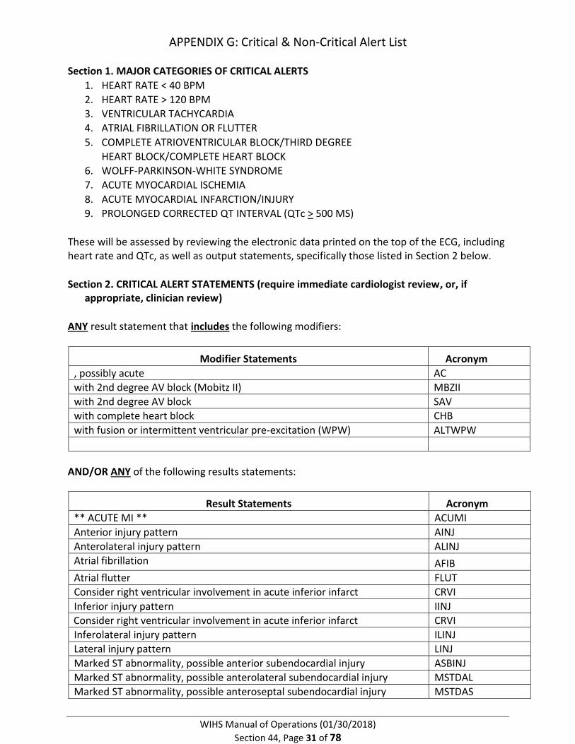
APPENDIX G: Critical & Non-Critical Alert List
Section 1. MAJOR CATEGORIES OF CRITICAL ALERTS
1. HEART RATE < 40 BPM
2. HEART RATE > 120 BPM
3. VENTRICULAR TACHYCARDIA
4. ATRIAL FIBRILLATION OR FLUTTER
5. COMPLETE ATRIOVENTRICULAR BLOCK/THIRD DEGREE
HEART BLOCK/COMPLETE HEART BLOCK
6. WOLFF-PARKINSON-WHITE SYNDROME
7. ACUTE MYOCARDIAL ISCHEMIA
8. ACUTE MYOCARDIAL INFARCTION/INJURY
9. PROLONGED CORRECTED QT INTERVAL (QTc > 500 MS)
These will be assessed by reviewing the electronic data printed on the top of the ECG, including heart rate and QTc, as well as output statements, specifically those listed in Section 2 below.
Section 2. CRITICAL ALERT STATEMENTS (require immediate cardiologist review, or, if appropriate, clinician review)
ANY result statement that includes the following modifiers:
Modifier Statements Acronym
, possibly acute AC
with 2nd degree AV block (Mobitz II) MBZII
with 2nd degree AV block SAV
with complete heart block CHB
with fusion or intermittent ventricular pre-excitation (WPW) ALTWPW
AND/OR ANY of the following results statements:
Result Statements Acronym
** ACUTE MI ** ACUMI
Anterior injury pattern AINJ
Anterolateral injury pattern ALINJ
Atrial fibrillation AFIB
Atrial flutter FLUT
Consider right ventricular involvement in acute inferior infarct CRVI
Inferior injury pattern IINJ
Consider right ventricular involvement in acute inferior infarct CRVI
Inferolateral injury pattern ILINJ
Lateral injury pattern LINJ
Marked ST abnormality, possible anterior subendocardial injury ASBINJ
Marked ST abnormality, possible anterolateral subendocardial injury MSTDAL
Marked ST abnormality, possible anteroseptal subendocardial injury MSTDAS

WIHS Manual of Operations (01/30/2018)
Section 44, Page 32 of 78
Marked ST abnormality, possible inferior subendocardial injury ISBINJ
Marked ST abnormality, possible inferolateral subendocardial injury MSTDIL
Marked ST abnormality, possible lateral subendocardial injury LSBINJ
Marked ST abnormality, possible septal subendocardial injury SSBINJ
Marked T wave abnormality, consider anterior ischemia MAT
Marked T wave abnormality, consider anterolateral ischemia MALT
Marked T wave abnormality, consider inferior ischemia MIT
Marked T wave abnormality, consider inferolateral ischemia MILT
Marked T wave abnormality, consider lateral ischemia MLT
Marked ST abnormality, possible anterior subendocardial injury ASBINJ
Marked ST abnormality, possible anterolateral subendocardial injury MSTDAL
Prolonged QT LNGQT
Septal injury pattern SINJ
ST depression, consider subendocardial injury or digitalis effect STDEP
ST elevation consider anterior injury or acute infarct AIOHAI
ST elevation consider anterolateral injury or acute infarct ALIHAI
ST elevation consider inferior injury or acute infarct IIOHAI
ST elevation consider inferolateral injury or acute infarct ILIHAI
ST elevation consider lateral injury or acute infarct LIOHAI
ST elevation, consider early repolarization, pericarditis, or injury SERYR1
ST elevation, consider injury or variant associated with LVH INJONV
T wave abnormality, consider anterior ischemia AT
T wave abnormality, consider anterolateral ischemia ALT
T wave abnormality, consider inferior ischemia IT
T wave abnormality, consider inferolateral ischemia ILT
T wave abnormality, consider lateral ischemia LT
Ventricular pre-excitation, WPW pattern type A WPWA
Ventricular fibrillation VFIB
Ventricular tachycardia VTACH
Ventricular pre-excitation, WPW pattern type B WPWB
Wolffe-Parkinson-White WPW
Section 3. NON-CRITICAL OUTPUT STATEMENTS (do NOT require immediate cardiologist review unless they are displayed in conjunction with any of the modifiers outlined in Section 2)
Result Statements Acronym
Aberant conduction ABCOND
Abnormal ECG AB
Abnormal left axis deviation ALAD
Abnormal QRS-T angle, consider primary T wave abnormality QRST
Abnormal right axis deviation ARAD
Abnormal right superior axis deviation RSAD
Accelerated ACCEL
Acute pericarditis PCARD
, Age undetermined AU

WIHS Manual of Operations (01/30/2018)
Section 44, Page 33 of 78
, and consecutive CSEC
and AND
Anterior infarct AMI
Anterior leads ANT
Anterolateral infarct ALMI
Anterolateral leads ANTLAT
Anteroseptal infarct ASMI
Anteroseptal leads ANTSEP
Anteroseptal injury pattern ASINJ
(Atrial rate=) ARAT
Atrial tachycardia ATAC
AV sequential or dual chamber electronic pacemaker AVPCK
Biatrial enlargement BAE
*** Bifascicular block*** BIFB
Biventricular hypertrophy BIVH
Blocked BLKED
Borderline ECG BORDE
Borderline BO
Cannot rule out CRO
Clockwise rotation of the heart, may invalidate criteria for
ventricular hypertrophy
CWRT
Coarse CRS
Counterclockwise rotation of the heart, may invalidate criteria for
v. hypertrophy
CCWRT
Deep Q wave in lead V6, QV6
Demand pacemaker; interpretation is based on intrinsic rhythm DPCK
Dextrocardia DXTRO
Early repolarization REPOL
Electronic atrial pacemaker APCK
Electronic ventricular pacemaker PCK
Fusion complexes FUS
In a pattern of bigeminy BIGEM
Incomplete left bundle branch block ILBBB
Incomplete right bundle branch block IRBBB
Increased R/S ratio in V1, consider early transition or posterior
infarct
QESPMI
Idioventricular rhythm IVR
Indeterminate axis INDAX
Inferior infarct IMI
Inferior leads INF
Inferior-posterior infarct IPMI
Inferolateral leads IFLAT
Inferoposterior leads INFPOS
Irregular IRR
Junctional bradycardia JUNBRAD

WIHS Manual of Operations (01/30/2018)
Section 44, Page 34 of 78
Junctional rhythm JUNCTR
Junctional ST depression, probably abnormal JST
Junctional ST depression, probably normal JSTN
Large LARG
Lateral infarct LMI
Lateral leads LAT
Left anterior fascicular block AFB
Left atrial bradycardia LABRAD
Left atrial enlargement LAE
Left atrial rhythm LAR
Left atrial tachycardia LATACH
Left axis deviation LAD3
Left bundle branch block LBBB
Left posterior fascicular block PFB
Left ventricular hypertrophy LVH2
Leftward axis LAD
** Less than 4 QRS complexes detected, no interpretation
possible **
ANLERR3
Low right atrial bradycardia RABRAD
Low right atrial rhythm RAR
Low right atrial tachycardia RATACH
Low voltage QRS LOWV
Marked sinus bradycardia MSBRAD
(masked by fascicular block?) MAFB
, maybe secondary to QRS abnormality SNDQA
** Memory allocation failure, no ECG interpretation possible ** ANLERR1
Minimal voltage criteria for LVH, may be normal variant QRSV
Moderate voltage criteria for LVH, may be normal variant LVH3
Moderate MOD
Narrow QRS tachycardia NQTACH
(No P- waves found) NOPF
** No QRS complexes found, no ECG analysis possible ** ANLERR2
Nonspecific intraventricular block IVCB
Nonspecific intraventricular conduction delay IVCD
Nonspecific ST abnormality NST
Nonspecific ST and T wave abnormality NSTT
Nonspecific T wave abnormality NT
Normal ECG NML
Normal sinus rhythm NSR
Northwest axis NWA
or OR
or digitalis effect ODIG
Otherwise normal ECG ABR
*** Pediatric ECG analysis *** PEDANL
, plus right ventricular enlargement RVE+
*** Poor data quality, interpretation may be adversely affected QCERR

WIHS Manual of Operations (01/30/2018)
Section 44, Page 35 of 78
Possible PO
Posterior infarct POSTMI
Posterior leads POS
premature atrial complexes PAC
premature ectopic complexes PEC
premature junctional complexes PJC
premature supraventricular complexes PSVC
premature ventricular and fusion complexes PVCF
premature ventricular complexes PVC
, probably digitalis effect PDIG
Prominent lateral voltage PLV
Prominent mid-precordial voltage, PMDPV
Prominent posterior voltage PPV
Pulmonary disease pattern PULD
*** QRS contour suggests infarct size is probably MISIZ
Right atrial enlargement RAE
Right axis deviation RAD4
Right bundle branch block -or-right ventricular hypertrophy RBBRVH
Right bundle branch block RBBB
Right superior axis deviation RAD5
Right ventricular hypertrophy RVH
Rightward axis RAD
RSR’ or QR pattern in V1 suggests right ventricular
conduction delay
RSR
S1-S2-S3 pattern, consider pulmonary disease, RVH, or normal
variant
S1S2S3
Septal infarct SMI
Septal leads SEP
Sinus/Atrial capture CAPUR
Sinus bradycardia SBRAD
Sinus rhythm SRTH
Sinus tachycardia STACH
Small SMA
ST & ST&
ST abnormality and STABAND
ST abnormality, possible digitalis effect STDIG
ST depression in STDPIN
ST elevation in STELIN
ST elevation, probably due to early repolarization SERYR2
Statement not found SNF
Supraventricular tachycardia SVT
*** Suspect arm lead reversal, interpretation assumes no
reversal
ARM
Undetermined rhythm UR
Unusual P axis and short PR, probable junctional bradycardia JBRAD

WIHS Manual of Operations (01/30/2018)
Section 44, Page 36 of 78
Unusual P axis and short PR, probable junctional rhythm JR
Unusual P axis and short PR, probable junctional tachycardia JTACH
Unusual P axis, possible ectopic atrial bradycardia EABRAD
Unusual P axis, possible ectopic atrial rhythm EAR
Unusual P axis, possible ectopic atrial tachycardia EATACH
T wave inversion in TINVIN
very large VLAR
very small VSMA
Voltage criteria for left ventricular hypertrophy LVH
with WITH
wide QRS rhythm WQR
wide QRS tachycardia WQTACH
with 1st degree AV block FAV
with 2:1 AV conduction W2T1
with 2nd degree AV block (Mobitz I) MBZI
with 2nd degree SA block (Mobitz I) SABI
with 2nd degree SA block (Mobitz II) SABII
with 3:1 AV conduction W3T1
with 4:1 AV conduction W4T1
with 5:1 AV conduction W5T1
with a competing junctional pacemaker CJP
with AV dissociation AVDIS
with frequent FREQ
with junctional escape complexes JESC
with marked sinus arrhythmia MSAR
with occasional OCC
with premature aberantly conducted complexes ABER
, with posterior extension PXT
with QRS widening and repolarization abnormality QRSW-2ST
with QRS widening QRSW
with rapid ventricular response RVR
with retrograde conduction RETC
with repolarization abnormality 2ST
with right ventricular involvement RVI
with short PR SPR
with sinus arrhythmia SAR
with sinus pause PAUSE
with slow ventricular response SVR
with strain pattern WSTR
with undetermined rhythm irregularity IRREG
with variable AV block VAVB
with ventricular escape complexes VESC