Embed Size (px)
Citation preview

Section 3
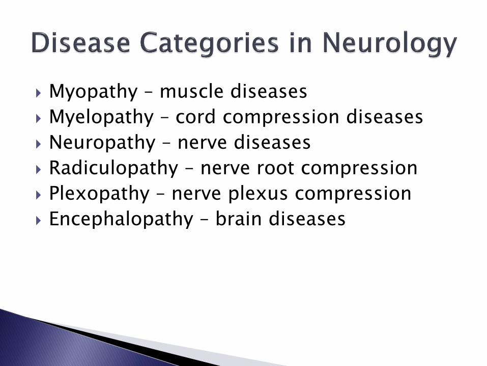
Myopathy – muscle diseases
Myelopathy – cord compression diseases
Neuropathy – nerve diseases
Radiculopathy – nerve root compression
Plexopathy – nerve plexus compression
Encephalopathy – brain diseases

Change in level of alertness & consciousness ◦ Ask “Where is this patient on the scale from being
drowsy to unconscious?”
Common metabolic causes ◦ Low or high glucose ◦ Low sodium ◦ Acidosis or alkalosis ◦ Low oxygen
Conditions that can cause consciousness change ◦ Psychosis ◦ Dementia, delirium ◦ Drug overdose ◦ Aphasia from stroke

Can range from mild weakness to total paralysis ◦ Ask “Is this specific muscle fatigue or weakness or are
all muscles weak?”
“If all muscles are weak, is it muscular, exhaustion or neurological?”
“Is it only one side of the body?”
“Is it upper motor neuron problem or lower motor neuron problem?”

An upper motor neuron lesion is a lesion of the neural pathway above the anterior horn cell or motor nuclei of the cranial nerves
Spasticity, increase in tone in the extensor muscles (lower limbs) or flexor muscles (upper limbs)
Weakness in the flexors (lower limbs) or extensors (upper limbs), but no muscle wasting
Babinski sign is present, where the big toe is raised (extended) rather than curled downwards (flexed) upon appropriate stimulation of the sole of the foot. The presence of the Babinski sign is an abnormal response in adulthood
Increase Deep tendon reflex (DTR)


With an upper motor neuron lesion, such as stroke, the muscles that are normally the weakest are the most affected
This will cause spasticity and contractures
Will also cause weakness of leg muscles on the opposite side of the stroke
Cortical lesions, such as stroke, usually cause a sensory loss and spasticity and weakness

A lower motor neuron lesion is a lesion which affects nerve fibers traveling from the anterior horn of the spinal cord to the relevant muscle(s) -- the lower motor neuron
One major characteristic used to identify a lower motor neuron lesion is flaccid paralysis - paralysis accompanied by muscle loss. This is in contrast to a upper motor neuron lesion, which often presents with spastic paralysis- paralysis accompanied by severe hypertonia
An example of a lower motor neuron lesion is an ulnar nerve neuropathy

SIGN UMN LMN
Weakness Yes Yes
Atrophy No * Yes
Faciculations No Yes
Reflexes Increased Decreased
Tone Increased Decreased
* May have mild atrophy due to disuse

Affects distal nerves in a glove like pattern
Paresthesias, weakness, sensory loss
Common in diabetes, RA, alcoholic abuse and B12 deficiency

These diseases cause proximal muscle weakness
Classic disease is muscular dystrophy ◦ Usually affects young boys with weakness of
pelvic and shoulder muscles
◦ The affected muscles are large and bulky, but very weak because the muscle cells do not contract properly
◦ Muscular dystrohy - mysterious disease
◦ Muscular Dystrophy Walking


Pain syndromes ◦ Neuritis or neuritic pain – pain due to nerve
dysfunction can be very severe pain
◦ Example is causalgia which comes on months after a crushing extremity injury
◦ The resulting pain is so severe that patient’s will often request amputation to relieve the pain
Sensory loss – can cause four things: ◦ Anesthesia – loss of sensation
◦ Hypoesthesia – decreased sensation
◦ Paresthesia –numbness, tingling, prickly
◦ Dysesthesia – uncomfortable burning sensation


Both are common in elderly
Gait disorders can be due to lower extremity problems or neurological problems
Balance problems may be caused by orthopedic dysfunction, low back problems,
cerebellar dysfunction or inner ear problems

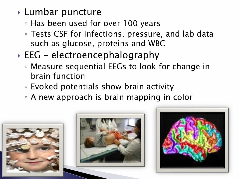
Lumbar puncture ◦ Has been used for over 100 years
◦ Tests CSF for infections, pressure, and lab data such as glucose, proteins and WBC
EEG – electroencephalography ◦ Measure sequential EEGs to look for change in
brain function
◦ Evoked potentials show brain activity
◦ A new approach is brain mapping in color


EMG - elctromyelography ◦ Valuable in diagnosing peripheral and muscular
disorders
ALS, Nerve root compression, thoracic outlet syndrome, neuropathy
Painful tests
Nerve conduction velocity studies ◦ Measure the transmission velocity in peripheral
nerves
◦ CTS, thoracic outlet syndrome, nerve entrapment syndromes

EMG NCVS


Neuroradiology ◦ CT scans
◦ MRI scans
◦ PET scans
◦ CT angiography
Angiography ◦ Injection of contrast dye
◦ The gold standard of brain vascular diagnosis
◦ Ruptured Brain Aneurysm
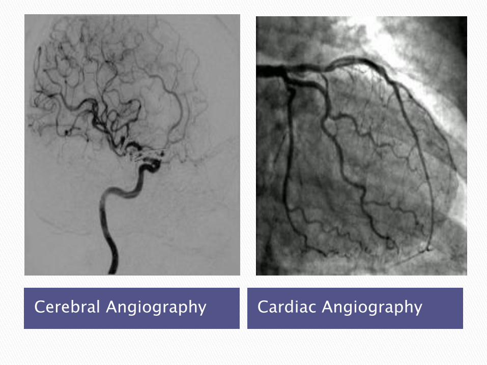
Cerebral Angiography Cardiac Angiography

Due to intrinsic dysfunction in the CNS
Migraine with aura
Migraine without aura
Cluster headaches
Tension headaches
Depression headaches

The result of problems outside the NS Cerebrovascular ischemia Embolism to a cerebral vessel Metabolic disorders Renal and liver failure Infectious processes TMJ problems Mass lesions Eye disorders CSF leak or increased pressure Endocrine dysfunction PMS Autoimmune disorders Fibromyalgia Hypertension

3:1 Female to male ratio
90% have a family history
Pathophysiology ◦ Trigeminal nerve mediated process of inflammation
which releases vasoactive neuropeptides that causes the extreme vasodilation of the cerebral vessels
◦ Headache results from the vasodilation
◦ The throbbing quality is similar to the vascular inflammation experienced anywhere in the body

Classic migraine with aura – 20% ◦ Aura occurs 30-60 minutes before headache ◦ Auras are usually visual followed by nausea,
numbness and tingling ◦ May last 4-6 hours
Migraine without aura – 80% ◦ Throbbing bilateral or unilateral pain without
warning ◦ Chronic sufferers average 15 days per month, or
in some cases every day
Diagnosis are by location, family history, pain characteristics, and age of the first attack

Rest in a dark room NSAID
Cafergot ◦ Oral or rectal
Vasoconstrictive drugs ◦ Midrin ◦ DHE – dihydroergotamine
Triptan drugs ◦ Blocks the serotonin receptors for severe sufferers ◦ Imitrex, Zomig, Relpax, Axert
Preventive drugs ◦ Beta-blockers, calcium channel blockers, serotonin
blockers, antidepressants
Unknown etiology, but thought to be related to melatonin and cerebral biorhythms ◦ Causes clusters of headaches lasting for several
days to weeks occurring several times per year
S & S ◦ Occurs behind one eye or over temple usually with
pupil constriction, unilateral nasal discharge, and conjunctiva redness
◦ One pupil can appear smaller with drooping eyelid ◦ Classically starts at night
Treatment ◦ Most care ineffective ◦ Imitrex and DHE often used
The most common headache ◦ AKA benign headache or muscle contraction headache
S & S ◦ Typically starts mid-afternoon ◦ Associated with tightness of head and neck muscles ◦ Band-like or vise-like pain
Treatment ◦ Efforts to reduce tension and manage stress ◦ Develop coping and relaxation strategies ◦ Migraine drugs are used ◦ Botulism toxin injections are used
Must be repeated every three months


S & S ◦ Depressed patient often awakens with headache
◦ Has other symptoms of depression, such as sleep problems, loss of appetite, feelings of worthlessness
◦ Loss of ability to feel pleasure & enjoyment in life
Anhedonia
◦ Headache symptoms similar to tension headaches
Treatment ◦ Simple analgesics, NSAIDs, depression treatment


3rd most common cause of death in the USA
Over 600,000 strokes per year
160,000 deaths per year ◦ 30% die in acute stage
◦ 30% - 40% severely disabled
Ischemic stroke – 80%
Hemorrhagic stroke – 20%
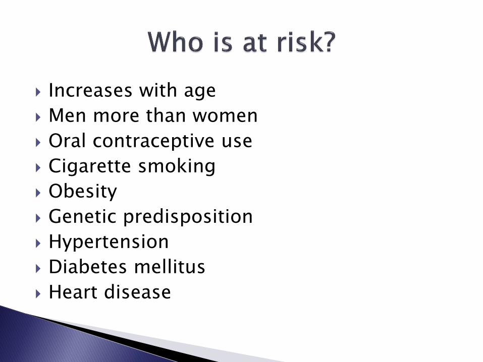
Increases with age
Men more than women
Oral contraceptive use
Cigarette smoking
Obesity
Genetic predisposition
Hypertension
Diabetes mellitus
Heart disease
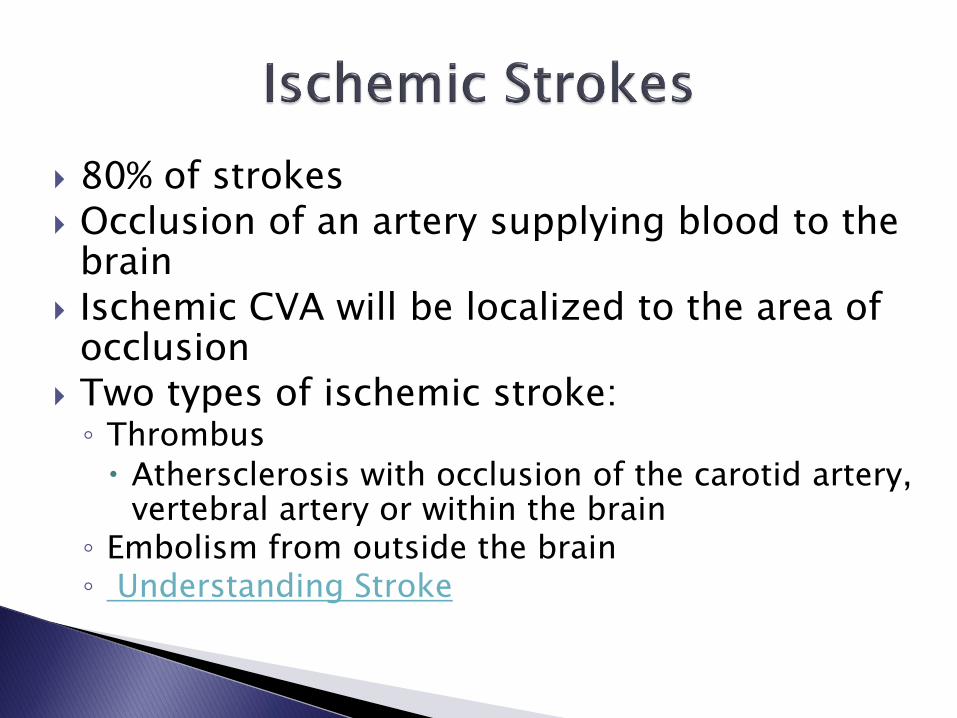
80% of strokes Occlusion of an artery supplying blood to the
brain Ischemic CVA will be localized to the area of
occlusion Two types of ischemic stroke: ◦ Thrombus
Athersclerosis with occlusion of the carotid artery, vertebral artery or within the brain
◦ Embolism from outside the brain ◦ Understanding Stroke

Blood clot from heart Platelets & fibrous debri from carotid artery Clumps of myoglobin can break from over
exerted muscle in extreme sports Fat can break off from a large bone fracture Nitrogen bubbles may build up in
bloodstream from scuba divers who decompress to fast
Amniotic fluid can get into the blood during childbirth
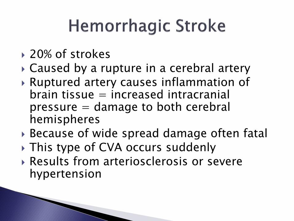
20% of strokes Caused by a rupture in a cerebral artery Ruptured artery causes inflammation of
brain tissue = increased intracranial pressure = damage to both cerebral hemispheres
Because of wide spread damage often fatal This type of CVA occurs suddenly Results from arteriosclerosis or severe
hypertension

Intracerebral bleeding ◦ Seen in elderly with high blood pressure and fragile
vessels, or in patients with bleeding disorders and those on anticoagulants
Subarachnoid bleeding ◦ Seen in 30-40 year olds and are mostly due to
congenital ateriovenous malformations
Subdural bleeding ◦ Often occurs in elderly who fall and strike their head
Epidural bleeding ◦ Usually from a ruptured temporal artery and is usually
caused by major head trauma
The actual precise symptoms depend on where the CVA was and how large it is
Sudden weakness, numbness or paralysis of one side of the body
Loss of consciousness Seizure may sometimes occur Sudden change in mental status, confusion
Slurred speech, dysarthria, aphasia Prognosis is more guarded if: ◦ loss of consciousness ◦ if a large part of the left side of the brain is affected
This is the dominant side for 95% of people

Ask the person to say a complete sentence
Ask the person to raise both hands above their heads
Ask the person to walk across the room ◦ Walk behind them to catch of unsteady
If any of the above are present – CALL 911
Controlling hypertension Manage and control diabetes Lower blood pressure Proper diet and exercise Stop smoking Anticholesterol drugs if lipids levels high 83mg ASA per day Any history of TIA ◦ Mini-stroke lasting 1-3 minutes with involvement of
face and speech ◦ Referral to vascular surgeon for carotid arteriography Mini Strokes (TIAs): Don't Ignore Symptoms, Act FAST

History is most important
CT scans present with 95% accuracy
Lumbar puncture if CT normal
CT with LP is 100% accurate diagnostically
MRI are used only if the diagnosis is still uncertain ◦ Open MRI is preferred
◦ Many patients have died in and older style MRI scanner which is enclosed and takes a long time for the test
Ischemic strokes ◦ Thrombolytic therapy - rtPA – recombinant tissue
plasma activator has revolutionized CVA tx
Must be administered within 3 hours ◦ Cerebral edema often follows post-stroke
Treated with IV steroids ◦ Heparin used after the initial three hours
Hemorrhagic strokes ◦ IV sodium nitroprusside to control blood pressure ◦ IV Vitamin K and fresh plasma if patient on Coumadin ◦ If ruptured aneurysm, then high risk brain stent is
used (50/50 chance of surgical)


Vertigo
Presyncope
Disequilibrium

Dysfunction of the inner ear False sensation of movement ◦ “I feel like the room is spinning.”
Causes ◦ Motion sickness ◦ Viral labyrhinthitis ◦ Benign positional vertigo
Calcified calcium crystals in the semicircular canals
Common in the elderly ◦ Meniere’s disease
Vertigo, tinnitus and hearing loss ◦ Rule out auditory nerve tumor ◦ TIA

Vertigo Treatment ◦ Depends on identifying the etiology
◦ VRT – Vestibular Rehabilitation Therapy
PT and OT specialty that minimizes dizziness, improves balance and prevents falls
Exercises designed to allow the brain to adapt and compensate for the cause of the vertigo
Success dependent on age, cognitive function, motor skills, overall health and physical strength
◦ Ear infections treated with antibiotics and myringotomy
◦ Antivert, benzodiazepines, clonazepam, antihistamines

Vertigo S & S ◦ Subjective vertigo – “I am moving.” ◦ Objective vertigo – “Things around me are moving.”
Vertigo Diagnosis ◦ DD asap to r/o CVA, tumor, hemorrhage, etc ◦ Questions:
What triggers the vertigo?
What other symptoms occur?
How long does the dizziness last?
What improves and worsens the dizziness? ◦ Physical exam and neurological exam ◦ CT & MRI ◦ ENG – electronystagmography – evaluates vestibular
system

Feeling of lightheadedness ◦ “I feel like I am going to faint.”
Causes ◦ PAT – Paroxysmal atrial tachycardia (160-200bpm) ◦ Coronary artery insufficiency ◦ Cardiac valve disease ◦ Blood pressure medications ◦ Vertebrobasilar artery insufficiency ◦ Hyperventilation syndrome
S & S ◦ Lightheadedness or giddiness ◦ Pallor, visual blurring, sweating, dyspnea ◦ Possible bitten tongue (associated with seizure)

History of any predisposing conditions ◦ Family history of sudden cardiac death ◦ Diabetes mellitus or hypoglycemia ◦ Parkinson’s disease ◦ Seizure disorder
Preceding or provocative events ◦ Prolonged standing (vasovagal syncope) ◦ Immediately on standing (orthostatic HTN) ◦ With exertion (CAD, cardiomyopathy, valve stenosis) ◦ After athletic exertion (vasovagal syncope) ◦ After Valsalva manuever ◦ Neck rotation or pressure ◦ Stressful event (vasovagal syncope) ◦ Use of arms (subclavian steal syndrome)

History and symptoms during an event: ◦ Nausea, chills & sweats (vasovagal syncope) ◦ Aura (migraine, seizure disorder) ◦ Slumping (CAD, arrthymia) ◦ Kneeling (orthostatic HTN) ◦ Loss of consciousness
Brief (arrhythmia)
> 5 minutes (neurological, metabolic, infectious) ◦ Chest pain (CAD, PE, aortic dissection) ◦ Incontinence urine or stool (seizure) ◦ Tonic-clonic movements
Movements occur before the fall (seizure disorder)
Movement occur after the fall ( vasovagal syncope)

Serum electrolytes and glucose
Hemoglobin or hematocrit
ECG and chest x-ray
BNP – brain natriuretic peptide
Other tests to consider ◦ Cardiac stress testing
◦ Holter monitor
◦ Echocardiogram
◦ Inpatient telemetry monitoring

Treat cause Treatment may only be education & support Instruct regarding postural hypertension and
dehydration Anticholinergic medication Alpha-adrenergic agents Always consider these three re: presyncope ◦ Frequency – alters the quality of life ◦ Recurrent & unpredictable and exposes patients to
high risk of trauma ◦ Can occur during high risk activity (driving, flying,
athletics, machine operation)
Due to dysfunction of balance processing ◦ “I feel I am going to fall over and hit my head.”
AKA ataxia ◦ 65% of over 60 have on a daily basis
Causes ◦ Inner ear problems
◦ Sensory disorders
◦ Joint and muscle problems
◦ Medications

S & S ◦ Loss of balance or feelings of unsteadiness
Diagnosis ◦ Careful history, physical and neurological exam ◦ This is the most serious form of vertigo and should be
immediately referred to a neurologist
Treatment ◦ Balance therapy ◦ Stress management ◦ Relaxation ◦ Rehabilitation

Degenerative disorder of the basal ganglia ◦ Usually in men over 50 ◦ One million cases in USA
S & S ◦ Four classic symptoms:
Resting muscle tremor
Slowness of voluntary movement – bradykinesia
Impaired postural reflexes – simian posture
Inability to maintain balance when being shoved or bumped
◦ Other symptoms:
Increased muscle tone or rigidity
Small “steppage” gaits
Frozen facial expression – “masked face”
Handwriting changes - micrographia

Diagnosis ◦ No classic diagnostic tests or lab studies
Treatment ◦ Dopamine is used for the first five years
◦ Anticholinergic drugs, MAO inhibitors, Symmetryl
◦ The meds do not stop the progression, they only provide symptomatic relief
◦ Surgical treatment is currently experimental
Implanting cadaver or fetal basal ganglion cells
Progress and Promise in Parkinson's Disease

Diagnosis of Alzheimer’s with DSM IV Criteria oMemory impairment – Amnesia
oOne or more of the following
• Aphasia (loss of word-finding)
• Apraxia (problems dressing)
• Agnosia (can’t recognize faces)
• Disturbance in executive functioning
• Ability to see the big picture
• Ability to “see the forest for the trees”
• They get lost and cannot get unlost
#1 Psychiatric condition in hospitals
Commonly caused by medications and UTI
Acute confused state
Changing level of consciousness / poor attention
Can be very subtle
Waxing and waning throughout the day
Disturbed sleep and awake cycle
Labile mood (tearful to giddy)
Frequent hallucination (visual) and dulusions
Urination in trash can

“My spouse is having and affair”
“Someone is stealing from me”
“I don’t know who this person is”

Must rule out other conditions
History based diagnosis
MRI
EEG
LP
Blood tests
The only final diagnosis of Alzheimer’s is made postmortem at autopsy

Family and community resources
Advocacy ◦ Advance directives, living wills, trusts, power of
attorney, and guardianship issue
Meds
Behavior management ◦ Agitation syndromes often occur
Rehabilitation ◦ Generally not effective

Progressive weakness, an autoimmune disease leading to dysfunction of neuromuscular junction
Antibodies attack the acetylcholine receptors of the motor end plate of the muscles
Results in LMN dysfunction with progressive weakness
More common in women
S & S ◦ Early symptoms related to the eyes, eyelids and eye muscles ◦ Weak hand grip ◦ Arm and leg weakness ◦ Difficulty speaking and swallowing
Diagnosis ◦ History and exam, EMG, blood tests
Treatment ◦ Prednisone, acetylcholine meds
Inflammatory disease of the CNS
400,000 cases in the USA
S & S ◦ Early signs
Disturbance of balance and gait
Visual loss and double vision ◦ Latter signs
Shaking and worsening balance problems
Inability to concentrate
Emotional lability (weeping, laughing), depression
Severe fatigue, muscle weakness, spasticity, hyper-reflexia
Intention tremor
Urinary urgency/incontinence
Loss of eye muscle coordination
Short term memory loss
Facial pain
Diagnosis of MS ◦ Suggestive history with onset of numbness, imbalance
and visual problems ◦ MRI is accurate 95% ◦ LP only with uncertain MRI findings
Treatment ◦ Symptom management
Deal with muscle spasticity with water therapy, stretching exercises, yoga
Antispasmodic agents
Antiepileptic agents – Dilantin
Narcotics for pain
Anti-depressants ◦ Modify the course of the disease
Beta-inteferon injections, steroids Multiple Sclerosis Breakthrough
Degenerative disease of UMN & LMN lesions Unknown cause autoimmune disorder Usually fatal in 1-2 years S & S ◦ Weakness of hands, loss of grip, tripping, falling ◦ Disease begins distally and works proximally ◦ No sensation loss, no pain, no mental loss ◦ Difficulty speaking and swallowing, drooling ◦ Death in 1-3 years from respiratory failure
Diagnosis ◦ History and muscle biopsy

Problem drinking ◦ Repetitive use of alcohol to deal with anxiety or
problems rather than social engagement ◦ 1/3 of adults are problem drinkers
Alcohol addiction ◦ Physiological dependence ◦ Withdrawal symptoms if intake is interrupted ◦ Addiction includes the development of tolerance ◦ Powerful compulsion to drink, even with strong
criticism and life disruptions ◦ 10% of adults are alcohol abusers
This 10% drink half of all the alcohol consumed

Drowsiness or lack of alertness
Altered sense of awareness
Impaired judgment
Loss of inhibition
Psychomotor dysfunction ◦ Pulling hair or repetitive movements
Dysarthria
Ataxia with nystagmus
Nausea and vomiting
Eventual prostration

Protection against atherosclerosis ◦ Kaiser Permanente study
◦ Framingham Study
◦ Albany Study
Protection against cognitive defects ◦ Ruitenberg Study
How much is best for health benefits? ◦ 1.5 drinks per day for men
1 serving= 5-6 ounces wine or 12 ounces beer or 1.5 ounces hard liquor
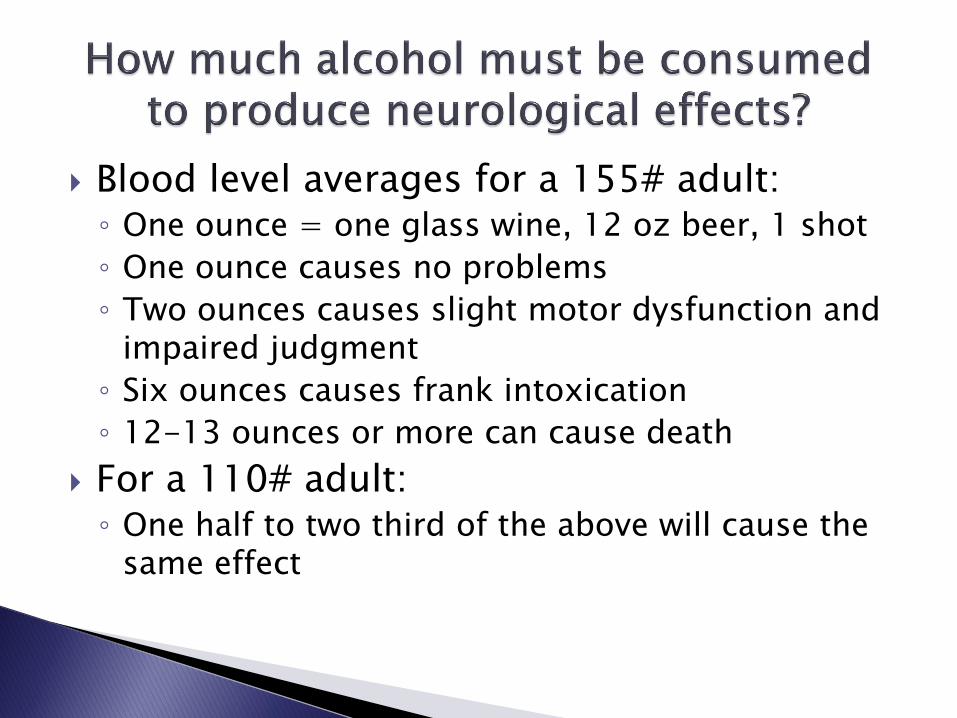
Blood level averages for a 155# adult: ◦ One ounce = one glass wine, 12 oz beer, 1 shot
◦ One ounce causes no problems
◦ Two ounces causes slight motor dysfunction and impaired judgment
◦ Six ounces causes frank intoxication
◦ 12-13 ounces or more can cause death
For a 110# adult: ◦ One half to two third of the above will cause the
same effect
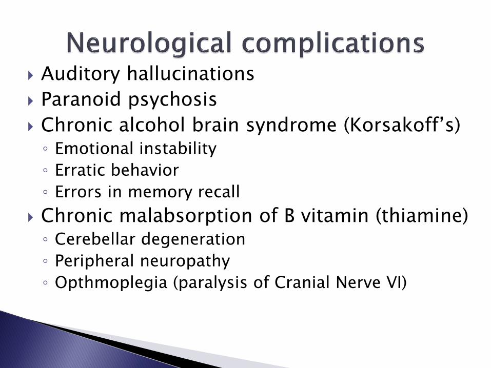
Auditory hallucinations
Paranoid psychosis
Chronic alcohol brain syndrome (Korsakoff’s) ◦ Emotional instability
◦ Erratic behavior
◦ Errors in memory recall
Chronic malabsorption of B vitamin (thiamine) ◦ Cerebellar degeneration
◦ Peripheral neuropathy
◦ Opthmoplegia (paralysis of Cranial Nerve VI)

Have you ever felt you should Cut down on your drinking?
Have people Annoyed you by criticizing your drinking?
Have you ever felt bad or Guilty about your drinking?
Have you ever had a drink first thing in the morning to steady your nerves or to get rid of a hangover (Eye opener)? ◦ Scoring - Item responses on the CAGE are scored 0 or
1, with a higher score an indication of alcohol problems ◦ >2 or greater is considered clinically significant.
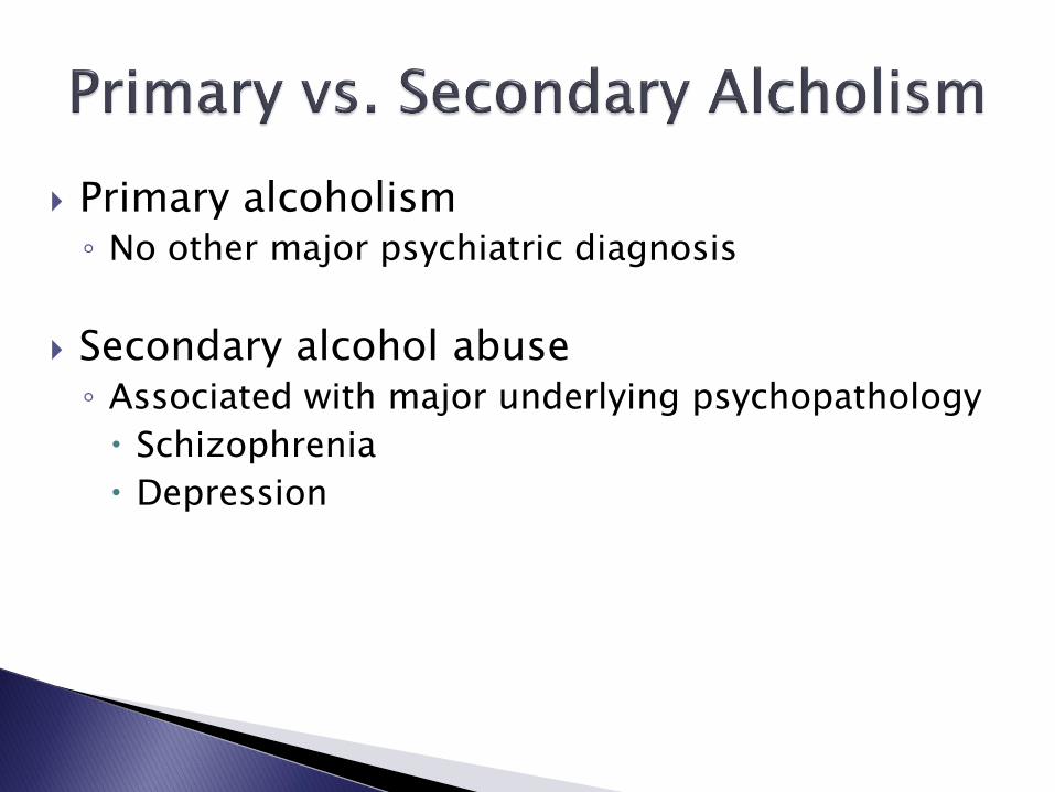
Primary alcoholism ◦ No other major psychiatric diagnosis
Secondary alcohol abuse ◦ Associated with major underlying psychopathology
Schizophrenia
Depression

Begins 8-24 hours after the last drink
Causes the opposite effects of alcohol ◦ Extreme anxiety
◦ Sleeplessness
◦ Terrifying nightmares & hallucinations
◦ Confusion
◦ Hypertension & tachycardia
◦ Generalized tremor
◦ Sweating and fever
◦ Seizures
◦ Potential death if withdrawal is severe

Overcome the patient’s denial
Family confrontation and intervention
Psychotherapy
Antabuse
There are over 100 approaches to therapy ◦ Acceptance of a severity of the problem
◦ Self-validation
◦ Realization of the need for others
◦ Self-control training
◦ Building a coping mechanism repertoire

Non REM Sleep ◦ Stage 1 – easily aroused ◦ Stage 2 – slightly deeper sleep ◦ Stage 3 – more difficult to arouse ◦ Stage 4 – difficult to arouse, low BP, respiration, heart
rate
REM Sleep ◦ Rapid eye movement sleep – restorative sleep
Sleep cycle ◦ It takes about 90 minutes to cycle through the above ◦ The cycle repeats 5-6 times per evening

50% of all adults have occasional insomnia
10% have chronic insomnia
Situational causes of insomnia ◦ Irregular schedules – circadian sleep disorder
◦ Anxiety and stress
◦ Consumption of heavy alcohol or food in evening
◦ Food allergies
◦ Depression
◦ Somatic complaints such as headache, back pain
Sleep onset delay ◦ A conditioned response
◦ Being “wired” - worried about not sleeping
Idiopathic insomnia ◦ No identifiable cause
Restless leg syndrome
Psychophysiological insomnia ◦ Depression
◦ Anxiety
◦ Bi-polar
Bright light therapy
OTC hypnotics ◦ Anti-histamines
Prescription medications ◦ Ambien, phenobarbital
Melatonin
Sleep hygiene ◦ Constitent sleep routine and bedtime rituals
Hypersomnia ◦ An increase by 25% of normal sleeping time
◦ Seen in clinical depression, encephalitis, tumor
Narcolepsy ◦ Experiences recurring, uncontrollable sleep
episodes in the waking hours
◦ Several episodes per day lasting 30-60 minutes


Incidence of seizures ◦ 2% of population have at least once ◦ <1% have recurrent seizures ◦ Half are idiopathic causes – unknown etiology ◦ Half may be caused by
Birth trauma
Brain infections
CNS toxins
Brain tumors
Head Injuries
Strokes
Reaction to inoculations
Alcoholism
Genetic - phenylketonuria

Febrile ◦ Occurs under two-years-old
◦ Associated with URI and fever > 105degree
Simple partial seizures ◦ Person is aware of what is happening
◦ Usually sensory symptoms (auditory, visual, smells)
Complex partial seizures ◦ Called psychomotor epilepsy
◦ Begins with an aura (visual, smells)
◦ Altered consciousness with staring and stupor
◦ Moving arms in purposeless ways

Absence seizure – petit mal seizure ◦ Usually in children aged 5-15 ◦ Momentary lapse kind of seizure ◦ Staring, facial twitching, and loss of consciousness ◦ Child is unaware that anything has occurred
Myoclonic seizures ◦ Brief, fast involuntary jerks ◦ Patient is aware of it but cannot control
Atonic seizures ◦ Called “drop attacks” ◦ In children with temporary loss of consciousness
Grand mal tonic-clonic seizures ◦ Classic full scale epileptic seizures ◦ Loss of consciousness with seizures for 1-2 minutes ◦ Awakens with no memory of the event
10 Truths About Epilepsy

Diagnosis ◦ Diagnosis is obvious from the history ◦ Must have a verifiable eye witness account ◦ Abnormal EEG shows areas (foci) of discharge
Treatment ◦ Anti-seizure meds for idiopathic seizures
Dilatin, phenobarbital, prinidone, carbamazepine ◦ Surgery is sometimes used for focal lesion, such as
brain tumor, abscess or vascular compression ◦ Valium and IV thiamine for alcoholic seizures

Severe headaches
S & S of stroke
Any rapidly progressive neurological symptoms ◦ Weakness, numbness, speaking problems, balance
troubles, thought problems, altered consciousness
Loss or change of consciousness
Acute vertigo or episodes of presyncope
Signs of alcoholic withdrawal
Seizures, not previously diagnosed or treated
S & S of Dementia or Alzheimer’s
Any patient with neurological symptoms not yet worked up, that is not emergent
Any new tremor, with or without symptoms of Parkinson’s disease
Tourette’s syndrome
Alcoholic dementia
Behavioral abnormalities for that person
Sleep disorders