Embed Size (px)
Citation preview

For almost 40 years, scientists inthe weapons program at LosAlamos have x-rayed, or radi-
ographed, implosions (hydrotests)using the giant PHERMEX (for pulsedhigh-energy radiographic machineemitting x-rays), which generated asingle, brief flash of x-rays that werethen recorded on film. Early on, theyrecognized that the design communityreally wanted an x-ray movie to betterunderstand the implosion process. Thevalue was obvious: One picture returnsposition; two pictures, velocity; threepictures, acceleration, and so on.Furthermore, because a movie recordsmultiple images of a single hydrotest,the desired information could be gath-ered at a reduced cost. The limitationwas the x-ray film.
Film has been used to record x-rayimages since the discovery of the x-ray, but despite over a century ofdevelopment, x-ray film still suffers
from certain drawbacks. It is relativelytransparent to x-ray photons—espe-cially those at higher energies—thatoften pass right through withoutimprinting any information. In addi-tion, film is essentially an analogrecording medium with limited sensi-tivity; that is, it must be exposed to aminimum amount of light before animage can be recorded. Normally, fora movie, separate images are recordedon separate pieces of film. Becausex-rays cannot be focused or reflectedlike visible light, no conventionaltechnology existed to perform thistask. Simply put, film cannot beadvanced fast enough to capture theextremely rapid explosions.
Interestingly, a filmless system wasproposed during the design phase ofPHERMEX by Doug Venable andRalph Stevens: “The PHERMEXdetection system will consist of amosaic of scintillation detectors that
will view pulses of . . . radiationthrough systems of interest . . .”(Stevens 1959). The scintillatorwould absorb the x-rays and convertthem to visible light, which couldrecord a limited number of channelselectronically. Berlyn Brixner and thelate Fred Doremire then expanded onthe original concept with proposalsfor a high-speed electronic camerathat had the potential of returningmultiple radiographs for each experi-ment. Unfortunately, these ideas wereahead of their time; it took another30 years for technology to catch upwith this initial vision.
First used in 1996, the PHERMEXx-ray camera (Watson et al. 1995)takes just two pictures—hardly amovie. Still, it was a solid-state, all-electronic system with no film, whichdemonstrated higher sensitivity andabsorbed more x-rays (that is, it hadhigher “quantum efficiency”) than
92 Los Alamos Science Number 28 2003
Scott A. Watson

film (see Figure 1). The large imageformat allowed us to see the entireimploding pit, and the increased sensi-tivity allowed us to see through densematerials for the first time. This revo-lutionary system changed forever theway we think about hydrotesting and,indeed, stockpile stewardship.
As modern hydrotesting facilitiessuch as the Dual-Axis RadiographicHydrotest (DARHT) come online, x-raycamera technology continues toadvance significantly with highlyoptimized components. In particular,the “scintillator” has evolved into alarge mosaic of inlaid crystals—much akin to Zuni jewelry, but withup to 350,000 pieces. Long (morethan 40 millimeters) square rods ofvery dense (greater than 7 grams percubic centimeter) scintillator crystalsare used to facilitate the x-rayabsorption process. Exotic manmadecrystals such as Lu2SiO5:Ce (LSO)
are also used because they exhibit arapid (50 nanoseconds) phosphores-cent decay between x-ray flashes sothat light from one image does notcorrupt its neighbors in the movie
sequence. These crystals are thenassembled into the mosaic by meansof stack lamination constructed fromhundreds of layers of photochemicallyetched stainless steel (see Figure 2).
Number 28 2003 Los Alamos Science 93
The DARHT Camera
Scintillatorarray
X-rays fromPHERMEX
Visible photons
Mirror
CooledCCD array
MCP Lens Lens
Photocathode Output phosphor
Figure 1. PHERMEX Two-Frame Camera System(a) X-rays coming from the target are converted into visible pho-tons by the scintillator. Photons emerging from the front of thescintillator follow one optical path and create one radiograph,while those emerging from the back create the second radi-ograph. The microchannel plate (MCP) in each pathway is the cru-cial electronic “shutter.”The MCP photocathode converts thephotons into electrons (which are then converted back into pho-tons by the output phosphor). By changing the voltage on theMCP, we can rapidly stop the flow of electrons and thus preventany light from reaching the cooled CCD detector. Appropriatetiming of the two MCP voltages allows us to take consecutive radiographs. (b) This photo is of the camera system. (c) The tworadiographs of H-1970, a VIPER shape-charge munition, were taken 17 µs (left) and 21 µs (right) after detonation. These are thefirst Los Alamos radiographs showing an explosive event at different times.
Figure 2. The DARHT Scintillator Lens(a) The LSO inlaid scintillator shown here has more than 135,000 focused pixel ele-ments. The blue color is a result of the natural emission spectrum of LSO, whichpeaks around 420 nm. (b) This schematic shows how the pixels are held in place toform the mosaic. The pixel pitch is 1.1 mm (1.0 mm LSO and 0.1 mm stainless steel).
X-rays
Scintillator “lens”
Visiblephotons
LSO crystalStainless steel
“layer”
(a) (b)
(a) (b)
(c)

Figure 4. Multistage CCD PixelA CCD pixel can be thought of as a bath-tub, complete with a faucet (DARHT) anddrain, that collects the photoelectronsproduced when light strikes the surfaceof the silicon pixel. (a) Thermal diffusionguides the photoelectrons to a “drain”region, where a local electric field cap-tures the photoelectrons in a potentialwell that is ultimately connected to thereadout electronics. The number of pho-toelectrons produced is proportional tothe number of photons striking the pixel.(b) Reversing the bias on the electrodesprevents the photoelectrons from reach-ing the collection drain. Thus, we canshutter the pixel and control the light sig-nal collected from that drain. (c) For theDARHT second-axis camera, each pixel isactually a superpixel with four separatedrains and four storage wells. Each drainregion has its own electrodes, whichallow us to open a “hole” in the bathtubover any selected well region. To capturethe first frame (i), drain A is openedwhereas the other drains are closed. Allthe photoelectrons generated in theentire superpixel region are collected bywell A. Thus, the device exhibits a 100%fill factor, giving increased sensitivity.After the first image is stored, we closedrain A and open drain B to collectcharge in region B for the second image(ii). This procedure is continued until allfour frames are collected. The chargefrom each region is then read out slowly(to minimize noise in the charge ampli-fier), bucket-brigade fashion from pixel topixel as in a conventional CCD.
94 Los Alamos Science Number 28 2003
The DARHT Camera
Scintillator
Visible light
Mirror
Lens CCDcamera
Insulatedliquid nitrogencontainer
Fiber-opticcable
X-rays
Computer and display screen
Figure 3. The DARHT First-AxisCameraThe camera consists of the scintillator,lens, and five optical lens/CCD sys-tems for capturing the scintillatorlight. The multiple cameras, with over-lapping fields of view, allow us toimage the entire scintillator with lessthan 1% geometric distortion.
VSD = 3 V VSD = 3 VVIA = 18 V
Drain/well
Photoelectron
Thermaldiffusion
Light input
VSD = 18 V VSD = 18 VVIA < 12 V
(a) Shutter Open (c) Four-Frame Capture
Superpixel
(b) Shutter Closed
Light input
Light input
(i)
(ii)
(iii)
(iv)
A
B
C
D
Because this special inlay techniqueallows each rod to point directly tothe x-ray source, the scintillatorexhibits no parallax blur despite thelong pixels used to construct it.
Converting the x-rays into a moreuseful visible light signal is only onechallenge. In photography, therequired sensitivity normally increaseswith higher frame rates, but unfortu-nately, the available sensitivity nor-mally decreases with higher framerates, and the net difference is madeup with bright movie lights. In ourcase, the movie light is DARHT,which cannot be made much brighter,so we must construct an extremelysensitive detector.
To construct the detector, weemploy a number of tricks. We use acustom f1.0 lens to collect as much ofthe scintillator light as possible andfocus that light on the largest, mostsensitive optical recording devicesavailable, namely, astronomy-gradecharge-coupled devices (CCDs),which are much like those on theHubble Space Telescope. Even thiscombination is not sensitive enough,so we must use multiple cameras in amosaic, as Figure 3 shows, and coolthe CCDs with liquid nitrogen toreduce electronic noise to the level of
a few electrons. At this point, wehave a remarkable camera system,which is easily 100 times more sensi-tive than film and 40 times more effi-cient at absorbing x-rays. This systemis now routinely used on the DARHTfirst axis (Watson et al. 2000).
To obtain multiple images, weemploy a unique CCD architecturejointly developed by Los Alamos andMassachusetts Institute of TechnologyLincoln Laboratories specifically forthe DARHT second axis (Reich et al.2003). This chip architecture retainsthe large format, low noise, and highsensitivity of astronomy-grade CCDsbut also records four images at a rateof two million frames per second.Because there is insufficient time totransfer data off the chip at this highframe rate, the information for eachframe must be stored locally on eachpixel and then slowly read off whenthe explosive experiment is over (seeFigures 4 and 5).
The next-generation camera(Watson et al. 2003) will employ atechnology in which the scintillatorlight is collected by an avalanche pho-todiode, amplified, and then pipelinedinto a dedicated high-speed digitizerfor every pixel. Although thisapproach requires a larger, more com-plex electronics package, the enhancedperformance should be astounding.Whereas the PHERMEX camera cantake two radiographs at 500 kilohertzand the DARHT camera can take fourradiographs at 2 megahertz, the next-generation camera will take thousandsof pictures at 20 megahertz. We hopethat the advanced camera will generateuseful results for the weapons commu-nity in a timely manner. �
Acknowledgments
This work was the result of an exten-sive decade-long collaboration. Theauthor thanks the RadiographicDetector Team from the
Hydrodynamics Group (DebraArchuleta, Steve Balzer, ChrisGossein, Mark Hoverson, HenryOlivas, Mike Ulibarri, Carl Vecere,and Chuck Vecere), MassachusettsInstitute of Technology LincolnLaboratories, Princeton Instruments,Bicron, Tecomet, and Spindler Hoyerfor significant contributions.
Further Reading
Reich, R. K., D. D. Rathman, D. M. O’Mara,D. J. Young, A. H. Loomis, E. J. Kohler et.al. 2003. High-Speed, ElectronicallyShuttered Solid-State Imager Technology.Rev. Sci. Instrum. 74 (3): 2027.
Stevens, R. R. 1959. “An Investigation of theStatistics Inherent in the Detection of SmallNumbers of X-Ray Quanta in the PHER-MEX System.” Los Alamos NationalLaboratory memorandum GMX-11-TM-141.
Watson, S. A., T. J. Kauppila, K. H. Mueller,and R. C. Haight. 1995. “Multiframe, High-Energy, Radiographic Cameras forSubmicrosecond Imaging.” Los AlamosNational Laboratory document LA-UR-95-3570.
Watson, S. A., C. A. Ekdahl Jr., S. J. Balzer, H.A. Bender, and A. Daiz. 2003. “Reliable,Low-Current, Continuous Cavity Imaging atDARHT.” Los Alamos National Laboratorydocument LA-UR-03-0908.
Watson, S. A., J. M. Gonzales, C. Gossein, M.Hoverson, and M. Ulibarri. 2000.“Quantum Efficiency, Noise PowerSpectrum, Linearity and Sensitivity of theDARHT g-Ray Camera.” Los AlamosNational Laboratory document LA-UR-00-0653.
Number 28 2003 Los Alamos Science 95
The DARHT Camera
Scott A. Watson came to Los Alamos NationalLaboratory as a summer student in 1986. Heholds a master’s degreein electrical engineeringfrom the University ofNew Mexico. He hasspent his entire careerimproving hydrotest radi-ography—at PHERMEXfirst and at DARHT morerecently. He enjoys stillphotography in his sparetime.
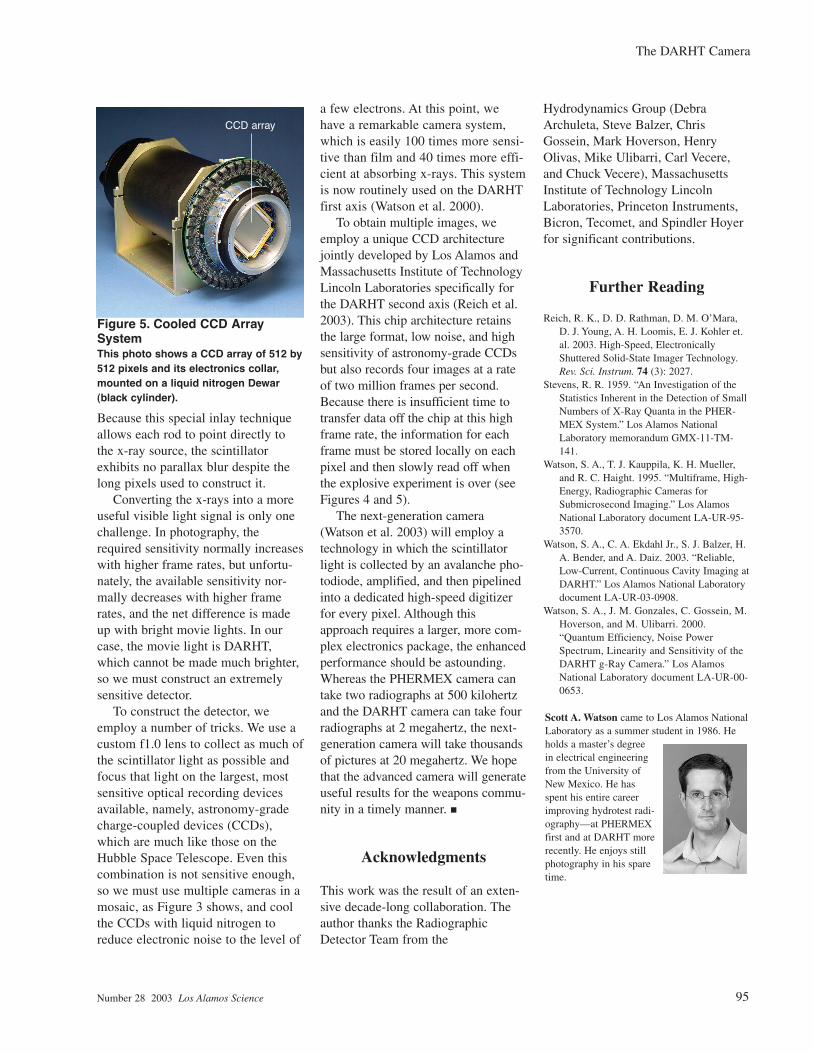
Figure 5. Cooled CCD ArraySystemThis photo shows a CCD array of 512 by512 pixels and its electronics collar,mounted on a liquid nitrogen Dewar(black cylinder).
CCD array