Embed Size (px)
Citation preview
Scintigraphic Demonstration of Tracheo-Esophageal Fistula
Eddy K. Dunn, Andre C. Man, Ke-jian Lin, Harley D. Kaufman, and Nathan A. Solomon
SUNY Downstate Medical Center-Kings County Hospital, Brooklyn, New York
A tracheo-esophageal fistula, developed following radiotherapy for an esopha-
geal carcinoma, was vividly demonstrated by radionuclide imaging. The abnormality was later confirmed by a barium esophagram and endoscopie examinations.The scintigraphic procedure, making use of a Tc-99m sulfur colloid swallow, ap
pears to be a simple alternative method that may be clinically useful for the diagnosis of such a condition.
J NucÃMed 24: 1151-1154,1983
Esophageal scintigraphy using Tc-99m sulfur colloid has been chemotherapy, together with a total of 6,000 rads of local externalproven most encouraging for evaluation of esophageal transit(1-3). Imaging with a similar protocol, even without computer
processing, has resulted in a clear illustration and eventual diagnosis of a tracheo-csophagcal fistula. The radionuclide study ispresented together with a brief review of the other currentlyavailable diagnostic procedures for this disease entity.
METHOD
The patient was positioned upright with his chest directly facingthe detector of the scintillation camera, which has a crystal 15 in.in diameter and •%in. thick. A parallel-hole, low-energy, high-
resolution collimator was used. Through a straw, the patient drewup 500 fid of Tc-99m sulfur colloid, in 3 ml of water, into hismouth, temporarily holding it there. (As a precaution againstcontamination through spilled or coughed-up material, he held alarge towel over his mouth.) With a multi-imager, serial, sequentialscintiphotos (3 sec/frame) were obtained, starting at the momentthat the patient was instructed to swallow. Subsequently several2.000-ID (information density) static images were made, with areaof interest positioned over the left lower bronchus.
CASE REPORT
A 54-yr-old black man was admitted because of persistent, severe, paroxysmal coughing and shortness of breath, while eatingand drinking, for 4 days. He also complained of severe thirst andorthostatic dizziness for a couple of days before admission. Fourmonths before this episode, following progressive dysphagia andloss of 55 Ib of weight in about half a year, a squamouscell carcinoma, located at the middle third of his esophagus, was diagnosedby esophagram (Fig. 1), csophagoscopy, and biopsy. For this hehad been treated with three courses of cisplatinum 20-mg infusion
Received June 2, 1983; revision accepted July I, 1983.For reprints contact: E.K. Dunn, MD, Dcpt. of Radiology Div. of
NucÃ.Med., SUNY Downstate Medical Center, 450 Clarkson Ave.,Brooklyn, NY 11203.
radiation in divided doses (180 rads/day, 5 days a week, givenevery other week). With the treatment, he showed improved ability
LAO RAO
FIG. 1. Circumferential mid-esophageal carcinoma seen as non-
distensible segmental contour defect (between arrowheads) onfull-column barium examination of esophagus.
Volume 24, Number 12 1151
by on December 25, 2018. For personal use only. jnm.snmjournals.org Downloaded from

DUNN, MAN. LIN, KAUFMAN, AND SOLOMON
AR L
UÈ-.LMB
LE-
0-3 sec 3-6 6-9 9-12
12-15
k15-18
LLB
18-21 21-24
B
ANI. A NT.
1MB
LE-
RLB
LLB
1 min 30 min
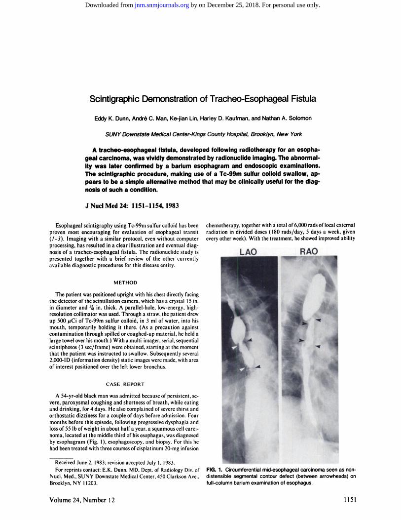
FIG. 2. Scintiphotos following Tc-99m sulfur colloid swallow. (A) Sequence of dynamic images at 3 sec per frame, beginning from time
the patient was instructed to swallow. Arrows mark locations of upper esophagus (UE), lower esophagus (LE), stomach (S), left mainbronchus (LMB), and left lower bronchi (LLB). Progression of swallowed bolus was evident, down esophagus into stomach, with simultaneousfilling of left bronchus and distal esophagus. (B) Subsequent scintiphotos, at 1 min after swallow, reveal presence of radioactive colloidin esophagus, stomach, and left main and lower-lobe bronchi. Half an hour later, aspiration of radionuclide resulted in delineation of rightlower-lobe bronchi (RLB) in addition.
to cat and drink until development of his current symptoms, immediately following his last dose of radiotherapy. Hence he wasreadmitted. Hsophageal seintigraphy was performed as describedabove. The findings clearly indicated the presence of a tracheo-
esophagcal fistula (Fig. 2). A subsequent esophagram confirmedthe diagnosis (Fig. 3). Bronchoscopy and csophagoscopy revealedtracheo-esophageal fistula from middle third of the esophagus to
left main bronchus. A Cclestin tube was inserted to bypass thefistula. His postoperative course was smooth, with only low-gradefever up to 101.8°F. His chest radiographs remained remarkably
clear throughout the hospitalizaron, with only mild pleuralscarring at the posterior left costophrcnic angle. Intravenous hy-
pcralimcntation was gradually tapered off and liquid diet wasstarted on the third postoperative day. He was able to tolerateregular diet on the eighth postoperative day. He was discharged10 days after the operation, to be followed in the oncologyclinic.
DISCUSSION
Tracheo-esophageal fistula can be congenital, encountered at
birth, or may develop later in life as a result of mechanical,chemical, or radiation injuries. Aside from the congenital form.
1152 THE JOURNAL OF NUCLEAR MEDICINE
by on December 25, 2018. For personal use only. jnm.snmjournals.org Downloaded from

CLINICAL SCIENCESCASE REPORTS
LAO RAO
FIG. 3. Barium swallow shows filling ofesophagus and bronchial ramifications withcontrast material (views from left and rightobliquity). Curved arrow indicates locationof tracheo-esophageal fistula.
infection and neoplasm with tissue destruction are quite frequentlycontributing factors in its formation. The standard and most directmethod of diagnosis for this condition, in patients who can followinstructions, appears to be through a barium esophagram. However, in pediatrie and nonrcsponsivc patients, contrast study withnasoesophageal intubation, or selective cathetcrization and en-doscopy under anesthesia, may be necessary. Not only becausethese procedures arc definitely more invasive but also becausespecial skill and equipment are involved, they are much less readilyavailable. Nevertheless, in elusive recurrence following surgicalrepair, both selective catheterization and endoscopy have beenshown to be highly useful (4-7).
Bivins et al., measuring the disappearance of Xc-133 introducedthrough a catheter into the esophagus in dogs with artificiallycreated tracheo-esophageal fistulas, demonstrated the potentialusefulness of radionuclide in the diagnosis of the problem (5). Sofar, however, no practical radiotracer method has yet been established for clinical application. Scintigraphic imaging of aTc-99m sulfur colloid swallow, as shown by the case presented, canbe performed just as easily and readily as an esophagram. Thecolloidal particles used in the procedure are less than l Min diameter and conceivably should provide sensitive detection of evena minute fistulous tract. Also, with the quantity of tcchnetiuminvolved, the scintiscanning procedure would impose a less significant radiation burden to the patient than the radiographiecontrast study, which requires fluoroscopic monitoring (9-12).It appears worthwhile to pursue and further evaluate, throughcorrelative studies with the other diagnostic procedures currentlyavailable, the efficacy and merit of the scintigraphic method in thedetection of tracheo-esophageal fistula.
REFERENCES
/. TOLIN RD, MALMUD LS, REILLEY J, et al: Esophagealscintigraphy to quantitate esophageal transit (Quantitationof esophageal transit). Gastroenterology 76:1402-1408,
19792. RUSSELL COM, HILL LD, HOLMES ER ni, et al: Radio
nuclide transit: A sensitive screening test for esophagealdysfunction. Gastroenterology 80:887-892, 1981
3. FISHER RS, MALMUD LS: Esophageal scintigraphy: Arethere advantages? Gastroenterology 80:1066-1067, 1981
4. KIRKS DR. BRILEY CA JR, CURRARINO G: Selectivecatheterization of tracheocsophageal fistula. AJR 133:763-764, 1979
5. FILSTON HC, RANKIN JS, KIRKS DR: The diagnosis ofprimary and recurrent tracheoesphageal fistulas. Value ofselective catheterization. J Pediatr Surg 17:144-148, 1982
6. BENJAMIN B: Endoscopy in congenital trachéalanomalies.J Pediatr Surg 15:164-171, 1980
7. BENJAMIN B: Endoscopy in esophageal atrcsia and tra-
cheoesophageal fistula. Ann Otol Rhinol Laryngol 90:376-382, 1981
8. BIVINS BA, REED MF, BELIN RP, et al: Diagnosis of tracheocsophageal fistula by radioscanning. J Surg Res 23:384-386,1977
9. SNYDERWS, FORDMR, WARNERGG, et al: "S," Absorbed dose per unii cumulated activity for selected radi-onuclides and organs, MIRI) Pamphlet No. II. New York,Society of Nuclear Medicine, 1975, pp 5-10, pp 150-151
10. EVE IS: A review of the physiology of the gastroinestinal tract
Volume 24, Number 12 1153
by on December 25, 2018. For personal use only. jnm.snmjournals.org Downloaded from
DUNN. MAN. LIN. KAUI MAN. AND SOLOMON
in relation to radiation doses from radioactive materials.HealthPhys 12:131-161, 1966
/ /. KEREIAKE.SJG, ROSENSTEINM: Handbook of RadiationDuxes in Nuclear Medicine and Diagnostic X-ray. Boca
Raton. Florida. CRC Press, 1980,pp 185-195, pp 212-21312. JOHNS HE, CUNNINGHAMJR: The Physics of Radiology.
Exposures to patient in diagnostic radiology, Springfield, Illinois.Charles C. Thomas, 1983,Chap. 16-21, pp 648-653
Cardiac Aneurysm Complicated by E. coli Abscess
Frederick E. Reinke, David L. Yuille, Leon J. Jackson, Howard J. Zeft, and Donald C. Mullen
Sf. Luke's Hospital, Milwaukee, Wisconsin
An E. coli myocardial abscess developed in the region of an old aneurysmalmyocardial scar. In spite of vigorous antibiotic therapy fever and positive bloodcultures persisted. A combination of In-111 WBC scanning and Tc-99m RBC gated
heart imaging located the infection in the aneurysmal scar. The abscess was resected and the patient survived.
J NucÃMed 24: 1154-1157, 1983
A myocardial abscessdevelopingin a pre-existing left-ventricular aneurysm is rare. There have been only seven previouslyreported cases (/-6). In contrast, there have been 15reported casesof an infected left-ventricularaneurysm in the setting of an acutemyocardial infarction (4,7.8). This report concerns a case of anabscess that developedin a pre-existinganeurysmof longduration,diagnosed preopcratively.
ReceivedMay 20. 1983;revisionacceptedAug. 1, 1983.[•'orreprintscontact:David!.. Yuille,MD,NuclearMedicineDcpt..
St. Luke's Hospital, 2900 West Oklahoma Ave., Milwaukee,Wl
53215.
CASE REPORT
A 73-yr-oldman washospitalizedwitha 3-wkprodromeof chillsand night sweats, and a 1-wk history of diarrhea and vomiting.Beforeadmission he had been treated for 5days with erythromy-cin. There was a history of a large anterior-wall myocardial infarction (AWM1) 4 yr before. Cardiac cathctcrization and chestradiograph at 1 mo after the infarction demonstrated a largeleft-ventricular (LV) aneurysm with total occlusion of the leftanterior descending (LAD) coronary and only minor changes inthe right and circumflexcoronaryarteries. He had beenon digoxinand diuretics since this episode, and was asymptomatic. The admission physical examination revealed bloodpressure of 130/70,
fcf
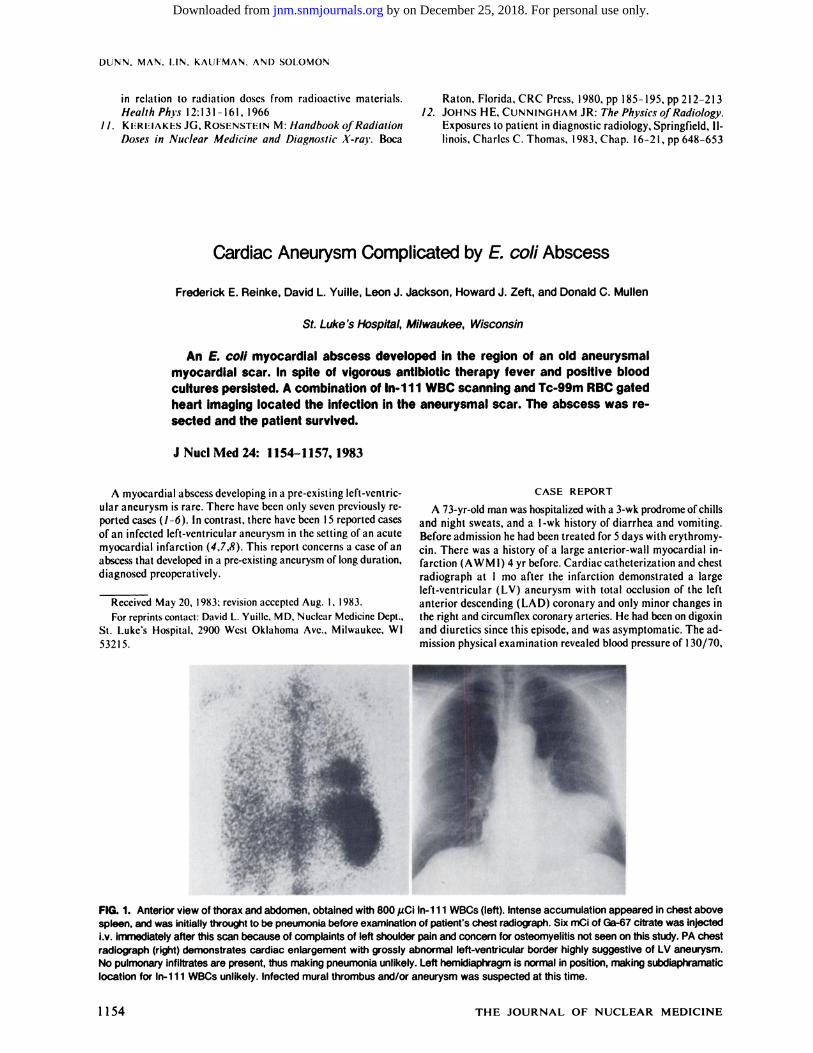
FIG. 1. Anterior view of thorax and abdomen, obtained with 800 juCi ln-111 WBCs (left). Intense accumulation appeared in chest abovespleen, and was initially through! to be pneumonia before examination of patient's chest radiograph. Six mCi of Ga-67 citrate was injected
i.v. immediately after this scan because of complaints of left shoulder pain and concern for osteomyelitis not seen on this study. PA chestradiograph (right) demonstrates cardiac enlargement with grossly abnormal left-ventricular border highly suggestive of LV aneurysm.
No pulmonary infiltrates are present, thus making pneumonia unlikely. Left hemidiaphragm is normal in position, making subdiaphramaticlocation for ln-111 WBCs unlikely. Infected mural thrombus and/or aneurysm was suspected at this time.
1154 THE JOURNAL OF NUCLEAR MEDICINE
by on December 25, 2018. For personal use only. jnm.snmjournals.org Downloaded from
1983;24:1151-1154.J Nucl Med. Eddy K. Dunn, André C. Man, Ke-jian Lin, Harley D. Kaufman and Nathan A. Solomon Scintigraphic Demonstration of Tracheo-Esophageal Fistula
http://jnm.snmjournals.org/content/24/12/1151This article and updated information are available at:
http://jnm.snmjournals.org/site/subscriptions/online.xhtml
Information about subscriptions to JNM can be found at:
http://jnm.snmjournals.org/site/misc/permission.xhtmlInformation about reproducing figures, tables, or other portions of this article can be found online at:
(Print ISSN: 0161-5505, Online ISSN: 2159-662X)1850 Samuel Morse Drive, Reston, VA 20190.SNMMI | Society of Nuclear Medicine and Molecular Imaging
is published monthly.The Journal of Nuclear Medicine
© Copyright 1983 SNMMI; all rights reserved.
by on December 25, 2018. For personal use only. jnm.snmjournals.org Downloaded from