Embed Size (px)
Citation preview
S c i e n t i f i c S y m p o S i u m 2012Building a Collaborative Research Community That Saves Lives
B o n e M a R R o w F a i L u R e D i S e a S e
Summary for patientS
The Aplastic Anemia & MDS International FoundationThAnkS Our 2012 SyMpOSIuM SpOnSOrS
With Support From
The Edward P. Evans Foundation
Office ofRare DiseasesResearchNational Institutes of Health
12012 Scientific Symposium | Aplastic Anemia & MDS International Foundation | www.AAMDS.org
Dear Friends, It is with great pleasure that we present this Summary for Patients of the Third AA&MDSIF Bone Marrow Failure Scientific Symposium held March 22 & 23, 2012 in Bethesda, Maryland. This symposium brought together virtually all of the world’s experts on the biology and treatment of aplastic anemia, myelodysplastic syndromes, paroxysmal nocturnal hemoglobinuria, and related disorders. It was a very special opportunity for us to focus on these diseases, consider what is known, and explore new and emerging ideas and directions. The Aplastic Anemia & MDS International Foundation (AA&MDSIF) is committed to providing patients and their families with answers, support and hope. While each of our programs and services touches all three, research is the program which most inspires hope. In addition to helping secure nearly $17 million in new federal research funding these past five years, we are proud to have awarded more than $3 million in research grants to over 50 researchers to advance the study of bone marrow failure. We are very pleased that several of these researchers attended the Scientific Symposium as both panelists and participants. We are most grateful to the co-chairs of this event, Richard Stone, MD and Neal Young, MD, for their leadership and to the outstanding committee with whom they worked to plan and organize this Symposium. In addition, we greatly appreciate the internationally respected group of speakers the committee assembled whose presentations stimulated discussion and provided new insights to enhance bone marrow failure research. This Symposium would not have been possible without the sponsorship of the National Heart, Lung, and Blood Institute, the NIH Office of Rare Diseases Research, and generous contributions from the Edward P. Evans Foundation and private industry. The collaborative effort of government, academia, private industry, and individuals, along with AA&MDSIF, demonstrates the mutual commitment to the discovery of new treatments for patients, and ultimately, cures for bone marrow failure diseases. We encourage you to read these summaries to learn more about bone marrow failure diseases and the most promising directions for future research. Sincerely,
Kevin Lyons-Tarr John M. HuberChairman, Board of Directors Executive Director
2 2012 Scientific Symposium | Aplastic Anemia & MDS International Foundation | www.AAMDS.org
table of contents
symposium information ......................................................3
General information ...........................................................4
Genetics and epidemioloGy of mds .................................5 Session Chair: Eva Hellstrom-Lindberg, MD, Karolinska Institute
• Molecular Genetics in Refractory Anemia with Ring Sideroblasts and its Clinical Relevance
Mario Cazzola, MD, University of Pavia• Prognostic Models in Myelodysplastic Syndromes Guillermo Garcia-Manero, MD, M.D. Anderson Cancer Center• Epidemiology and Management of 1,000 Patients with
Low- and Intermediate-1 Risk Myelodysplastic Syndromes (MDS) in the European LeukemiaNet MDS Registry
Eva Hellstrom-Lindberg, MD, Karolinska Institute• U2AF1 Mutations in MDS Matthew Walter, MD, Washington University in St. Louis • Genetics and MDS Therapy Benjamin Ebert, MD, PhD, Dana-Farber Cancer Institute
pathophysioloGy / molecular tarGets in mds .........................7Session Chair: Jaroslaw Maciejewski, MD, PhD, Cleveland Clinic
• Molecular Pathogenesis of the 5q- Syndrome Jacqueline Boultwood, MD, Oxford University• Immune Dysregulation and the Effects of DNA
Methyltransferase Inhibition in Myelodysplastic Syndromes Ghulam Mufti, DM, FRCP, Kings College Hospital• Novel Pathway Mutations in Myelodysplasia Seishi Ogawa, MD, PhD, University of Tokyo
Genetics / immunobioloGy of aplastic anemia & pnh.......9Session Chair: Jean Soulier, MD, PhD, Hôpital Saint-Louis
• Aplastic Anemia: Concepts of Clonality Jaroslaw Maciejewski, MD, PhD, Cleveland Clinic• Genomic Instability, Bone Marrow Failure, and Clonal
Evolution in Patients with Fanconi Anemia
Jean Soulier, MD, PhD, Hôpital Saint-Louis• Ribosomopathies in Bone Marrow Failure Akiko Shimamura, MD, Fred Hutchinson Cancer Research
Center• Telomere Disease Neal Young, MD, National Heart, Lung, and Blood Institute• Shwachman-Diamond Syndrome Alan Warren, MD, PhD, Cambridge University
summary of day 1 ....................................................................................12Sharon Savage, MD, FAAP, National Cancer Institute
non-transplant treatments for aplastic anemia & pnh ........................................................13Session Chair: Antonio Risitano, MD, PhD, University of Naples
• Immunosuppression for Aplastic Anemia Judith Marsh, MD, Kings College Hospital• Eltrombopag in Severe Aplastic Anemia Neal Young, MD, The National Heart, Lung, and Blood
Institute
• Pathogenesis and Pathophysiology of Paroxysmal Nocturnal Hemoglobinuria
Lucio Luzzatto, MD, University of Firenze• Complement: Taming the Double-Edged Sword for
Therapeutic Benefit Michael Holers, MD, University of Colorado• Complement Modulation in PNH: From the Bench
to the Bedside Antonio Risitano, MD, PhD, University of Naples
hematopoietic cell transplantation for aplastic anemia, mds & pnh…………… ............................16Session Chair: H. Joachim Deeg, MD, Fred Hutchinson Cancer Research Center
• Alternative Donor Bone Marrow Transplantation for Aplastic Anemia and Paroxysmal Nocturnal Hemoglobinuria
Robert Alan Brodsky, MD, Johns Hopkins• Hematopoietic Progenitor Cell Transplants for Severe
Aplastic Anemia in Children: Are We Ready for “Prime Time”? David Margolis, MD, Medical College of Wisconsin• Stem Cell Transplantation for Acquired Aplastic Anemia in
Adults Andrea Bacigalupo, MD, Ospedale S. Martino• Stem Cell Transplantation for Myelodysplastic Syndromes Bart Scott, MD, Fred Hutchinson Cancer Research Center• Can We Decrease Myelodysplastic Syndromes/Acute
Myelogenous Leukemia Relapse Rates after Allogeneic Transplantation?
Marcos de Lima, MD, M.D. Anderson Cancer Center
non-transplant treatments for mds ..............................19Session Chair: Mikkael Sekeres, MD, MS, Cleveland Clinic
• Azacitdine and Decitabine in Myelodysplastic Syndromes: Lone Wolves or Social Networkers?
Rena Buckstein, MD, FRCPC, Sunnybrook Health Sciences Center
• Lenalidomide in 2012: Where Have We Been and Where Are We Going?
Aristoteles Giagounidis, MD, PhD, St. Johannes Hospital• What I Find Exciting about the Future in MDS Therapies David Steensma, MD, Dana-Farber Cancer Institute
summary of day 2 ....................................................................................21Michael Pulsipher, MD, University of Utah
Glossary ......................................................................................................22
aa&mdsif online learninG center ..................................................24
Funding for this conference was made possible in part by 1R13 HL112578-01 from the National Institutes of Health, National Heart, Lung, and Blood Institute and the Office of Rare Disease Research. The views expressed in written conference materials or publications and by speakers or moderators do not necessarily reflect the official policies of the Department of Health and Human Services; nor does mention of trade names, commercial practices, or organizations imply endorsement by the U.S. Government
32012 Scientific Symposium | Aplastic Anemia & MDS International Foundation | www.AAMDS.org
symposium information
purpose of the symposiumThe Third AA&MDSIF Bone Marrow Failure Disease Scientific Symposium, “Building a Collaborative Research Community That Saves Lives,” was held on March 22-23, 2012 in Bethesda, Maryland. The symposium gathered over 100 participants to hear more than 30 of the world’s leading researchers on aplastic anemia, MDS and PNH share the latest findings, discuss current areas of controversy, and propose specific recommendations for the highest priority directions for basic and clinical research needed to advance the field.
Aplastic anemia, myelodysplastic syndromes (MDS), and paroxysmal nocturnal hemoglobinuria (PNH) are rare diseases that all result in bone marrow failure. Once considered distinct, these three diseases are now believed to be linked by similar pathophysiologies. Exploration of current research issues in aplastic anemia, MDS, and PNH would greatly benefit from increased collaboration between basic and clinical scientists and between scientists studying the individual diseases. Increased understanding of the molecular events driving these diseases and of the response to treatment are needed to define at-risk populations and improve current therapies.
The exchange of ideas and opinions on current research that occurred at the symposium is resulting in collaboration among investigators toward new understanding of the diseases, networks to further individual research, and new approaches on how to transform findings into treatments.
Key findingsSeveral important clinical and scientific advances made recently in bone marrow failure disease research were identified at the symposium. These included:
• Genetic and epigenetic defects identified in MDS that may lead to novel targeted therapies in the near future;
• New techniques to study MDS patients’ genetic information to better predict disease progression and identify the best treatment options;
• New directions for immune therapy in aplastic anemia and MDS;
• Better understanding of the immune and non-immune disease mechanism in aplastic anemia, especially in the field of telomere biology;
• Improvement in the outcome of stem cell transplantation in aplastic anemia;
• The promising effectiveness of the anti-complement treatment for PNH and a new drug that may further enhance outcomes;
• The role of stem cell transplantation as a curative option for PNH patients;
• Stem cell transplantation as an option for more MDS patients than previously considered;
• New pathways for the development of combination therapies for patients with MDS.
These findings and other new insights resulting from the symposium will have an impact on both research and improved patient care for years to come.
co-chair richard m. stone, MDProfessor of Medicine, Harvard Medical School, Dana-Farber Cancer Institute Boston, MA
co-chair neal young, MD Chief, Hematology Branch National Heart, Lung, and Blood InstituteDirector, Trans-NIH Center for Human Immunology, Autoimmunity and Inflammation
Bethesda, MD
h. Joachim deeg, MD Member, Fred Hutchinson Cancer Research CenterProfessor of Medicine, University of WashingtonSeattle, WA
eva hellstrom-lindberg, MDProfessor and Senior PhysicianKarolinska InstituteStockholm, Sweden
Jaroslaw maciejewski, MD, PhDChairman and Professor of Medicine Department of Translational Hematology & Oncology ResearchTaussig Cancer Institute, Cleveland Clinic
Cleveland, OH
regis peffault de latour, MD, PhDSenior Staff Member, Hopital Saint-LouisParis, France
antonio risitano, MD, PhDDivision of Hematology, Federico II University of NaplesNaples, Italy
mikkael sekeres, MD, MSAssociate Professor of MedicineDirector of Leukemia Program, Cleveland ClinicCleveland, OH
symposium planning committee
4 2012 Scientific Symposium | Aplastic Anemia & MDS International Foundation | www.AAMDS.org
General information
aplastic anemia & mds international foundationThe Aplastic Anemia & MDS International Foundation is the world’s leading non-profit health organization dedicated to supporting patients and their families living with aplastic anemia, MDS, PNH and related bone marrow failure diseases. AA&MDSIF provides answers, support and hope to thousands of patients and their families around the world. Founded in 1983, the Aplastic Anemia & MDS International Foundation is celebrating nearly 30 years of service as a recognized and respected leader in patient education, advocacy and research.
What aa&mdsif does• Provides education and support to patients and their families through news updates and plain
language materials, Online Learning Center, Peer Support Network, conferences, community events, and more
• Funds medical research to find better treatments and cures for aplastic anemia, MDS and PNH
• Advocates for increased federal funding of bone marrow failure disease research
• Promotes public awareness of bone marrow failure diseases
• Educates medical professionals on the most up-to-date information about these diseases, their diagnosis and treatment
Dear Patients, Families, and Friends,
Through your generosity, you can help make more research grants and programs possible.
Consider donating $5,000 or $10,000 to the AA&MDSIF Research is Hope Fund, or pledging to raise $60,000 as part of a fund named for you/your loved one to fund a grant in its entirety.
Consider giving a leadership gift of $2,500, $5,000 or more to support educational programs and support services that help the 50+ people each day who are newly diagnosed, and the thousands of people who are fighting these diseases – and surviving like you or your loved one! Please call to talk about your interests in making an impact on the lives of patients and family members through AA&MDSIF. Thank you. Many thanks to all the patients, families, and friends who contributed to make this research possible, and to help AA&MDSIF bring understandable and useful research information directly to patients, families, and health professionals.
Sandra Walter-Steinberg, Director of Development(301) 279-7202 x104 • [email protected]
Current and past AA&MDSIF grantees who participated in the 2012 Scientific Symposium (left to right): Dr. Lisa Minter, Dr. Seth Corey, Dr. Cristian Bellodi, Dr. Kim-Hien Dao, Dr. David Araten, Dr. Jarek Maciejewski, Dr. Antonio Risitano.
Want to fund research into better treatments and a cure for aplastic anemia, mds or pnh?
52012 Scientific Symposium | Aplastic Anemia & MDS International Foundation | www.AAMDS.org
summary for patients
thursday, march 22
Genetics and epidemiology of mdsSession Chair: Eva Hellstrom-Lindberg, MD Karolinska Institute, Sweden
molecular Genetics in refractory anemia with ring sideroblasts and its clinical relevance _________________________________________
Mario Cazzola, MD, University of Pavia, Italy
Refractory anemia with ring sideroblasts (RARS) is a type of myelodysplastic syndrome (MDS) characterized by 15% or more ring sideroblasts in the bone marrow. Ring sideroblasts are immature red cells with iron-loaded mitochondria visualized by Prussian blue staining as a perinuclear ring of blue granules.
Dr. Cazzola has been studying RARS for many years. In the 1980s, he showed that ineffective erythropoiesis is the major mechanism of anemia in this type of MDS, and that patients who have RARS and don’t need regular red blood cell transfusions tend to survive longer than patients who need regular transfusions.
Dr. Cazzola and his colleagues have now sequenced the whole exome, or coding regions of all genes, in eight patients with RARS. They identified 62 gene mutations (DNA changes) in these patients. These mutations are not inherited and these patients cannot pass these mutations to their children. Six of the eight patients had a mutation in the SF3B1 gene. A follow-up study in 533 patients found a mutation in the SF3B1 gene in 28% of patients with MDS and 72% of patients with RARS.
The protein encoded by the SF3B1 gene plays a key role in the RNA splicing machinery and more precisely is a core component of the spliceosome. Messenger RNA contains the genetic coding information needed to make proteins, and cells must splice together certain DNA sequences in RNA to form a messenger RNA molecule that can produce a protein that works properly.
The SF3B1 mutation seems to be responsible for the formation of ring sideroblasts. People with this mutation tend to have a benign clinical course, mainly characterized by anemia and iron overload. They produce less hepcidin, a liver hormone that helps control iron levels and hose deficiency may result in iron accumulation in tissues. Investigations are currently ongoing in order to develop drugs that can counteract the effect of SF3B1 mutation on erythropoiesis and iron metabolism.
prognostic models in myelodysplastic syndromes _________________________________________
Guillermo Garcia-Manero, MD, M.D. Anderson Cancer Center, Houston, TX
Experts have created several prognostic scoring systems for myelodysplastic syndrome (MDS). Doctors and researchers use these systems to identify which patients need treatment, predict how long patients are likely to survive with or without treatment, and choose the best treatment for each patient.
Doctors often use the International Prognostic Scoring System (IPSS) to choose treatments for their patients, but its predictions aren’t always precise enough. For example, the IPSS doesn’t take into account many factors that affect patient survival, including percentage of blasts (young white blood cells) and cytogenetics, or abnormalities in chromosomes.
The World Health Organization classification-based prognostic scoring system (WPSS) classifies MDS into five risk groups based on how long patients are likely to survive and their likelihood of developing leukemia. But the WPSS can’t be used with patients who develop MDS as a result of prior cancer treatment, and it uses the same cytogenetic categories as the IPSS, which might not be the best ones.
Dr. Garcia-Manero developed the Global MDS model to categorize MDS. This model assigns different weight to various patient characteristics, such as performance status (ability to perform routine activities), age, bone marrow blasts, and prior blood transfusions. This model gives more accurate predictions than the IPSS. Patients with fewer comorbidities, or health problems other than MDS, tend to survive longer than those with more comorbidities. So Dr. Garcia-Manero has incorporated a comorbidity score into his prognostic scoring system to make it more accurate.
6 2012 Scientific Symposium | Aplastic Anemia & MDS International Foundation | www.AAMDS.org
summary for patients
A group of experts recently created a revised IPSS (IPSS-R). The IPSS-R is based on the same factors as the IPSS, but emphasizes cytogenetic abnormalities more than high blast counts and includes more information than the IPSS. This system will probably become the standard prognostic model in the near future.
In the past, doctors often didn’t treat patients with lower-risk MDS, but many of these patients didn’t survive long. Dr. Garcia-Manero therefore developed a more accurate prognostic model for patients with lower-risk MDS. This system takes into account cytogenetics, age, hemoglobin and platelet counts, and bone marrow blasts. This lower-risk prognostic scoring system accurately predicts survival in patients with lower-risk MDS according to the IPSS.
Prognostic scoring system research is now focusing on ways to predict which patients will respond to certain treatments (such as stem cell transplants) based, for example, on mutations in genes. epidemiology and management of 1,000 patients with low- and intermediate-1 risk myelodysplastic syndromes (mds) in the european leukemianet mds registry _________________________________________
eva hellstrom-lindberg, MD Karolinska Institute, Sweden
The European Myelodysplastic Syndromes (MDS) Registry was designed for studies of the natural history and clinical management of European patients with low-risk or intermediate-1 risk MDS according to the International Prognostic Scoring System (IPSS). The registry collects data on 1,000 patients from 14 European countries starting within three months of their MDS diagnosis. Nurses and doctors enter information on patients into a web-based form every six months. The information they supply includes disease characteristics, treatments, comorbidities (diseases and conditions other than MDS), blood cell levels, and quality of life. Some of the doctors also send in blood and urine samples from their patients.
Patient ages range from 19 to 95 years, with a median age of 74. Almost all (94%) patients are Caucasian. The types of MDS that patients have varies quite a bit by country. Most (78%) patients
have good or intermediate cytogenetics, while 22% with otherwise good prognostic features lack cytogenetic analysis. Of patients in the registry, 18% have diabetes mellitus, 12% have thyroid disease, and 12% have arrhythmia. Almost half (45%) of patients are taking anti-hypertensive drugs to lower their blood pressure.
Quality of life as measured by physicians is higher in some countries, including the Czech Republic, Denmark, and Greece, than in other countries, such as France and Poland. Quality of life is very different in patients who do and don’t need regular blood transfusions.
On average, 25% of patients need regular blood transfusions at any time point. Over time, the percentage of patients undergoing MDS treatment has risen. By far the most common treatment is erythropoietin-stimulating agents (ESAs), used in more than a third of the patients. Doctors use ESAs to treat anemia in patients with lower-risk MDS and delay the need for red blood cell transfusions. Although ESAs are often used in the United States, their use is less common in Europe and it varies by country. Researchers will be able to use data from the European registry to find out whether ESA treatment benefits patients.
So far, 16% of patients have died. Slightly less than half of these deaths appear to be related to MDS. Some of the factors that seem to affect survival are number of transfusions, IPSS score, and blood iron levels.
u2af1 mutations in mds _________________________________________
Matthew Walter, MD Washington University in St. Louis, MO
The majority of blood-forming cells in the bone marrow of patients with myelodysplastic syndromes (MDS) come from abnormal clones, or copies. In MDS bone marrow samples, there is a founding clone that is more common than the others. All of the other unhealthy clones that form MDS are descendants of this founding clone. Using samples from seven patients with MDS that had evolved into acute myelogenous leukemia (AML), Dr. Walter identified mutations (changes) in several genes that were associated with a founding clone. Every founding clone and its subclones, or daughter clones, had a mutation in at least one of these genes.
72012 Scientific Symposium | Aplastic Anemia & MDS International Foundation | www.AAMDS.org
summary for patients
In one bone marrow sample, a mutation in the STAG2 gene happens in the founding clone very early in the development of MDS and is present in the subsequent daughter clone. In this patient, clones with this mutation made up 80% of the bone marrow at the time of diagnosis. Later on, a cell from this clone gained additional mutations in the PTPN11 and RUNX1 genes. By the time the patient developed AML, 50% of the bone marrow consisted of clones with all three mutations, while all AML cells had the STAG2 mutation. Dr. Walter speculates that identifying and targeting gene mutations that occur in the founding clone may provide a rational approach to specifically kill MDS cells.
In another recent study, 9% of 150 patients with newly diagnosed MDS had a mutation in the U2AF1 gene. The U2AF1 gene plays a role in splicing the precursor to messenger RNA. Messenger RNA contains the genetic coding information needed to make proteins. Cells must splice together certain sequences in RNA to form a messenger RNA molecule that can produce a protein that works properly. Importantly, the U2AF1 genetic mutation seems to occur in the MDS founding clone.
Many laboratories are now studying mutations in genes that splice pre-messenger RNA. Researchers are also studying several drugs that can modify splicing genes. For example, a splicing modulator drug appears to cause the selective death of bone marrow cells from mice with the U2AF1 mutation, but has less affect on normal cells. More research is needed to find out whether drugs that modify splicing genes can be used to treat MDS.
Genetics and mds therapy _________________________________________
Benjamin Ebert, MD, PhD Dana-Farber Cancer Institute, Boston, MA
Doctors use the International Prognostic Scoring System (IPSS) to estimate the prognosis of patients with myelodysplastic syndromes (MDS) and to select appropriate treatments. While the IPSS score is very effective, patients within IPSS categories remain highly heterogeneous. For example, among patients with low-risk or intermediate-1 risk MDS according to the IPSS, patients have dramatically different survival. In a study of 289 patients with lower-risk MDS, a quarter of patients had much poorer survival than other “lower-risk” patients. Up to 70% of these patients had a mutation, or change, in at least one gene that is often mutated in patients with MDS.
Some of the most commonly mutated genes (SF3B1, SRSF2, and U2AF1) in MDS play a role in splicing pre-messenger RNA. Messenger RNA contains the genetic coding information needed to make proteins. Cells must splice together certain initial RNA molecule sequences into a mature RNA to form a messenger RNA molecule that can produce a protein that works properly. In these “lower-risk” patients, survival was significantly worse in patients with an SRSF2 or U2AF1 mutation and seemed to be slightly better in those with an SF3B1 mutation.
Developing treatments that can interfere with the genetic mutations involved in MDS is challenging. Unhealthy hematopoietic stem cells, which make blood cells in bone marrow, can’t easily be studied when they’re removed from a patient with MDS. This is partly because the microenvironment, or surroundings of the stem cells, affects the functioning of these cells, and researchers can’t replicate this microenvironment when they remove the cells to study them. Dr. Ebert has used a technique called pooled, in vivo, short hairpin RNA (shRNA) screening to examine the function of key genes for MDS or leukemia in a mouse bone marrow, thereby overcoming some of these problems.
Using shRNA, Dr. Ebert found that leukemia cells depend on the ITGB3 gene to grow. Knocking out the function of this gene stopped leukemia cells from growing in mice and led to a loss of activity of a protein known as spleen tyrosine kinase, or Syk. Knocking down Syk activity, or inhibiting this activity with a drug, slowed down the growth and survival of leukemia cells. These results show that the activity of the ITGB3 gene, including the role of Syk, might be a target for a future AML treatment.
pathophysiology/molecular targets in mdsSession Chair: Jaroslaw Maciejewski, MD, PhD Cleveland Clinic, Cleveland, OH
Myelodysplastic syndromes (MDS) start with unstable chromosomes that become abnormal. As a result of these abnormalities, as well as DNA damage, patients develop mutations (changes) in certain genes. The abnormal chromosomes and genetic mutations cause genes that normally suppress tumors to become inactive, genes that cause cancer to become active, and other genes to act in abnormal
8 2012 Scientific Symposium | Aplastic Anemia & MDS International Foundation | www.AAMDS.org
summary for patients
ways. Patients develop epigenetic changes, or changes that are not due to changes in DNA structure, leading to abnormal DNA methylation, a chemical process that helps control gene activity. Some of the later steps in this process exacerbate the damage caused by the earlier steps.
Researchers have now identified different types of mutations in several types of genes in MDS. Some of these genes regulate epigenetic processes, for example, whereas others play a role in splicing pre-messenger RNA, which is an important step in gene activity. Researchers are using whole-genome sequencing to study these genes. A person’s genome has all of his or her DNA, including all of that person’s genes. In genome-sequencing studies, researchers scan genomes of many patients to find differences in genes that are more common in people with the disease. These studies show that most patients with MDS have mutations in many genes, and the mutation patterns are different in acute myelogenous leukemia, MDS, and chronic myelomonocytic leukemia.
molecular pathogenesis of the 5q- syndrome _________________________________________
Jacqueline Boultwood, PhD Oxford University, United Kingdom
Patients with 5q- syndrome, a type of myelodysplastic syndrome (MDS), have a deletion (loss) of the long (q) arm of chromosome 5. One of the commonly deleted regions of the del5q chromosome in patients with 5q- syndrome contains approximately 40 genes. Most of these genes have lower levels of expression, or activity, in patients with 5q- syndrome. This low activity level shows that these genes are haploinsufficient, meaning that they have lost one of their two alleles (copies). Two genes that are haploinsufficient in patients are SPARC, a gene that normally suppresses tumor formation, and RPS14, which is part of the ribosomes in cells that make proteins. Reduced expression of RPS14 decreases production of red blood cells in bone marrow cells from patients with 5q- syndrome, and forcing RPS14 to function normally increases red blood cell production.
Several genes that play a role in the formation of active proteins are deregulated in people with 5q- syndrome, so they cannot form healthy proteins.
Some patients with MDS who do not have 5q- syndrome also have deregulations in these genes.
About a quarter of all patients with Diamond-Blackfan anemia (DBA) also have haploinsufficiency of RPS19, a ribosomal protein that is closely related to RPS14. As a result of this genetic deficiency, patients with 5q- syndrome or DBA have macrocytic anemia, meaning that their red blood cells are fragile and larger than normal.
Haploinsufficiency in ribosomal genes activates the p53 protein, resulting in defective bone marrow cells and increasing the death rate of bone marrow cells that form blood cells. In a study in bone marrow cells, inhibiting p53 with the chemical compound pifithrin-alpha rescued the cells’ ability to form healthy red blood cells.
The ribosomes in the bone marrow cells of patients with 5q- syndrome probably can’t translate genetic information correctly to form healthy proteins. L-leucine is an amino acid (protein “building block”) that helps ribosomes translate genetic information. In studies of bone marrow cells from people with 5q- syndrome, L-leucine improved the translation of genetic information and the formation of healthy blood cells.
immune dysregulation and the effects of dna methyltransferase inhibition in myelodysplastic syndromes _________________________________________
Ghulam Mufti, DM, FRCP Kings College Hospital, United Kingdom
The immune system allows the body to defend itself from foreign substances (such as viruses and bacteria). The immune system does not seem function properly in people with myelodysplastic syndromes (MDS). In the 1980s, Dr. Mufti showed that many patients with MDS had auto-antibodies, or immune system proteins, that fight the person’s own proteins. These patients also had abnormal levels of immunoglobulin, an antibody used by the immune system to fight foreign objects. People with MDS often have autoimmune diseases, meaning that their bodies produce antibodies against their own cells and tissues. Also, patients with MDS have abnormal numbers of T cells, a type of white blood cell involved in the immune system, and the T cells they have don’t work properly.
92012 Scientific Symposium | Aplastic Anemia & MDS International Foundation | www.AAMDS.org
summary for patients
T regulatory (Treg) cells control the activity of other T cells. Patients with high-risk MDS tend to have abnormally high numbers of Treg cells, but those with low-risk MDS have normal numbers of Treg cells. Patients with different types of MDS might have different types of Treg cells in the bone marrow and the blood that circulates throughout the body.
Dr. Mufti studied the effects of azacitidine (Vidaza®) treatment on the immune systems of 70 patients with intermediate-2 or high-risk MDS and ten healthy people. MDS patients had higher levels of Treg cells than healthy patients at first. But by nine months, Treg cell levels were similar in MDS patients and healthy participants. Patients who did not respond to the azacitidine treatment had higher Treg levels than those who did respond.
Dr. Mufti stimulated cells from four MDS patients and four healthy donors with CD3 CD28 molecules, which activate T cells. He then treated the cells with azacitidine. The numbers of T helper cells, which help other immune cells carry out their functions, were similar in treated and untreated cells. However, the treated MDS cells had significantly fewer Treg cells than the untreated MDS cells.
These studies and others show that in people with MDS, the immune system stops the bone marrow from making healthy blood cells. Azacitidine seems to affect the functioning of immune system cells. But the Treg cells of patients who have been treated with azacitidine are still very different from the Treg cells of healthy people.
novel pathway mutations in myelodysplasia _________________________________________
Seishi Ogawa, MD, PhD University of Tokyo, Japan
Many of the gene mutations (changes) that are common in acute myelogenous leukemia (AML) are even more common in myelodysplastic syndromes (MDS). Dr. Ogawa is particularly interested in finding the mutations that are unique to people with MDS.
Dr. Ogawa sequenced the whole exome, or coding regions of all genes, in samples from 29 patients with MDS and related myeloid neoplasms such as chronic myelomonocytic leukemia (CMML) and secondary AML. He identified 271 mutations in the coding sequences of genes. These parts of genes contain codes for making proteins. On average, each patient sample had 9.3 mutations. This is similar to the number of mutations per sample in other blood cancers, including AML, but much lower than the number per sample in other types of cancer, such as lung cancer.
Dr. Ogawa identified 13 genetic mutations in more than one patient with MDS. Researchers had previously identified seven of these genes, but five were new: BCOR, SRSF2, STAG2, U2AF35, and ZRSR2. About 60% of patients with MDS had a mutation in the SF3B1, U2AF35, SRSF2, or ZRSR2 genes. Some of these genetic mutations were especially common in patients with certain types of MDS. For example, 75% of patients with sideroblastic anemia had an SF3B1 mutation. These genes are involved in splicing pre-messenger RNA, which is an important step in gene activity (expression). Haploinsufficiency (loss of one of two copies, or alleles) of SF3B1 causes a platelet shortage in mice. Future research needs to shed light on other biological consequences of splicing gene mutations.
Mutations in splicing genes are relatively rare in patients with monosomy 7 (a loss of one of two number 7 chromosomes). However, patients with monosomy 7 who have mutations in certain splicing genes seem to have a better prognosis. Studies to date on whether splicing gene mutations affect the prognosis of patients with other forms of MDS have had mixed results. The relationship between splicing gene mutations and prognosis in patients with MDS needs to be explored further.
Drugs that target the splicing process in MDS genes might offer a new way to treat MDS. For example, SF3b1 inhibitors interfere with pre-messenger RNA splicing. This type of drug might be useful for MDS, chronic lymphocytic leukemia, and other cancers that involve mutations in splicing genes.
10 2012 Scientific Symposium | Aplastic Anemia & MDS International Foundation | www.AAMDS.org
summary for patients
Genetics/immunobiology of aplastic anemia and paroxysmal nocturnal hemoglobinuriaSession Chair: Jean Soulier, MD, PhD Hôpital Saint-Louis, France
aplastic anemia: concepts of clonality _________________________________________
Jaroslaw Maciejewski, MD, PhD Cleveland Clinic, Cleveland, OH
Aplastic anemia sometimes evolves to myelodysplastic syndromes (MDS). One of the signs that a patient with aplastic anemia has developed MDS is a drop in the counts of all types of blood cells. Survival is better in patients with aplastic anemia that has not evolved to MDS than in patients whose aplastic anemia has evolved.
Figuring out whether a patient has aplastic anemia or hypocellular MDS at diagnosis is important for choosing the right treatment. Like patients with aplastic anemia, those with hypocellular MDS have too few cells in the bone marrow, but they also have abnormal clones, or copies, of bone marrow cells that turn into abnormal blood cells. In true MDS, the number of abnormal clones grows, the bone marrow has more unhealthy clones than healthy ones, and the patient often has abnormal chromosomes. When a patient has aplastic anemia, the abnormal clones in the bone marrow do not survive long and the chromosomes are normal. Doctors can find out whether a patient has MDS or aplastic anemia by studying the patient’s chromosomes and blood cells in the bone marrow.
Dr. Maciejewski studied the chromosomes of 120 patients with aplastic anemia using a technique—single nucleotide polymorphism array karytoping—that could only identify abnormal chromosomes if at least 20% of cells in the bone marrow were abnormal clones. The most common abnormality found using this technique is monosomy 7, a type of MDS. Patients with monosomy 7 have only one copy, not the usual two copies, of chromosome 7. When Dr. Macijewski used another technique, metaphase cytogenetics, to study chromosomes from people with aplastic anemia, he found that some patients with aplastic anemia had monosomy 7 and others had a deletion in chromosome 7, meaning that part or all of the chromosome was missing.
Dr. Maciejewski has been following more than 200 patients with large granular lymphocyte (LGL) leukemia. In this rare type of cancer, the number of T lymphocytes, a type of white blood cell that helps fight infections, increases slowly. Like patients with aplastic anemia, those with LGL leukemia have low counts of different types of blood cells. But people with LGL leukemia have more abnormal clones than people with aplastic anemia. Patients with LGL leukemia often have mutations, or changes, in the STAT3 gene. A few patients with aplastic anemia, MDS, and paroxysmal nocturnal hemoglobinuria also have STAT3 gene mutations.
Genomic instability, bone marrow failure, and clonal evolution in patients with fanconi anemia _________________________________________
Jean Soulier, MD, PhD Hôpital Saint-Louis, France
In patients with Fanconi anemia, a rare inherited disease, the bone marrow does not make enough red blood cells, white blood cells, or platelets. Researchers have identified 15 genes which can be mutated in patients with Fanconi anemia. Every patient with this disease has at least one of these genetic mutations.
The first signs of Fanconi anemia—such as short size; abnormal thumbs; a small head; and heart, lung, and kidney problems—are present at birth, but Fanconi anemia is often diagnosed during childhood, when their bone marrow doesn’t make healthy blood cells, or even later during their teens or adults if they have myelodysplastic syndromes (MDS), acute myelogenous leukemia (AML), or other cancers.
Fanconi anemia is difficult to study because it is so rare. Patients with Fanconi anemia have very few stem cells available in their bone marrow to make healthy blood cells. Stem cell counts are already low soon after birth, showing that the disease starts very early. In Fanconi anemia patients, the p53 protein, which normally finely tunes the stem cell pool and suppresses tumor development, is abnormally active. Researchers recently bred Fanconi anemia mice that have lower counts of hematopoietic stem cells, like patients, and they have confirmed (by reducing the activity of the p53 protein in various experiments), that the p53 hyperactivation impairs the development of more blood-forming stem cells in the bone marrow. Studies in cells from people with Fanconi anemia had similar results.
112012 Scientific Symposium | Aplastic Anemia & MDS International Foundation | www.AAMDS.org
summary for patients
Based on these studies, it appears that DNA damage and cellular stress in Fanconi anemia lead to increased activity of p53 and other proteins like p21 in the bone marrow stem cells that form blood cells. These stem cells age more quickly and die more often than healthy stem cells, leading to bone marrow failure. A similar process seems to happen, for other reasons, in patients with Diamond-Blackfan anemia and dyskeratosis congenita, which are also rare bone marrow failure disorders.
When Fanconi anemia evolves to MDS or AML, patients often develop abnormalities of chromosomes 1, 3, and 7. As the number of chromosome abnormalities increases, the bone marrow loses more of its ability to make healthy blood cells but evolves into a MDS and/or into AML. However, transplanting hematopoietic, or blood-forming, stem cells into patients with Fanconi anemia can prevent or treat MDS and AML.
ribosomopathies in bone marrow failure _________________________________________
Akiko Shimamura, MD, PhD Fred Hutchinson Cancer Research Center Seattle, WA
Ribosomes are cell structures that process the cell’s genetic instructions to create proteins. To function properly, the parts (known as subunits) of ribosomes must join together. Patients with certain rare, inherited bone marrow failure disorders—including dyskeratosis congenita, Diamond-Blackfan anemia, and Schwachman-Diamond syndrome (SDS)—have defects in the genes that give instructions for making the proteins in ribosomes.
SDS is passed down through families through a mutated, or abnormal, gene on one of the numbered, or non-sex, chromosomes. Patients with SDS are at risk of bone marrow failure, meaning that their bone marrow stops making enough healthy blood cells, and acute myelogenous leukemia. Some people with SDS have neutropenia, or a shortage of certain white blood cells.
People only develop SDS if they have two copies of the mutated gene. More than 90% of patients with SDS have a mutation in the SBDS gene. This gene is involved in the formation and growth of ribosomes, and it helps ribosome subunits join together. The gene also plays a key role in blood cell formation. The ribosome subunits of patients with SDS do not join together properly.
Initiation factors are proteins that bind to part of a ribosome when it starts forming proteins. The initiation factor eIF6 helps control the ability of ribosome subunits to join together. Reducing the activity of eIF6 allows ribosome subunits in cells from patients with SDS to join together properly but does not improve the bone marrow’s ability to form healthy blood cells.
telomere disease _________________________________________
Neal Young, MD National Heart, Lung, and Blood Institute Bethesda, MD
Some patients with aplastic anemia, whether acquired (resulting from exposure to certain environmental factors) or constitutional (inherited), have very short telomeres. Telomeres are located at the ends of chromosomes and help keep chromosomes stable. As people age, their telomeres become shorter, and this shortening is fastest in the first 20 years of life.
In patients with aplastic anemia, the immune system destroys the stem cells in the bone marrow that would normally form blood cells. This lack of stem cells in the bone marrow is probably one reason why hematopoietic stem cell transplant (which involves the infusion into the patient of stem cells from a healthy donor) can cure aplastic anemia. About 15% of patients with aplastic anemia develop acute myelogenous leukemia (AML) over the long term as a result of immune system activity. Children apparently have more stem cells than adults, and children with aplastic anemia do better with treatments to suppress their immune system (immunosuppression) than do adults. These and other evidence suggest a link between the number of stem cells (and perhaps their ability to regenerate) response to immunosuppressive therapy, and likelihood of later developing AML.
TERT and TERC are genes that produce components of the telomerase repair complex that helps control telomere length. A small but significant minority of patients with aplastic anemia have mutations that decrease the activity of telomerase, an enzyme that maintains telomere length. These mutations seem to increase the risk of AML.
Many members of a large Mennonite family that Dr. Young has studied have a TERT mutation. Several of these individuals have aplastic anemia, and some of these patients also have fibrotic
12 2012 Scientific Symposium | Aplastic Anemia & MDS International Foundation | www.AAMDS.org
scarring in the liver and lungs. Others have the TERT mutation, but their blood counts are normal. In humans, and also in mouse models, short telomeres can be inherited from a mutant parents, but in the absence of a mutation in the offspring, there is no apparent telomere disease. Accelerated telomere attrition may also be a response to stem cell depletion, reflecting the increased mitotic activity of limited numbers of stem cells.
More research is needed to understand the effects of TERT and TERC mutations on different organs in the body, as well as the reasons why these mutations have such diffferent effects in members of the same families. Other research topics are why some families with short telomeres don’t have a strong cancer history and what the link is between short telomeres and cancer.
shwachman-diamond syndrome _________________________________________
Alan Warren, MD, PhD Cambridge University, United Kingdom
Shwachman-Diamond syndrome (SDS) is a rare inherited disease that affects the bone marrow and other organs. In these patients, the bone marrow doesn’t make one or more types of blood cells. Once they reach their 30s, more than a third of patients with SDS develop myelodysplastic syndromes or acute myelogenous leukemia. SDS is caused by mutations in a gene called SBDS.
Ribosomes are cellular structures that process the cell’s genetic instructions to create proteins. The ribosomal assembly pathway is very complex, involving the assembly of 80 ribosomal proteins by the action of around two hundred different factors. Very little is currently known about the mechanisms of ribosome assembly.
Dr. Warren believes that SDS is caused by a defect in the maturation of ribosomes. Initiation factor 6 (eIF6) is a protein that must be released from maturing ribosomes to allow cells to start making proteins. The release of eIF6 provides a mechanism to control the ability of ribosomal subunits (components) to join together. Dr. Warren believes that the role of the SBDS protein is to trigger release of eIF6 from the large ribosomal subunit.
Dr. Warren has used nuclear magnetic resonance imaging to identify the structure of SBDS and the consequences of its mutations. With this technique,
he can identify mutations that change surface epitopes (parts of proteins that are immune system targets) or protein stability.
When Dr. Warren used a genetic tool to delete the SBDS gene in mice, the effect on the mice was dramatic. The mice developed abscesses on the surfaces of their liver, and they had abnormal cells in the liver and areas with dead cells. By examining the liver cells under a microscope, Dr. Warren could tell which animals had a mutation in the SBDS gene. Cells from the livers of mice with the SBDS mutation also had abnormal ribosomal subunit profiles consistent with the idea that the mice had a defect in ribosome assembly. Cells from these mice also accumulated large amounts of eIF6 protein on the large ribosomal subunit.
Using cells from patients, Dr. Warren confirmed that SBDS mutations are associated with a defect in the joining of ribosomal subunits to create proteins. Further experiments showed that the mutation interrupts ribosome assembly, activating the p53 protein that normally suppresses tumor development. The activated p53 protein then stops bone marrow cells from dividing, leading to the death of these cells.
summary of day 1
Sharon Savage, MD, FAAP National Cancer Institute Bethesda, MD
Lessons from the presentations on the first day of the meeting include the need for studies to learn about the patterns of bone marrow failure disorders in different populations. Following patients over the long term is critical to understand how bone marrow failure disorders develop and how these disorders change over time.
The studies that use genomic techniques to study all of the hereditary information in patients with bone marrow failure disorders are exciting. Researchers are now using these techniques to predict the course of a patient’s disease and help doctors choose the best treatments for their patients. One challenge for researchers is narrowing down the vast amount of information on genetics that studies are generating.
Researchers are using new techniques to study issues that have been looked at in the same way for a long time. These new approaches have helped experts identify potential targets, such as spliceosomes, or groups of proteins involved in splicing pre-messenger RNA, for new treatments.
summary for patients
132012 Scientific Symposium | Aplastic Anemia & MDS International Foundation | www.AAMDS.org
friday, march 23
non-transplant treatments for aplastic anemia & pnhSession Chair: Antonio Risitano, MD, PhD University of Naples, Italy
immunosuppression for aplastic anemia _________________________________________
Judith Marsh, MD Kings College Hospital, United Kingdom
The standard first treatment for aplastic anemia consists of drugs to weaken the patient’s immune system and stop it from attacking the bone marrow. This treatment, known as immunosuppressive therapy, helps the bone marrow make more healthy blood cells. Immunosuppressive treatment usually involves a combination of horse antithymocyte globulin (ATG) and cyclosporine. Survival rates with ATG and cyclosporine treatment improved dramatically in the last decades of the 20th century, but have not changed much over the last two decades.
Doctors sometimes add granulocyte colony-stimulating factor (G-CSF), a drug that might help the bone marrow form new blood cells, to immunosuppressive treatment. A clinical trial found that survival was similar in patients with severe aplastic anemia treated with a combination of ATG, cyclosporine, and G-CSF or just ATG and cyclosporine. But patients treated with G-CSF had fewer infections and may spend less time in the hospital.
A form of horse ATG (ATGAM®) is available in the United States, but over the last few years, rabbit ATG has been the only form of ATG that can be used in most other countries. Most data show that fewer patients respond to rabbit ATG than horse ATG, and survival with rabbit ATG isn’t as good as with horse ATG.
Investigators have studied other immunosuppressive agents in aplastic anemia. For example, 71% of previously untreated patients with severe aplastic anemia responded to high-dose cyclophosphamide (Cytoxan®) and G-CSF, and 48% of patients who had not responded to other treatments also responded to cyclophosphamide. But patients had serious side effects, such as shortages of several types of blood cells for very long periods of time.
Another immunosuppressive treatment that investigators are studying is alemtuzumab (Campath®). This drug reduces the number of lymphocytes (a type of white blood cell) in the blood even more than ATG, and it works in other autoimmune diseases. A few small studies have found that alemtuzumab and cyclosporine seems to benefit patients with aplastic anemia who had not responded to horse or rabbit ATG in the past or whose disease had relapsed after ATG treatment. The largest study to date included 90 patients with severe aplastic anemia. For the part of the study involving newly diagnosed patients, the study closed early, after just 16 patients had enrolled, because responses to alemtuzumab were low and less than with rabbit ATG. Alemtuzumab is more effective when given to patients whose aplastic anemia has returned after previously responding to ATG.
eltrombopag in severe aplastic anemia _________________________________________
Neal Young, MD National Heart, Lung, and Blood Institute, Bethesda, MD
Patients with aplastic anemia have very few hematopoietic stem cells, which make blood cells, in the bone marrow, so they have low blood cell counts. About two-thirds of people with aplastic anemia respond to treatment with horse antithymocyte globulin (ATG), which suppresses the patient’s immune system and stops it from attacking the bone marrow, and cyclosporine, another immunosuppressant drug. Survival rates in patients with aplastic anemia has increased over time.
Patients with aplastic anemia who don’t respond to standard treatment often have infections, and they might need one or two blood transfusions every week. Therefore, new treatments are needed for patients who don’t respond to horse ATG and cyclosporine.
A patient’s likelihood of responding to treatment for aplastic anemia likely depends on how many stem cells are in the patient’s bone marrow. Children with aplastic anemia are more likely to respond to treatment than adults, and children tend to have more stem cells than adults. Also, patients with a complete response to treatment, as measured by blood counts, have a lower risk of developing a blood cancer or dying.
summary for patients
14 2012 Scientific Symposium | Aplastic Anemia & MDS International Foundation | www.AAMDS.org
Eltrombopag (Promacta®) offers an alternative to immunosuppressive therapy in patients with aplastic anemia. This drug mimics thrombopoietin, a hormone that controls platelet production in the bone marrow. This process increases the number of platelets and decreases bleeding risk. Patients take the drug by mouth, and most tolerate the drug well.
NIH researchers have studied eltrombopag in 25 patients with aplastic anemia who hadn’t responded to repeated immunosuppressive treatment. Blood cell counts in about half the patients increased dramatically. The largest increases happened after patients had been on eltrombopag for at least three months, and there were very few serious side effects. Immune system markers didn’t change in the patients who responded, so the researchers believe that eltrombopag helps mobilize stem cells to form healthy blood cells.
Several trials of eltrombopag are ongoing or scheduled to start soon. A new study in the Hematology Branch of NHLBI will use a combination of eltrombopag and immunosuppressive treatment, and another will administer eltrombopag to patients with low- or intermediate-risk myelodysplastic syndromes.
pathogenesis and pathophysiology of paroxysmal nocturnal hemoglobinuria _________________________________________
Lucio Luzzatto, MD Istituto Toscano Tumori and University of Firenze, Italy
Patients with paroxysmal nocturnal hemoglobinuria (PNH) have an abnormal clone, or population of cells, originating from one of the stem cells in the bone marrow that makes blood cells. Because of a mutation, or change, in the PIG-A gene, the cells belonging to this clone don’t have proteins that would normally be attached by glycosylphosphatidylinositol (GPI), a fat molecule found on many cells that anchors proteins to cell surfaces. In addition, patients with PNH have little normal hematopoiesis, or formation of healthy blood cells in the bone marrow.
Dr. Luzzatto described a patient with severe aplastic anemia who needed regular red blood cell transfusions. Eventually, her blood cell counts
improved, and she needed fewer red blood cell transfusions. Testing showed that she had developed PNH cells. Dr. Luzzatto suddenly realized that the evolution of this patient’s bone marrow cells to PNH cells was actually nature’s way of curing her aplastic anemia because the PNH cells could form a lot of blood cells, although the red cells are not quite normal, as they tend to be destroyed through a normal component of the blood called complement.
The link between PNH and aplastic anemia was first identified in the 1970s. Studies have shown that patients with PNH, like those with aplastic anemia, have very few normal stem cells that form blood cells in the bone marrow. Thus, patients with PNH have severe bone marrow failure, but this is largely compensated by the blood cells produced by the PNH clone. Although the blood cells produced by the PNH clone are not quite normal, the patient is much better off than if they were not produced, as in aplastic anemia.
Dr. Luzzatto has explored the relationships between abnormal bone marrow cells and bone marrow failure in PNH. He theorized that patients with PNH have a few stem cells in the bone marrow with a PIG-A mutation.
When the patient’s immune system attacks the bone marrow, which happens in aplastic anemia, the attack targets GPI-linked proteins or GPI itself. Thus, paradoxically, the PNH cells can withstand the attack because they lack the GPI-linked proteins or GPI that are the targets of the immune attack.
When researchers tried to find evidence to support this theory, they found that even healthy people have a very small number of cells with the PIG-A mutations. Therefore, the mutation alone doesn’t cause PNH. Also, normally clones with the PIG-A mutation are less likely to reproduce than cells without the mutation. After reviewing evidence from several different studies, Dr. Luzzatto concluded that it is the special bone marrow environment present in patients with PNH that supports the reproduction and growth of clones with the PIG-A mutation. Currently, we can say that PNH is ‘a blessing in disguise’, because this PIG-A mutation enables the bone marrow to still produce blood cells, and today we have a drug, eculizumab, that can help ameliorate the tendency of PNH red cells to be destroyed by complement.
summary for patients
152012 Scientific Symposium | Aplastic Anemia & MDS International Foundation | www.AAMDS.org
complement: taming the double-edged Sword for Therapeutic Benefit _________________________________________
Michael Holers, MD University of Colorado, Denver, CO
Complement proteins are part of the immune system, which protects the body from disease. When a person is injured or attacked by a virus, the complement system recruits enzymes and other mediators to fight the invader. Certain complement proteins normally protect red blood cells from destruction by complement. But patients with paroxysmal nocturnal hemoglobinuria (PNH) have abnormal red blood cells that don’t have two important complement-regulating proteins. As a result, the complement system destroys red blood cells prematurely.
Eculizumab (Soliris®) is a drug that blocks complement protein attacks on blood cells and prevents hemolysis (premature destruction of red blood cells) in people with PNH. Research on eculizumab in PNH shows that a complement activation pathway, known as the “tickover” or alternative pathway, plays an important role in the complement system and in PNH. The tickover pathway allows the immune system to respond immediately to viruses or bacteria, and it helps the body determine whether a substance comes from the body or is a foreign invader. Although other complement pathways can become active in the presence of a trigger (usually some kind of infectious agent), the tickover pathway always has low-level activity. This activity is the first step in the development of PNH.
The complement system interacts with the coagulation system, which forms blood clots. For example, the complement system improves the coagulation system’s ability to form clots. And proteases, which are enzymes that break down proteins, in the coagulation system can activate complement proteins.
In developing treatments for PNH and other diseases caused by abnormal complement activity, researchers have to balance the potential risks and benefits of interfering with the complement system. For example, treatments should affect the site of complement activation only, leaving the rest of the immune system alone so that it can still fight infections.
Studies in mice of complement inhibitors that target complement inhibitors to sites of activation using
CR2, a C3 fragment receptor in the complement system, show that these inhibitors are more effective than untargeted inhibitors. These inhibitors act locally, leaving the rest of the complement system intact, and they don’t appear to increase the risk of infection.
complement modulation in pnh: from the bench to the bedside _________________________________________
Antonio Risitano, MD, PhD University of Naples, Italy
Complement proteins are part of the immune system. Patients with paroxysmal nocturnal hemoglobinuria (PNH) have abnormal red blood cells that don’t have two important proteins that normally regulate complement. In these patients, the complement system destroys red blood cells prematurely, a process known as hemolysis.
Eculizumab (Soliris®) is a drug that blocks complement protein attacks on blood cells and prevents hemolysis in people with PNH. Once patients start eculizumab treatment, their lactate dehydrogenase (LDH) levels drop. High levels of this enzyme mean that the red blood cells are breaking down, so the drops in LDH with eculizumab treatment show that the drug efficiently blocks hemolysis in the blood vessels– the so-called intravascular hemolysis. In patients with PNH, eculizumab dramatically reduces the need for blood transfusions and the number of thromboembolisms, or clots that block blood vessels. Eculizumab also improves survival in people with PNH.
Dr. Risitano has recently studied the benefits of eculizumab on the blood systems of patients with PNH. About a third of patients treated with eculizumab have normal red blood cell counts. Although most of the other patients don’t need regular red blood cell transfusions, their red blood cell counts are persistently low. Unlike untreated patients with PNH, a substantial proportion of treated patients have C3 (a protein in the complement system) on the surface of their red blood cells. C3 may appear on red blood cells of treated patients because eculizumab acts downstream C3, thus it is not designed to prevent early events in complement activation. Patients with more C3-positive cells have more immature red blood cells and a poorer response to eculizumab. This finding suggests that C3 plays a role in red blood cell formation.
summary for patients
16 2012 Scientific Symposium | Aplastic Anemia & MDS International Foundation | www.AAMDS.org
Researchers are developing several new complement inhibitors, including inhibitors of C3, for PNH treatment. Dr. Risitano has shown that TT30, a recombinant protein which merged two normal human proteins, inhibits C3 activation and the deposition of C3 on PNH cells. As a result, TT30 prevents hemolysis in cells from patients with PNH, with a dose-dependent effect (the higher dose of TT30, the larger inhibition of hemolysis). In addition, by inhibiting early complement activation on PNH red blood cells, TT30 may prevent alternative mechanism of red blood cell destruction (the so-called extravascular hemolysis) that in some patients may limit the benefit from eculizumab. A clinical trial of TT30 has started enrolling patients in the United States and Europe. Dr. Risitano hopes that TT30 might offer a cure for PNH, or least a better option than current treatments.
hematopoietic cell transplantation for aplastic anemia, myelodysplastic syndromes, and paroxysmal nocturnal hemoglobinuriaSession Chair: H. Joachim Deeg, MD Fred Hutchinson Cancer Research Center Seattle, WA
alternative donor bone marrow transplantation for aplastic anemia and paroxysmal nocturnal hemoglobinuria _________________________________________
Robert Alan Brodsky, MD Johns Hopkins University School of Medicine Baltimore, MD
Hematopoietic stem cell transplant (HSCT) can potentially cure aplastic anemia and paroxysmal nocturnal hemoglobinuria (PNH). This procedure involves the infusion of healthy blood-forming (hematopoietic) stem cells from a healthy donor with the same HLA (immune system) markers as the patient. The donor’s stem cells (known as a graft) enter the bone marrow, where they form healthy blood cells. HSCT works best in young patients who have a perfectly HLA-matched sibling donor; however, less than 30% of aplastic anemia patients will have such a donor. Patients who lack a matched sibling (brother or sister) donor are usually treated with
immunosuppressive drugs, but the response rate is only 60% and some of the responders will eventually relapse. Many of these patients will require HSCT, but most won’t have an HLA-matched sibling donor.
Researchers are studying the use of “alternative donor” bone marrow transplantation in aplastic anemia and PNH. This procedure uses stem cells from a donor whose HLA markers do not completely match those of the patient. In the past, survival was much better after HSCT using stem cells from HLA-identical siblings than from related, HLA-mismatched donors or unrelated, HLA-matched donors. Alternative donor HSCT has had better outcomes in recent years, but many patients develop severe graft-versus-host disease (GVHD), a common complication of HSCT, and up to half of patients die as a result of the transplant.
In haploidentical stem cell transplant, the donor’s HLA markers match half of the patient’s HLA markers. The major advantage of this approach is that the majority of patients will be able to find a donor for HSCT. This is because this approach allow doctors to use children, parents, siblings or half siblings as donors for HSCT. Cyclophosphamide, administered on days three and four after bone marrow transplant can improve engraftment and decrease the risk of GVHD in patients who have undergone HSCT from a haploidentical, related donor. In a study of 13 patients with refractory severe aplastic anemia or refractory PNH treated with HSCT followed by high dose cyclophosphamide, 12 patients entered complete remission with no GVHD.
hematopoietic progenitor cell transplants for severe aplastic anemia in children: are We ready for “prime time”? _________________________________________
David Margolis, MD Medical College of Wisconsin Milwaukee, WI
Dr. Margolis discussed treatments for children with severe aplastic anemia using examples of four patients.
CASE 1: 15-year-old boy who has a sibling with matching HLA (immune system) markers.
Children with aplastic anemia who undergo bone marrow transplant (BMT) from an HLA-matched sibling have high survival rates and low rates of graft-versus-host disease (GVHD), a common complication of BMT.
summary for patients
172012 Scientific Symposium | Aplastic Anemia & MDS International Foundation | www.AAMDS.org
Some doctors use drugs that suppress the immune system (also known as immunosuppressive drugs) as first-line therapy for children with aplastic anemia because they reason that these patients can undergo BMT later if necessary. Immunosuppressive drugs stop the body’s immune system from attacking the bone marrow and allow the bone marrow to form healthy blood cells. However, immunosuppressive treatment before BMT from a related donor increases the risk that the patient’s body will reject the donated cells. Therefore, up-front BMT from an HLA-matched sibling is the best choice for Case 1.
case 2: 11-year-old girl who was treated with immunosuppressive therapy, needs regular blood transfusions, and has several HLA-matched, but unrelated donors.
In a study of children with aplastic anemia who had failed immunosuppressive therapy in the past, about 95% of the children survived at least five years after BMT from a matched, unrelated donor, and very few developed severe GVHD. BMT from a matched, unrelated donor is the best choice for Case 2.
case 3: 11-year-old girl who was treated with immunosuppressive therapy, needs regular blood transfusions, and has no HLA-matched, unrelated donors.
Several small studies have shown that the transplantation of umbilical cord blood from an unrelated donor can be a good treatment option in children with severe aplastic anemia who haven’t responded to immunosuppressive treatment. Most children in these studies survive and stop needing regular blood transfusions. A cord blood transplant is a good option for Case 3.
case 4: 11-year-old girl who was treated with immunosuppressive therapy, needs regular blood transfusions, has no HLA-matched, unrelated donors, and has frozen blood from her umbilical cord.
The literature on the use of the patient’s own frozen umbilical cord blood for transplantation is very limited, although these types of transplantation are becoming more common. The existing reports suggest that transplantation with the patient’s own cord blood might benefit children with aplastic anemia, but more research is needed. A transplant of the patient’s cord blood might help Case 4.
stem cell transplantation for acquired aplastic anemia in adults _________________________________________
Andrea Bacigalupo, MD Ospedale S. Martino, Italy
Adults with aplastic anemia who have a related donor with the same HLA (immune system) markers as the patient should undergo hematopoietic stem cell transplantation (HSCT) as soon as possible. Survival with HSCT within 100 days of diagnosis is better than if the patient has the HSCT at a later time. Survival is also better and the risk of graft-versus-host disease (GVHD, a common complication of HSCT) is lower if the stem cell source is bone marrow (BM) rather than G-CSF mobilized peripheral blood (PB). The standard conditioning regimen for transplantation from an HLA identical sibling includes antithymocyte globulin (ATG) and cyclophosphamide (Cytoxan®) to prevent the patient’s immune system from attacking the transplanted cells. Survival in the age group 0-20 years exceeds 85%, and in patients aged 21-30 years it is 80% .
The results are less encouraging for patients over the age of 30, with transplant related mortality in the order of 30% or more. One study showed that adding fludarabine (Fludara®) to cyclophosphamide and ATG seems to improve survival after HSCT in patients older than 30. Another study found that a combination of alemtuzumab (Campath®), fludarabine, and cyclophosphamide seems to reduce the risk of chronic GVHD (starting more than three months after HSCT) in patients older than 50.
HSCT from an HLA-matched, unrelated donor is becoming more common and survival after this procedure is improving. Large donor registries are now available, so patients have a better chance of finding a matched donor than in the past. Fewer patients develop severe GVHD after this procedure, and better treatments are available for HSCT complications (such as infections). Better conditioning regimens are also available, including combinations of low doses of total body irradiation with cyclophosphamide with or without ATG and/or cyclophosphamide. Although survival after HSCT from an HLA-matched, unrelated donor has not been as good as with HSCT from a matched sibling in the past, the survival differences are smaller today. In the last quinquennium (2005-2009), the survival differences disappear if the HSCT takes
summary for patients
18 2012 Scientific Symposium | Aplastic Anemia & MDS International Foundation | www.AAMDS.org
place beyond six months from diagnosis. Survival from matched, unrelated donor HSCT also improves if the stem cells come from the donor’s BM instead of peripheral blood (bloodstream) and the patient undergoes ATG conditioning.
In conclusion, transplantation both from identical siblings, or unrelated donors, has greatly improved in the past decade and should be considered for all patients with aplastic anemia under the age of 50. For older patients, up to the age of 65 , transplantation can be considered, but only after having failed a course of immunosuppressive therapy (ATG). The use of cord blood or family mismatched donors, is still considered experimental.
stem cell transplantation for myelodysplastic syndromes _________________________________________
Bart Scott, MD Fred Hutchinson Cancer Research Center Seattle, WA
Hematopoietic stem cell transplant (HSCT) is the only cure for myelodysplastic syndromes (MDS).
Whether patients with MDS should undergo HSCT as soon as their MDS is diagnosed, or wait for months, or even years, depends on their IPSS score. Delaying HSCT after diagnosis improves survival in patients with low-risk or intermediate-1 MDS, but survival in patients with intermediate-2 or high-risk MDS is best when they have HSCT right away. Also, survival after HSCT is better when patients proceed to transplant prior to progression to AML.
Cytogenetics, or the study of abnormal chromosomes, can also affect HSCT outcomes. People with secondary MDS, caused by a previous treatment for another disease or disorder, tend to have more abnormal chromosomes and worse outcomes after HSCT than people with primary MDS. An MDS scoring system based on cytogenetic features accurately predicts the probability of relapse and survival after HSCT in patients with MDS.
Chemotherapy with cytarabine before HSCT leads to a complete response (no signs of MDS) in up to 60% of patients, especially those with normal cytogenetics. But this treatment can have serious complications and even when it works, the remission often lasts only a few months. Also, more intensive pre-transplant chemotherapy treatments have more serious side effects than less intensive regimens.
Patients must receive some form of immune suppression prior to HSCT in order to allow for engraftment of donor stem cells. Initially, only high dose of chemotherapy or radiotherapy was used to condition patient prior to HSCT, but recent newer methods have been employed with lower doses of chemo/radio therapy. This decrease some of the toxicity of transplant but may also leaed to a higher risk of relapse. Retrospective studies have shown equivalent overall survival in patients who are in remission at time of transplant and receive either high dose or low dose conditioning prior to HSCT. These retrospective studies do not take into account inherent bias that may be present when assigning patients to a given conditioning regimen. A Phase III clinical trial is currently comparing the effects of more and less intensive chemotherapy treatment before HSCT on 18-month survival in patients with MDS or AML.
can We decrease myelodysplastic syndromes/acute myelogenous leukemia relapse rates after allogeneic transplantation? _________________________________________
Marcos de Lima, MD M.D. Anderson Cancer Center Houston, TX
Although hematopoietic stem cell transplant (HSCT) is the only cure for myelodysplastic syndromes (MDS), most patients with MDS are older than 55 and the risk of relapse (not responding to HSCT) increases with age. Patients are often treated with high doses of chemotherapy before HSCT to suppress the immune system and stop it from rejecting the donated cells. But older patients often have serious side effects from this chemotherapy, and using lower doses of chemotherapy to decrease the side effects can increase the risk of relapse.
Azacitidine (Vidaza®) is a hypomethylating drug that kills unhealthy cells in the bone marrow of people with MDS. Initially, Dr. de Lima gave low doses of the drug to 17 patients with acute myelogenous leukemia (AML) or MDS to prevent or treat relapse after HSCT and demonstrated the feasibility and potential activity of this approach, which was very well tolerated.
Subsequently, Dr. de Lima designed a Phase I clinical trial to find the safest dose and schedule of azacitidine for high-risk patients with MDS or
summary for patients
192012 Scientific Symposium | Aplastic Anemia & MDS International Foundation | www.AAMDS.org
summary for patients
AML who have had HSCT. Patients were treated with up to four monthly cycles of azacitidine after transplant to prevent relapse. Chemotherapy for preparative regimen for transplant was fludarabine, melphalan, and mylotarg. To prevent graft-versus-host disease (GVHD, a common HSCT complication), patients were treated with tacrolimus (Hecoria® or Prograf®). The donated stem cells came from a related donor or an unrelated donor with the same HLA (immune system) markers as the patient. Median patient age was 60, and most had serious comorbidities, or diseases and conditions other than their MDS or AML. Patients treated with more cycles of azacitidine, regardless of dose, had less GVHD, and the patients tolerated the drug well.
T regulatory (Treg) cells control the activity of other T cells. In a study, low-dose azacitidine treatment after HSCT increased the number of Treg cells in 27 patients with AML. The patients tolerated monthly azacitidine treatments well and had a lower risk of GVHD. Currently, a randomized study is ongoing comparing maintenance therapy with azacitidine (Vidaza®) for one year versus no maintenance after allogeneic HSCT. Dr. de Lima is developing a short Phase I clinical trial to compare the effects of taking azacitidine by mouth to injections of azacitidine under the skin using a short needle. He then plans to launch a larger, multicenter clinical trial of oral azacitidine to prolong remission after HSCT.
non-transplant treatments for myelodysplastic syndromesSession Chair: Mikkael Sekeres, MD, MS Cleveland Clinic, Cleveland, OH
azacitidine and decitabine in myelodysplastic syndromes: lone Wolves or social networkers? _________________________________________
Rena Buckstein, MD, FRCPC Sunnybrook Health Sciences Center Canada
Methylation and deacetylation, which are chemical processes that silence genes in patients with myelodysplastic syndromes (MDS), prevent them from carrying out their normal activities. Luckily, this process can be reversed with a combination of
histone deacetylase (HDAC) inhibitors (which help restore gene expression) and hypomethylating agents (which both restore gene expression and kill unhealthy cells in the bone marrow), including the hypomethylating agents azacitidine (Vidaza®) and decitabine (Dacogen®).
Patients who have MDS and too much methylation of several genes have a poorer prognosis and a higher chance of developing acute myelogenous leukemia (AML). Patients more likely to respond to azacitidine treatment are those whose methylation levels drop after treatment, although this is not always enough. Factors associated with a poorer response to azacitidine include abnormal chromosomes, previous low dose cytarabine chemotherapy and bone marrow blasts of > 15%. Patients treated with azacitidine have shorter survival if they have poor performance status (ability to do routine daily tasks), abnormal blasts in the blood (young white blood cells), higher risk chromosomes and a heavy dependence on red blood cell transfusions. Patients with mutations (changes) in certain genes (such as DNMT3A, ASXL1, and TET-2) are more likely to respond to azacitidine or decitabine, but they don’t survive longer than patients without these mutations.
Researchers have combined several other drugs with azacitidine or decitabine to treat MDS and AML. One of the most exciting combinations is azacitidine and lenalidomide (Revlimid®), which slows down the growth of blood vessels that feed abnormal cells as one mode of action. In a multicenter Phase I/II clinical trial of azacitidine and lenalidomide in 36 patients with high-risk MDS, 71% of patients responded to the treatment and 42% had a complete response (meaning that their blood had no signs of MDS). None of the patients had side effects that were so severe that they had to stop the treatment.
Another promising study focused on a combination of azacitidine and vorinostat (Zolinza®), an HDAC inhibitor. Of 28 patients with MDS or AML, 82% responded to the combination treatment and 41% had a complete response. Patients had very few serious side effects, and none of the side effects stopped them from continuing treatment.
The MDS treatment field is entering an era of social networking, in which a single-agent hypomethylating agent may not be the way to go in the future. Small studies to date suggest that combination treatments may have better

20 2012 Scientific Symposium | Aplastic Anemia & MDS International Foundation | www.AAMDS.org
summary for patients
complete and overall response rates and faster response times. However, these findings will need to be verified in larger randomized trials involving many centers before they can be uniformly adopted. In the absence of a clinical trial, the standard of care is still single agent azacitidine or decitabine for patients with higher-risk MDS.
lenalidomide in 2012: Where have We been and Where are We Going? _________________________________________
Aristoteles Giagounidis, MD, PhD St. Johannes Hospital, Germany
Patients with 5q- syndrome, a type of myelodysplastic syndrome (MDS), have a deletion (loss) of the long (q) arm of chromosome 5. This disease affects more women than men, and patients have macrocytic anemia, which means that their red blood cells are larger than normal. Unlike other types of MDS, 5q- syndrome rarely evolves to acute myelogenous leukemia (AML).
Lenalidomide (Revlimid®) is a drug that slows down the growth of the blood vessels that feed abnormal cells and kills abnormal cells in the bone marrow. In a Phase II clinical trial in 205 patients with low- or intermediate-risk 5q- syndrome according to the International Prognostic Scoring System, 67% of patients treated with lenalidomide stopped needing regular blood transfusions. The most common severe side effects were neutropenia, or a shortage of certain white blood cells, and thrombocytopenia, or a shortage of platelets. After four years, about 30% of patients developed AML.
Patients with less than 5% blasts, or abnormal young white blood cells in the bone marrow, have a significantly lower risk of AML than those with 5% or more blasts. Similarly, patients who don’t need regular red blood cell transfusions are less likely to develop AML than those who do need transfusions. Patients with low-risk 5q- syndrome who develop a mutation, or change in the TP53 gene at a very early stage of the disease, are more likely to develop AML and less likely to have a complete response to lenalidomide, meaning that their blood had no signs of MDS.
A combination of lenalidomide and azacitidine (Vidaza®), a hypomethylating drug that kills unhealthy cells in the bone marrow, seems to be more effective than either drug alone in patients
with higher-risk MDS. Lenalidomide combined with 500 μg romiplostim (Nplate®), a protein used to treat low platelet counts, was more effective in a study of patients with MDS than lenalidomide plus placebo or lenalidomide plus 750 μg romiplostim. Another combination that researchers are exploring is lenalidomide and L-leucine, an amino acid (protein “building block”).
What i find exciting about the future in myelodysplastic syndromes _________________________________________
David Steensma, MD Dana-Farber Cancer Institute, Boston, MA
The revolution in genomics has allowed researchers to study all of a person’s DNA (including all of that person’s genes) more easily, more rapidly and at a lower cost. Hopefully, this revolution will drive the development of new therapies for myelodysplastic syndromes (MDS) by increasing knowledge of how these diseases work. But genetics doesn’t tell the whole story in MDS. For example, people with MDS have abnormal clones, or copies, of stem cells in the bone marrow that make blood cells. Potentially, new treatments could target the genetic mutations (changes) that drive the development of abnormal clones, but this will require a better understanding of the genetic mutations that actually “drive” the disease, because some of the genetic mutations that are detected are only “passengers” – present, but along for the ride and not controlling the progress of the disease.
Researchers are studying combinations of several different treatments added to azacitidine (Vidaza®), a hypomethylating drug that was FDA approved for MDS in 2004 and that kills unhealthy cells in the bone marrow of people with MDS, but which does not cure the disease. Another MDS research priority is developing better treatments for the symptoms of MDS. Sometimes, patients whose blood cells respond well to MDS treatments still have troubling symptoms, like fatigue. MDS research is also aimed at speeding up the translation of clinical trial results to patients.
Many new treatments are being tested in clinical trials for MDS, including:
• Nucleoside analogs, or artificial copies of nucleosides, which are the building blocks of DNA: clofarabine (Clolar®) and sapacitabine
212012 Scientific Symposium | Aplastic Anemia & MDS International Foundation | www.AAMDS.org
• Kinase inhibitors, which interfere with cell communication and growth: rigosertib (Estybon®), ARRY-614, MLN4924, KB004 antibody, and CYT387
• Epigenetic modifiers, which interfere with changes in gene activity that are not due to changes in DNA structure: oral azacitidine, SGI-110, 5-fluoro-2’-deoxycytidine with tetrahydrouridine, panobinostat, vorinostat (Zolinza), belinostat, and entinostat
• Monoclonal antibodies, or artificial antibodies, that target cancer cells: siltuximab, etanercept (Enbrel®), alemtuzumab (Campath®), and lintuzumab
• Proteasome/amino acid recycling inhibitors, which kill abnormal blast cells (young white blood cells): tosedostat and bortezomib (Velcade®)
• Immunomodulators and immune tolerance disruptors, which alter the immune system
and promote its recognition of MDS cells as abnormal, thereby promoting their clearance from the body: vaccines, various innovative transplantation strategies including transplantation of mesenchymal (embryonic) stem cells from umbilical cord blood or placentas, AS-101, and maitake mushroom extract
• Other agents: ezatiostat (TELINTRA®), BP100-1.01, OXi4503, ganetespib, TXA127, CPI-613, omacetaxine/homoharringtonine (OMAPRO™), and plerixafor (Mozobil®)
Patients need hope, and an important activity of the Aplastic Anemia & MDS International Foundation is maintaining hope in patients. Hope is a fragile entity, and researchers who conduct clinical trials must be careful not to oversell the promise of these studies. But researchers must also continue to work hard to find new treatments for MDS.
summary for patients
summary of day 2
Michael Pulsipher, MD University of Utah Salt Lake City, UT
Research has made tremendous advances in understanding the genetics and biology of myelodysplastic syndromes (MDS), and many wonderful studies in this area were presented at this symposium. Research has also shown the relationships between many biological features of MDS and outcomes in patients. This biological research is important because it leads to targeted therapies that will benefit patients. A downside is that the outcomes of many new therapies aren’t good enough, so more research is needed. One challenge is to better understand the roles of treatment before hematopoietic stem cell transplant (HSCT), HSCT itself, and treatment after HSCT.
Some wonderful research has been done which had shed light on why paroxysmal nocturnal hemoglobinuria (PNH) occurs, and a very active agent— eculizumab (Soliris®)—is now available for this disease. Further research has found a possible second drug which can add to the very good outcomes with eculizumab. Many patients can have significant improvement in their symptoms and a very good quality of life when they respond to these medicines. A challenge in this field is the high cost of eculizumab treatment, but hopefully this problem can be solved.
Good therapies are available for aplastic anemia, and these treatments keep patients alive longer than in the past. The major challenge in aplastic anemia is that experts don’t understand the biology of the disease well enough. More biological markers are needed to make progress in this field. A disappointment is the lack of major progress with treatments that suppress the immune system. The possibility that eltrombopag (Promacta®) may expand existing stem cells is exciting, but not enough patients have responded to this treatment in studies so far, and further studies of patients earlier in their therapy are needed to see if this medication will improve patient outcomes.
Tremendous advances have been made in HSCT for aplastic anemia, and its outcomes have improved dramatically in recent years. Problems with HSCT, such as graft-versus-host disease (a common HSCT complication), still need to be solved, but some nice progress is being made in this area. Researchers are now trying to figure out the best time to do HSCT and how to maximize its chances of success.
One way to make progress in all of these areas is for researchers to work together. This is especially important because all of these diseases are rare. Many researchers are now doing this, as shown by the successful European LeukemiaNet MDS Registry and other groups.
22 2012 Scientific Symposium | Aplastic Anemia & MDS International Foundation | www.AAMDS.org
Glossary
alemtuzumab (campath®) - A monocolonal antibody - a medicine that is engineered to look for a specific substance in the body. Alemtuzumab attaches to and kills white blood cells called lymphocytes. In certain types of aplastic anemia, lymphocytes are responsible for attacking the bone marrow stem cells. Alemtuzumab is in clinical trials for treating aplastic anemia. It is approved by the U.S. Food and Drug Administration (FDA) for treating certain types of leukemia and is helpful in other conditions that require immunosuppressive therapy.
antithymocyte globulin (atG, atgam®, thymoglobulin®) - ATG is an immunosuppressant, a drug that lowers the body’s immune response. Scientists believe that aplastic anemia happens when the immune system attacks and destroys bone marrow stem cells. ATG kills the specific cells that are attacking the bone marrow stem cells. This allows the bone marrow to grow and make new blood cells. ATG may be used to treat other bone marrow failure diseases in some cases. ATG is approved by the U.S. Food and Drug Administration (FDA) for treating moderate and severe aplastic anemia. ATG is commonly used with another drug called cyclosporine.
bone marrow - The soft, spongy tissue inside most bones. Blood cells are formed in the bone marrow.
cord blood transplant - A procedure where umbillical cord stem cells are given to the patient through an intravenous (IV) line. Stem cells are collected from an umbilical cord right after the birth of a baby. They are kept frozen until needed. In time, donated stem cells given to the patient begin making new, healthy blood cells.
cyclosporine (neoral, sandimmune) - An immunosuppressant that lowers the body’s immune response. Cyclosporine is used along with antithymocyte globulin (ATG), another immunosuppressant, for treating aplastic anemia and some other forms of bone marrow failure.
cytarabine (ara-c) - a chemotherapy drug that slows the growth of cancer cells by inhibiting the division of cells by interfering with DNA replication.
cytogenetics - (sie-toe-juh-neh-tiks) - The study of chromosomes (DNA), the part of the cell that contains genetic information. Some cytogenetic abnormalities are linked to different forms of myelodysplastic syndroms (MDS).
de novo - (di-no-vo) - Brand new, referring to the first time something occurs. MDS that is untreated or that has no known cause is called de novo MDS.
diamond-blackfan anemia - A rare form of pure red cell aplasia that can be passed down from parent to child. Diamond-Blackfan anemia (DBA) is characterized by low red blood cell counts detected in the first year of life. Some people with DBA have physical abnormalities such as small head size, low frontal hairline, wide-set eyes, low-set ears. Genetic testing is used to diagnose DBA.
dyskeratosis congenita - A rare disease beginning from birth that is characterized by symptoms similar to geriatrics such as abnormal skin pigmentation, irregular nail development, and patches on the tongue or mouth. It is accompanied by a predisposition for malignancy and bone marrow failure.
epidemiology - The study of patterns and causes of disease in groups of people. Epidemiology researchers study how many people have a disease, how many new cases are diagnosed each year, where patients are located, and environmental or other factors that influence disease.
epigenetic - Heritable modifications to a genome that affect the expression of a gene without altering the genetic sequence.
epoetin alfa (epogen®, procrit®) - Epoetin alfa can help improve red blood cell counts in bone marrow failure disease patients whose natural erythropoietin levels are low. It is given by injection under the skin (subcutaneous) or in the vein (intravenous). Epoetin alfa is approved by the U.S. Food and Drug Administration (FDA) for treating anemia. It is in a class of drugs called growth factors (cytokines).
erythropoietin-stimulating agents (esa) - A man-made version of a naturally occurring substance in the body (erythropoietin) that encourages the bone marrow to make more red blood cells.
erythropoietin - (i-rith-row-poy-uh-tun) - A protein made by the kidneys. Erythropoietin, also called EPO, is created in response to low oxygen levels in the body (anemia). EPO causes the bone marrow to make more red blood cells. A shortage of EPO can also cause anemia.
etiology - The study of the cause or origin of a disease.
232012 Scientific Symposium | Aplastic Anemia & MDS International Foundation | www.AAMDS.org
fanconi anemia - A rare inherited disorder that happens when the bone marrow does not make enough blood cells: red cells, white cells, and platelets. Fanconi anemia is diagnosed early in life. People with Fanconi anemia have a high likelihood of developing cancer. Genetic testing is used to diagnose Fanconi anemia.
fludarabine (Fludara®) - A chemotherapy drug that interferes with the growth of cancer cells. It is usually used to treat chronic lymphocytic leukemia (CLL).
genomics - A sub-discipline of genetics that is concerned with studying the genome (the entirety of the genetic sequence).
graft-versus-host disease (GVhd) - Also called GVHD, it is a common complication of bone marrow/stem cell transplantation. It is caused when the donor’s immune cells, now in the patient, begin to see the the patient’s body as foreign and mount an immune response. GVHD most commonly affects the recipient’s skin, intestines, or liver. Severity can range from mild to very severe. In some cases, GVHD can be prevented or treated with immunosuppressive drug therapy.
hematopoiesis - (hi-mat-uh-poy-EE-suss) The process of making blood cells in the bone marrow.
immunobiology - A branch of science concerned with the immune system of organisms.
immunsuppresion - Deliberate prevention or reduction of the immune system’s response, e.g. stopping the body from rejecting a bone marrow transplant.
monosomy 7 - A rare chromosomal disorder characterized by deletion of a portion of chromosome 7 that is seen in a variety of hematologic disorders.
pathogenesis - The mechanism or process that causes a condition.
pathophysiology - The study of the changes or abnormal functions that occur in a body that is in a disease state.
platelet - The smallest type of blood cell. Platelets help the blood to clot and stop bleeding. Also called a thrombocyte.
prognosis - The probable or predicted outcome of a condition or disease.
red blood cell - The most numerous type of blood cell in healthy people. Red blood cells contain hemoglobin, a protein that picks up oxygen in the lungs and brings it to cells in all parts of the body. Also called erythrocyte, RBC.
refractory - Not responsive to treatment or cure. For example, refractory anemia is a low red blood cell count that doesn’t respond to standard treatments.
ribosomal gene - A gene that develops ribosomes, which build or synthesize proteins to support cell functions.
secondary mds - A type of MDS that is caused by a previous treatment for another disorder or disease. Treatments typically associated with secondary MDS include radiation therapy and chemotherapy used to treat cancer. Also called therapy-related MDS, T-MDS.
splicing genes - The process of modifying a genetic sequence by removing nonsense sequences (genetic sequences that do not code for the desired proteins) and joining together the functional sequences.
stem cells - Cells in the body that develop into other cells. There are two main sources of stem cells. Embryonic stem cells come from human embryos and are used in medical research. Adult stem cells in the body repair and maintain the organ or tissue in which they are found. Blood-forming (hemapoietic) stem cells are found in the bone marrow. These cells make copies of themselves and develop into red cells, white cells, and platelets.
t cells - A principal type of white blood cell that completes maturation in the thymus and that has various roles in the immune system, including the identification of specific foreign antigens in the body and the activation and deactivation of other immune cells.
white blood cell - Cells in the body that fight disease and infection by attacking and killing germs. There are several types of white blood cells including neutrophils, eosinophils, basophils, lymphocytes and monocytes. Each type of cell fights a different kind of germ. Also called WBC, leukocyte.
whole genome sequencing - The process of determining the entire sequence of an organism’s DNA.
Glossary
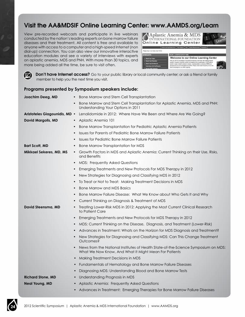
24 2012 Scientific Symposium | Aplastic Anemia & MDS International Foundation | www.AAMDS.org
Visit the AA&MDSIF Online Learning Center: www.AAMDS.org/LearnView pre-recorded webcasts and participate in live webinars conducted by the nation’s leading experts on bone marrow failure diseases and their treatment. All content is free and available to anyone with access to a computer and a high-speed Internet (non dial-up) connection. You can also view our innovative interactive education modules and see a variety of interviews with experts on aplastic anemia, MDS and PNH. With more than 30 topics, and more being added all the time, be sure to visit often.
Don’t have Internet access? Go to your public library or local community center; or ask a friend or family member to help you the next time you visit.
programs presented by symposium speakers include:
Joachim deeg, md • Bone Marrow and Stem Cell Transplantation
• Bone Marrow and Stem Cell Transplantation for Aplastic Anemia, MDS and PNH: Understanding Your Options in 2011
aristoteles Giagounidis, md • Lenalidomide in 2012: Where Have We Been and Where Are We Going?
david margolis, md • Aplastic Anemia 101
• Bone Marrow Transplantation for Pediatric Aplastic Anemia Patients
• Issues for Parents of Pediatric Bone Marrow Failure Patients
• Issues for Pediatric Bone Marrow Failure Patients
bart scott, md • Bone Marrow Transplantation for MDS
mikkael sekeres, md, ms • Growth Factors in MDS and Aplastic Anemia: Current Thinking on their Use, Risks, and Benefits
• MDS: Frequently Asked Questions
• Emerging Treatments and New Protocols For MDS Therapy in 2012
• New Strategies for Diagnosing and Classifying MDS in 2012
• To Treat or Not to Treat: Making Treatment Decisions in MDS
• Bone Marrow and MDS Basics
• Bone Marrow Failure Disease: What We Know about Who Gets It and Why
• Current Thinking on Diagnosis & Treatment of MDS
david steensma, md • Treating Lower-Risk MDS in 2012: Applying the Most Current Clinical Research to Patient Care
• Emerging Treatments and New Protocols for MDS Therapy in 2012
• MDS: Current Thinking on the Disease, Diagnosis, and Treatment (Lower-Risk)
• Advances in Treatment: Whats on the Horizon for MDS Diagnosis and Treatment?
• New Strategies for Diagnosing and Classifying MDS: Can This Change Treatment Outcomes?
• News from the National Institutes of Health State-of-the-Science Symposium on MDS: What We Now Know, And What It Might Mean For Patients
• Making Treatment Decisions in MDS
• Fundamentals of Hematology and Bone Marrow Failure Diseases
• Diagnosing MDS: Understanding Blood and Bone Marrow Tests
richard stone, md • Understanding Prognosis in MDS
neal young, md • Aplastic Anemia: Frequently Asked Questions
• Advances in Treatment: Emerging Therapies for Bone Marrow Failure Diseases
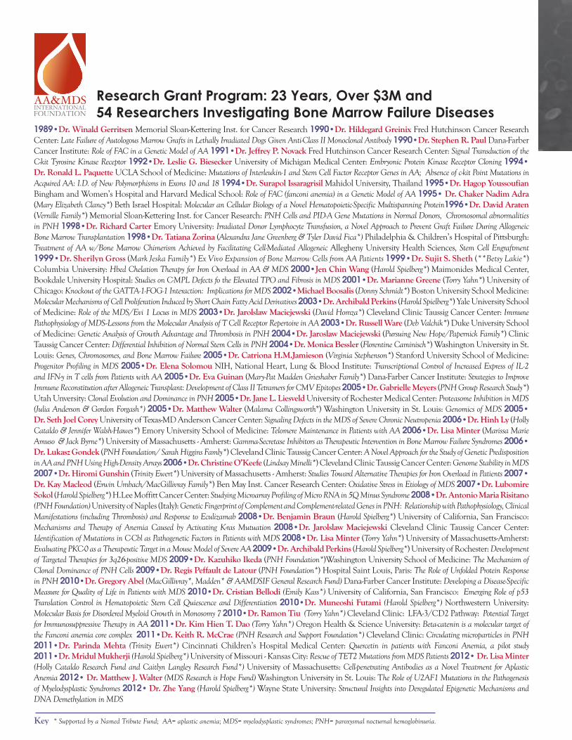
1989•Dr. Winald Gerritsen Memorial Sloan-Kettering Inst. for Cancer Research 1990•Dr. Hildegard Greinix Fred Hutchinson Cancer Research Center: Late Failure of Autologous Marrow Grafts in Lethally Irradiated Dogs Given Anti-Class II Monoclonal Antibody 1990•Dr. Stephen R. Paul Dana-Farber Cancer Institute: Role of FAC in a Genetic Model of AA 1991•Dr. Jeffrey P. Novack Fred Hutchinson Cancer Research Center: Signal Transduction of the C-kit Tyrosine Kinase Receptor 1992•Dr. Leslie G. Biesecker University of Michigan Medical Center: Embryonic Protein Kinase Receptor Cloning 1994• Dr. Ronald L. Paquette UCLA School of Medicine: Mutations of Interleukin-1 and Stem Cell Factor Receptor Genes in AA; Absence of c-kit Point Mutations in Acquired AA: I.D. of New Polymorphisms in Exons 10 and 18 1994•Dr. Surapol Issaragrisil Mahidol University, Thailand 1995•Dr. Hagop Youssoufian Bingham and Women’s Hospital and Harvard Medical School: Role of FAC (fanconi anemia) in a Genetic Model of AA 1995• Dr. Chaker Nadim Adra (Mary Elizabeth Clancy*) Beth Israel Hospital: Molecular an Cellular Biology of a Novel Hematopoietic-Specific Multispanning Protein1996•Dr. David Araten (Vernille Family*) Memorial Sloan-Kettering Inst. for Cancer Research: PNH Cells and PID-A Gene Mutations in Normal Donors, Chromosomal abnormalities in PNH 1998•Dr. Richard Carter Emory University: Irradiated Donor Lymphocyte Transfusion, a Novel Approach to Prevent Graft Failure During Allogeneic Bone Marrow Transplantation 1998•Dr. Tatiana Zorina (Alexandra Jane Greenberg & Tyler David Fica*) Philadelphia & Children’s Hospital of Pittsburgh: Treatment of AA w/Bone Marrow Chimerism Achieved by Facilitating Cell-Mediated Allogeneic Allegheny University Health Sciences, Stem Cell Engraftment 1999•Dr. Sherilyn Gross (Mark Jeska Family*) Ex Vivo Expansion of Bone Marrow Cells from AA Patients 1999•Dr. Sujit S. Sheth (**Betsy Lakie*) Columbia University: Hbed Chelation Therapy for Iron Overload in AA & MDS 2000•Jen Chin Wang (Harold Spielberg*) Maimonides Medical Center, Bookdale University Hospital: Studies on C-MPL Defects fo the Elevated TPO and Fibrosis in MDS 2001•Dr. Marianne Greene (Torry Yahn*) University of Chicago: Knockout of the GATTA-1-FOG-1 Interaction: Implications for MDS 2002•Michael Boosalis (Donny Schmidt*) Boston University School Medicine: Molecular Mechanisms of Cell Proliferation Induced by Short Chain Fatty Acid Derivatives 2003•Dr. Archibald Perkins (Harold Spielberg*) Yale University School of Medicine: Role of the MDS/Evi 1 Locus in MDS 2003•Dr. Jarolslaw Maciejewski (David Homza*) Cleveland Clinic Taussig Cancer Center: Immune Pathophysiology of MDS--Lessons from the Molecular Analysis of T Cell Receptor Repertoire in AA 2003•Dr. Russell Ware (Deb Valchik*) Duke University School of Medicine: Genetic Analysis of Growth Advantage and Thrombosis in PNH 2004•Dr. Jaroslaw Maciejewski (Pursuing New Hope/Papernick Family*) Clinic Taussig Cancer Center: Differential Inhibition of Normal Stem Cells in PNH 2004•Dr. Monica Bessler (Florentine Caminisch*) Washington University in St. Louis: Genes, Chromosomes, and Bone Marrow Failure 2005•Dr. Catriona H.M.Jamieson (Virginia Stephenson*) Stanford University School of Medicine: Progenitor Profiling in MDS 2005•Dr. Elena Solomou NIH, National Heart, Lung & Blood Institute: Transcriptional Control of Increased Express of IL-2 and IFN-y in T cells from Patients with AA 2005•Dr. Eva Guinan (Mary-Pat Madden Grieshaber Family*) Dana-Farber Cancer Institute: Strategies to Improve Immune Reconstitution after Allogeneic Transplant: Development of Class II Tetramers for CMV Epitopes 2005•Dr. Gabrielle Meyers (PNH Group Research Study*) Utah Unversity: Clonal Evolution and Dominance in PNH 2005•Dr. Jane L. Liesveld University of Rochester Medical Center: Proteasome Inhibition in MDS (Julia Anderson & Gordon Forgash*) 2005•Dr. Matthew Walter (Malama Collingsworth*) Washington University in St. Louis: Genomics of MDS 2005• Dr. Seth Joel Corey University of Texas-MD Anderson Cancer Center: Signaling Defects in the MDS of Severe Chronic Neutropenia 2006•Dr. Hinh Ly (Holly Cataldo & Jennifer Walsh-Hawes*) Emory University School of Medicine: Telomere Maintenance in Patients with AA 2006•Dr. Lisa Minter (Marissa Marie Amuso & Jack Byrne*) University of Massachusetts - Amherst: Gamma-Secretase Inhibitors as Therapeutic Intervention in Bone Marrow Failure Syndromes 2006• Dr. Lukasz Gondek (PNH Foundation/ Sarah Higgins Famly*) Cleveland Clinic Taussig Cancer Center: A Novel Approach for the Study of Genetic Predisposition in AA and PNH Using High-Density Arrays 2006•Dr. Christine O’Keefe (Lindsay Minelli*) Cleveland Clinic Taussig Cancer Center: Genome Stability in MDS 2007•Dr. Hiromi Gunshin (Trinity Ewert*) University of Massachusetts - Amherst: Studies Toward Alternative Therapies for Iron Overload in Patients 2007• Dr. Kay Macleod (Erwin Umbach/MacGillivray Family*) Ben May Inst. Cancer Research Center: Oxidative Stress in Etiology of MDS 2007•Dr. Lubomire Sokol (Harold Spielberg*) H.Lee Moffitt Cancer Center: Studying Microarray Profiling of Micro RNA in 5Q Minus Syndrome 2008•Dr. Antonio Maria Risitano (PNH Foundation) University of Naples (Italy): Genetic Fingerprint of Complement and Complement-related Genes in PNH: Relationship with Pathophysiology, Clinical Manifestations (including Thrombosis) and Response to Eculizamab 2008•Dr. Benjamin Braun (Harold Spielberg*) University of California, San Francisco: Mechanisms and Therapy of Anemia Caused by Activating K-ras Mutuation 2008•Dr. Jarolslaw Maciejewski Cleveland Clinic Taussig Cancer Center: Identification of Mutations in C-Cbl as Pathogenetic Factors in Patients with MDS 2008•Dr. Lisa Minter (Torry Yahn*) University of Massachusetts-Amherst: Evaluating PKC-0 as a Therapeutic Target in a Mouse Model of Severe AA 2009•Dr. Archibald Perkins (Harold Spielberg*) University of Rochester: Development of Targeted Therapies for 3q26-positive MDS 2009•Dr. Kazuhiko Ikeda (PNH Foundation*)Washington University School of Medicine: The Mechanism of Clonal Dominance of PNH Cells 2009•Dr. Regis Peffault de Latour (PNH Foundation*) Hospital Saint Louis, Paris: The Role of Unfolded Protein Response in PNH 2010•Dr. Gregory Abel (MacGillivray*, Madden* & AAMDSIF General Research Fund) Dana-Farber Cancer Institute: Developing a Disease-Specific Measure for Quality of Life in Patients with MDS 2010•Dr. Cristian Bellodi (Emily Kass*) University of California, San Francisco: Emerging Role of p53 Translation Control in Hematopoietic Stem Cell Quiescence and Differentiation 2010•Dr. Muneoshi Futami (Harold Spielberg*) Northwestern University: Molecular Basis for Disordered Myeloid Growth in Monosomy 7 2010•Dr. Ramon Tiu (Torry Yahn*) Cleveland Clinic: LFA-3/CD2 Pathway: Potential Target for Immunosuppressive Therapy in AA 2011•Dr. Kim Hien T. Dao (Torry Yahn*) Oregon Health & Science University: Beta-catenin is a molecular target of the Fanconi anemia core complex 2011•Dr. Keith R. McCrae (PNH Research and Support Foundation*) Cleveland Clinic: Circulating microparticles in PNH 2011•Dr. Parinda Mehta (Trinity Ewert*) Cincinnati Children’s Hospital Medical Center: Quercetin in patients with Fanconi Anemia, a pilot study 2011•Dr. Mridul Mukherji (Harold Spielberg*) University of Missouri - Kansas City: Rescue of TET2 Mutations from MDS Patients 2012• Dr. Lisa Minter (Holly Cataldo Research Fund and Caitlyn Langley Research Fund*) University of Massachusetts: Cell-penetrating Antibodies as a Novel Treatment for Aplastic Anemia 2012• Dr. Matthew J. Walter (MDS Research is Hope Fund) Washington University in St. Louis: The Role of U2AF1 Mutations in the Pathogenesis of Myelodysplastic Syndromes 2012• Dr. Zhe Yang (Harold Spielberg*) Wayne State University: Structural Insights into Deregulated Epigenetic Mechanisms and DNA Demethylation in MDS
research Grant program: 23 years, Over $3M and 54 researchers Investigating Bone Marrow Failure Diseases
Key * Supported by a Named Tribute Fund; AA= aplastic anemia; MDS= myelodysplastic syndromes; PNH= paroxysmal nocturnal hemoglobinuria.
100 park Avenue | Suite 108 | rockville, MD 20850(800) 747-2820 | (301) 279-7202 | (301) 279-7205 (Fax) [email protected] | www.AAMDS.org
Fighting Bone Marrow Failure Diseases Through patient Support and
research Since 1983