Embed Size (px)
Citation preview

Scientific adviser: ass.prof Makharynska O.S
Head of department: prof. Yabluchansky M.I.

Structure:
Acute Renal failure (ARF) – definition
Anatomy and physiology of kidneys
ARF diagnostic criterias
ARF - classification
Etiology and pathophysiology
Diagnostic evaluation
Prerenal ARF
Intrinsic ARF
Postrenal ARF
Differential diagnosis
Complications
Management: prevention and treatment

Acute kidney injury, previously known
as acute renal failure, encompasses a wide
spectrum of injury to the kidneys, not just
kidney failure, and characterized by the sudden
impairment of kidney function resulting in
retention of nitrogenous and other waste
products normally cleared by the kidneys.
Acute kidney injury: NICE guideline DRAFT (March 2013)
Harrison’s principles of internal medicine, 19 Ed, 2015
• AKI complicates 5-7% of acute care hospital admissions and up to 30% of admissions to
the intensive care unit
• AKI is associated with a marked-increased risk of death in hospitalized individuals
• Risk factors for developing ARF: age, IDDM, CKD, LV dysfunction
• AKI may be community-acquired (volume depletion, adverse effects of medications,
obstruction of the urinary tract) or hospital-acquired (sepsis, major surgical procedures,
liver failure, intravenous iodinated contrast administration, and nephrotoxic medication).

Structural organization
renal parenchyma:
- cortex
- medulla
nephrons:
- cortical
- juxtamedullary

Renal blood supply:
the kidneys receive 20% of the cardiac output vascular supply:
- renal arteries
- interlobar arteries
- arcuate arteries
- interlobular arteries
- afferent arterioles
- glomerular capillaries
- efferent arterioles
- peritubular capillaries quizlet.com

Basic Renal Physiology
Nephron is
the functional unit of thekidney, capable of formingurine has two majorcomponents: glomerulus
tubule:○ proximal
○ loop of Henle
○ distal
○ collecting

Adjusting the resistances of the afferent and efferent
arterioles, the kidneys can regulate both the hydrostatic
pressures in the glomerular and peritubular capillaries,
changing the rate of glomerular filtration and/or tubular
reabsorption in response to homeostatic demands.
Determinants of renal blood flow (RBF)
renal artery pressure - renal vein pressure
total renal vasculature resistanceRBF=

Glomerular filtration rate (GFR)- is widely accepted as the best overall index of kidney
function in health and disease. Depends on the interplaybetween hydrostatic and oncotic pressures within thenephron
hydrostatic pressure is usually higher in the glomerulus thanwithin the tubule, forcing filtrate out of the capillary bed into thetubule
oncotic pressure is generated by non-filtered proteins: it helps toretain fluid in the intravascular space
GFR: Kf* (hydrostatic pressure - oncotic pressure)
Normal GFR: 100 ml/min/1.72m2
*Kf - filtration coefficient in the glomerulus

Evidence of decreasing renal function
(reduced GFR):
Rising BUN (blood urea nitrogen) – nl about 10 mg/dL (nr
= 7-25 mg/dL)
Rising creatinine – nl about 1 mg/dL (nr = 0.6 – 1.2 mg/dL)
Note! normal BUN: creatinine ratio = 10-20:1


The RIFLE criteria for AKI
ARF, acute renal failure; GFR, glomerular filtration rate; Screat, serum creatinine concentration; UO, urine output.
Acute kidney injury: NICE guideline DRAFT (March 2013)
The acronym RIFLE stands for the increasing severity classes Risk, Injury, and Failure; and the two
outcome classes, Loss and End-Stage Renal Disease (ESRD).

AKIN criteria for AKI
Abrupt (within 48 h) reduction in kidney function currently
defined as an absolute increase in serum creatinine of 0.3
mg/dL or more (≥26.4 μmol/L) or
A percentage increase in serum creatinine of 50% or more
(1.5-fold from baseline) or
A reduction in urine output (documented oliguria of < 0.5
mL/kg/h for >6 h)
http://emedicine.medscape.com/article/1925597-overview
The RIFLE criteria are defined as changes within 7 days, while the AKIN criteria
suggest using 48 hours.

Acute kidney injury: NICE guideline DRAFT (March 2013), http://www.kidney-international.org

KDIGO criteria for AKIAKI is defined as any of the following (Not Graded):
K Increase in SCr by ≥0.3 mg/dl (≥26.5 lmol/l) within 48 hours;
or
K Increase in SCr to ≥1.5 times baseline, which is known or
presumed to have occurred within the prior 7 days; or
K Urine volume ˂0.5 ml/kg/h for 6 hours.
http://www.kidney-international.org

1. PRERENALImpaired renal perfusion(shock, hypovolemia, volumeshifts, ↑CO, ↑PVR(pulmonaryvascular resistance), renalartery obstruction)
2. INTRARENALInvolves parenchymalchanges (renal trauma, acutetubular necrosis, infectiousdiseases, glomerulonephritis)
3. POSTRENALObstruction to urinary tractprostate disease, obstruction,spinal cord injury, pelvictrauma)
Types of Acute Kidney Injury

Classification of the major causes of
acute kidney injury
Harrison’s principles of internal medicine, 19 Ed, 2015
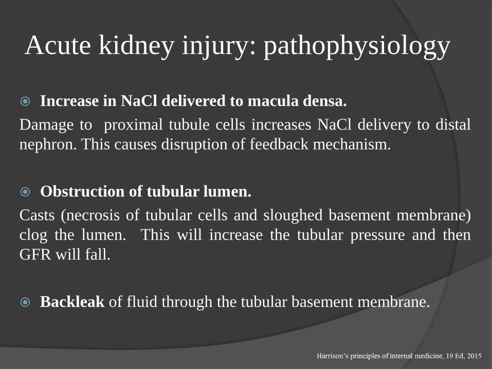
Acute kidney injury: pathophysiology
Increase in NaCl delivered to macula densa.
Damage to proximal tubule cells increases NaCl delivery to distal
nephron. This causes disruption of feedback mechanism.
Obstruction of tubular lumen.
Casts (necrosis of tubular cells and sloughed basement membrane)
clog the lumen. This will increase the tubular pressure and then
GFR will fall.
Backleak of fluid through the tubular basement membrane.

1. INITIATING PHASE
Begins at time of insult until S&S seen (hours to days)
2. OLIGURIC or ANURIC PHASE
Oliguria caused by GFR decrease
Begins 1-7 days after insult depending on cause
Usually lasts usually 10-14 days (may last up to 8 weeks)
Longer the phase, poorer prognosis of renal recovery
Manifestations are changes in UOP, fluid & electrolyte balances, & uremiain serum levels of urea, creatinine, uric acid, K+ & Mg
Acute Kidney Injury Stages (1)

3. DIURETIC PHASE
-Gradual increase of UOP can reach 1-2 (or more) L per day.Nephrons are still not fully functional
-Caused by osmotic diuresis and inability of tubules to concentrate.
-Recovered ability to excrete wastes, but not concentrate.
-Monitor for hypokalemia, hyponatremia & dehydration
-Hypovolemia and hypotension can occur Lasts 1-3 weeks
-Acid-base, electrolyte and waste product levels begin to normalize.
4. RECOVERY PHASE
Begins when GFR increases allowing BUN and creatinine to reach aplateau and decrease
May still have glycosuria and decreased ability to concentrate urine
Major improvements first 1-2 weeks but may take 12 months tostabilize.
Acute Kidney Injury Stages (2)
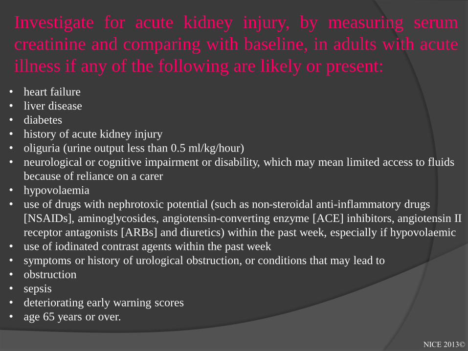
Investigate for acute kidney injury, by measuring serum
creatinine and comparing with baseline, in adults with acute
illness if any of the following are likely or present:
• heart failure
• liver disease
• diabetes
• history of acute kidney injury
• oliguria (urine output less than 0.5 ml/kg/hour)
• neurological or cognitive impairment or disability, which may mean limited access to fluids
because of reliance on a carer
• hypovolaemia
• use of drugs with nephrotoxic potential (such as non-steroidal anti-inflammatory drugs
[NSAIDs], aminoglycosides, angiotensin-converting enzyme [ACE] inhibitors, angiotensin II
receptor antagonists [ARBs] and diuretics) within the past week, especially if hypovolaemic
• use of iodinated contrast agents within the past week
• symptoms or history of urological obstruction, or conditions that may lead to
• obstruction
• sepsis
• deteriorating early warning scores
• age 65 years or over.
NICE 2013©

Monitoring and preventing deterioration
in patients with or at high risk of acute
kidney injury
• Detecting acute kidney injury with the RIFLE, AKIN or KDIGO
definitions
• Identifying the cause(s) of acute kidney injury:
- Urinalysis
- Ultrasound (useful for obstructive forms)
- Doppler (to assess renal blood flow)
- Nuclear Medicine Scans
• Managing acute kidney injury:
- Relieving urological obstruction
- Pharmacological management
- Referring for renal replacement therapy
- Referring to nephrology

Acute kidney injury: common
clinical features azotemia
hypervolemia
electrolytes abnormalities:
K+ phosphate
Na+ calcium
metabolic acidosis
hypertension
oliguria – anuria (a biomarker of
tubular injury)
atheroembolies in small vessels

Urine Findings
Complete anuria early in the course of AKI is uncommon except in
the following situations: complete urinary tract obstruction, renal
artery occlusion, overwhelming septic shock, severe ischemia
(often with cortical necrosis), or severe proliferative
glomerulonephritis or vasculitis.
Oliguria, defined as <500 mL/24 h) usually denotes more severe
AKI (i.e., lower GFR) than when urine output is preserved.
Proteinuria > 1 g/d in AKI suggests damage to the glomerular
ultrafiltration barrier or excretion of myeloma light chains
Extremely heavy proteinuria ("nephrotic range“ >3.5 g/d) can
occasionally be seen in glomerulonephritis, vasculitis, or
interstitial nephritis

Acute Kidney Injury: Urine Volume Anuria (< 100 ml/24h)
Acute bilateral arterial or venous occlusion
Bilateral cortical necrosis
Acute necrotizing glomerulonephritis
Obstruction (complete)
ATN (very rare)
Oliguria (100-500 ml/24h)
Pre-renal azotemia
ATN
Non-Oliguria (> 500 ml/24h)
ATN
Obstruction (partial)

Interpretation of urinary sediment
findings in acute kidney injury
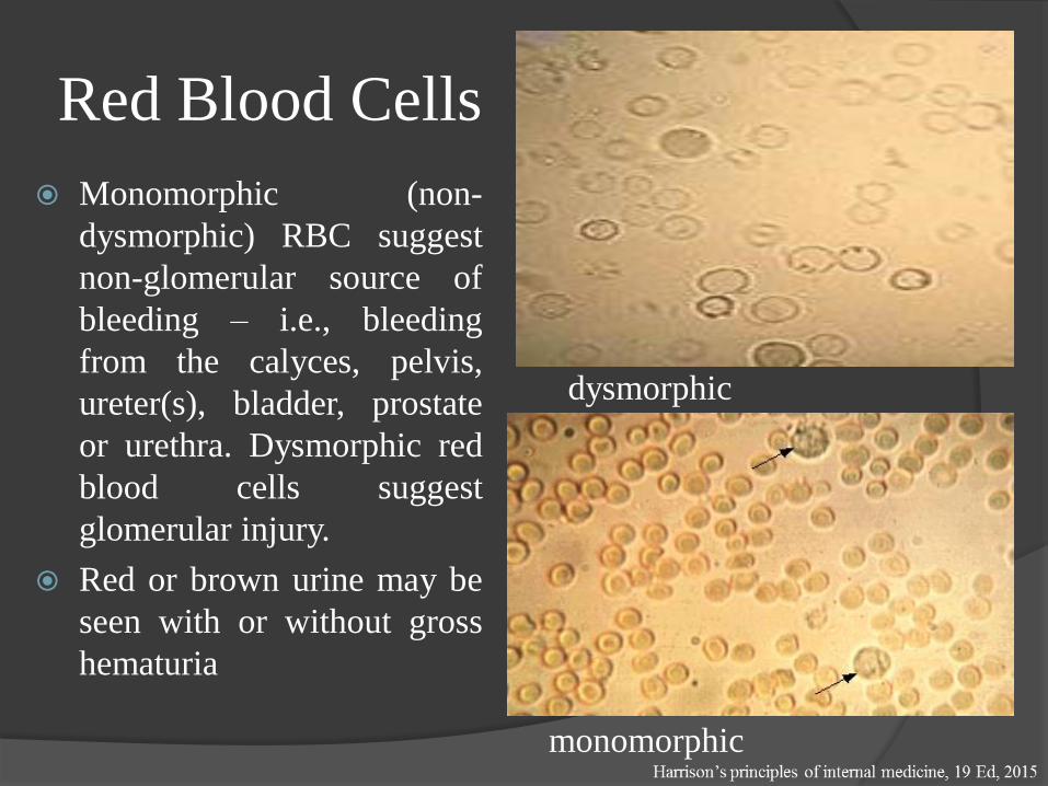
Red Blood Cells
Monomorphic (non-
dysmorphic) RBC suggest
non-glomerular source of
bleeding – i.e., bleeding
from the calyces, pelvis,
ureter(s), bladder, prostate
or urethra. Dysmorphic red
blood cells suggest
glomerular injury.
Red or brown urine may be
seen with or without gross
hematuria
dysmorphic
monomorphic
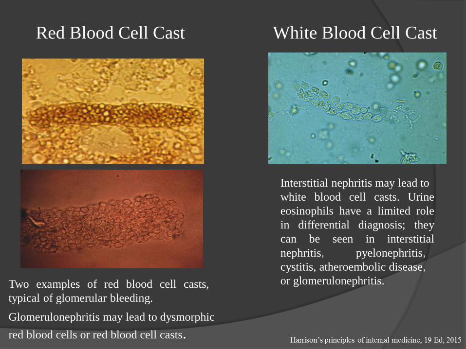
Red Blood Cell Cast
Two examples of red blood cell casts,
typical of glomerular bleeding.
White Blood Cell Cast
Glomerulonephritis may lead to dysmorphic
red blood cells or red blood cell casts.
Interstitial nephritis may lead to
white blood cell casts. Urine
eosinophils have a limited role
in differential diagnosis; they
can be seen in interstitial
nephritis, pyelonephritis,cystitis, atheroembolic disease,or glomerulonephritis.

Renal Tubular Epithelial
Cell CastPigmented Granular
Casts
Pigmented granular (“muddy brown”)
casts are characteristic of acute tubular
necrosis

Renal Failure IndicesSeveral indices have been used to help differentiate prerenal azotemia
from intrinsic AKI when the tubules are malfunctioning.
Renal Failure Index (RFI)
• The fractional excretion of sodium (FeNa) is the fraction of the filtered
sodium load that is reabsorbed by the tubules, and is a measure of both
the kidney's ability to reabsorb sodium as well as endogenously and
exogenously administered factors that affect tubular reabsorption.
• Urine osmolality - in the patient not taking diuretics and with good
baseline kidney function, urine osmolality may be above 500
mOsm/kg in prerenal azotemia. Loss of concentrating ability is
common in septic or ischemic AKI, resulting in urine osmolality
below 350 mOsm/kg
urine [Na]
urine creatinine / serum creatinine=

Urine ChemistriesFractional Excretion of Na and Urea
Since urinary indices depend on urine sodium concentration,they should be interpreted cautiously if the patient has receiveddiuretic
Spot urine Na may be affected (raised) by diuretic use andbaseline impaired kidney function (chronic renal disease wheremaximum urine Na reabsorption is impaired)
Fractional excretion of Na accounts for this by including creatinine:
FENa= urine [Na] ÷ plasma [Na] X 100
urine creatinine ÷ plasma creatinine
FxExurea – substitute urine urea nitrogen for Na useful if patient receiving diuretics

Normal kidney on ultrasound Hydronephrosis on ultrasound
AKI: Ultrasound Findings
Kidney Cancer Ultrasound Diabetic Kidney Ultrasound
http://yandex.ua/clck/jsredir?from=yandex.ua%3Bimages%2Fsearch%3Bimages%3B%3B&text

AKI: Novel Biomarkers
BUN and creatinine are functional biomarkers of glomerular
filtration rather than tissue injury biomarkers
Kidney injury molecule-l (KIM-1) is a type 1 transmembrane
protein that is abundantly expressed in proximal tubular cells
injured by ischemia or nephrotoxins such as cisplatin, can be
detected shortly after ischemic or nephrotoxic injury in the urine
Neutrophil gelatinase associated lipocalin (NGAL, also known as
lipocalin-2 or siderocalin) - a protein in granules of human
neutrophils, can be detected in the plasma and urine within 2 h of
cardiopulmonarγ bypass-associated AKI.
Interleukin (IL) 18 - a pro-inflammatory cytokine of the IL- l
superfamily that may mediate ischemic proximal tubular injury
L-type fatty acid binding protein - from ischemic proximal tubule
cells

Acute Kidney Injury: Pre-renal Causes
Decreased effective perfusion without cellular injury
Hemorrhage
Sodium depletion
Pump failure
Increased vascular capacity e.g., sepsis
Increased renal vascular resistance (hepatorenal, NSAIDs)
Vasoconstriction drugs (radio contrast, cyclosporine, amphotericin B)
Decreased intraglomerular pressure (ACEI, ARB)
Redistribution of ECF
“Third space” accumulation
Edematous disorders
Drugs
Renal tubular and glomerular functions are intact
Reversible if underlying cause is corrected
Prerenal azotemia (from "azo" meaning nitrogen, and "- emia") is the most common
form of AKI.

Pre-Renal Azotemia Pathophysiology Renal hypoperfusion
Decreased RBF (renal blood flow) and GFR
Increased filtration fraction (GFR/RBF)
Increased Na and H2O reabsorption
Oliguria, high Uosm, low UNa
Elevated BUN/Cr ratio
In the absence of renal artery stenosis, renal arterial
pressure (RAP) is the same as systemic mean arterial
pressure (commonly referred to by nephrologists as
“renal perfusion pressure”).
Renal venous pressure (RVP) is usually, but not
always, low and relatively constant.
The glomerular afferent and efferent arterioles are the
major sites of renal vascular resistance (Raff and Reff,
respectively); changes in either will affect renal blood
flow (RBF).

Pre-renal AKI Physical examination – focus on volume status
Vital signs – current and preceding the development of AKI
Neck veins, lungs, heart, mucous membranes
Edema – presacral and extremity
Laboratory studies
BUN : creatinine ratio – elevated in pre-renal; >10-20:1
Unremarkable urinary sediment, high specific gravity
May see hyaline casts
Urine dipstick negative (no blood or protein)
There is no intrinsic kidney damage in pre-renal ARF; rising BUN and
creatinine occur because the kidneys are inadequately perfused.

Pre-renal AKI Urine Electrolytes
Intact renal tubular function in the setting of impaired renal
perfusion (due either to volume loss, pump failure, renal
vasoconstriction , etc) results in avid tubular reabsorption of
sodium.
Therefore, low urine Na (<20 mEq/L) and low fractional
excretion of Na (<1%) and of urea (<35%) in pre-renal ARF.
Therefore, normal renal physiologic responses occur,
manifested in urine electrolytes that reflect intact kidney tubular
function.
Prerenal azotemia and ischemic tubular necrosis represent a
continuum. Azotemia progresses to necrosis when blood flow
is sufficiently compromised to result in the death of tubular
cells.

AKI: Renal or IntrinsicThe most common causes of intrinsic AKI are sepsis, ischemia and nephrotoxins,
both endogenous and exogenous


Renal or Intrinsic AKI
In all types of intrinsic ARF, BUN : creatinine ratio preserved
(10-20:1)
The history, PE, and especially, urine analysis will help to
differentiate
Classified according to primary site of injury:
Vascular
Glomerular (acute glomerulonephritis)
Tubular (acute tubular necrosis or ATN)
Interstitial (acute interstitial nephritis)

Renal or Intrinsic AKI – Vascular
Type Large vessels – must be bilateral
Renal vein thrombosis
Renal artery stenosis
Urine eosinophils
Low C3
Small vessels
Vasculitis
Atheroembolic
Malignant hypertension
Thrombotic microangiopathies.

Small vessels – atheroembolic
Relatively common
Risk factors = catheter manipulation and anticoagulation in
the setting of atherosclerosis
PE may reveal livedo reticularis
Progressive rise then stabilization of BUN, creatinine usually
without significant recovery of kidney function
Cholesterol clefts occ seen on kidney biopsy but transient so
bx not suggested; Dx clinically

Renal or Intrinsic AKI – Glomerular
Type History – systemic or primary kidney
– edema
PE – BP (usually hypertensive)
– edema
BUN : creatinine ratio preserved
Urine analysis: + protein, blood (RBCs, RBC casts)
Often will require kidney biopsy
RBC Casts
Renal or Intrinsic AKI – Interstitial
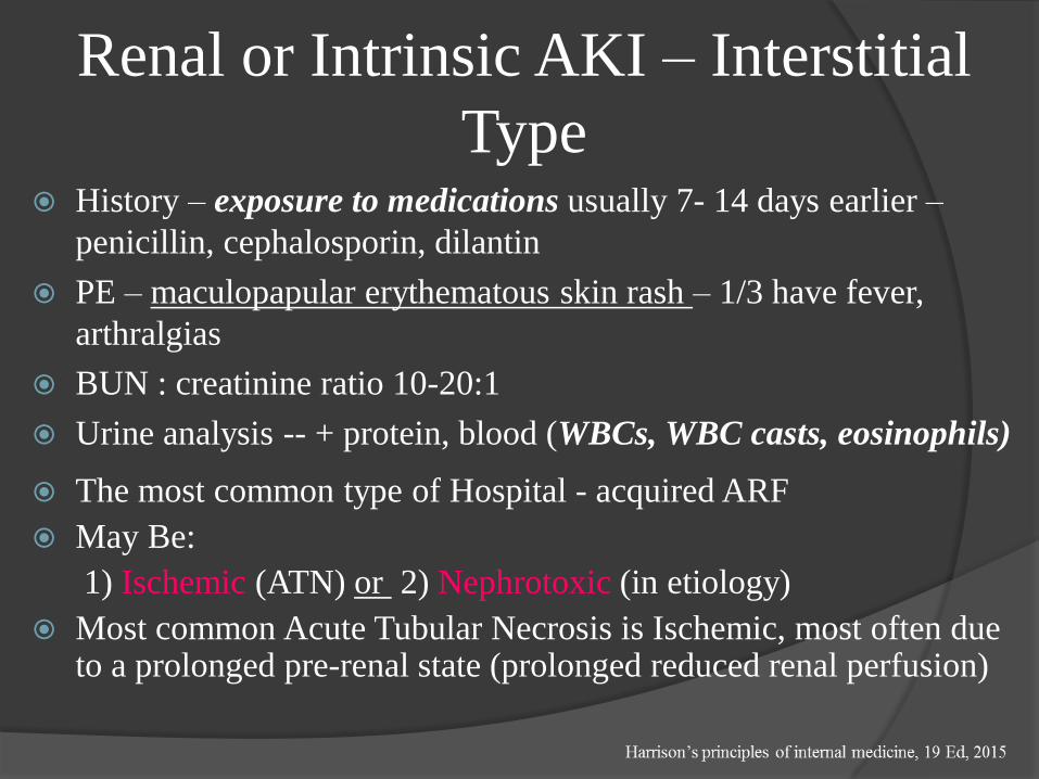
Type History – exposure to medications usually 7- 14 days earlier –
penicillin, cephalosporin, dilantin
PE – maculopapular erythematous skin rash – 1/3 have fever,
arthralgias
BUN : creatinine ratio 10-20:1
Urine analysis -- + protein, blood (WBCs, WBC casts, eosinophils)
The most common type of Hospital - acquired ARF
May Be:
1) Ischemic (ATN) or 2) Nephrotoxic (in etiology)
Most common Acute Tubular Necrosis is Ischemic, most often due to a prolonged pre-renal state (prolonged reduced renal perfusion)

Renal or Intrinsic AKI – Acute
Tubular Necrosis History – prolonged pre-renal state
exposure to nephrotoxin
aminoglycoside antibiotics
ethylene glycol
pigments (myoglobin, hemoglobin)
PE – volume status (to exclude pre-renal ARF)
BUN:creat ratio preserved (10-20:1)
Urine analysis – usually negative protein, blood
- granular casts (dirty brown casts)
- renal tubular epithelial cells
Urine chemistries – urine Na>40 meq/L

Prior to the article by Anderson , it was believed that most casesof ATN were oliguric. Today, we know that ATN can presentwith oliguria or non-oliguria and that both presentations arecommon. Any cause of ATN can present with nonoliguria ; non-oliguria is more likely with nephrotoxic causes of ATN such asaminoglycosides, contrast media, cis-platinum, andamphotericin.
The increased incidence of non-oliguric ATN during the past 25
years is most likely due to the increased usage of nephrotoxins,
more frequent chemical testing, and more aggressive use of
fluids, potent diuretics, and vasodilators in the management of
ATN. The reduced mortality of non-oliguric compared to
oliguric ATN is probably not because of the increased urine
volume but rather due to a lower associated mortality of the
conditions causing non-oliguric compared to oliguric ATN.
(Data from Anderson et al: Non-oliguric Acute Renal Failure.
New Engl J Med 296:134, 1977.)

Acute Tubular Necrosis

Acute Tubular Necrosis
Acute tubular necrosis
showing focal loss of tubular
epithelial cells (arrows) and
partial occlusion of tubular
lumens by cellular debris (D)
(H&E stain).
Tubular epithelialdegeneration and hyalineamphophilic casts (positivewith immunologic stains formyoglobin) in a patient withrhabdomyolysis andmyoglobinuric acute tubularnecrosis.
The kidney biopsy can provide definitive diagnostic and
prognostic information about acute kidney diseases and CKD.
Acute Interstitial NephritisCauses:
Allergic interstitial nephritis
Drugs (aminoglycosides, cisplatin and carboplatin)
Infections
Bacterial (sepsis)
Viral
Sarcoidosis
Radiation nephritis
Iodinated contrast agents
Risk factors for nephrotoxicity include older age, chronic
kidney disease, and prerenal azotemia

Acute Interstitial NephritisClinical Characteristics
Fever
Rash
Arthralgias
Eosinophilia
Urinalysis
Microscopic hematuria
Sterile pyuria
Eosinophiluria
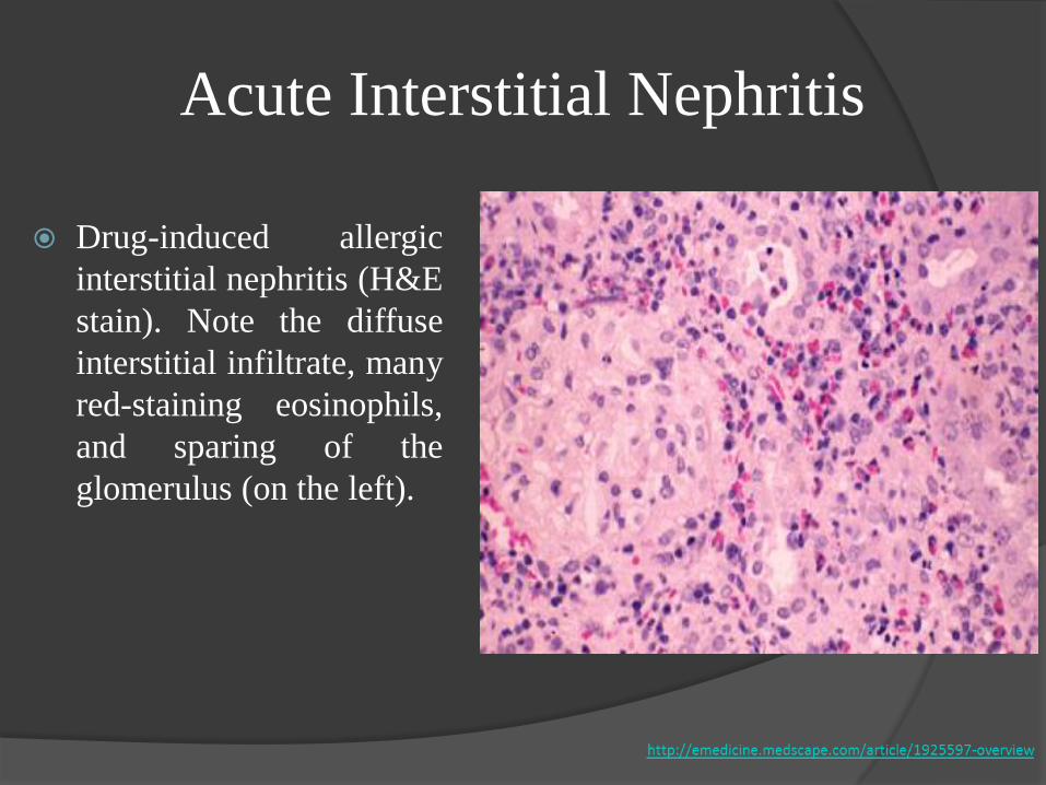
Acute Interstitial Nephritis
Drug-induced allergic
interstitial nephritis (H&E
stain). Note the diffuse
interstitial infiltrate, many
red-staining eosinophils,
and sparing of the
glomerulus (on the left).
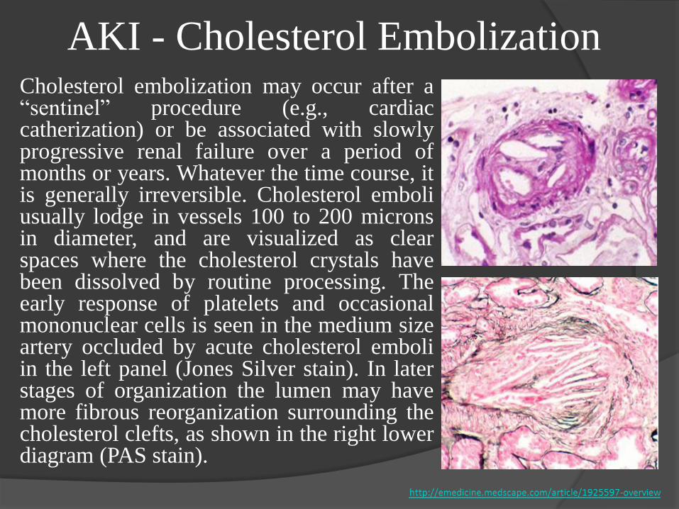
AKI - Cholesterol EmbolizationCholesterol embolization may occur after a“sentinel” procedure (e.g., cardiaccatherization) or be associated with slowlyprogressive renal failure over a period ofmonths or years. Whatever the time course, itis generally irreversible. Cholesterol emboliusually lodge in vessels 100 to 200 micronsin diameter, and are visualized as clearspaces where the cholesterol crystals havebeen dissolved by routine processing. Theearly response of platelets and occasionalmononuclear cells is seen in the medium sizeartery occluded by acute cholesterol emboliin the left panel (Jones Silver stain). In laterstages of organization the lumen may havemore fibrous reorganization surrounding thecholesterol clefts, as shown in the right lowerdiagram (PAS stain).

Acute Kidney Injury: Post-renal
Causes Intra-renal Obstruction
Acute uric acid nephropathy
Drugs (e.g., acyclovir)
Extra-renal Obstruction
Renal pelvis or ureter ;must be
bilateral unless solitary
kidney (e.g., stones, clots,
tumors, papillary necrosis,
retroperitoneal fibrosis)
Bladder (e.g., BPH,
neuropathic bladder)
Urethra (e.g., stricture)

Acute Kidney Injury: Post-renal
Postrenal AKI occurs when the normally directional flow of urine is
acutely blocked either partially or totally, leading to increased retrograde
hydrostatic pressure and interference with glomerular filtration.
Obstruction to urinary flow - from the renal pelvis to the tip of the
urethra. For AKI to occur in healthy individuals, obstruction must
affect both kidneys unless only one kidney is functional, in which case
unilateral obstruction can cause AKI.
Elevated pressure in urinary conduits results in renal parenchymal
destruction if unrelieved - an initial period of hyperemia from afferent
arteriolar dilation is followed by intrarenal vasoconstriction from the
generation of angiotensin II, thromboxane A2, and vasopressin, and
a reduction in NO production
Important to rule out quickly:
potential for recovery of renal function is often inversely related to the
duration of the obstruction

History – symptoms (frequency, hesitancy, etc)
- carcinoma, DM, stones, medications in anamnesis
PE – distended bladder, prostatic enlargement, pelvic masses, lymphnodes
Laboratory studies
-- elevated BUN:creat ratio
-- unremarkable urine sediment
-- variable urine chemistries
Bladder catheterization
Renal ultrasound – hydronephrosis
Treatment is to relieve the obstruction:
- Bladder catheterization
- Nephrostomy tubes

AKI: Differential Diagnosis
Acute kidney injury: NICE guideline DRAFT (March 2013), http://www.kidney-international.org
AKD, acute kidney diseases and disorders; AKI, acute kidney injury; CKD, chronic kidney disease;
GFR, glomerular filtration rate; NKD, no known kidney disease; SCr, serum creatinine.

Prerenal azotemia vs Renal azotemia
Urine sediment: hyaline
and fine granular casts
Urinary to plasma
creatinine ratio: high
Urinary Na: low
FENa: low
Increased urine output in
response to hydration
Urine sediment: brown
granular casts and
tubular epithelial cells
Urinary to plasma
creatinine ratio: low
Urinary Na: high
FENa: high

Prenal Renal
BUN/Cr >20 <20
FeNa <1% >1%
RFI <1% >1%
UNa (mEq/L) <20 > 40
Specific gravity high low

AKI vs progression of Chronic Kidney
Disease Distinction important (etiology, prognosis, therapy differ)
Past BUN, creatinine values; course of BUN, creatinine rise
Half and half nails
Kidney size and echogenicity by ultrasound (10 cm lower
limit of normal; normal size usually 11-12 cm depending on
height)

AKI: Complications
Uremia
Hypo- or hypervolemia
Hyponatremia
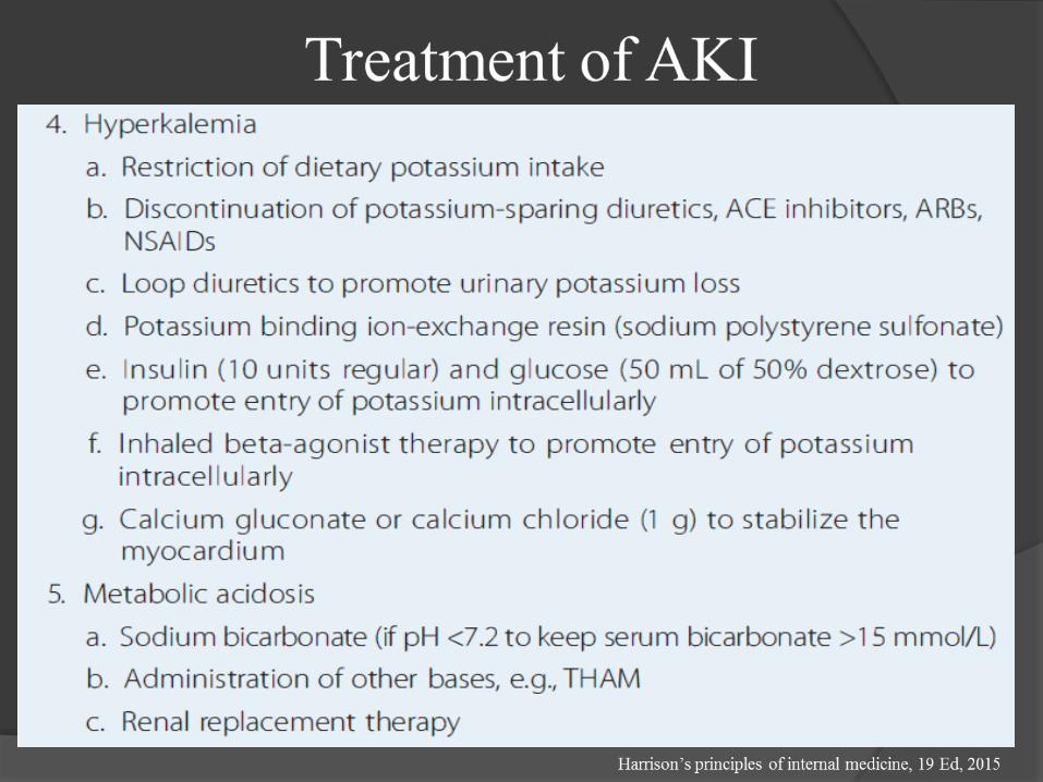
Hyperkalemia
Acidosis
Hyperphosphatemia and hypocalcemia
Bleeding
Infections
Cardiac complications
Malnutrition

Acute Kidney Injury: Prevention
Recognize Patients At Risk (Postoperative States, CardiacSurgery, Septic Shock)
The management of individuals with and at risk for AKI variesaccording to the underlying cause
Prevent Progression From Prerenal To Renal
Preserve Renal Perfusion:
Isovolemia, Cardiac Output, Normal Blood Pressure
Avoid Nephrotoxins (Aminoglycosides, NSAIDS,Amphotericin)

Treatment of AKI
Based on type/etiology of AKI (acute kidney injury) i.e., pre-renal,post-renal, or intrinsic renal initially
Pre-renal – volume, improve renal perfusion
Post-renal – relieve obstruction
Intrinsic – glomerular, tubular, interstitial, vascular depends ontype
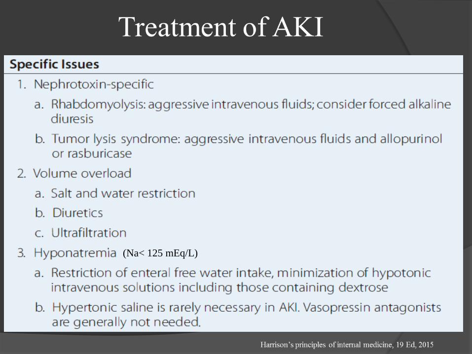
(Na< 125 mEq/L)


Hemoglobinuria:
Transfusion Reactions, HUS (Hemolytic Uremic Syndrome),ECMO (Extra-corporeal Membrane Oxygenation)
Myoglobinuria:
Crush Injuries, Rhabdomyolysis
Urine (+) Blood, But (-) Red Blood Cells
CFK, K+
Treatment
Aggressive Hydration + Urine Alkalinization,
Mannitol / Furosemide
AKI: Hemoglobinuria + Myoglobinuria

Acute Kidney Injury: fluid therapy
If patient is fluid overloaded
○ fluid restriction (insensible losses)
○ attempt furosemide 1-2 mg/kg
○ Renal replacement therapy (see later)
If patient is dehydrated:
○ restore intravascular volume first
○ then treat as euvolemic (below)
If patient is euvolemic:
○ restrict to insensible losses (30-35 ml/100kcal/24 hours)+ other losses (urine, chest tubes, etc)

AKI – Indications for Dialysis
A – acidosis
E – electrolyte disturb., usually hyperkalemia
I – intoxications (lithium, ethylene glycol, etc)
O – overload (volume overload)
U – uremia (symptoms, signs)
Dialysis is indicated when medical management fails to control volume overload,
hyperkalemia, or acidosis. The timing of dialysis is still a matter of debate. Late
initiation of dialysis carries the risk of avoidable volume, electrolyte and metabolic
complications of AKI. On the other hand, initiating dialysis too early may
unnecessarily expose individuals to intravenous lines and invasive procedures,with the attendant risks of infection, bleeding, procedural complications, a n d
hypotension. The initiation of dialysis should not await the development of a life-
threatening complication of renal failure.