Embed Size (px)
Citation preview

1
Bioanalytical Chemistry Fall 2010Schedule (Mondays 2:30-5:30 pm)September
13
Lecture 1September
20
Lecture 2September 27
Lecture 3October 4
Lecture 4October 11
No classes –
Reading WeekOctober 18
Lecture 5October 25
Lecture 6November 1
Midterm exam (30%)November 8
Lecture 7November 12
Last date to drop courses without receiving a gradeNovember 15
Lecture 8November 22
Lecture 9November 29
Lecture 10December 6
Project presentation (30%)December 12
Exams start, Final exam (40%)
http://www.chem.yorku.ca/profs/krylov/Teaching
2
Homeostasis of Multi-cellular Organisms Homeostasis -
definition
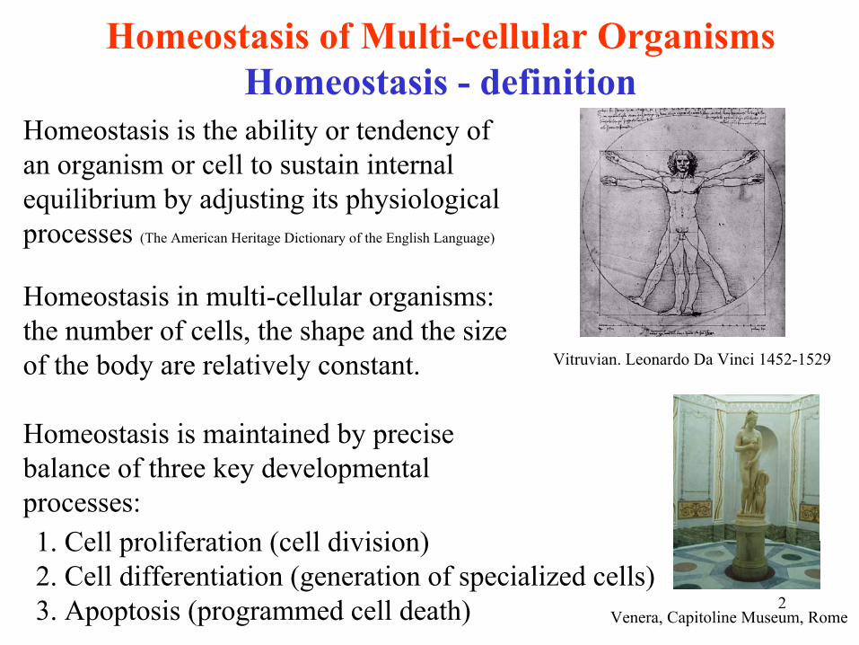
Homeostasis is the ability or tendency of an organism or cell to sustain internal equilibrium by adjusting its physiological processes (The American Heritage Dictionary of the English Language)
Homeostasis in multi-cellular organisms: the number of cells, the shape and the size of the body are relatively constant.
Homeostasis is maintained by precise balance of three key developmental processes:
Vitruvian. Leonardo Da
Vinci 1452-1529
1. Cell proliferation (cell division)2. Cell differentiation (generation of specialized cells)3. Apoptosis (programmed cell death) Venera, Capitoline Museum, Rome
3
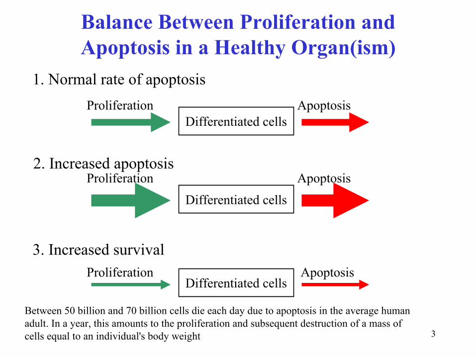
Balance Between Proliferation and Apoptosis in a Healthy Organ(ism)
Proliferation Apoptosis
Proliferation Apoptosis
Proliferation Apoptosis
1. Normal rate of apoptosis
2. Increased apoptosis
3. Increased survival
Differentiated cells
Differentiated cells
Differentiated cells
Between 50 billion and 70 billion cells die each day due to apoptosis in the average human adult. In a year, this amounts to the proliferation and subsequent destruction of a mass of cells equal to an individual's body weight
4
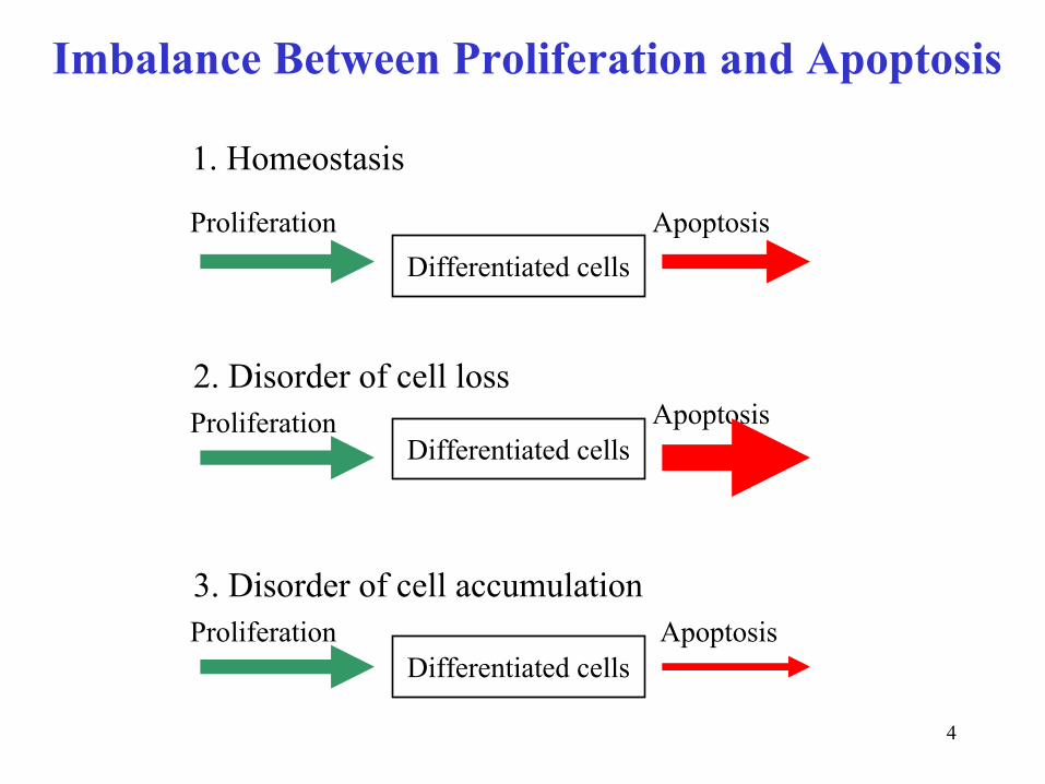
Imbalance Between Proliferation and Apoptosis
Proliferation Apoptosis
Proliferation Apoptosis
Proliferation Apoptosis
1. Homeostasis
2. Disorder of cell loss
3. Disorder of cell accumulation
Differentiated cells
Differentiated cells
Differentiated cells

5
Homeostasis DisordersIn adult organisms, if Proliferation Rate ≠
Apoptosis Rate
then we
deal with Homeostasis Disorders:
Cell loss disordersapoptosis > proliferation
Cell accumulation disordersproliferation > apoptosis
- AIDS-
Neurodegradation
-
Ischemic injuries
-
Autoimmunity-
Viral Infections
- Cancer
Being able to treat these disorders requires that: 1.
The molecular mechanisms of cell proliferation, cell differentiation and apoptosis be well understood
2.
Interplay of the three mechanisms be well understood (intuitively we feel that this interplay can be achieved through
common molecules participating in the three processes)

6
Most of cells in our organism do not and cannot proliferate. Such cells are called differentiated cells.Three types of cells can proliferate: 1) germ cells, 2) stem cells and 3) tumor cells. Non-germ cells proliferate through a mitotic cycle (or cell cycle for simplicity):
Cell Proliferation
RNA and protein synthesis occurs continuously,DNA synthesis occurs only during the S phaseThis feature can be used to determine cell’s position in the cell cycle
7
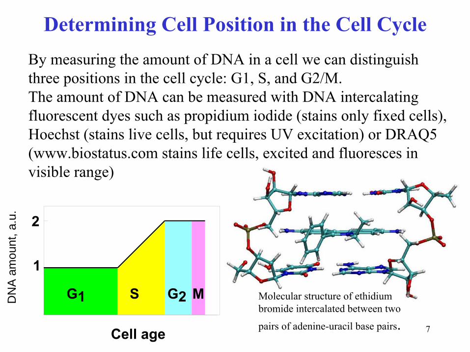
Determining Cell Position in the Cell CycleD
NA
am
ount
, a.u
.
By measuring the amount of DNA in a cell we can distinguish three positions in the cell cycle: G1, S, and G2/M.The amount of DNA can be measured with DNA intercalating fluorescent dyes such as propidium
iodide (stains only fixed cells),
Hoechst (stains live cells, but requires UV excitation) or DRAQ5 (www.biostatus.com
stains life cells, excited and fluoresces in
visible range)
Molecular structure of ethidium
bromide intercalated between two
pairs of adenine-uracil
base pairs.

8
Key Molecular Players in the Cell Cycle (all of them are proteins)
Kinases
are the main engines of the cell cycle machineryKinase
-
enzyme catalyzing phosphorylation
(in contrast to
phosphatase
that catalyzes de-phosphorylation)
Cyclin
Dependent Kinase
(CDK) Machinery
CDK itself catalytic subunit (Engine)serine/threonine
protein kinase
always present in the cellsCyclin
regulatory subunit (Gas pedal)associates with and activates CDKaccumulates during cell cycle (origin of the name) and destroyed during mitosis
CKI
Cyclin
Dependent Kinase
Inhibitor (Brake)
9
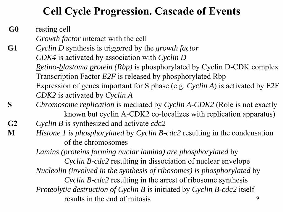
Cell Cycle Progression. Cascade of EventsG0
resting cellGrowth factor interact with the cell
G1
Cyclin D synthesis is triggered by the growth factorCDK4 is activated by association with Cyclin DRetino-blastoma protein (Rbp) is phosphorylated by Cyclin D-CDK complexTranscription Factor E2F is released by phosphorylated RbpExpression of genes important for S phase (e.g. Cyclin A) is activated by E2FCDK2 is activated by Cyclin A
S
Chromosome replication is mediated by Cyclin A-CDK2 (Role is not exactly known but cyclin A-CDK2 co-localizes with replication apparatus)
G2
Cyclin B is synthesized and activate cdc2M
Histone 1 is phosphorylated by Cyclin B-cdc2 resulting in the condensationof the chromosomes
Lamins (proteins forming nuclar lamina) are phosphorylated by Cyclin B-cdc2 resulting in dissociation of nuclear envelope
Nucleolin (involved in the synthesis of ribosomes) is phosphorylated by Cyclin B-cdc2 resulting in the arrest of ribosome synthesis
Proteolytic destruction of Cyclin B is initiated by Cyclin B-cdc2 itself results in the end of mitosis
10
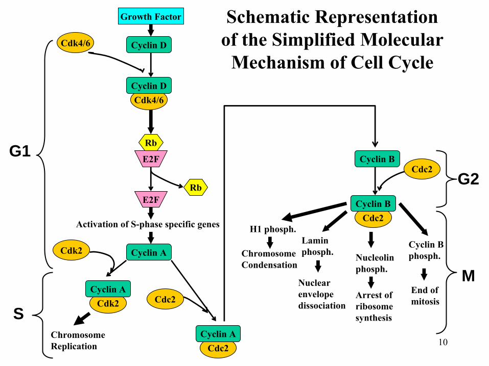
Schematic Representation of the Simplified Molecular
Mechanism of Cell Cycle
Cdc2
Cdc2
Cdk2
Cdk4/6
Cyclin D
Growth Factor
Cdk4/6
Cyclin D
Rb
E2F
E2FRb
Activation of S-phase specific genes
Cyclin ACdk2
Cyclin A
ChromosomeReplication
Cdc2
Cyclin A
Cyclin B
Cyclin B
H1 phosph.
ChromosomeCondensation
Laminphosph.
Nuclearenvelopedissociation
Nucleolinphosph.
Arrest of ribosomesynthesis
Cyclin B phosph.
End ofmitosis
G1
S
G2
M
Cdc2

11
Kinetics of cyclins during the cell cycle
12
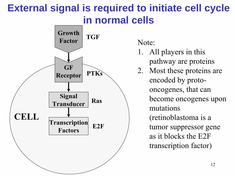
External signal is required to initiate cell cycle in normal cells
GrowthFactor
GFReceptor
SignalTransducer
TranscriptionFactors
CELL
TGF
PTKs
Ras
E2F
Note: 1.
All players in this pathway are proteins
2.
Most these proteins are encoded by proto-
oncogenes, that can become oncogenes upon mutations (retinoblastoma is a tumor suppressor gene as it blocks the E2F transcription factor)
13
Generalization: The Role of External Factors1.
External factors are proteins that are secreted by cells to induce a proper response in neighboring cell.
2.
External factors (they are often called ligands) work through interaction with cellular receptors
3.
Cellular receptors are membrane proteins4.
Upon interaction with external factors, cellular receptors start
a
cascade of reactions that lead to the expression of specific genes.
In general, the cells need external factors to start proliferation, differentiation or apoptosis

14
Control of Cell CycleRestriction point
Point of decision making -
to enter the S phase or not to enter (to divide or not to divide)After the decision to divide is made, the abortion is not allowed and if a serious problem is encountered, the cell should undergo apoptosis
CheckpointsControl loops which make initiation of one event dependent on successful completion of an earlier event.
Examples ofCheckpoint working in specific phases:
1. Completion of previous mitosis before passing R point (G1)2. Completion of DNA replication before entering G2 (S)
Checkpoint working in all phases (externally-induced damages)1. DNA damage by radiation2. Oncogene activation3. DNA tumor viruses4. Hypoxia

15
Manipulations with the Cell Cycle are required to generate many cells in the same cell-cycle position for the
analysis of chemical contents of cells in this positionSynchronizationNatural synchronization Stimulated synchronizationSynchrony of embryo development
1. ArrestLasts for several cycle cycles
2. ReleaseBecomes asynchronous suddenly
Lasts for less than a cycle
Traditional Ways of Cell Cycle ArrestPhase MethodG0
Growth to confluence, Contact InhibitionG1
L-mimosine
(a rare plant amino acid, inhibitor of serine hydroxymethyltransferases)
S
Inhibitors of synthesis of deoxyribonucleotide
triphosphate
(Thymidine)G2
DNA topoisomerase
II inhibitors (Hoechst 33432)M
Inhibitors of tubulin
assembly (Calcimine)
Note:
Physical arrest (contact inhibition) does not disturb normal biochemistry, Chemical arrest (inhibition of one normal cellular process which occurs only at a
specific stage of cell cycle, checkpoint controls arrest the cycle at this point)
disturbs normal cellular biochemistry, often results in cell deathIt is better to work with single cells so that synchronization and arrest are not needed but working with single cells requires advanced bioanalytical methods

16
Cell DifferentiationExample: Differentiation in Dictyostelium discoideum
Dictyostelium discoideum, mexamoebae
(social amoebae [ə
mē′bə])
part-time multi-cellular organism
Lives on decaying logsEats bacteria
In shortage of food supplyThousands of single amoebae aggregateThey form a multi-cellular organism with cells performing different functions
It can move and find new food supplyPart of cells differentiate to form spores. Spores are disseminatedEvery spore gives live to new mexamoebae

17
Mechanism and Some Clues and Conclusions-
Starving cells release cAMP
-
Cells move against the gradient of cAMP•
Differentiation is initiated by external signal
-
cAMP
initiates expression of new proteins -
These proteins are responsible for:
cell adhesion cell differentiation
•
Differentiation is the production of different proteins in different types of cells
-
Several types of cells differentiate from identical ancestor-cells•
All single mexamoebae cells have identical genes
•
Only parts of genes are expressed•
Expression pattern can be changed in response to external
signals – Differential Gene Expression
Cyclic-adenosine-monophosphate
18
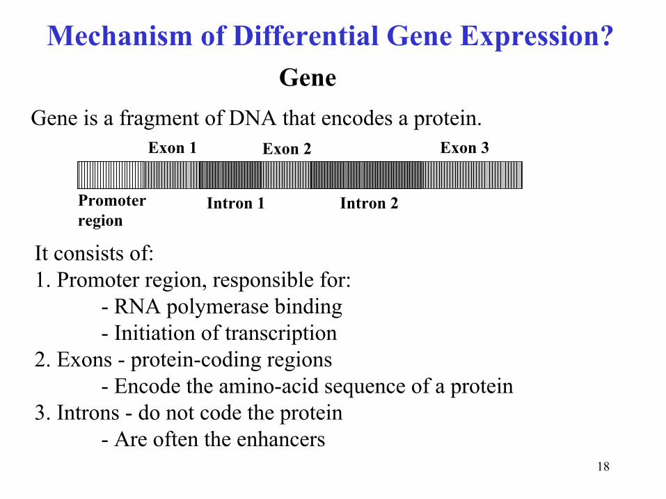
Mechanism of Differential Gene Expression?
It consists of:1. Promoter region, responsible for:
-
RNA polymerase binding-
Initiation of transcription
2. Exons
-
protein-coding regions-
Encode the amino-acid sequence of a protein
3. Introns
-
do not code the protein-
Are often the enhancers
Promoterregion
Exon
1
Intron
1 Intron
2
Exon
2 Exon
3
Gene is a fragment of DNA that encodes a protein.
Gene

19
External signal is required to initiate cell differentiation
ExternalFactor
Receptor
SignalTransducer
TranscriptionFactors
CELL
Note: The signal transduction pathway is in general similar to that of cell cycle initiation
20
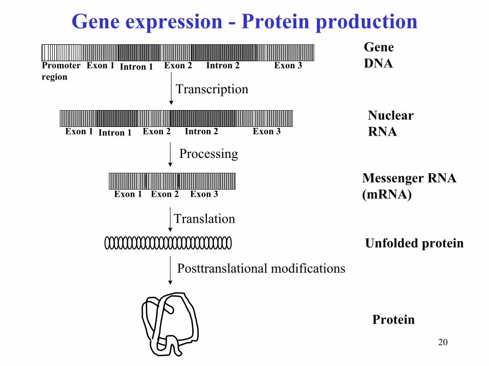
Gene expression -
Protein production
Promoterregion
Exon
1 Intron
1 Intron
2Exon
2 Exon
3
GeneDNA
Exon
1 Intron
1 Intron
2Exon
2 Exon
3NuclearRNA
Exon
1 Exon
2 Exon
3Messenger RNA(mRNA)
Transcription
Processing
Translation
Unfolded protein
Posttranslational modifications
Protein
21
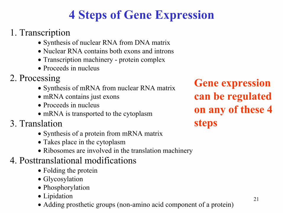
4 Steps of Gene Expression1. Transcription
•
Synthesis of nuclear RNA from DNA matrix•
Nuclear RNA contains both exons
and introns•
Transcription machinery -
protein complex•
Proceeds in nucleus2. Processing
•
Synthesis of mRNA from nuclear RNA matrix•
mRNA contains just exons•
Proceeds in nucleus•
mRNA is transported to the cytoplasm3. Translation
•
Synthesis of a protein from mRNA matrix•
Takes place in the cytoplasm•
Ribosomes
are involved in the translation machinery4. Posttranslational modifications
•
Folding the protein•
Glycosylation•
Phosphorylation•
Lipidation•
Adding prosthetic groups (non-amino acid component of a protein)
Gene expression can be regulated on any of these 4 steps

22
Transcriptional regulation1. Promoters and enhancers
•
Regulatory DNA sequences
•
Promoters bind RNA polymerase
•
Enhancers activate the use of the promoters
•
Enhancers regulate tissue-specific transcription
2. Transcription factors•
Regulatory proteins
•
Bind to the enhancer and/or promoter regions
•
Regulated by phosphorylation
3. DNA Methylation•
5th base in DNA, 5-methylcytosine
•
Differences between male and female pronuclei
are due to
differences in their DNA methylation patterns

23
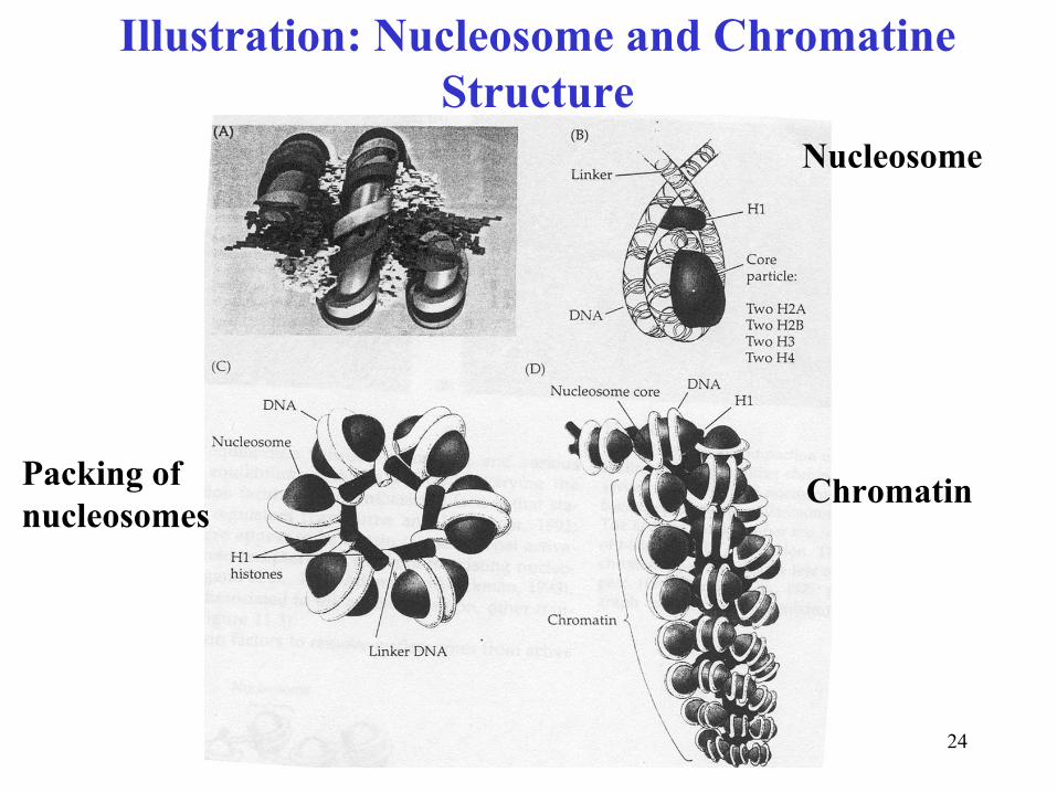
Transcriptional regulation contd.
•
Chromatin is DNA-protein complex•
DNA is tightly and regularly packed in chromatin
• Chromatin consists of nucleosomes•
Nucleosome
is composed of a histone
core, H1 histone, and
two loops of DNA (~140 bp)•
Nucleosome
bids are linked with ~60 bp
DNA
•
DNA in chromatin is not accessible for transcriptional factorsunless chromatin is “activated”
Hypotheses of chromatin “activation”:
1. Transcription factors compete with histone
H1 for specific DNA sequences2. Transcriptional activators (proteins) disrupt nucleosome
so
that transcription factor can reach the promoter region.
4. The activation of chromatin
24
Illustration: Nucleosome
and Chromatine Structure
Nucleosome
Packing ofnucleosomes
Chromatin

25
Regulation by RNA processing1. Control of processing of nuclear RNA into mRNA-
Not all the genes transcribed to nuclear RNAs
have corresponding mRNAs
in the cytoplasm
2. Splicing different combination of potential exons
leads to a variety of related proteins-
Thus, one gene may create a family of related proteins
-
Spliceosomes
(RNA-protein complexes) cut introns
and splice exons. Differential regulation of spliceosomes
can change the RNA processing
pattern.
Note: There are only less then 25,000 human genes encoding proteins instead of ~160,000 thought a couple of decades ago. Alternative splicing is an important way to enrich the pool of proteins with this “small”
genome.
Analysis of splice isoforms of proteins is difficult due to their similarities in structures
26
Translational regulation
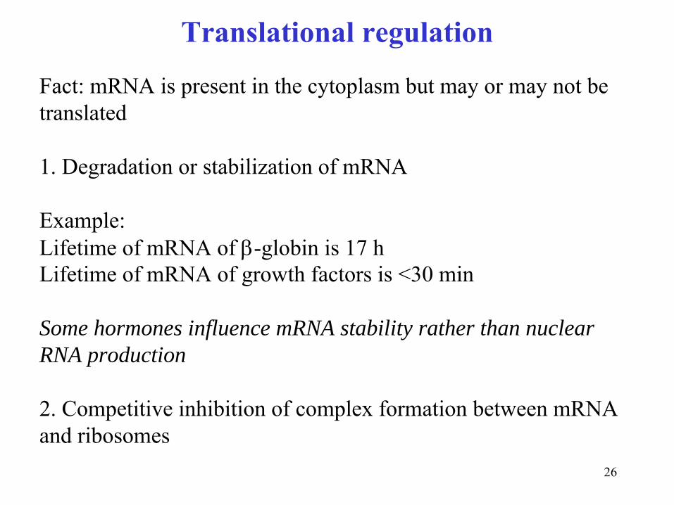
Fact: mRNA is present in the cytoplasm but may or may not be translated
1. Degradation or stabilization of mRNA
Example:Lifetime of mRNA of β-globin
is 17 h
Lifetime of mRNA of growth factors is <30 min
Some hormones influence mRNA stability rather than nuclear RNA production
2. Competitive inhibition of complex formation between mRNA and ribosomes

27
Differentiation in Embryo Development
EggSperm Cleavage
Blastomere
A
Blastomere
B
Blastula
Blastula development
28
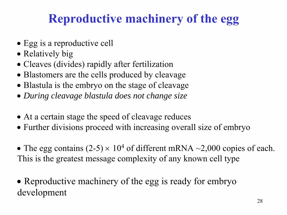
Reproductive machinery of the egg
•
Egg is a reproductive cell
•
Relatively big
•
Cleaves (divides) rapidly after fertilization
•
Blastomers
are the cells produced by cleavage
•
Blastula is the embryo on the stage of cleavage
•
During cleavage blastula does not change size
•
At a certain stage the speed of cleavage reduces
•
Further divisions proceed with increasing overall size of embryo
•
The egg contains (2-5) ×
104
of different mRNA ~2,000 copies of each. This is the greatest message complexity of any known cell type
•
Reproductive machinery of the egg is ready for embryo development
29
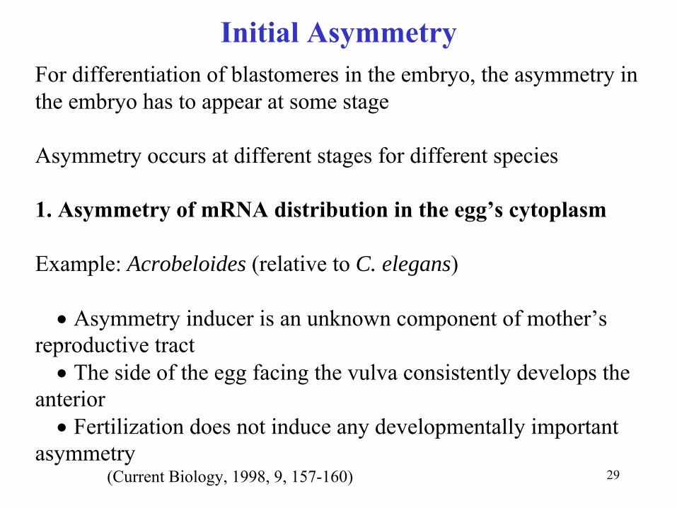
Initial AsymmetryFor differentiation of blastomeres
in the embryo, the asymmetry in
the embryo has to appear at some stage
Asymmetry occurs at different stages for different species
1. Asymmetry of mRNA distribution in the egg’s cytoplasm
Example: Acrobeloides (relative to C. elegans)
•
Asymmetry inducer is an unknown component of mother’s reproductive tract•
The side of the egg facing the vulva consistently develops the
anterior•
Fertilization does not induce any developmentally important
asymmetry(Current Biology, 1998, 9, 157-160)
30
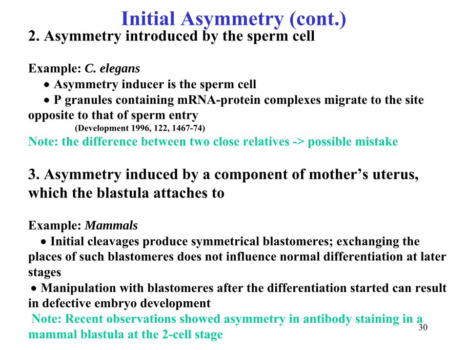
Initial Asymmetry (cont.)2. Asymmetry introduced by the sperm cell
Example: C. elegans•
Asymmetry inducer is the sperm cell•
P granules containing mRNA-protein complexes migrate to the site opposite to that of sperm entry
(Development 1996, 122, 1467-74)Note: the difference between two close relatives -> possible mistake
3. Asymmetry induced by a component of mother’s uterus, which the blastula attaches to
Example: Mammals•
Initial cleavages produce symmetrical blastomeres; exchanging the places of such blastomeres
does not influence normal differentiation at later stages•
Manipulation with blastomeres
after the differentiation started can result in defective embryo developmentNote: Recent observations showed asymmetry in antibody staining in a
mammal blastula at the 2-cell stage

31
Anterior-posterior asymmetryAll three considered above scenarios deal with an anterior-posterior asymmetry or “single-dimensional asymmetry”
In C. elegans three genes are involved in anterior-posterior axis formation:
par, Wnt, lit-1 (Nature, 1997, 390, 294-298)
•
Anterior-posterior asymmetry can produce only axially-symmetric organisms with only asymmetry along the AP axis
•Mammals have two-dimensional asymmetry (one dimensional symmetry: right-left)
•
One extra asymmetry initiator has to participate in differentiation Science (2002), 298(5600), 1946-1948.
32
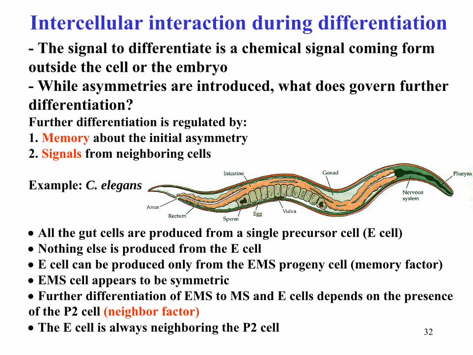
Intercellular interaction during differentiation-
The signal to differentiate is a chemical signal coming form
outside the cell or the embryo-
While asymmetries are introduced, what does govern further
differentiation? Further differentiation is regulated by:1. Memory
about the initial asymmetry2. Signals
from neighboring cells
Example: C. elegans
•
All the gut cells are produced from a single precursor cell (E cell)•
Nothing else is produced from the E cell•
E cell can be produced only from the EMS progeny cell (memory factor)•
EMS cell appears to be symmetric•
Further differentiation of EMS to MS and E cells depends on the presence of the P2 cell (neighbor factor)•
The E cell is always neighboring the P2 cell

33
Early embryogenesis in
C. ElegansFormation of founder cells. Anterior is to the left. The founder cell names and theircontributions to the embryo are indicated. Clones of cells from each founder are indicated by shading. Asymmetrically dividing cells P0
, P1
, P2
, EMS, and P3
are unshaded, with blastomere
names inside the cell borders. The 16-cell embryo at the bottom is highly schematized for simplicity; it shows only approximate relative positions of blastomeres
and does not reflect the fourth ABcleavage, which happens at roughly the same time as the P3 division.

34
Differentiation in a mature organismDifferentiated cells do not proliferate
Most of somatic cells are differentiated
Stem cells do proliferate. There should be many stem cells in tissues undergoing continuous renewal (skin, intestine, blood)
Example: proliferation, differentiation and apoptosis of blood cells
Stemcell
Stemcell
Blood cellProliferation
Differen
tiation Apoptosis
Blood cell
Stemcell

35
Science, September 5, 2003
36
Differentiation and cancerWhy does cancer kill?
•
Cancer cells are undifferentiated•
They do not produce tissue-specific proteins•
They do not serve as the normal tissue cells•
They suppress survival of normal cells•
Undifferentiated cancer cells establish colonies in new tissues where normal differentiated cells would not survive
How does undifferentiation
happen?•
Cancer destroys anti-mutation machinery•
Tissue-specific genes are also mutated•
Cancer cells up-regulate and down-regulate the expression of the intact genes•
Undifferentiation
of cancer cells is often reversible•
Differentiation in cancer cells can be induced by hormones etc.
37
Example: colon cancer
Downregulation
of:fatty-acid binding proteinscytokeratincarbonic anhydraseguanylinuroguanylin
Upregulation
of:growth factorsenzymes involved in glycolysis
Science 1997, 276, 1268-1271
38
The role of Cell Differentiation in cancer diagnostic and therapy
•
Differentiation of cancer cells is an important prognostic factor
•
More differentiated cancer cells are more susceptible to
therapies and easier undergo apoptosis
•
Pregnancy protects from breast cancer through the induction of
a complete differentiation of the mammary gland
•
Retinoic acid is known as differentiation inducer in cancer cells
•
It is a potential anticancer drug

39
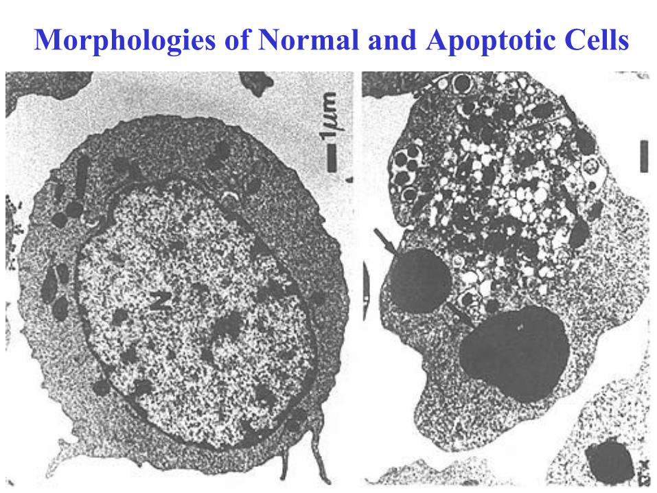
APOPTOSISTwo means of cell death
Necrosis
(Pathological cell death caused by acute cell injury)•
Destruction of cellular membrane•
Leakage of cytoplasmic contents•
Inflammatory response
Apoptosis (Programmed cell death through controlled autodigestion of the cell)
•
Integrity of cellular membrane is maintained•
Endogenous proteases digest important proteins •
Endogenous nucleases degrade DNA•
Disassembled cells brake down into apoptotic bodies•
Apoptotic bodies are phagocytosed by neighboring cells•
No inflammatory response
40
Morphologies of Normal and Apoptotic Cells
41
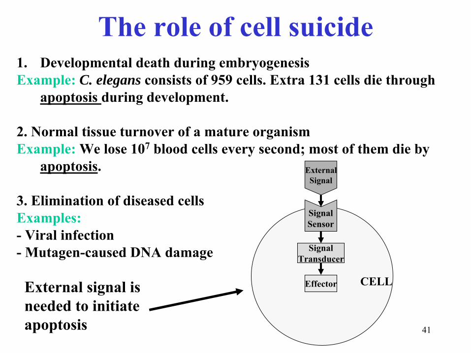
The role of cell suicide1.
Developmental death during embryogenesis
Example:
C. elegans consists of 959 cells. Extra 131 cells die through apoptosis during development.
2. Normal tissue turnover of a mature organismExample:
We lose 107
blood cells every second; most of them die by apoptosis.
3. Elimination of diseased cellsExamples:-
Viral infection
-
Mutagen-caused DNA damage
ExternalSignal
SignalSensor
SignalTransducer
Effector CELLExternal signal is needed to initiate apoptosis

42
Suicide machinery
DeathLigand
Caspases
ProteasesEndonucleases
Mutagen
DNA
p53
Granzyme
B(serine protease)
Caspases
Caspases
Cytotoxic T cellsMacrophages
RadiationChemotherapeutics
CELL
DeathReceptor
43
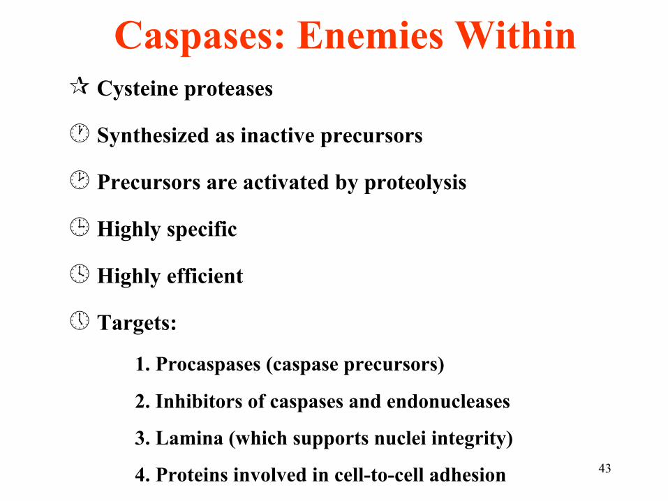
Caspases: Enemies WithinCysteine proteases
Synthesized as inactive precursors
Precursors are activated by proteolysis
Highly specific
Highly efficient
Targets:
1. Procaspases
(caspase
precursors)
2. Inhibitors of caspases
and endonucleases
3. Lamina (which supports nuclei integrity)
4. Proteins involved in cell-to-cell adhesion
44
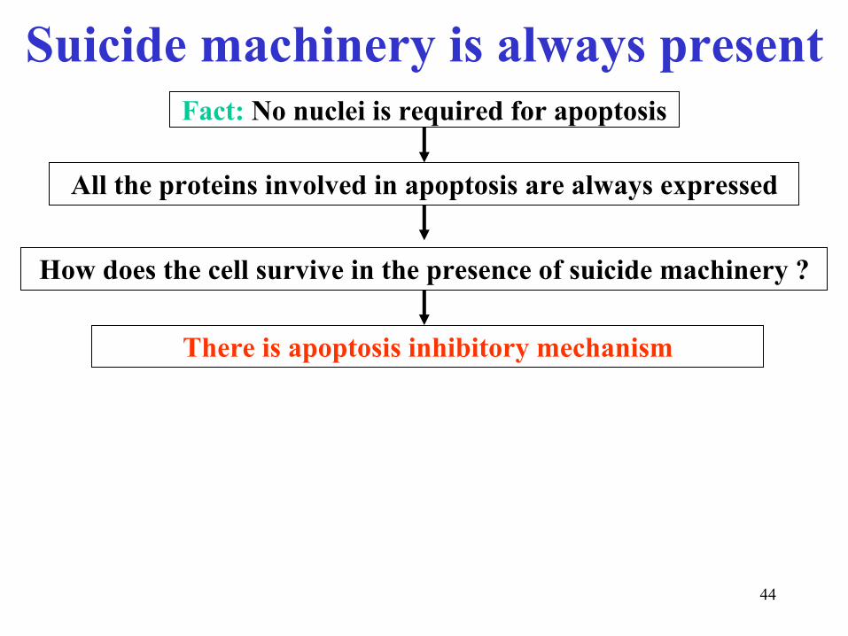
Suicide machinery is always presentFact:
No nuclei is required for apoptosis
All the proteins involved in apoptosis are always expressed
How does the cell survive in the presence of suicide machinery
?
There is apoptosis inhibitory mechanism

45
Inhibition of apoptosis
TNF
Caspases
CaspasesEndonucleases
Mutagen
DNA
p53
Granz.B
Caspases
Caspases
T cellsMacrophages
RadiationChemotheur.
TNFR
Survival factor
Sensor
Transducer
Apoptosis inhibitor (Bcl-2)
46
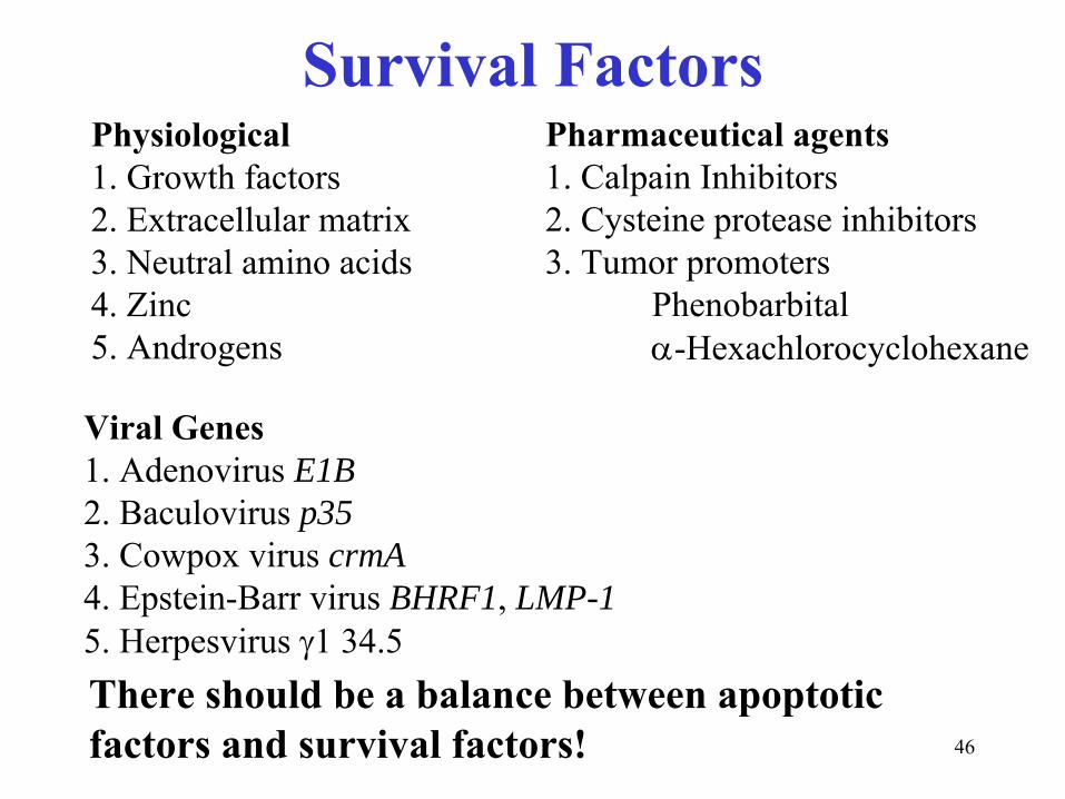
Survival FactorsPhysiological1. Growth factors2. Extracellular matrix3. Neutral amino acids4. Zinc5. Androgens
Viral Genes1. Adenovirus E1B2. Baculovirus p353. Cowpox virus crmA4. Epstein-Barr virus BHRF1, LMP-15. Herpesvirus γ1 34.5
Pharmaceutical agents1. Calpain Inhibitors2. Cysteine protease inhibitors3. Tumor promoters
Phenobarbitalα-Hexachlorocyclohexane
There should be a balance between apoptotic factors and survival factors!