Embed Size (px)
Citation preview

Satin leaf (Chrysophyllum oliviforme) Extract Mediated Green Synthesis of Silver Nanoparticles:
Antioxidant and Anticancer Activities Anju Varghese.R, Anandhi.P, Arunadevi.R, Boovisha.A, Sounthari.P, Saranya.J, Parameswari.K, Chitra.S*
Department of Chemistry, PSGR Krishnammal College for Women,
Coimbatore -641 004, India.
Abstract The recent development and implementation of new technologies have led to new era, the nano-revolution which unfolds role of plants in bio and green synthesis of nanoparticles which seem to have drawn quite an unequivocal attention with a view of synthesizing stable nanoparticles. The biosynthesis of nanoparticles has been proposed as a cost effective and environmental friendly alternative to chemical and physical methods. Plant mediated synthesis of nanoparticles is a green chemistry approach that interconnects nanotechnology and plant biotechnology. In the present study, synthesis of silver nanoparticles (AgNPs) has been demonstrated using extracts of Chrysophyllum oliviforme reducing aqueous silver nitrate. The synthesized nanosilver was characterized by IR, UV, XRD and SEM-EDS. The synthesized nano silver have been screened for antioxidant and anticancer activities. Keywords: Nanosilver, antioxidant, anticancer, biosynthesis, satin leaf.
1. INTRODUCTION
Nanomaterials are cornerstones of nanoscience and nanotechnology. Nanostructure science and technology is a broad and interdisciplinary area of research and development activity that has been growing explosively worldwide in the past few years. It has the potential for revolutionizing the ways in which materials and products are created and the range and nature of functionalities that can be accessed. Nanoscale materials are defined as a set of substances where at least one dimension is less than approximately 100 nanometers. Currently, sustainability initiatives that use green chemistry to improve and/or protect our global environment are focal issues in many fields of research. The development of cost efficient and ecologically benign methods of synthesis of nanomaterial remains a scientific challenge as metal nanoparticles are of use in various catalytic applications, viz electronics, biology and biomedical applications, material science, physics, environmental remediation fields. It is well known that the toxicity of nanomaterials essentially depends on the structural features such as size, shape, composition and the surface chemistry. To prolong the life span of metal nanoparticles it is vital to select stabilizing agents and pathways that are environmentally friendly, non-toxic and easy to implement. Novel methods of ideally synthesizing NPs are thus being thought which are formed at ambient temperatures, neutral pH, low costs and environmentally friendly fashion. Keeping these goals in view nanomaterials have been synthesized using various routes. Among the biological alternatives, plants and plant extracts seem to be the best option. Plants are nature’s “chemical factories”. They are cost efficient and require little or no maintenance. A vast repertoire of secondary metabolites is found in all plants which possess redox capacity and can be exploited for biosynthesis of nanoparticles. As a wide range of
metabolites are presented in the plant products/extracts, nanoparticles produced by plants are more stable and the rate of synthesis is faster in comparison to microorganisms. Thus, the advantages of using plant and plant-derived materials for biosynthesis of metal nanoparticles have instigated researchers to investigate mechanisms of metal ions uptake and bio reduction by plants, and to understand the possible mechanism of metal nanoparticle formation in and by the plants. Silver nanoparticles receive enormous scientific, technological, and commercial attention due to their unique size and shape dependent properties. Extensive research has been devoted to explore the applications of silver nanoparticles in diversified fields including healthcare/ biomedical, sensors, spectroscopy and catalysis. One of the challenging tasks in the synthesis of nanostructured materials is the precise control of size and shape. Especially, silver nanoparticles exhibit drastic variation in their physicochemical properties with the size, shape, and their conjugation with other organic/ biological substances. The synthesis processes of silver nanoparticles play a major role in the control of their size and shape, thus wide range of physical, chemical, as well as biological methods have been established and reported. Among them, biological processes that are based on bacteria, fungus, bio-derived chemicals, and plant extracts are extensively investigated due their eco-friendly protocol and better morphological control. Using “green” methods in the synthesis of silver nanoparticles has increasingly become a topic of interests as conventional chemical methods are expensive and require the use of chemical compounds/organic solvents as reducing agents. Present research has prompted for further exploration in the use of plant extracts for the synthesis of silver nanoparticles.
Anju Varghese.R et al /J. Pharm. Sci. & Res. Vol. 7(6), 2015, 266-273
266
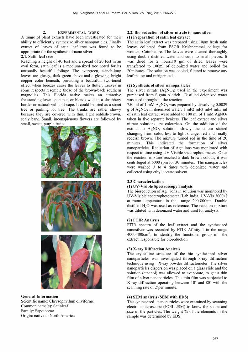
2. EXPERIMENTAL WORK A range of plant extracts have been investigated for their ability to efficiently synthesize silver nanoparticles. Finally extract of leaves of satin leaf tree was found to be appropriate for the synthesis of nano silver. 2.1. Satin leaf tree Reaching a height of 40 feet and a spread of 20 feet in an oval form, satin leaf is a medium-sized tree noted for its unusually beautiful foliage. The evergreen, 4-inch-long leaves are glossy, dark green above and a glowing, bright copper color beneath, providing a beautiful, two-toned effect when breezes cause the leaves to flutter. Leaves in some respects resemble those of the brown-back southern magnolias. This Florida native makes an attractive freestanding lawn specimen or blends well in a shrubbery border or naturalized landscape. It could be tried as a street tree or parking lot tree. The trunks are rather showy because they are covered with thin, light reddish-brown, scaly bark. Small, inconspicuous flowers are followed by small, sweet, purple fruits.
General Information Scientific name: Chrysophyllum oliviforme Common name(s): Satinleaf Family: Sapotaceae Origin: native to North America
2.2. Bio reduction of silver nitrate to nano silver (1) Preparation of satin leaf extract The satin leaf extract was prepared using 10gm fresh satin leaves collected from PSGR Krishnammal college for women, Coimbatore. The leaves were cleaned thoroughly using double distilled water and cut into small pieces. It was dried for 2 hours.10 gm of dried leaves were transferred to 100ml of deionized water and boiled for 20minutes. The solution was cooled, filtered to remove any leaf matter and refrigerated. (2) Synthesis of silver nanoparticles The silver nitrate (AgNO3) used in the experiment was purchased from Sigma Aldrich. Distilled deionized water was used throughout the reaction. 750 ml of 1 mM AgNO3 was prepared by dissolving 0.0029 g of AgNO3 in deionized water. 1 ml/2 ml/3 ml/4 ml/5 ml of satin leaf extract were added to 100 ml of 1 mM AgNO3 taken in five separate beakers. The leaf extract and silver nitrate solutions are colourless. On the addition of the extract to AgNO3 solution, slowly the colour started changing from colourless to light orange, red and finally reddish brown. The mixture turned red in the time of 20 minutes. This indicated the formation of silver nanoparticles. Reduction of Ag+ ions was monitored with respect to time using UV-Visible spectrophotometer. Once the reaction mixture reached a dark brown colour, it was centrifuged at 6000 rpm for 30 minutes. The nanoparticles were washed 3 to 4 times with deionized water and collected using ethyl acetate solvent. 2.3 Characterization (1) UV-Visible Spectroscopy analysis The bioreduction of Ag+ ions in solution was monitored by UV-Visible spectrophotometer [Lab India, UV-Vis 3000+] at room temperature in the range 200-800nm. Double distilled H2O was used as reference. The reaction mixture was diluted with deionized water and used for analysis. (2) FTIR Analysis FTIR spectra of the leaf extract and the synthesized nanosilver was recorded by FTIR Affnity 1 in the range 4000-400cm-1, to identify the functional group in the extract responsible for bioreduction (3) X-ray Diffraction Analysis The crystalline structure of the bio synthesized silver nanoparticles was investigated through x-ray diffraction technique using X-ray powder diffractometer. The silver nanoparticles dispersion was placed on a glass slide and the solution (ethanol) was allowed to evaporate, to get a thin film of silver nanoparticles. This thin film was subjected to X-ray diffraction operating between 10˚ and 80˚ with the scanning rate of 2˚per minute. (4) SEM analysis (SEM with EDS) The synthesized nanoparticles were examined by scanning electron microscope (JOEL JSM) to know the shape and size of the particles. The weight % of the elements in the sample was determined by EDS.
Anju Varghese.R et al /J. Pharm. Sci. & Res. Vol. 7(6), 2015, 266-273
267
2.4 Antioxidant activity DPPH Assay[1] DPPH Radical Scavenging Activity The free radical scavenging capacity of the methanolic extracts of silver nanoparticles (MAP) was determined using DPPH. DPPH solution (0.004% w/v) was prepared in 95% methanol. Methanol extract of MAP was mixed with 95% methanol to prepare the stock solution (10 mg/100mL). The concentration of this MAP solution was 10 mg /100 ml or 100µg/ml. this solution were taken in five test tubes & by serial dilution with same solvent was made the final volume of each test tube up to 10 ml whose concentration was then 20µg/ml, 40µg/ml, 60µg/ml, 80µg/ml & 100µg/ml respectively. Freshly prepared DPPH solution (0.004%w/v) was added in each of these test tubes containing MAP (20 µg/ml, 40µg/ml, 60µg/ml, 80µg/ml, 100µg/ml) and after 10 min, the absorbance was taken at 517nm using a spectrophotometer (HACH 4000 DU UV–visible spectrophotometer). Ascorbic acid was used as a reference standard and dissolved in distilled water to make the stock solution with the same concentration (10 mg/100mL) of methanolic extracts of silver nanoparticle. Control sample was prepared containing the same volume without any extract and reference ascorbic acid. 95% methanol was used as blank. Percentage scavenging of the DPPH free radical was measured using the following equation,
2.5 Anticancer activity [2, 3] IN VITRO CYTOTOXICITY ASSAY METHODOLOGY The human cervical cancer cell line (Hela) was obtained from National Centre for Cell Science (NCCS), Pune and grown in Eagles Minimum Essential Medium (EMEM) containing 10% fetal bovine serum (FBS). All cells were maintained at 370 C, 5% CO2, 95% air and 100% relative humidity. Maintenance cultures were passaged weekly, and the culture medium was changed twice a week. Cell treatment procedure The monolayer cells were detached with trypsin-ethylenediaminetetraacetic acid (EDTA) to make single cell suspensions and viable cells were counted by tryphan blue exclusion assay using a hemocytometer. The cell suspension was diluted with medium containing 5% FBS to give final density of 1x10⁵ cells/ml. one hundred microlitres per well of cell suspension were seeded into 96-well plates at plating density of 10,000 cells/well and incubated to allow for cell attachment at 370C, 5% CO2, 95% air and 100% relative humidity. After 24 h the cells were treated with serial concentrations of the test samples. The samples were dispersed in phosphate buffered saline (PBS) and diluted to twice the desired final maximum test concentration with serum free medium. Additional four, 2 fold serial dilutions were made to provide a total of five sample concentrations. Aliquots of 100 µl of these different
sample dilutions were added to the appropriate wells already containing 100 µl of medium, resulted the required final sample concentrations. Following drug addiction the plates were incubated for an additional 48 h at 370 C, 5% CO2, 95% air and 100% relative humidity. The medium containing without samples were served as control and triplicate was maintained for all concentrations. MTT assay 3-[4,5-dimethylthiazol-2-yl]2,5-diphenyltetrazolium bromide (MTT) is a yellow water soluble tetrazolium salt. A mitochondrial enzyme in living cells, succinate-dehydrogenase, cleaves the tetrazolium ring, converting the MTT to an insoluble purple formazan. Therefore, the amount of formazan produced is directly proportional to the number of viable cells. After 48 h of incubation, 15 µl of MTT (5 mg/ml) in phosphate buffered saline (PBS) was added to each well and incubated at 37 0C for 4 h. The medium with MTT was then flicked off and the formed formazan crystals were solubilized in 100 µl of DMSO and then measured the absorbance at 570 nm using micro plate reader. The percentage cell viability was then calculated with respect to control as follows
100 x [A]Control
[A]Test viabilityCell %
3. RESULTS AND DISCUSSION
In recent years, noble metal nanoparticles have been the subject of focussed research due to their unique optical, electronic, mechanical, magnetic and chemical properties that are significantly different from those of bulk materials. These special and unique properties could be attributed to their small size and large surface areas. Hence metallic nanoparticles find applications in different fields such as catalysts, photonics and electronics. Silver nanoparticles has attracted considerable attention due to their diverse properties and uses like magnetic and optical polarizability, electrical conductivity, catalysis, anti-microbial activity, DNA sequencing, etc [4]. Many techniques for the synthesis of silver nanoparticles such as chemical reduction of silver ions with or without stabilizing agents [5], thermal decomposition in organic solvents[6], chemical reduction and photoreduction in reverse micelles[7] and radiation chemical reduction[8] have been reported in literature. Most of these methods are extremely expensive and also involve the use of toxic, hazardous chemicals which may pose potential environmental and biological risks. Since noble metal nanoparticles are widely used in medicine, there is a need to develop environmentally friendly nanoparticle synthesis that does not use toxic chemicals. A quest for an environmentally sustainable synthesis process has led to a few biomimetic approaches. Biomimetics refers to applying biological principles in material formation. One of the fundamental processes in biomimetic synthesis involves bioreduction. Biological methods of nanoparticle synthesis using microorganisms, enzymes, fungus and plant extracts have been suggested as possible eco-friendly alternatives to chemical and physical methods.
Anju Varghese.R et al /J. Pharm. Sci. & Res. Vol. 7(6), 2015, 266-273
268

In the present work an attempt was made to synthesize nanosilver particles by bioreduction of AgNO3 using satin leaf extract. 1-5ml of the satin leaf extract was added to 100 ml of 1mM AgNO3 solution taken in five separate beakers and the colour change was noted. The colourless solution turned light orange, red and fully dark brown. Appearance of red colour is an indication of formation of silver nanoparticles.
The satin leaf extract is rich in reducing sugars and phenolic compounds. It also contains flavonoids, saponins, catechuic tannins and traces of anthraquinones. These compounds are responsible for bioreduction. Fig 1 shows the photographs of leaf extract, 1 mM AgNO3, 1mM AgNO3 with 1-5 ml of leaf extract at different time intervels.
(a)
(b)
(c)
(d)
(e)
(f)
Fig. 1(a) Leaf extract (b) 1mM AgNO3 (c) 1mM AgNO3+1-5ml extract after 5minutes
(d) 1mM AgNO3+1-5ml extract after 10minutes (e) 1mM AgNO3+1-5ml extract after 20minutes (f) 1mM
AgNO3+1-5ml extract after 30minutes
3.1 UV-Vis Spectral analysis: Fig 2(a) shows UV-Vis spectra recorded at different time intervals (24 hrs, 48 hrs, 72 hrs) for 1 mM aqueous solution of silvernitrate containing 1ml satin leaf extract. The samples displayed an optical absorption band at about 442 nm (fig 2a) typical of Ag nanoparticle. This is due to the electron oscillations that collectively gather around the surface of metal particles. With increase in time, the peak becomes sharper with an increase in intensity. This increase in intensity could be due to increasing number of nanoparticles formed as a result of reduction of Ag ions present in aqueous solution. The absorption band round 200-350 nm is due to the presence of several organic compounds present in the plant extract.
2 (a)
2 (b)
Fig. 2 UV-Vis spectra at different time intervals 2(a) - (24 hrs, 48 hrs, 72 hrs) and different concentrations of
the extracts 2(b) – (1ml, 2ml, 3ml, 4ml, 5ml)
Satin leaf
Leaf extract
100 ml of 1 mM AgNO3 + 1 ml leaf extract
Silver Nanoparti
cles
Anju Varghese.R et al /J. Pharm. Sci. & Res. Vol. 7(6), 2015, 266-273
269

Of the three metals (Ag,Au,Cu) that display plasmon resonance in the visible spectrum, silver exhibits the highest efficiency of plasmon excitation [9]. Optical excitation of plasmon resonance in nanosize silver particles is the most efficient mechanism by which light interacts with silver more efficiently than a particle of the same dimension composed of any organic or inorganic chromophore. The light-interaction cross- section for silver can be about ten times that of the geometric cross-section, which indicates that the particles capture much more light than is physically incident on them [10]. Silver is the only material whose plasmon resonance can be tuned to any wavelength in the visible spectrum. In the present work, UV-Visible absorption spectrum of nano silver particles showed a peak at 442 nm which corresponds to the characteristic surface plasmon resonance of Ag nanoparticles. The plasmon band is symmetrical which indicates that the solution does not contain many aggregated particles. 3.2 FT-IR Analysis The FT-IR spectrum of the leaf extract was recorded using FT-IR Affinity1 Spectrophotometer to identify the functional groups in the leaf extract responsible for bioreduction. Fig 3 shows the FT-IR spectrum of the leaf extract. The spectrum shows a broad band at 3341.82 cm-1 which may be due to presence of -OH/-N-H/H2O molecules or a combination of all the above. Similarly a peak is observed at 1643.42 cm-1 characteristic of >C=N- group. A small peak is observed at 1243.18 cm-1 characteristic of phenolic C-O-H stretching. From the IR spectrum it can be concluded that –OH/>N-H groups, >C=N groups in the extract are responsible for reduction of silver nitrate to nanosilver.
Fig.3 FT-IR of Satin leaf extract
3.3 i. SEM Analysis: Fig.4 shows the SEM images of silver nanoparticles. The shape and size of the nanosilver particles were determined by recording the SEM images of the sample using Scanning Electron Microscope (JOEL, JSM 6360). From the SEM image it is evident that the synthesized nano silver particles have flower shape. The calculated crystallite size has been found to be 25nm.
Fig.4 SEM images of silver nanoparticles
ii. Energy Dispersive Spectroscopy (EDS) During EDS analysis, the spectrum is bombarded with an electron beam inside the scanning electron microscope. The bombarding electrons collide with the specimen atoms own electrons, knocking some of them off in the process. A position vacated by an ejected inner shell electron is eventually occupied by a higher energy electron from an overshell. This is possible, provided the transferring outer electron gives up some of its energy by emitting an X-ray. The amount of energy released by transferring electron depends on which shell it is transferred and to which shell it is transferring. The atom of every element releases X-rays with unique amounts of energy during the transferring process. Thus by measuring the amount of energy present in the X-rays being released by a specimen during electron beam bombardment, the identity of the atom from which the X-ray was emitted can be established.
Fig.5 EDS image of silver nanoparticles
Anju Varghese.R et al /J. Pharm. Sci. & Res. Vol. 7(6), 2015, 266-273
270

Fig.5 shows the energy dispersive absorption spectroscopy photograph of the derived silver nanoparticles. Silver nanocrystallites display an optical absorption band peak at approximately 3 KeV, which is typical of the absorption of metallic silver nanocrystallites[11]. Small peaks for C,O,Cl and K correspond to the elements present in the extract which might have capped the nanosilver particles. Table 1 gives the Wt% of the elements present in the nanosilver sample. Table 1: Wt% of the elements in the nanosilver sample
from EDS spectrum. Element Wt% O Cl Ag Total
15.05 9.65 75.3 100
3.4 X-ray diffraction study The crystalline structure of the biosynthesized silver nano particles was investigated by XRD analysis and the obtained x-ray diffraction pattern is shown in Fig.6. The obtained diffraction peaks at 32.1909, 44.2215 and 64.3399 are respectively assigned to (111), (200) and (311) planes which indicate that the synthesized Ag nanoparticles are crystallised in face centered cubic (fcc) symmetry [12]. The average crystallite size of the silver nanoparticles was calculated using a line broadening profile of (111) peak at 37.7076 and Scherer’s formula
where λ is the wavelength (1.5406), β is the full width half intensity (FWHM)0.5018 of the corresponding peak and θ is the angle of the diffraction peak (2θ = 37.7076). The average crystallite size of the silver nanoparticles is 158nm
Fig.6 XRD pattern of silver nanoparticles synthesized
using satin leaf extract
3.5 Antioxidant property [1] The DPPH free radical scavenging assay showed potent inhibitory capacity for the synthesized silver nanoparticles. The % of inhibition increased with increase in concentration of silver nanoparticles. DPPH is a stable free radical and shows a characteristic absorption at 517nm whose colour changes from violet to yellow upon reduction. The antioxidant (silver nanoparticles) react with DPPH and convert it to 1,1-diphenyl-2-picryl hydrazine with decolouration. The antioxidant activity of silver nanoparticles is based on electron transfer reaction between Ag and 1,1-diphenyl-2-picryl hydrazyl radical. Silver nanoparticles quenched the activity of DPPH by donating electrons. Silver nanoparticles posses potential antioxidant activity as compared to ascorbic acid with IC50 99.47 for silver nanoparticles and 66.26 for ascorbic acid.(Table. 2, Fig.7)(IC50 concentration at which 50% radicals are quenched).
Control: 0.4335
Serial number
Concentration (µg/ml)
Absorbance (nm) Test Standard
1 20.000 0.41013 0.40809 0.4123 0.30883 0.30879 0.313 2 40.000 0.34893 0.34189 0.3551 0.28693 0.28989 0.3031 3 60.000 0.26593 0.26789 0.2821 0.27503 0.26699 0.2812 4 80.000 0.26313 0.25309 0.2653 0.18893 0.17989 0.1921 5 100.000 0.20593 0.20089 0.2121 0.12693 0.13189 0.1431
Anju Varghese.R et al /J. Pharm. Sci. & Res. Vol. 7(6), 2015, 266-273
271

Table 2: Antioxidant activity of Ag nanoparticles
Fig.7: DPPH radical Scavenging activity
Fig.8: DPPH Radical Scavenging Activity Cytotoxic activity evaluation by MTT Assay The invitro cytotoxicity of silver nanoparticles was evaluated for human cervical cancer cell line (Hela) at different concentration. Our cytotoxicity analysis shows a direct dose response relationship (Table.7). Cytotoxicity increases at higher concentrations (Fig.9). The results showed that Hela cells proliferation were significantly inhibited by silver nanoparticles with an IC50 value of 40.365μg/ml (Fig. 10) (IC50 concentration at which 50% cells are dead).
Table 7: Effect of silver nanoparticles on Hela cell Viability
Concentration of silver nanoparticles (μg/ml)
Hela cells
6.25 92.6943 12.5 85.346 25 74.678 50 40.365 100 20.986
Fig. 9: Effect of silver nanoparticles on Hela cell
Viability
4. CONCLUSIONS In this work the synthesis of silver nanoparticles mediated by Satin leaf extract has been described. The silver nanoparticles were synthesized by adding 1-5ml of satin leaf extract to 100ml 1mM AgNO3. Change of colour from colourless to red confirmed the formation of silver nanoparticles. The synthesis of silver nanoparticles was characterized by IR, UV, XRD and SEM-EDS. The antioxidant and anticancer activity of silver nanoparticles were evaluated. The silver nanoparticles were found to be a potent antioxidant and anticancer agent.
ACKNOWLEDGEMENT Publication cost of this paper was supported by the Korean Chemical Society
REFERENCES [1] I.I. Koleva, T.A.Van Beek, J.P.H.Linssen, A. De Groot, L.N.
Evstatieva, Screening of plant extracts for antioxidant activity: a comparative study on three testing methods, Phytochem. Anal., 13, 8-17 (2002).
[2] T. Mosmann, Rapid colorimetric assay for cellular growth and survival: Application to proliferation and cytotoxicity assays, J. Immunol. Methods., 65, 55-63 (1983).
[3] A. Monks, D. Scudiero, P. Skehan, R. Shoemaker, K. Paul, K, D. Vistica, C. Hose, J.Langley, P. Cronise, A. Vaigro-Wolff, M. Gray-Goodrich, H. Campbell, J. Mayo, Boyd, Feasibility of high flux anticancer drug screen using a diverse panel of cultured human tumour cell lines, J Natl Cancer Inst., 83, 757-766 (1991).
[4] M. Forough, K. Farhadi, Biological and green synthesis of silver nanoparticles, Turkish J Eng Env Sci., 34, 281-287 (2010).
[5] L.M. Liz-Marzan, I. Lado-Tourino, Reduction and Stabilization of Silver Nanoparticles in Ethanol by Nonionic Surfactants, Langmuir., 12 3585-3589 (1996).
Serial number
Concentration (µg/ml)
% of activity Test Standard
MEAN MEAN 1 20.000 0.5640404 28.44137 2 40.000 15.48121 32.33987 3 60.000 34.06707 36.69973 4 80.000 36.84687 56.8689 5 100.000 49.98626 69.09496
IC50
=99.47 IC50
=66.26
Anju Varghese.R et al /J. Pharm. Sci. & Res. Vol. 7(6), 2015, 266-273
272

[6] K. Esumi, T. Tano, K. Torigoe, K. Meguro, Preparation and Characterization of Biometallic Pd-Cu Colloids by Thermal Decomposition of Their Acetate Compounds in Organic Solvents, J. Chem. Mater., 2564-2567 (1990).
[7] M.P.Pileni, Fabrication and physical properties of self-organized silver nanocrystals, Pure Appl. Chem., 72, 53-65 (2000).
[8] A. Henglein, Reduction of Ag(CN)(2)(-) on silver and platinum colloidal nanoparticles, Langmuir., 17, 2329-2333 (2001).
[9] K.Kneipp, H.Kneipp, I.Itzkan, R.R.Dasari, M.S.Feld, Ultra-sensitive Chemical analysis by Raman spectroscopy, J.Phys., 14 R597-R624 (2002).
[10] K.Esumi, R.Isono, T.Yoshimura, Preparation of PAMAM- and PPI-metal (silver, platinum and palladium) nanocomposites and their catalytic activities for reduction of 4-nitrophenol, Langmuir., 20 237-243 (2004).
[11] Naheed Ahmad, Seema Sharma, Green Synthesis of Silver Nanoparticles Using Extracts of Ananas comosus, GSC., 2 141-147 (2012).
[12] Cynthia Mason, Singaravelu Vivekanandhan, Manjusri Misra, Amar kumar Mohanty, Switchgrass (Panicum virgatum) Extract Mediated Green Synthesis of Silver Nanoparticles, World J. Nano Sci. and Eng., 2, 47-52 (2012).
Anju Varghese.R et al /J. Pharm. Sci. & Res. Vol. 7(6), 2015, 266-273
273


