Embed Size (px)
Citation preview

i
UNIVERSIDAD SAN FRANCISCO DE QUITO
Colegio de Postgrados
Vigilancia Molecular del Dengue en un Área Remota de la Costa Norte del
Ecuador usando Muestras de Suero y Sangre Capilar Tomada en Papel
Filtro
Molecular Surveillance of Dengue Fever in a Remote Area of the Northern
Coast of Ecuador Using Serum and Capillary Blood Samples on Filter
Paper
Sara Gabriela Cifuentes Rodríguez, M.D.
Tesis de grado presentada como requisito
para la obtención del título de Magíster en Microbiología
Quito, Mayo del 2012

ii

iii
© Derechos de autor
Sara Gabriela Cifuentes Rodríguez, M.D.
2012

iv
ACKNOWLEDGMENTS
This work would not have been possible without the initiative, experience, and writing
contribution of Dr. Manuel Baldeón, Thesis Director and Professor of Immunology of the
Master’s degree program of Microbiology.
Dr. Gabriel Trueba, director of the Master’s degree program in Microbiology, at Universidad
San Francisco de Quito (USFQ), contributed with their experience in research, teaching and
the revision of this document.
Dr. Marco Fornasini, Epidemiology Professor of the Master’s degree program in
Microbiology, contributed with their experience in research, and the revision of this
document.
Faculty members of the program in Microbiology at USFQ offered their knowledge to my
training.
Dr. Joseph Eisenberg, Professor and Researcher at University of Michigan School of Public
Health, contributed with academic and economic support for the development of this work.
Meghan Milbrath, PhD student in Environmental Health Sciences at the University of
Michigan contributed with experience and previous information about seroprevalence of
dengue in Borbón Ecuador.
Personnel of Ecodess Project supported the development of this work in the field.
Dra. Mary Regato, researcher and expert in dengue of the Instituto Nacional de Higiene (INH)
Leopoldo Izquieta Perez, contributed with technical advice in RT-PCR technique.

v
Isabel Real, Gardenia Freire and Guido Quiñonez, laboratory technicians of Hospital Civil of
Borbón and SNEM laboratory respectively, provided technical support in blood samples
collection.
My friends, parents and sisters, were my infinite support during my Masters Microbiology
Program.

vi
ABSTRACT
In the last 20 years, dengue has suffered a geographic expansion from urban to rural settings.
Currently, the methods for the diagnosis of dengue infections in the field include antibody
detections with ELISA rapid test. The objective of this research was to apply a
retrotranscriptase polymerase chain reaction (RT-PCR) technique in blood collected from
febrile patients for the diagnosis and identification of dengue serotypes circulating in an
endemic area in the Northern Coast of Ecuador. Two types of samples were collected and
analyzed, serum and capillary blood on filter paper from febrile patients at Hospital Civil de
Borbón (HCB) and National Service of Arthropod Borne Diseases Control (SNEM)
respectively. A total of 77 samples (36 serum and 41 blood spots samples) were collected
from July 2010 to February 2011. Six (17%) serum samples and 7 (17%) blood spots samples
were positive for dengue infection by RT-PCR. Nucleotide sequences of the amplicons
indicated the presence of DENV-2 and DENV-3 serotypes in these two types of samples.
This is the first report of DENV-3 since 2009 in Ecuador. In addition, this is the first time that
RNA from blood samples on filter paper has been used successfully to study dengue virus
infection in Ecuador.
Keywords: Dengue, ELISA (Enzyme-linked immunosorbent assay), RT-PCR (Polymerase
chain reaction- retrotranscriptase), blood spots on filter paper, surveillance, DENV-3.

vii
RESUMEN
En los últimos 20 años el dengue ha sufrido una expansión geográfica desde zonas urbanas a
zonas rurales. Actualmente, los métodos para el diagnóstico de infecciones por el virus del
dengue en el campo incluyen pruebas rápidas de ELISA. El objetivo de ésta investigación fue
aplicar una técnica de laboratorio por la técnica de Reacción en Cadena de la Polimerasa-
Retrotranscriptasa (RT- PCR) en muestras de sangre colectadas de pacientes febriles para el
diagnostico e identificación de los serotipos circulantes del dengue en un área endémica en la
costa norte del Ecuador. Dos tipos de muestras fueron obtenidas y analizadas, suero y sangre
capilar en papel filtro de pacientes febriles del Hospital Civil de Borbón (HCB) y del Servicio
Nacional de Control de Enfermedades Transmitidas por Artrópodos (SNEM)
respectivamente. Un total de 77 muestras (36 muestras de suero y 41 de sangre capilar)
fueron obtenidas entre Julio del 2010 a Febrero del 2011. Seis (17%) muestras de suero y 7
(17%) muestras de sangre capilar fueron positivas para la infección por el virus del dengue
por RT-PCR. Las secuencias de nucleótidos de los amplicones indicaron la presencia de
DENV-2 y DENV-3 en estos dos tipos de muestras. Este es el primer reporte de DENV-3
desde el 2009 en el Ecuador. Esta es la primera vez que el RNA de muestras de sangre en
papel filtro ha sido utilizado exitosamente para estudiar infecciones por el virus del dengue en
Ecuador.
Palabras clave: Dengue, ELISA (Enzyme-linked immunosorbent assay), RT-PCR
(Polymerase chain reaction-retrotranscriptase), sangre capilar en papel filtro, vigilancia,
DENV-3.

viii
CONTENTS
ACKNOWLEDGMENTS ........................................................................................................................................................ iv ABSTRACT .................................................................................................................................................................................... vi RESUMEN ..................................................................................................................................................................................... vii
PART I. ..................................................................................................................................................1
DENGUE VIRUS INFECTION ................................................................................................................. 1 INTRODUCTION ........................................................................................................................................................................ 1 EPIDEMIOLOGY ........................................................................................................................................................................ 2 TRANSMISSION.......................................................................................................................................................................... 3 DENGUE CASE CLASSIFICATION ................................................................................................................................. 4 LABORATORY DIAGNOSIS................................................................................................................................................ 6 SURVEILLANCE ........................................................................................................................................................................ 6
Epidemiological Surveillance ................................................................................................................................................ 6 Entomological Surveillance ................................................................................................................................................... 8
DEFICIENCIES OF DENGUE DIAGNOSIS IN ECUADORIAN REMOTE COMMUNITIES .......... 8 VACCINE DEVELOPMENT ............................................................................................................................................... 10
Part II. ................................................................................................................................................. 12
SHORT REPORT: MOLECULAR SURVEILLANCE OF DENGUE FEVER IN A REMOTE
AREA OF THE NORTHERN COAST OF ECUADOR USING SERUM AND CAPILLARY
BLOOD SAMPLES ON FILTER PAPER ........................................................................................... 12
INTRODUCTION ...................................................................................................................................................................... 12 MATERIALS AND METHODS .......................................................................................................................................... 14
Study Site and Sample Collection ...................................................................................................................................... 14 Serum and blood samples ..................................................................................................................................................... 15 RNA Extraction ........................................................................................................................................................................ 15 Reverse Transcription and PCR Amplification (RT-PCR) ....................................................................................... 16
RESULTS ....................................................................................................................................................................................... 17 RT-PCR Detection and Typing of Dengue Virus ......................................................................................................... 17
DISCUSSION ............................................................................................................................................................................... 18 CONCLUSIONS ......................................................................................................................................................................... 23 RECOMMENDATIONS ......................................................................................................................................................... 23
BIBLIOGRPHY................................................................................................................................. 25
GLOSSARY ..................................................................................................................................................... 31
APPENDIX ....................................................................................................................................................... 35

ix
TABLE LIST
Table 1. Serotype identification by RT-PCR in serum and blood spots on filter paper samples. ------------------- 35
Table 2. Crosstab to compare ELISA and RT-PCR results agreements. -------------------------------------------------- 36

PART I.
DENGUE VIRUS INFECTION
INTRODUCTION
Dengue fever (DF) is a vector borne disease with wide distribution in tropical and sub-
tropical regions around the world. In the last 10 years, the geographic expansion of this
disease has been from urban to rural settings (WHO, 2009). Currently, it is estimated that 50-
100 million cases of DF occur annually worldwide, and approximately 40% of world
population are at risk because they live in dengue endemic areas (WHO, 2009; Stephenson,
2005). Dengue virus (DENV) is member of Flaviviridae family, Flavivirus genus (Henchal
& Putnak, 1990; Martina, Koraka, & Osterhaus, 2009). It consists of a single-stranded RNA
genome surrounded by an icosahedral nucleocapsid covered by a lipid envelope, and the
complete virion is about 50 nm in diameter (WHO, 2009; Henchal & Putnak, 1990). Four
DENV serotypes were established based on neutralization assay data: DENV-1, DENV-2,
DENV-3, and DENV-4 (Martina, Koraka, & Osterhaus, 2009). Infection with one of these
four serotypes does not provide complete cross-protective immunity; this means that people
who are living in a dengue endemic area can become infected with DENV four times in their
lifetime (Gubler & Clark, 1995).
DENVS are transmitted among humans by Aedes mosquitoes (the only natural
mosquito hosts), mainly Aedes aegypti and Aedes albopictus (Henchal & Putnak, 1990;
Martina, Koraka, & Osterhaus, 2009). An important event related with the dissemination
of the vector and the virus around the world was the Second World War, it produced
economic disruption and human population migration during and after that conflict. DF

2
became disseminated beyond of common endemic areas (Stephenson, 2005). On the other
hand, in the 1950s, 1960s, and most of the 1970s, the American region reported very few
cases of DF because Central and South America had eradicated Aedes aegypti. However, in
the 1970s the eradication program was discontinued, and the vector reached many areas in
which the mosquito was not present before (Gluber, 1998). The big changes in the world
population and humanity´s life styles during the second half of the twentieth century increased
the intercontinental movement of people, and decreased the public health measures worsening
the situation about the control of this infectious disease (Stephenson, 2005).
EPIDEMIOLOGY
The first epidemics of dengue-like disease were recorded between 1779-80 in Asia,
Africa and North America. It was not until the Second World War when the global pandemic
of dengue in Southeast Asia began and with that the spread of the disease beyond its usual
geographical locations. (Gluber, 1998; Stephenson, 2005). Outbreaks of classic DF occurred
in the Caribbean and in the northern part of South America in 1963-64, 1968-69, 1972-75 and
1977-78 (WHO, 1997) Dengue hemorrhagic fever (DHF) was first described in the 1950s in
the Philippines and Thailand, but it was not until an outbreak in Cuba in 1981 that DHF
became a health problem in the Americas (CDC, 2010). During this epidemic, 344.203 cases
of dengue were reported with 158 deaths. After that, other epidemic occurred in Venezuela in
1989 to 1993 where 11.260 cases were reported with 136 deaths. Three DENV serotypes were
isolated during these events, DENV-1, 2 and 4 (WHO, 1997). In recent decades, DF/DHF has
become a major public health problem in Ecuador. The first dengue epidemic in Ecuador
occurred in 1988 and it was caused by DENV-1. DENV-2 was detected two years later,

3
DENV-4 appeared in 1993 and DENV-3 in the year 2000 (Regato, Mosquera, Coloma,
Mosquera, & Alava, 2006). The first cases of DHF were reported in 2001. Year 2001 was the
year with the highest level activity of DF ever recorded in the world (Stephenson, 2005).
However, according to the information given by the Ministry of Public Health of Ecuador
(personal contact), between 1998 and 2010, the peak observed in the major number of cases
was in 2000 with 22.958 cases (Aguilar, 2010).
TRANSMISSION
Gluber has described three types of transmission cycles of dengue virus (Gluber,
1998): a) enzootic transmission cycle in the rain forest of Asia and Africa; b) rural epidemic
transmission cycle; and c) urban endemic /epidemic cycle, the most important transmission
cycle in public health. Different Aedes (Stegomyia) spp. may act as a vector in the enzootic
and rural cycles; however, A. aegypti is the main vector in urban cycles. A. aegypti is a highly
domesticated tropical mosquito; it lives closer to humans, and prefers to lays its eggs in
artificial containers within and around homes. The adult mosquito prefers to rest indoors, this
means that people, rather than mosquitoes, spread the virus between communities (WHO,
2009; Gluber, 1998). Female mosquitoes bite preferably 2 to 3 hours after daybreak and
several hours before dark. Humans are infected with DENV when a female infective
mosquito bites a susceptible person. Then, start the intrinsic incubation period of 3 to 14 days,
after which, the person experience fever and other nonspecific symptoms and signs. While a
person is in the febrile period, the circulating virus in the peripheral blood may be taken by
the biting of other mosquito and start the extrinsic period of 8 to 12 days; after that, this
mosquito can transmit the virus to other uninfected person (Gluber, 1998).

4
DENGUE CASE CLASSIFICATION
Classic DF can result in clinically silent infection and most of patients recover
(Stephenson, 2005), it usually occurs as an acute febrile disease characterized by headaches,
bone, joint, and muscular pain, rash and leucopenia. Epidemiological studies from endemic
areas show that 14%-87% of all dengue infections manifest few or atypical clinical symptoms
(Jelinek, et al., 2002). DHF is a potential complication of DF, and may have case fatality
rates of 1% or higher, especially in infants and young children (Stephenson, 2005). It is
manifested specially by high fever, hemorrhagic phenomena, frequently with hepatomegaly
and signs of circulatory failure when the condition is severe. The patients with DHF can
develop dengue shock syndrome (DSS) characterized by hypovolemic shock consequential
from plasma leakage (WHO, 1997). Halstead found that DHF and DSS were 15–80 times
more likely in secondary infections than in primary infections and were positively associated
with pre-existing dengue-virus-specific antibodies; this shows an increased immune response
(Halstead, 1982).
Traditionally the WHO has classified dengue infections into Dengue fever (DF) and
dengue hemorrhagic fever (DHF). Dengue Fever: DF is characterized by the presence of
acute febrile illness with two or more of the following manifestations, frontal headache, retro-
ocular pain, muscle and joint pain, rash and hemorrhagic manifestations and leucopenia.
These criteria are in agreement with the WHO and CDC guidelines (WHO, 1997; CDC,
2010). Dengue Haemorrhagic Fever: DHF is characterized by the presence of fever (or
history of acute fever) lasting two to seven days; hemorrhagic signs (positive "Tourniquet

5
test"1, petechiae, ecchymoses or purpura; bleeding from oral mucosa and gastrointestinal tract
(haematemesis or melena), or other locations; thrombocytopenia (100 000 cells per mm3 or
less); evidence of plasma leakage manifested by elevated haematocrit (≥ 20% above average
for sex, age and population), serous effusion (pleural effusion, ascites and hypoproteinaemia.
When DHF is accompanied of circulatory failure manifested by rapid and weak pulse, narrow
pulse pressure (<20 mm Hg) or hypotension, cold skin, and restlessness the patient will be
diagnosed of DSS (WHO, 1997). However, changes in the epidemiology of dengue have
lead to problems with the use of the existing WHO classification (WHO, 2009). A systematic
review (Bandyopadhyay, Lum, & Kroeger, 2006) has suggested that the current WHO case
classification system should be reviewed and if necessary modified owing to difficulties in
applying the criteria for DHF in the clinical situation, together with the increase in clinically
severe dengue cases which did not fulfill the strict criteria of DHF.
According to a group of expert, the new suggested dengue classification is as: non-
severe (with warning signs or without warning signs) and severe dengue (WHO, 2009). A
probable dengue case is identified when a subjects presents a febrile disease with two or more
of the following criteria: nausea, vomiting, rash, aches and pains, a positive “Tourniquet test”,
leucopenia and any warning signs (abdominal pain or tenderness, persistent vomiting, clinical
fluid accumulation, mucosal bleed, lethargy, restlessness, liver enlargement and a laboratory
test showing increase in hematocrit levels concurrent with rapid decrease in platelet count).
Confirmation of dengue infection is done by laboratory diagnosis. Severe dengue is
considered when the patient presents the following signs and symptoms: a) severe plasma
1 Positive Tourniquet test. It is a clinical diagnostic method to assess fragility of capillary walls showing the presence of thrombocytopenia.
A blood pressure cuffs is applied on the upper arm and inflated to a point between the systolic and diastolic blood pressures for five minutes. The test is positive if there are more than 20 petechiae per 2.5 cm area square are observed (Teixeira & Barreto, 2009; Cao, et al., 2002).

6
leakage that could lead to a Dengue Shock Syndrome (DSS) or a respiratory distress; b)
severe bleeding and c) severe organ involvement (WHO, 2009; Teixeira & Barreto, 2009).
However, currently the old WHO classification into DF/DHF/DSS continues to be widely
used (WHO, 2009).
LABORATORY DIAGNOSIS
Dengue infection confirmatory diagnosis require three types of procedures: a) isolation
of the virus, b) identification of the presence of dengue antibodies in serum, and c)
identification of the presence of viral antigen or nucleic acids (RNA) in the serum or tissue
(WHO, 2009; WHO, 2011). Techniques for virus isolation, RNA or antigen detection can be
used for 4 to 5 days after the onset of the illness because the virus is circulating in peripheral
blood. Once the acute phase of infection has finished, serology is the method of choice for
diagnosis (WHO, 2011).
Lindergren et al., demonstrated that the combination of serology method as IgM ELISA and
nucleic acid detection method as RT-PCR assays, dengue diagnosis can be identified in as
many as 85% of single acute-phase samples (Lindegren, Vene, Lundkvist, & Falk, 2005)
SURVEILLANCE
The management of a dengue epidemic is carried out by the implementation of effective
strategies involving prevention, control and timely response when emerge new dengue cases.
These strategies are part of dengue surveillance system which must involve both
epidemiological surveillance and entomological surveillance (WHO, 2009).
Epidemiological Surveillance

7
This type of surveillance involves an ongoing systematic collection, recording,
analysis, interpretation and diffusion of data used as public health tools for dengue prevention
and control (WHO, 2009; WHO/SEARO, 1999). The surveillance system has three
components: passive surveillance, active surveillance and event-based surveillance. Passive
surveillance involves reporting of dengue cases according to a standardized way of case
identification (WHO classification into DF/DHF/DSS) by private physicians, clinics, health
centers and hospitals that provides medical attention to the population at risk. On the other
hand, active surveillance involves a proactive search of dengue infections, especially when
they could be attributed to other infectious agents as, influenza or rubella in periods of low
transmission. Such surveillance requires adequate laboratory diagnosis support (WHO, 2009).
Finally, the event-based surveillance is conducted to investigate cases of fever of unknown
etiology without a routine data collection, but requires interdisciplinary action of an
epidemiologist, an entomologist and a microbiologist. The three described methods need
laboratory support and should be used collectively for accurately predict epidemic activity
(WHO, 2009; WHO, 2011). Laboratory support is a critical component in surveillance
because should be able to identify the presence of dengue virus, the serotype, the severity of
illness, and a primary or secondary infection. The prediction of epidemics should be based on
the analysis of genetic sequence of the viruses circulating, both during and between epidemics
(Ooi, Gluber, & Nam, 2007). Unfortunately, most endemic countries do not have effective
surveillance system, neither a mosquito control program. A literature review about
effectiveness of epidemiological surveillance systems in dengue endemic countries showed
that Ecuador has a weak passive surveillance system, it does not have active surveillance

8
program, and the infrastructure and laboratory capabilities are rudimentary (Gubler D. ,
2002).
Entomological Surveillance
Good measures for vector control are based on surveillance of A. aegypti.
Entomological surveillance allows health authorities to determine the distribution, population
density, major larval habitats, and spatial and temporal risk factors related to dengue
transmission, and levels of insecticide susceptibility or resistance. For vector control there are
several methods for the detection and monitoring of larval and adult populations, for example,
larval surveys, pupal/ demographic surveys, and adult surveys. The selection of an
appropriate method depends on surveillance objectives, levels of infestation, and availability
of resources (WHO, 2011; Ooi, Gluber, & Nam, 2007).
DEFICIENCIES OF DENGUE DIAGNOSIS IN ECUADORIAN REMOTE
COMMUNITIES
Esmeraldas is a coastal province of Ecuador that is endemic for DF. There is limited
information about the epidemiology of DF and DHF in this region. The largest number of DF
cases in Esmeraldas was 680 in 2001, without DHF cases. In 2004, 1 case of DHF was
reported for first time and, 4 cases of DHF in 2005, since then, there have not been any other
report of DHF until 2009. In 2008, 110 DF cases were reported and in 2009, 231 cases were
the two lowest numbers of cases reported in the last 5 years (Aguilar, 2010). However, a study
conducted in summer 2007 in Borbón (Borbón-Esmeraldas located in a remote region of the
northern coast of Ecuador near the Colombian border) established a 92% of seroprevalence

9
for dengue infection. Based on the resent serology data, dengue has a substantial presence in
rural northern coastal Ecuador (Connors, Coloma, Beatty, Cevallos, & Eisenberg, 2008). The
Hospital Civil de Borbón (HCB) in Borbón reflexed the weakness in the passive surveillance
system. For example, when a febrile patient was classified as a probable case of DF based on
clinical symptoms, following a hospital protocol, blood samples should be collected for serum
analysis. The serum samples were subjected to an IgM ELISA rapid test (Panbio, Cat. E-
DEN01D). When the onset of fever and other nonspecific symptoms and signs occured
recently (less than a week), febrile patients were invited to provide a blood sample in a week
time, but patients seldom comply the medical indication and the probable dengue case were
lost (Unpublished data).
On the other hand, record keeping at the hospital level was deficient. Between July
2010 to February 2011 the HCB registered 5 DF cases, but only 3 were reported to the
national surveillance system (Direccion Provincial de Salud de Esmeraldas, 2011).
Additionally, in the same period of time, 10 of 36 serum samples analyzed by the laboratory
of HCB were found positive for IgM ELISA rapid test (Panbio, Cat. E-DEN01D), showing a
significant loss of data that could be reported to national surveillance system. Similar situation
has been described in Bandung-Indonesia, where a study designed to evaluate the dengue
reporting system found that only 31% of the 650 hospitalized DHF/DSS cases were reported
to the Municipal Health Authorities. They recommended that DHF/DSS cases should be
reported according to a diagnosis made during hospitalization sooner than a serological
confirmation is obtained (Chairulfatah, Setiabudi, Agoes, van Sprundel, & Colebunders,
2001). In Texas, USA government undertook an assessment of under diagnosis of dengue
through a review of medical records between 23 July and 20 August 1999. The data showed

10
significant and alarming results, 50% of suspected case-patient (a person aged >5 years with a
temperature of >38.3 C° and rash of any duration or fever for >3 days without cough or
diarrhea) had undiagnosed dengue infection (CDC, 2001). Furthermore, dengue infection
cases could be missed if they appear in the setting of other febrile illnesses with similar signs
or symptoms as rubella (Bustosa, Hamdan, Loroño, Montero, & Gómez, 1990), riketsiosis
(Zavala-Velazquez, Yu, & Walker, 1996), measles (Oliveira, et al., 2001), etc.
Therefore, it is important to establish a strong epidemiological surveillance system at HCB,
and educate the medical personnel and administrative staff on the importance of an accurate
and opportune report. Gluber has considered that most DF/DHF endemic countries including
Ecuador, acknowledge the need to carry out surveillance programs of this disease, but not all
have the capacity to develop a program planning owing to deficiencies in the laboratory
capabilities and the lack of active surveillance (Gubler D. , 2002). However, it is possible to
have good surveillance systems; Singapore has showed to be the only one endemic country
where the surveillance is effective to plan, response, prevent and control a DF/DHF outbreak.
VACCINE DEVELOPMENT
The complexity of developing a dengue vaccine is attributed among other things to the
distribution of serotype/genotype of dengue epidemics. Additionally there are
serotype/genotype variants from year to year a phenomenon that is somewhat unpredictable
(WHO, 2009). Despite or this, progress has been made in the development of a vaccine that
immunize against all four virus serotypes simultaneously. Currently, there are six tetravalent
candidate vaccines in Phase I–III trials; probably a new tetravalent vaccine will be licensed in
few years (Gubler D. , 2011). According to Hombach, the type of candidate vaccines that are

11
in advanced stages of development are live attenuated, live recombinant, and subunit and
inactivated vaccines (Hombach, 2007). The advantages in developing live attenuated vaccines
for dengue are because they are based on other live flavivirus vaccines that have been
previously successful as yellow fever vaccine. Other advantages are focused in that live
attenuated viral vaccines have a track record of producing lasting antibody responses and
protection, and the cost of production should be less than other vaccine technologies.
Until the availability of a licensed vaccine, it is important to focus in disease
surveillance. Vector control remains the key strategy in dengue prevention and control.
Unfortunately, the extensive and often indiscriminate use of insecticides has resulted in a
global pandemic of insecticide resistance (Wilder-Smith, Ooi, Vasudevan, & Gubler, 2010;
Ng, 2011).

12
PART II.
SHORT REPORT: MOLECULAR SURVEILLANCE OF DENGUE
FEVER IN A REMOTE AREA OF THE NORTHERN COAST OF
ECUADOR USING SERUM AND CAPILLARY BLOOD SAMPLES ON
FILTER PAPER
INTRODUCTION
Dengue is the most important arthropod borne viral disease in the world.
Approximately 2.5 billon people are at risk to get the infection. The World Health
Organization (WHO) considers that dengue is a public health problem that not only affects
urban groups as traditionally observed but also rural populations as seen in the last two
decades (WHO, 2009). There are four serotypes that cause the disease and are antigenically
related (DENV 1 – 4) (WHO, 2009; Teixeira & Barreto, 2009; Gubler D. , 1997).
Dengue is transmitted through the bite of infected female Aedes spp. mosquitoes
(Gubler D. , 1997). Humans, lower primates, and mosquitoes are considered the only natural
hosts for DENV infections (Henchal & Putnak, 1990). Clinical disease follows unpredictable
clinical evolution ranging from unapparent or mild febrile to severe and fatal hemorrhagic
disease. According to a group of experts, dengue is classified as: non-severe (with warning
signs or without warning signs) and severe dengue (WHO, 2009). Severe dengue is
considered when the patient presents the following signs and symptoms: a) severe plasma

13
leakage that could lead to a Dengue Shock Syndrome (DSS) or a respiratory distress; b)
severe bleeding and c) severe organ involvement (WHO, 2009; Teixeira & Barreto, 2009).
Outbreaks of classic DF occurred in the Caribbean and, in the northern part of South
America in 1963-64, 1968-69, 1972-75 and 1977-78 (WHO, 1997). DHF was first described
in the 1950s in the Philippines and Thailand but, it was not until an outbreak in Cuba in 1981
that DHF became a health problem in the Americas. In recent decades, DF/DHF has become
a major public health problem in Ecuador. The first dengue epidemic in Ecuador occurred in
1988 and it was caused by DENV-1. DENV-2 was detected two years later, DENV-4
appeared in 1993 and DENV-3 in the year 2000 (Regato, Mosquera, Coloma, Mosquera, &
Alava, 2006) and the first cases of DHF were reported in 2001. Year 2001 was the year with
the highest level activity of DF ever recorded in the world (Stephenson, 2005). It has been
estimated that an increase in disease incidence will continue indefinitely in the future around
the world owing to demographic, societal and technical changes (Gubler D. , 2002). Gluber
has suggested implementing efficient prevention and control strategies to fight against the
emergence of the more frequent and larger epidemics of DF/DHF.
Surveillance is an important support of any prevention and control program (Gubler &
Casta-Valez, 1991). Ecuador as endemic country has neither an effective surveillance system
nor an effective mosquito control program (Gubler D. , 2002). Ecuadorian surveillance system
is based on passive surveillance. It means that health authorities wait until the medical
community recognizes the dengue transmission. Unfortunately, this type of surveillance is
very insensitive, because in the absence of epidemic transmission there is a low index of
suspicion among physicians, and dengue is rarely diagnosed and generally is too late to

14
implement effective preventive measures that can throw back the course of the epidemic
(Gubler & Casta-Valez, 1991).
In remote areas as Borbón (a northwest town of Esmeraldas) with poor economic and
health services development, the passive surveillance is less effective and critical. A previous
study conducted in the summer of 2007 in the same town, established a 92% of sero-
prevalence for dengue infection (Connors, Coloma, Beatty, Cevallos, & Eisenberg, 2008),
indicating that dengue was endemic in this region and that the weakness in the surveillance
system contribute to mask the real number of DF cases specially during inter-epidemic
transmission. The objective of the present study was to establish a laboratory technique for
RT-PCR using blood samples collected on filter paper from febrile patients for the diagnosis
and identification of dengue serotypes circulating in an endemic area in the Northern Coast of
Ecuador.
MATERIALS AND METHODS
Study Site and Sample Collection
The study was conducted at Hospital Civil de Borbón (HCB) and at the National
Service of Arthropod Borne Diseases Control (SNEM) laboratory, in Borbón-Esmeraldas
located in the Northern coast of Ecuador. Two types of samples were obtained to study the
presence of dengue RNA. Serum samples and whole blood spotted on filter paper from febrile
patients that attended HCB or SNEM laboratory respectively. Both groups of samples were
from different sources of patients. On the one hand, part of the routine diagnosis of febrile
patients at HCB is the collection of venous blood for clinical laboratory test, an aliquote of
serum from these samples was used to look for dengue RNA. On the other hand, blood spots

15
were collected form subjects referred to or spontaneously seeking diagnosis for malaria at
SNEM laboratories. All the participants accepted an oral informed consent approved by
Bioethics committee USFQ and Institutional Review Board of University of Michigan.
Serum and blood samples
Venous blood was obtained from the arm, and sera were stored at -20°C on the HCB
laboratory, and then transported on liquid nitrogen to the laboratory of USFQ and stored at
the same conditions until analysis. Blood drops (2-4) were collected on filter paper (Whatman
903 Specimen Collection Paper, Model 10538017) from patients showing symptoms of
malaria. Filter papers were left to dry over night and then stored at 0ºC in a zipper bag to
avoid humidity until their transport to the laboratory of USFQ.
RNA Extraction
Total RNA from blood spots on filter paper and from sera was isolated by the Trizol
method (Ambion by life technologies, Cat.10296-010). To prepare the samples on the filter
paper (Prado, et al., 2005), six punches (6mm diameter) were collected in a 1.5 ml
microcentrifuge tube and eluted with 400ul of RNase-free water (GIBCO) at 37ºC for 30
minutes. Subsequently, 250ul aliquot of the filter paper eluent or 250ul of serum were
transferred to a 1.5 ml microcentrifuge tube, mixed with 750ul of Trizol and 200ul of
chloroform and incubated at -20ºC for 10 minutes. Samples were centrifuged at 4ºC for 15
minutes at 18000g and 400ul of the aqueous phase was transferred to a new 1.5 ul
microcentrifuge tube and treated with an equal volume of cold isopropyl alcohol. After 10
minutes incubation at -20ºC, samples were centrifuged at 4ºC for 25 minutes at 18000 g The

16
precipitate was washed with 1ml of cold 75% ethanol and then centrifuged at 4ºC for 5
minutes at 18000 g. The pellet was dried for 1 hour and resuspended in 25 ul of RNase-free
water and stored in liquid nitrogen until used.
Reverse Transcription and PCR Amplification (RT-PCR)
A 5ul aliquote of RNA extract was subjected to retrotranscriptase PCR using
SuperScript III One-Step RT-PCR System with Taq DNA Polymerase (Invitrogen Life
Technologies) following manufacturer instructions. The reaction mix contained 12.5ul of
Reaction mix 2x (Invitrogen Life Technologies), 0.2mM primer D1, 0.2mM primer TS1,
0.2mM primer TS2, 0.2mM primer TS3, 0.2mM primer DEN4, SuperScript III RT/Platinum
Taq Mix and, RNase-free water (GIBCO). Reverse transcription was conducted at 52°C for
60 min, followed by 40 amplification cycles of 94°C for 30 seconds, 55°C for 1 minute, and
72°C for 2 minutes and a final extension at 72°C for 5 min. The amplification protocol and
primers used was previously described with some modifications (Harris E, 1998). The
sequence primers and amplicon sizes are as follows: D1: 5´-TCA ATA TGC TGA AAC GCG
CGA GAA ACC G, TS1: 5´-CGT CTC AGT GAT CCG GGG G (482 pb, D1-TS1), TS2: 5´-
CGC CAC AAG GGC CAT GAA CAG (119 pb, D1-TS2), TS3: 5´-TAA CAT CAT CAT
GAG ACA GAG C (290 pb, D1-TS3), DEN4: 5´-TGT TGT CTT AAA CAA GAG AGG TC
(389 PB, D1-DENV4).
cDNA amplification products were tested by electrophoresis in a 1.5% agarose gels
using 12ul of the reaction. Gels were stained with SYBR® Safe DNA Gel Stain (1:10,000)
and evaluated under UV light. PCR products size was estimated according to the migration

17
patterns of a Trackit 100 bp DNA Ladder (Invitrogen Life Technologies). In addition, part of
the PCR product was sent to Functional Biosciences (Wisconsin, USA) for sequencing.
As positive controls enriched media from Dengue cultures (DENV 1 to 4) were kindly
donated by Instituto Nacional de Higiene, Leopoldo Izquieta Pérez, Guayaquil-Ecuador. RNA
isolation and RT-PCR analysis from cell culture media were performed as indicated above.
Quality of RNA extracts was tested using β-actin gene amplification (primer forward, 5' CGG
AAC CGC TCA TTG CC 3' and, reverse: 5' ACC CAC ACT GTG CCC ATC TA)
STATISTICAL ANALYSIS
PASW Statistics 18 (version 18.0, 2012) statistics software was used to determine the
concordance and kappa index.
RESULTS
RT-PCR Detection and Typing of Dengue Virus
A total of 77 serum and blood spots samples from febrile patients were collected from
July 2010 to February 2011. Thirty-six serum samples and 41 blood samples on filter paper
were collected. Thirty-nine samples were from male and 38 were from female patients.
Positive PCR reactions were evident in 6 (17 %) of serum samples and seven (17 %) from
blood samples (Table 1). Of the six positive serum samples 5 were from male patients and 1
from a female patient. Seven positive samples were from whole blood spots (3 men and 4
female). It was shown the amplification of DENV-2 and DENV-3 serotypes in the two types
of samples, proved by sequencing. One patient showed PCR amplification of DENV-2 and
DENV-3 sequences at the same time, probably owing to a co-infection. Samples of two

18
laboratories were from different patients, except two patients that assisted to both laboratories
in the same day; both patients were negative for Malaria microscopy, but only one was
positive for RT-PCR test.
It is important to note that it was possible to isolate total RNA from all blood samples
from filter paper. To our knowledge, this is the first time that RNA from blood samples on
filter paper has been used successfully to study dengue virus infection in Ecuador.
On the other hand, the HCB laboratory personnel carried out IgM ELISA rapid test
(Panbio, Cat.E-DEN01D) using serum samples. Comparision of ELISA and RT-PCR using
Cohen´s kappa index (Table 2) showed that there was no concordance (Cohen´s kappa index
was 0.030, p= 0.837.
DISCUSSION
The use of an inexpensive transport method and a PCR procedure allowed it to detect
DENV-2 and DENV-3 in blood samples of people from July 2010 to February 2011 in the
northern coast of Ecuador. During the same period of time DENV 1, 2 and 4 were detected in
other coastal cities located in the center and southern region of the Ecuadorian coast (Regato,
Personal contact, 2010). This results showed the reintroduction of DENV-3 in Ecuador, which
was not reported since 2009 (PAHO, 2012). Thus, during 2010 and 2011, 4 serotypes of
dengue virus were circulating in the Ecuadorian coast.
In the present study, samples were collected from a remote region in the northern
Ecuadorian Coast that limits with Colombia. The proximity of both regions has allowed the
commerce and exchange of individuals and goods through time though waterway. Of

19
relevance for the present report is that during the time this research was carried out, Colombia
reported the four serotypes of virus in its territory (PAHO, 2012); opening the possibility of
an interchange of the different dengue viruses including DENV-3 between both countries.
Mosquitoes frequently fly approximately 100 to 200 meters around the breeding house (Muir
& Kay, 1998); therefore, DENV-3 could be introduced by people as reservoirs or by
mosquitoes brought by transport medium. Alternatively, is important to consider the
possibility of sylvatic transmission, because this region is surrounded by rain forest. Dengue
viruses circulate in two types of cycles, a sylvatic cycle involving non-human primates and
various species of Aedes mosquito (such as Ae. furcifer, Ae. luteocephalus and Ae.
taylori), and in an urban and rural cycle involving humans which principal vector is Aedes
aegypti (Cardosa, et al., 2009).However, only in Africa and Asia has been described this
cycle (Wang, et al., 2000). Although, in Brazil for first time between 1999 and 2005 was
reported the possibility that DENV-1 could be involved in a sylvatic cycle (Mario LG de
Figueiredo, 2010).
Previous studies have identified a dynamic movement of dengue viruses in different
regions of the world associated with more severe forms of dengue infection, particularly with
the appearance of DENV-3. A study conducted in India, has shown that replacement of
circulating DENV-2 (subtype IV) by a DENV-3 (subtype III) could be the reason for the
increased incidence of DHF/DSS in that country (Dash, et al., 2006) . The authors of the study
indicate that the major dengue outbreaks in northern India were the result of the appearance of
DENV-3 (subtype III). Lanciotti, et al., hypothesized that a genetic shift in DENV-3 may
have been responsible for the emergence of severe form of the disease in some countries in
Africa, Asia and Puerto Rico in America between 1956 to 1992 (Lanciotti, Lewis, Gubler, &

20
Trent, 1994), however studies that associate DHF to the abundance of DENV-3 in an endemic
region of Colombia did not show an increase in the severity of the disease. During 2007,
DENV-3 was more abundant than DENV-1 and 2 and the presence of DHF was lower than in
2008 where DENV-1 and 2 were more common (Gomez, Villabona, Torres, Miranda, &
Ocazionez, 2008). The authors of the study conclude that the genetic characteristics of dengue
viruses determine their pathogenicity. In addition, competitive displacement between dengue
strains may play an important role in the dynamics of dengue infection. Invading more
virulent strains may compete with local strains and could infect and disseminate within the
vector making it more efficient in the transmission of the virus (Hanley, Nelson, Schirtzinger,
Whitehead, & Hanson, 2008). Thus, it is important to improve laboratory capabilities in
dengue endemic areas to determine the dynamics of dengue infection and the identification of
different serotypes and genotypes associated with dengue outbreaks. Good laboratory support
is a critical component of dengue epidemiological surveillance, it allows the diagnosis and
identification of dengue virus serotypes, their severity, the recurrence, the genetic
characteristics, and could also help in the prediction of future epidemics (Ooi, Gluber, &
Nam, 2007). Evidence of the importance of good laboratory capabilities was demonstrated in
the present study when out of 41 samples that were taken only for the diagnosis of malaria,
the 41 were diagnosed as free of malaria and 7 were positive for dengue infections The
laboratory diagnosis allowed the identification of 7 cases of dengue that could have never
been detected if molecular techniques would not have been used. . One reason for the lack of
diagnosis of dengue infection could be owing to dengue´s non-specific signs and symptoms
and the low index of suspicious between heath providers personnel. In addition, the
community where this study was carried out has the perception that malaria and dengue are

21
the same disease. It is important to understand that a patient with an undifferentiated febrile
illness in an endemic area of tropical diseases should be studied to rule out all possible
infectious agents Leptospira , Plasmodium , Rickettsia , dengue fever, Brucella, and other
viral infections including viral encephalitis (Manock, et al., 2009). Shirtcliffe et al., stated that
dengue virus infection should be considered in all febrile travelers from developed countries
who have recently returned from areas where the disease is endemic and in whom tests for
malaria are negative (Shirtcliffe, Cameron, Nicholson, & Wiselka, 1998) .
An important method for dengue infection in the clinical setting is ELISA that identifies
immunoglobulin M and G (in paired sample) as a marker of acute infection (WHO, 2009).
Hospital records of ELISA analysis of serum samples also analyzed by RT-PCR in the present
study identified 10 positive samples. Out of these 10 samples, only one sample was positive
with both techniques, ELISA and RT-PCR. It is important to consider the natural course of
dengue infection and the immune response. After the onset of illness, during the viremia for
approximately 2 to 7 days that roughly corresponding to the period of fever (Shu & Huang,
2004) there are absence of specific antibodies in a primary infection. On the other hand, an
important humoral, antibody mediated immune response appears only after 5 to 7 days of the
onset of illness (WHO, 2009). These characteristics of dengue infection would explain the
differences in the detection of infection by RT-PCR and ELISA in the present study. The
detection of viral RNA by RT-PCR could be done between 3 to 6 days (data not shown in
results), and the virus specific serum immunoglobulin M (IgM) was detected by ELISA
between 5 to 13 days after the onset of fever (data not shown in results), it is according to
previously described information (WHO, 2009). The individual that was positive for dengue
infection by RT-PCR and ELISA presumably was re-infected at the time of the laboratory
22
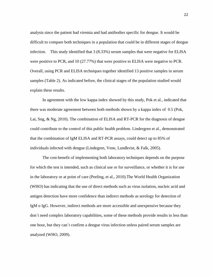
analysis since the patient had viremia and had antibodies specific for dengue. It would be
difficult to compare both techniques in a population that could be in different stages of dengue
infection. This study identified that 3 (8.33%) serum samples that were negative for ELISA
were positive to PCR, and 10 (27.77%) that were positive to ELISA were negative to PCR.
Overall, using PCR and ELISA techniques together identified 13 positive samples in serum
samples (Table 2). As indicated before, the clinical stages of the population studied would
explain these results.
In agreement with the low kappa index showed by this study, Pok et al., indicated that
there was moderate agreement between both methods shown by a kappa index of 0.5 (Pok,
Lai, Sng, & Ng, 2010). The combination of ELISA and RT-PCR for the diagnosis of dengue
could contribute to the control of this public health problem. Lindergren et al., demonstrated
that the combination of IgM ELISA and RT-PCR assays, could detect up to 85% of
individuals infected with dengue (Lindegren, Vene, Lundkvist, & Falk, 2005).
The cost-benefit of implementing both laboratory techniques depends on the purpose
for which the test is intended, such as clinical use or for surveillance, or whether it is for use
in the laboratory or at point of care (Peeling, et al., 2010).The World Health Organization
(WHO) has indicating that the use of direct methods such as virus isolation, nucleic acid and
antigen detection have more confidence than indirect methods as serology for detection of
IgM o IgG. However, indirect methods are more accessible and unexpensive because they
don´t need complex laboratory capabilities, some of these methods provide results in less than
one hour, but they can´t confirm a dengue virus infection unless paired serum samples are
analyzed (WHO, 2009).

23
CONCLUSIONS
This report describes the reintroduction of DENV-3 in the northern coast of Ecuador between
2010 and 2011, and the utility of an inexpensive transport method to stabilize RNA from
blood spots samples to conduct RT-PCR procedures to detect dengue serotypes. This
procedure may improve epidemiological surveillance in remote Ecuadorian communities
where febrile patients are only tested for malaria (Chairulfatah, Setiabudi, Agoes, van
Sprundel, & Colebunders, 2001; CDC, 2001). Blood collection on filter paper has improved
the management of other tropical disease like malaria in many endemic countries (Taylor, et
al., 2011; Al-Harthi & Jamjoom, 2008; Maeno, et al., 2008). Blood filter paper collection
offers a safe to handle and easy to storage, and transport samples (Matheus & al., 2008;
Matheus & al., 2012).
RECOMMENDATIONS
To implement the blood filter paper collection to transport samples from remote areas
to a reference laboratory as the Instituto Nacional de Higiene (INH) Leopoldo Izquieta
Perez.
To improve laboratory capabilities in dengue endemic areas to determine the dynamics
of dengue infection and the identification of different serotypes and genotypes
associated with dengue outbreaks.
To study an undifferentiated febrile illness in an endemic area of tropical diseases to
rule out all possible infectious agents as leptospirosis, malaria, rickettsioses, dengue
fever, brucellosis, and other viral infections as encephalitis.

24
To implement a strong epidemiological and entomological surveillance system in
Ecuador to improve the way to control, to prevent and to response during dengue
outbreaks.
To carry out other studies with a bigger sample of people and the comparisons
between a gold standard technique as virus isolation and RT-PCR and ELISA rapid
test to test the sensitivity, specificity and predictive value of this procedures in the
field.

25
BIBLIOGRPHY
Aguilar, E. (2010). Datos Reportados por Subsecretaria Regional de Salud Costa e Insular
Año 2004 - 2009. Casos confirmados. EPI-2. Ministerio de Salud Pública del Ecuador,
Epidemiología, Quito.
Al-Harthi, S., & Jamjoom, M. (2008). PCR assay in malaria diagnosis using filter paper
samples from Jazan region, Saudi Arabia. J Egypt Soc Parasitol , 38 (3), 693-706.
Bandyopadhyay, S., Lum, L., & Kroeger, A. (2006). Classifying dengue: a review of the
difficulties in using the WHO case classification for dengue haemorrhagic fever. Trop. Med.
Int.Health , 11 (8), 1238- 1255.
Bustosa, J., Hamdan, A., Loroño, M., Montero, M., & Gómez, B. (1990). Serologically
proven acute rubella infection in patients with clinical diagnosis of dengue. Epidemiol Infect ,
104 (2), 297–302.
Cao, X., Ngo, T., Wills, B., Kneen, R., Nguyen, T., Ta, T., et al. (2002). Evaluation of the
World Health Organization standard tourniquet test and a modified tourniquet test in the
diagnosis of dengue infection in Vietnam. Trop Med Int Health , 7 (2), 125-132.
Cardosa, J., Ooi, M., Tio, P., Perera, D., Holmes, E., Bibi, K., et al. (2009). Dengue Virus
Serotype 2 from a Sylvatic Lineage Isolated from a Patient with Dengue Hemorrhagic Fever.
PLoS Negl Trop Dis , 3 (4), e423.
CDC. (2010, October). Centers for Disease Control and Prevention. Retrieved December
2011, from Dengue: www.cdc.gov
CDC. (2001). Underdiagnosis of dengue-Laredo, Texas, 1999. Retrieved 2011 йил December
from MMWR Morb Mortal Wkly Rep. Centers for Disease Control and Prevention:
www.cdc.gov
Chairulfatah, A., Setiabudi, D., Agoes, R., van Sprundel, M., & Colebunders, R. (2001).
Hospital based clinical surveillance for dengue haemorrhagic fever in Bandung, Indonesia
1994-1995. Acta Trop , 80 (2), 111-115.

26
Connors, K., Coloma, J., Beatty, R., Cevallos, W., & Eisenberg, J. (2008). The effect of road
access on the transmission of dengue fever in rural Ecuador. Cornell University, New York.
Dash, P., Parida, M., Saxena, P., Abhyankar, A., Singh, C., Tewari, K., et al. (2006).
Reemergence of dengue virus type-3 (subtype-III) in India: implications for increased
incidence of DHF & DSS. Virol J. , 3, 55.
Direccion Provincial de Salud de Esmeraldas. (2011). Personal contact .
Gluber, D. (1998). Dengue and Dengue Hemorrhagic Fever. Clin. Microbiol. Rev , 11 (3).
Gomez, S., Villabona, C., Torres, F., Miranda, D., & Ocazionez, R. (2008). Dengue virus
serotype 3 (genotype III) from Colombia: a perspective of its pathogenic potential. Dengue
Bulletin , 32, 126-137.
Gubler, D. (2011). Emerging vector-borne flavivirus diseases:are vaccines the solution?
Expert Rev. Vaccines , 10 (5), 563–565.
Gubler, D. (1997). Epidemic dengue/dengue hemorrhagic fever: A global public heath
problem in 21st century. Dengue Bulletin , 21, 1-15.
Gubler, D. (2002). How effective is epidemiological surveillance used for dengue program
planning and epidemic response. Dengue Bulletin , 26, 96-106.
Gubler, D., & Casta-Valez, A. (1991). A Program for Prevention and Control of Epidemic
Dengue and Dengue Hemorrhagic Fever in Puerto Rico and the U.S. Virgin Islands. Bull Pan
Am Health Organ , 25 (3), 237-247.
Gubler, D., & Clark, G. (1995). Dengue/Dengue Hemorrhagic Fever:The Emergence of a
Global Health Problem. Emerg Infect Dis , 1 (2), 55-57.
Halstead, S. (1982). Immune enhancement of viral infection. Prog Allergy , 31, 301-64.
Hanley, K., Nelson, J., Schirtzinger, E., Whitehead, S., & Hanson, C. (2008). Superior
infectivity for mosquito vectors contributes to competitive displacement among strais of
dengue virus. BMC Ecology , 8, 1.

27
Harris E, R. T. (1998). Typing of dengue viruses in clinical specimens and mosquitoes by
single-tube multiplex reverse transcriptase PCR. J Clin Microbiol , 36 (9), 2634-2639.
Henchal, R., & Putnak, R. (1990). The Dengue Viruses. Clin Microbiol Rev , 3 (4), 376-396.
Hombach, J. (2007). Vaccines against dengue: a review of current candidate vaccines at
advanced. Pan Am J Public Health , 21 (4), 254-260.
Jelinek, T., Mühlbergerr, N., Harms, G., Corachán, M., Grobusch, M., Knobloch, J., et al.
(2002). Epidemiology and clinical features of imported dengue fever in Europe: sentinel
surveillance data from TropNetEurop. Clin Infect Dis , 35 (9), 1047-1052.
Lanciotti, R., Lewis, J., Gubler, D., & Trent, D. (1994). Molecular evolution and
epidemiology of dengue-3 viruses. J Gen Virol , 75 (1), 65-75.
Lindegren, G., Vene, S., Lundkvist, A., & Falk, K. (2005). Optimized Diagnosis of Acute
Dengue Fever in Swedish Travelers by a Combination of Reverse Transcription-PCR and
Immunoglobulin M Detection. J Clin Microbiol , 43 (6), 2850–2855.
Maeno, Y., Nakazawa, S., Dao le, D., Yamamoto, N., Giang, N., Van Hanh, T., et al. (2008).
A dried blood sample on filter paper is suitable for detecting Plasmodium falciparum
gametocytes by reverse transcription polymerase chain reaction. Acta Trop , 107 (2), 121-127.
Manock, S., Jacobsen, K., de Bravo, N., Russell, K., Negrete, M., Olson, J., et al. (2009).
Etiology of Acute Undifferentiated Febrile Illness in the Amazon Basin of Ecuador. Am. J.
Trop. Med. Hyg , 81 (1), 146–151.
Mario LG de Figueiredo, A. d. (2010). Mosquitoes infected with dengue viruses in Brazil.
Virology Journal , 7, 152.
Martina, B., Koraka, P., & Osterhaus, A. (2009). Dengue Virus Pathogenesis: an Integrated
View. Clin Microbiol Rev , 564–581.
Matheus, S., & al., e. (2008). Short Report: Dengue-3 outbreak in Paraguay: Investigations
using capillary blood samples on filter paper. 79 (5), 685-687.

28
Matheus, S., & al., e. (2012). Virological surveillance of dengue in Saint Martin and Saint
Barthélemy, French West Islands, using blood samples on filter paper. 86 (1), 159-165.
Muir, L., & Kay, B. (1998). Aedes aegypti survival and dispersal estimated by mark-release-
recapture in northern Australia. Am J Trop Med Hyg , 58 (3), 277-282.
Ng, L. (2011). Challenges in dengue surveillance and control. Western Pacific Surveillance
and Response Journal , 2 (2).
Oliveira, S., Siqueira, M., Camacho, L., Nogueira, R., Spinetti, C., Cubel Garcia, R., et al.
(2001). The aetiology of maculopapular rash diseases in Niterói, State of Rio de Janeiro,
Brazil: implications for measles surveillance. Epidemiol Infect , 127 (3), 509–516.
Ooi, E., Gluber, D., & Nam, V. (2007). Dengue research needs related to surveillance and
emergency response. Report of scientific working group on Dengue, WHO, Geneva.
PAHO. (2012). Dengue Regional Information: Number of Cases. Retrieved May 01, 2012,
from Pan American Health Organization:
http://new.paho.org/hq/index.php?option=com_content&task=view&id=264&Itemid=363&la
ng=es
Peeling, R., Artsob, H., Pelegrino, J., Buchy, P., Cardosa, M., Devi, S., et al. (2010).
Evaluation of diagnostic tests: dengue. Nat Rev Microbiol , 8 (12), S30-S38.
Pok, K., Lai, Y., Sng, J., & Ng, L. (2010). Evaluation of nonstructural 1 antigen assays for the
diagnosis and surveillance of dengue in Singapore. Vector Borne Zoonotic Dis , 10 (10),
1009-1016.
Prado, I., Rosario, D., Bernardo, L., Alvarez, M., Rodríguez, R., Vázquez, S., et al. (2005).
PCR detection of dengue virus using dried whole blood spotted on filter paper. J Virol
Methods , 125 (1), 75-81.
Regato, M. (2010). Personal contact.

29
Regato, M., Mosquera, C., Coloma, J., Mosquera, C., & Alava, A. (2006). Aplicación de la
RT-PCR de un solo paso en el diagnósticoy tipificación de las cepas circulantes del virus del
dengue en el Ecuador. Rev Ecuat Hig Med Trop , 43 (2), 11-18.
Shirtcliffe, P., Cameron, E., Nicholson, K., & Wiselka, M. (1998). Don't forguet dengue!
Clinical features of dengue fever in returning travellers. J R Coll Physicians Lond , 32 (3),
235-237.
Shu, P., & Huang, J. (2004). Current advances in dengue diagnosis. Clin Diagn Lab Immuno ,
11 (4), 642-650.
Stephenson, J. (2005). Understanding dengue pathogenesis: implications for vaccine design.
Bulletin of the World Health Organization , 83 (4).
Taylor, B., Martin, K., Arango, E., Arango, E., Agudelo, O., Maestre, A., et al. (2011). Real-
time PCR detection of Plasmodium directly from whole blood and filter paper samples. Malar
J , 10, 244.
Teixeira, M., & Barreto, M. (2009). Diagnosis and management of dengue. BMJ , 339, 1189-
1193.
Wang, E., Ni, H., Xu, R., Barrett, A., Watowich, S., Gubler, D., et al. (2000). Evolutionary
relationships of endemic/epidemic and sylvatic dengue viruses. J Virol , 74 (7), 3227-3234.
WHO. (2011). Comprehensive Guidelines for Prevention and Control of Dengue and Dengue
Haemorrhagic Fever - Revised and expanded edition. New Delhi: World Health
Organization, Regional Office for South-East Asia.
WHO. (1997). Dengue haemorrhagic fever: diagnosis, treatment, prevention and control
(2nd edition ed.). Geneva: World Health Organization.
WHO. (2009). Dengue: guidelines for diagnosis, treatment, prevention and control (New
ed.). Geneva: World Health Organization.
WHO/SEARO. (1999). Prevention and Control of Dengue and Dengue Haemorrhagic Fever
-Comprehensive Guidelines. WHO. New Delhi: WHO Regional Publication.

30
Wilder-Smith, A., Ooi, E., Vasudevan, S., & Gubler, D. (2010). Update on Dengue:
Epidemiology, Virus Evolution, Antiviral Drugs, and Vaccine Development. Curr Infect Dis
Rep , 12, 157–164.
Zavala-Velazquez, J., Yu, X., & Walker, D. (1996). Unrecognized Spotted Fever Group
Rickettsiosis Masquerading as Dengue Fever in Mexico. Am J Trop Med Hyg , 55 (2), 157-
159.

31
GLOSSARY
Active surveillance involves a proactive search of dengue infections, especially when they
could be attributed to other infectious agents as, influenza or rubella in periods of low
transmission. Such surveillance requires adequate laboratory diagnosis support.
Aedes mosquitoes are the largest genus of the subfamily Culicinae, they are found in all
habitats, ranging from the tropics to the Arctic.
Antigen is any molecule that can bind specifically to an antibody. When an antigen can
produce by itself the production of antibodies is called immunogen.
Ascites is pathologic fluid collection within the abdominal cavity.
Assay: An assay is an analysis done to determine the biological or pharmacological potency
of a drug, or the presence of a substance and the amount of that substance.
Brucellosis is a highly contagious zoonosis caused by ingestion of Brucella spp. in
unsterilized milk or meat from infected animals or close contact with their secretions.
Dengue hemorrhagic fever (DHF) is a potential complication of DF, and may have case
fatality rates of 1% or higher, especially in infants and young children. It is manifested
specially by high fever, hemorrhagic phenomena, frequently with hepatomegaly and signs of
circulatory failure when the condition is severe. The patients with DHF can develop dengue
shock syndrome (DSS) characterized by hypovolemic shock consequential from plasma
leakage.
Dengue is also known as Dengue fever (DF), an acute mosquito-borne viral disease of
sudden onset that usually follows a benign course with fever and other signs and symptoms as
headaches, bone, joint, and muscular pain, rash and leucopenia.
Ecchymoses is a subcutaneous purpura larger than 1 centimeter or hematoma.
Encephalitis is the inflammation of the brain.
Endemic is an epidemiologic term used to describe that a disease is present in a community at
all times but in relatively low frequency.
Envelope in a virus is a coat typically derived from portions of the host cell membranes when
the virus is maturing.
Enzootic is equivalent to endemic term, but in a non-human setting.
Enzyme-linked immunosorbent assay (ELISA), is a analytic biochemistry assay that uses
one sub-type of heterogeneous, solid-phase enzyme immunoassay (EIA) to detect the
presence of a substance (as proteins) in a liquid sample or wet sample.
Epidemic is the occurrence of more cases of a disease than would be expected in a
community or region during a given time period.

32
Epidemiologic surveillance involves an ongoing systematic collection, recording, analysis,
interpretation and diffusion of data used as public health tools for dengue prevention and
control
Event-based surveillance is conducted to investigate cases of fever of unknown etiology
without a routine data collection, but requires interdisciplinary action of an epidemiologist, an
entomologist and a microbiologist.
Extrinsic incubation period in a vector, it is the time between entrance of an pathogen into
the vector and the time when that vector can transmit the infection to other host.
Fever is any body temperature above 37°C. However, in practice a person is usually not
considered to have a significant fever until the temperature is above 38°C.
Flaviviridae are a family of viruses that are primarily spread through arthropod vectors.
Flavus means yellow in Latin.
Flavivirus is a genus of the family Flaviviridae, including West Nile virus, dengue virus, tick-
borne encephalitis virus, yellow fever virus, and other virus that cause encephalitis.
Genotype is the genetic makeup of a cell, an organism, or an individual usually with
reference to a specific character under consideration.
Haematemesis is the presence of blood in vomiting.
Haematocrit is the ratio of the volume occupied by red blood cells in the total volume of
blood, expressed as a percentage.
Hepatomegaly is the enlargement of the liver.
Hypoproteinaemia reflex the abnormally low level of protein in the blood.
Hypotension is the blood pressure that is below the normal. This term is the opposite of
hypertension (abnormally high blood pressure).
Hypovolemic shock refers to a medical or surgical condition in which rapid fluid loss results
in multiple organ failure due to inadequate circulating volume and subsequent inadequate
perfusion. Most often, it is secondary to rapid blood loss plasma leakage.
Immunity is the ability to resist to any infections. Cross-protective immunity is the reaction
between an antibody and an antigen that differs from the inmunogen.
Immunoglobulin, it is also known as antibody. It is a protein produced by plasma cells and B
lymphocytes after activation. The classes of immunoglobulins are termed immunoglobulin A
(IgA), immunoglobulin G (IgG), immunoglobulin M (IgM), immunoglobulin D (IgD) and
immunoglobulin E (IgE).
Influenza is a condition commonly referred to as the flu, and it is producer by Influenza
virus. Influenza A, B, and C viruses are the only members of the Orthomyxoviridae family,

33
and only influenza A and B viruses cause significant human disease. The orthomyxoviruses
are enveloped and have a segmented negative-sense RNA genome.
Insecticide is a pesticide used to combat insects.
Intrinsic incubation period is the time that takes to a pathogen multiply in a host, and then it
can be transmitted to or infect to other host.
Leptospirosis is an infectious disease caused by a spirochete transmitted by rats as well as by
skunks, opossums, raccoons, foxes, and other vermin.
Lethargy is the inability to continue functioning at the level of one's normal abilities.
Leucopenia describes the situation in which there are fewer leucocytes in the blood than
normal.
Malaria is an infectious disease caused by parasites from the Plasmodium family that can be
transmitted by the Anopheles mosquito or by a contaminated needle or transfusion.
Melena is the sign of black feces (digested blood) that are associated with gastrointestinal
hemorrhage.
Neutralization is the process of inactivation of a virus or a toxin molecule through
neutralizing antibodies.
Nucleic acids are biological molecules including DNA(deoxyribonucleic acid) and RNA
(ribonucleic acid).
Nucleocapsid is a unit of viral structure, consisting of a capsid with the enclosed nucleic
acid.
Outbreak is an epidemiologic term used to describe an occurrence of disease greater than
would otherwise be expected at a particular time and place.
Pandemic is an epidemic of infectious disease that has spread through human populations
across a large region; for instance multiple continents, or even worldwide.
Passive surveillance involves reporting of dengue cases according to a standardized way of
case identification by private physicians, clinics, health centers and hospitals that provides
medical attention to the population at risk.
Petechiae are a minor hemorrhage. It is small (1-2mm) red or purple spot on the body.
Platelet is a irregular, disc-shaped element in the blood that assists in blood clotting. They are
fragments of large bone marrow cells called megakaryocytes.
Polymerase chain reaction (PCR) is a molecular technique designed to amplify a single or a
few copies of a piece of DNA, generating thousands to millions of copies of a particular DNA
sequence. Retrotrancriptase (RT) is a DNA polymerase enzyme that transcribes single-
stranded RNA into single-stranded DNA.

34
Purpura is a small hemorrhage (> 3mm in diameter) in the skin, mucous membrane, or
serosal surface.
Rash is an eruption of the skin, typically referred to as an exanthema.
Rickettsioses are severe infectious diseases caused by rickettsiae and rickettsia-like
organisms. The best-known rickettsial diseases infect humans and are usually transmitted by
parasitic arthropod vectors.
Rribonucleic acid (RNA) is a nucleic acid molecule similar to DNA but containing ribose
rather than deoxyribose.
Sero-prevalence is the number of persons in a population who test positive for a specific
disease based on serology specimens; often presented as a percent of the total specimens
tested or as a proportion per 100,000 persons tested.
Serotype is the kind of microorganismcharacterized by serologic typing.
Serum is the clear liquid that can be separated from clotted blood.
Thrombocytopenia is the term for a reduced platelet count.
Tourniquet test is a clinical diagnostic method to assess fragility of capillary walls showing
the presence of thrombocytopenia. A blood pressure cuffs is applied on the upper arm and
inflated to a point between the systolic and diastolic blood pressures for five minutes. The test
is positive if there are more than 20 petechiae per 2.5 cm area square are observed
Vaccination is the deliberate induction of adaptative immunity to a pathogen by injecting a
vaccine, a dead or attenuated (non pathogenic) from the pathogen.
Vector borne disease A vector-borne disease is one in which the pathogenic microorganism
is transmitted from an infected individual to another individual by an arthropod or other agent,
sometimes with other animals serving as intermediary hosts.
Vector in epidemiology is any agent (person, animal or microorganism) that carries and
transmits an infectious pathogen into another host.
Virus is a microorganism smaller than bacteria, they can replicate only in a living cell owing
to metabolic machinery that viruses don’t have. It is composed of a nucleic acid genome
enclosed in a protein coat.
Yellow fever is an acute systemic illness caused by a Flavivirus. The viral infection causes a
high fever, bleeding into the skin, and necrosis of cells in the kidney and liver. Severe
jaundice gives the name to the disease.

35
APPENDIX
TABLE 1. SEROTYPE IDENTIFICATION BY RT-PCR IN SERUM AND
BLOOD SPOTS ON FILTER PAPER SAMPLES.
Samples
Character
istics
Number
of
samples
RT-PCR DENV-1 DENV-2 DENV-3 DEN
V-4
POSITIVE (%)
Serum 36 6 17 0 5 1 0
Blood
spots
41 7* 17 0 3 5 0
* A patient shown co-infection with both serotypes

36
TABLE 2. CROSSTAB TO COMPARE ELISA AND RT-PCR RESULTS
AGREEMENTS.
RT-PCR Total
Positive Negative
ELISA** Positive 1 9 10
Negative 3 21 24
Total 4 30 34*
*Two samples were excluded from 36 serum samples, because didn’t have complete
information about positive or negative result in ELISA test.
** ELISA test was carried out by HCB laboratory personnel.


![virus y priones [Modo de compatibilidad]ssanchez/virus y priones.pdf · Retrotranscriptasa o transcriptasa inversa. Enzimas Los virus pueden contener una mínima cantidad de enzimas](https://img.dokumen.tips/doc/110x75/5ba2912909d3f210318c4abc/virus-y-priones-modo-de-compatibilidad-ssanchezvirus-y-retrotranscriptasa.jpg)