Embed Size (px)
Citation preview

ADVANCED LUNG DIAGNOSTICS
SAMIR S. MAKANI, MD, FCCP
DIRECTOR
INTERVENTIONAL PULMONARY AND BROCHOSCOPY
UNIVERSITY OF CALIFORNIA SAN DIEGO MEDICAL CENTER
SAN DIEGO, CA
Dr. Samir Makani is an Associate professor of Medicine and the Director of Interventional Pulmonology and Bronchoscopy at the University of California, San Diego. He completed his Pulmonary and Critical Care fellowship at the University of California, San Diego followed by a fellowship in Interventional Pulmonology at Henry Ford Hospital in 2009. OBJECTIVES:
Participants should be better able to:
1. Gain knowledge and understanding of Navigational Bronchoscopy including its applications and limitations;
2. Gain knowledge and understanding of endobronchial ultrasound including advanced applications of lung mass fine needle aspiration and fiducial placement.
SATURDAY, MARCH 14, 2015 8:45 AM
1

ADVANCES IN PULMONARY
DIAGNOSTICS
Samir Makani, MD, FCCP Director, Interventional Pulmonology
Associate Professor of Medicine University of California, San Diego
DISCLOSURES
• Consultant for Covidien 2015 • Consultant for Olympus 2015 • Consultant for Carefusion 2015
2
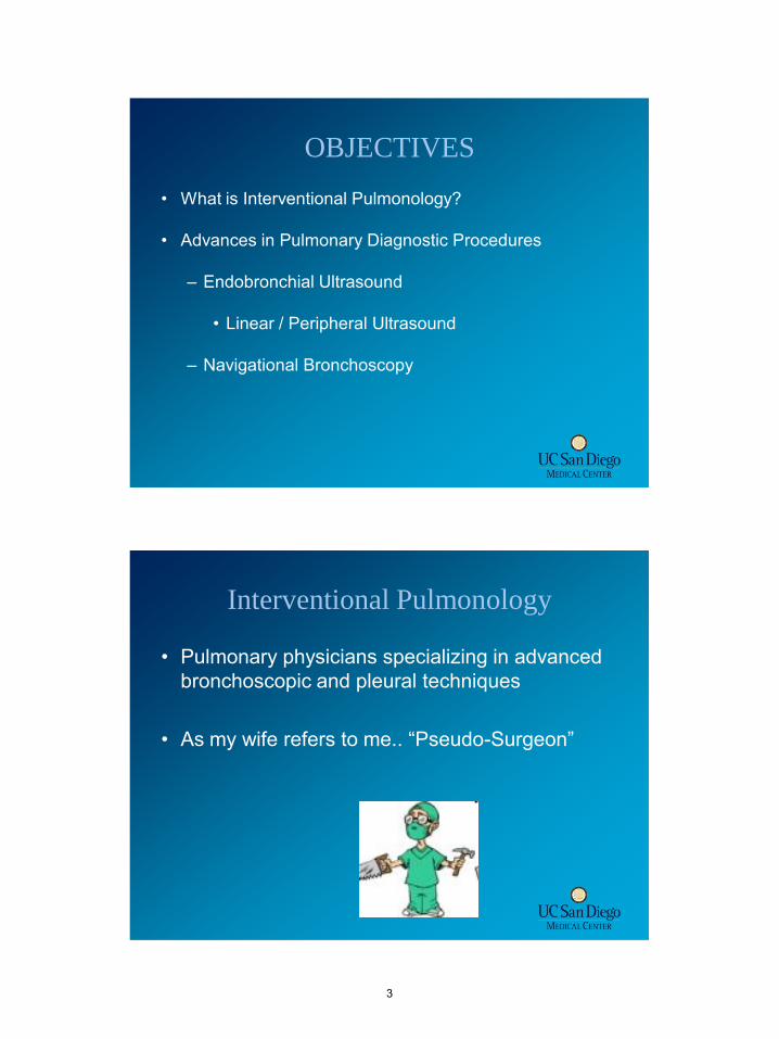
OBJECTIVES
• What is Interventional Pulmonology?
• Advances in Pulmonary Diagnostic Procedures – Endobronchial Ultrasound
• Linear / Peripheral Ultrasound
– Navigational Bronchoscopy
Interventional Pulmonology
• Pulmonary physicians specializing in advanced bronchoscopic and pleural techniques
• As my wife refers to me.. “Pseudo-Surgeon”
3

ENDOBRONCHIAL
ULTRASOUND
Beyond Lymph Node Staging
BACKGROUND
• Lung Cancer remains the #1 cause of cancer related death worldwide
• Appropriate staging of lung cancer is important as it guides treatment options
• Mediastinal lymph node sampling is essential for adequate staging and avoiding unnecessary thoracotomy
4

MEDIASTINAL LN ANATOMY
1 cm in Short
Axis on CT
“N” STAGE
N2 N3 N1
•Ipsilateral Hilar LN •10,11,12
•Stage IIA - IIB
•Surgical
•Ipsilateral Med LN •2,3,4,5,6,7,8,9
•Stage IIIA •Non – Surgical
•? 5,6
•Contralateral Med/Hilar LN
•Stage IIIB •Non - Surgical
5

“The prospect of cure
depends on stage.” Stefano Gasparini,
MD Heidelberg, Sept.
2002
“If you don’t look at lymph
nodes, everyone has stage I
non-small cell lung cancer.” Malcome DeCamp, Jr.
MD Beth Israel
Deaconess
MEDIASTINAL LYMPH NODE EVALUATION
• Non – Invasive – CT, PET, PET/CT
• No confirmatory tissue • False Positive
• Invasive – Mediastinoscopy – “The Gold Standard”
• Cervical / TEMLA • Chamberlain
– VATS/Thoracotomy • Minimally Invasive
– Standard TBNA
– EBUS-TBNA – EUS- FNA – Combined EBUS/EUS FNA
6

False Positive: • Infectious:
– Endemic mycoses – Tuberculosis
• Inflammatory: – Rheumatoid nodules – Sarcoidosis
False Negative • Bronchioloalveolar cell
carcinoma • Carcinoid tumors • Mucinous adenocarcinoma
POSITIVE EMISSION TOMOGRAPHY
STAGING PROCEDURES
Gomez and Silvestri. Proc Am Thorac Soc 2009
7

MEDIASTINOSCOPY
• “Gold Standard”
• Sensitivity – 78 - 90%
• Invasive procedure • General Anesthesia • Limited access to the posterior and inferior
mediastinum • 2% risk of major morbidity • Cost
MINIMALLY INVASIVE TECHNIQUES
• Minimally Invasive – Standard TBNA
– EBUS-TBNA
– EUS- FNA
– Combined EBUS/EUS FNA
8

• TBNA needles
– 13 mm long
– 22 gauge
– 19 / 21 gauge
• Knowledge of node anatomical position
• Blind procedure
• Low Senstivity/Specificity
Standard Transbronchial Needle Aspiration
Types of Endobronchial Ultrasound
• Radial (Peripheral) Probe
– Passes through the working channel of a bronchoscope
– Fitted with a water-inflatable balloon at the tip
– Enables the evaluation of the airway wall and the adjacent mediastinal structures
• Convex Probe
– Incorporated into the tip of a dedicated bronchoscope
– Allows real-time imaging and sampling of tissue
9

Radial Probe EBUS
• Peripheral EBUS – 20 MHZ
• Visualization of airway wall and pulmonary parenchyma to the chest wall
Convex Probe EBUS
• The transducer is incorporated into the tip of the bronchoscope
• Dedicated for the real-time guidance of transbronchial needle aspiration (EBUS-TBNA)
• Frequency of 7.5 MHZ - 12 MHZ (lower resolution but deeper penetration)
Yasufuku, K. et al. Chest 2004;126:122-128
10

Convex Probe EBUS Bronchoscope
• Outer diameter of bronchoscope: 6.7 mm
• Outer diameter of tip: 6.9 mm
• Diameter of working channel: 2.0 mm
• Dedicated disposable needles are used (21 and 22 G)
Working Channel
Optic
US Probe
US Aspects
• Contact method: direct contact of the probe with the bronchial wall or by inflating the balloon
• Scanning direction: linear curved array (scans parallel to the insertion direction)
• Generates a 50o image parallel to the long axis of the bronchoscope
• Has Doppler capability to differentiate vascular structures from tissue
Sheski FD. Chest 2008;133:264-270
11

LYMPH NODE POSITIONING
4R 10R
Vessel involvement
RADIOLOGY
12

Clinical Applications of EBUS-TBNA
• Lymph node staging in lung cancer patients (stations 1, 2, 3, 4, 7, 10, and 11)
• Diagnosis of unknown hilar and mediastinal lymphadenopahty
• Diagnosis of mediastinal tumors
13

MEDIASTINAL LYMPH NODE ANATOMY
Cervical Mediastinoscopy EBUS-TBNA EUS-FNA
Mountain. Chest 1997
EBUS-TBNA in the Staging of Lung
Cancer- Experience to Date
• A large body of data exists (over 300 publications)
• Meta-analysis of EBUS-TBNA in the staging of lung cancer – 11 studies with 1299 patients met criteria and
included in the study – Overall sensitivity 93% – Overall specificity 100%
Eur J Cancer. 2009 May;45(8):1389-96
14

ACCP PRACTICE GUIDELINES
DIAGNOSIS AND MANAGEMENT OF LUNG CANCER 3rd ED 2013
• 4.4.4.3. In patients with high suspicion of
N2,3 involvement, either by discrete
mediastinal lymph node enlargement or PET
uptake (and no distant metastases), a needle
technique (endobronchial ultrasound [
EBUS]-needle aspiration[ NA], EUS-NA or
combined EBUS/EUS-NA) is recommended
over surgical staging as a best first test
(Grade 1B) .
The Role of EBUS in the Diagnosis of
Other Mediastinal Diseases
• Is EBUS-TBNA a good diagnostic study to diagnose? – Lymphoma – Sarcoidosis – Other benign diseases (infectious)
1) Yes 2) No
15

Is EBUS-TBNA a good diagnostic study to diagnose? - Lymphoma - Sarcoidosis - Other benign diseases (infectious)
1. Yes 2. No
1. 2.
21%
79%
The Role of EBUS in the Diagnosis of Other Mediastinal Diseases
The Role of EBUS-TBNA in the Diagnosis of
Lymphoma • Two studies exist:
– Retrospective review of 25 patients referred for mediastinal lymphadenopathy
• 13 had a prior history of lymphoma and 12 had mediastinal lymphadenopathy of unknown etiology
– Sensitivity: 90.9%, specificity: 100% – Comments:
• The presence of prior lymphoma diagnosis greatly facilitates diagnosis • Flow cytometry is essential for making the diagnosis of lymphoma
– Another study prospectively evaluated 98 patients with
suspected lymphoma • EBUS-TBNA sensitivity: 57%
• Specificiy: 100%
• Surgical biopsy was avoided in a significant proportion of such patients (76%)
Kennedy M. Thorax 2008;63:360-365
Steinfort, DP. J Thorac Oncol. 2010;5(6):804-9
16

The Role of EBUS-TBNA in the
Diagnosis of Sarcoidosis • Study from MUSC
– 50 consecutive patients referred for suspected sarcoidosis – EBUS-TBNA used to sample lymph nodes: average size 16 mm
(range, 4-40 mm) – Diagnosis was established by seeing non-caseating granuloma – Sensitivity was 85%
• A randomized controlled trial of standard vs. EBUS
guided TBNA in patients with suspected sarcoidosis – 24 patients randomized to EBUS-TBNA – 26 patients randomized to conventional TBNA – Diagnostic yield: 83.3% vs. 53.8% in favor of EBUS (p <0.05)
Garwood S. Chest 2007;132:1289-
302
Tremblay, A. Chest 2009;136:340-346
RADIOLOGY – CASE 2
17
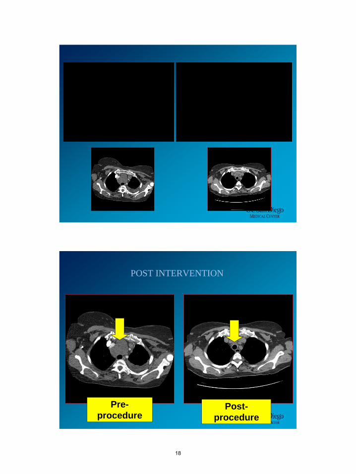
POST INTERVENTION
Pre-
procedure Post-
procedure
18

EBUS-TBNA Complications
• Overall very safe technique with no reported complications
• A few reports of infectious complications have emerged: – Infectious pericarditis/pericardial effusion after EBUS-TBNA
of subcarinal mass – Tumor bed infection after EBUS-TBNA of right lung mass
posterior to the bronchus intermedius – One case of mediastinal abscesses – Once case of ascending mediastinitis
• Possible explanations:
• Deposition of oral contaminants into the lymph node or tumor mass caused the infection
• Reporting bias of complications in a new technology
Hass, AR. Eur Respir J 2009;33:935-38
Moffatt-Bruce, SD. J Cardiothoracic Surg. 2010;5:33
Parker KL. Ann Thoracic Surg 2010,89:1271-2
EBUS-TBNA Complications
• Incidence of bacteremia following EBUS-TBNA – 43 patients undergoing EBUS-TBNA had blood
cultures within 60 seconds of the puncture – Incidence of bacteremia: 7%
• Similar to bacteremia reported following routine flexible bronchoscopy
• No clinical features suggestive of infections
Steinfort, DP. Eur Respir J 2009
19

EBUS and the SPN
• Peripheral EBUS – Advantages
• Confirmation of position
– Limitations • Actual biopsy performed with standard technique • Lack of directional biopsy
• Linear EBUS
– Advantages • Direct visualized biopsy
– Limitations • Diameter / Flexibility of endoscope • “Straight path”
CASE
• 76 y/o male with squamous cell lung cancer s/p XRT with increased lesion size.
20

EBUS and Stereotactic Radiotherapy
• Fiducial Placement – Lymph Nodes
– Peripheral Pulmonary nodules / masses
21

CASE
• 77 y/o female with single enlarged hilar lymph node and history of squamous cell carcinoma.
22

EBUS BRONCHOSCOPY
• Advantages – Good Sensitivity and diagnostic yield – Access to majority of mediastinal / hilar LN – Airway evaluation – Other pulmonary procedures including FNA – Very safe
• Limitations
– AP Window / Sub Diaphragmatic LN / Adrenal – Pulmonary status
CONCLUSIONS
• EBUS is an accurate, safe, and cost-effective primary procedure for mediastinal staging in lung cancer
• “A diagnosis should be obtained by whatever
method is easiest in patients who are presumed to have SCLC or who have very clear evidence of advanced NSCLC” – Rivera et al CHEST 2007
23

NAVIGATIONAL BRONCHOSCOPY
BACKGROUND
• Lung Cancer is the #1 cause of cancer related death, and the #2 cause of mortality overall in the U.S.
• More than 150,000 pts/yr present with the diagnostic dilemma of a Solitary Pulmonary Nodule (SPN)
• Early detection and treatment of lung cancer can lead to increased 5 year survival
24

THE SOLITARY PULMONARY NODULE
• <3 cm in short axis • Surrounded by aerated
lung and not visible beyond the visual segmental bronchi
• Location: – Inner 2/3 of lung – Outer 1/3 of lung – Lobe / Segment
Inner 2/3
Outer 1/3
THE SOLITARY PULMONARY NODULE
12mm
X-ray and Fluoro Invisible
Adenocarcinoma Adenocarcinoma
25

NLST
• National Lung Cancer Screening Trial – Began enrollment in 2002 – Low Dose CT vs. CXR – Annual examination for 3 years
• Inclusion Criteria – Current or former smokers aged 55-74
NLST • Results
– 20.3 % reduction in lung cancer mortality with CT – Actual Numbers
• 53,500 participants from 33 centers • 354 deaths in CT group • 442 deaths in the CXR group
• Implications – – Large false-positive rate with screening CT – National Comprehensive Cancer Network (NCCN)
• New guidelines for lung cancer screening
• Low dose CT in select patient populations based on
NLST data
– USPSTF – Grade B Recommendation 2014
26
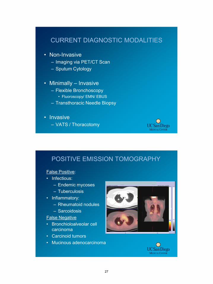
CURRENT DIAGNOSTIC MODALITIES
• Non-Invasive – Imaging via PET/CT Scan – Sputum Cytology
• Minimally – Invasive – Flexible Bronchoscopy
• Fluoroscopy/ EMN/ EBUS
– Transthoracic Needle Biopsy
• Invasive – VATS / Thoracotomy
False Positive: • Infectious:
– Endemic mycoses – Tuberculosis
• Inflammatory: – Rheumatoid nodules – Sarcoidosis
False Negative • Bronchioloalveolar cell
carcinoma • Carcinoid tumors • Mucinous adenocarcinoma
POSITIVE EMISSION TOMOGRAPHY
27

TRANSTHORACIC NEEDLE ASPIRATION (TTNA)
• Sensitivity rate = 80-95%2
• Specificity rate = 50-88%2
• False negative rate = 3-29%2
• Cannot be used in all cases due to co-morbidities, or lesion location
• Complications: – Pneumothorax (5-50%)
• Rate affected by size, distance, passes performed
• 4% necessitates chest tube insertion
– Hemoptysis and hemorrhage (up to 10%)
1. Shulman, L, et al. Curr Opin Pulm Med 2007; 13:271-277
2. Ost, et al, NEJM 2003; 348:2535-42
VATS / THORACOTOMY
• Highly invasive procedure
• Not suitable for patients with
advanced disease or significant
co-morbidities
• Associated with higher morbidity
and mortality rates
28

BRONCHOSCOPY FOR SOLITARY PULMONARY NODULES
• Transbronchial biopsies with fluoroscopic guidance – > 2 cm
• Yield 63%
– < 2 cm • Yield <34%
Unchanged for previous 20 years
Rivera Chest 2007
• Peripheral lesions are beyond bronchoscopic visualization
• Sampling techniques are guided using fluoroscopy
• Solid Lesions that are < 1 cm not visible with fluoroscopy
TRANSBRONCHIAL BIOPSY OF SPN
Scope
Bx Forceps
29

Lung Anatomy
FACTORS INCREASING YIELD OF FLEXIBLE BRONCHOSCOPY
• Size of the lesion – > 2cm increased yield to 63%
• Use of fluoroscopy • Number of biopsies
– 1 biopsy – 45% – 6 biopsies – 70%
• Visible airway to lesion (“Bronchus Sign”) – 60% vs 25%
• Endobronchial Ultrasound • Electromagnetic Navigation
Rivera Chest 2007
30

SuperDimension Electromagnetic Navigation
•Navigational System which utilizes GPS-like navigation and the patient’s natural airways
to access peripheral pulmonary nodules
31

ELECTROMAGNETIC LOCATION BOARD
• Location board underneath mattress generates Electromagnetic Field
REAL TIME LOCATION
32

• Miniaturized sensor delivers accurate information
• x, y, z, roll, pitch, and yaw
• 166 times/second
REAL TIME INFORMATION
ELECTROMAGNETIC NAVIGATION BRONCHOSCOPY (ENB) PROCEDURE OVERVIEW
CT-Scan DICOM CD Planning Software Planned Pathway File Navigation Biopsy Treatment
33

CT SCAN
DICOM CD
Specific Parameters for Slice Thickness and Interval
ELECTROMAGNETIC NAVIGATION BRONCHOSCOPY (ENB)
Planning Phase:
• Visualize lung anatomy and
airways in 3D – CT planar views, 3D map,
and virtual bronchoscopy
• Mark the target and plan pathway
34

PROCEDURE PLANNING
PROCEDURE PLANNING
35

ELECTROMAGNETIC NAVIGATION BRONCHOSCOPY (ENB)
Procedure Phase:
• Automatic Registration (CT-
to-body matching)
• Real-time virtual bronchoscopy navigation
Locatable Guide
Slide Courtesy of Michael J. Simoff, M.D.
36

EDGE CATHETER
PROCEDURE
37

PROCEDURE
EBUS AND PERIPHERAL LESIONS
38

APPROACH TO DIAGNOSTIC MANEUVERS
•EMN used to navigate to lesion
•pEBUS through EWC to confirm lesion location
•Fluoroscopy to confirm position
•Transbronchial biopsies
•3 biopsies
•Confrmation with pEBUS
•3 more biopsies
•pEBUS
•Brushings / TBNA
•40 cc Normal Saline instilled for bronchial washing
EMN – Yield, Safety, and Efficacy
Study N Statistics and Efficacy
Becker et al. JOB 2005 30 •Registration accuracy (AFTRE) 3mm
•Diagnostic Yield = 69%
•No serious complications related to use of device
Gildea et al. AJRCCM 2006 60 •Diagnostic Yield = 74% in peripheral lesion
•Diagnostic Yield = 100% in lymph nodes
•57% of lesions were < 2cm in diameter
Makris et al. ERJ 2007 40 •Diagnostic Yield = 62.5%
•Diagnostic Yield 77.2% when divergence ≤ 4mm
Eberhardt et al. Chest 2007 89 •Positive biopsy obtained for 67% of lung lesions
•88% of positive biopsies obtained at RML location
Eberhardt et al. AJRCCM 2007 120 •Positive biopsy obtained in 69% of EBUS group
•Positive biopsy obtained in 59% of ENB group
•Positive biopsy obtained in 88% of EBUS + ENB group
•Yield in independent of lesion size or lobar distribution
Wilson, et al. JOB 2007 248 •ENB successfully reached 95% of peripheral lesion and 94.3% of
lymph nodes
•On day of procedure, 65% of patients received a definitive
malignant or plausible non-malignant diagnosis; with follow-up
another 5% confirmed as non-malignant
Bertoletti et al. Respiration 2009 54 •Diagnostic success rate of 71.4% with median tumor size of 28mm
•4% pneumothorax rate; 1 requiring chest tube
Edell et al. New Current Procedural Terminology Codes
Effective 2010
- •Summary of 600 patients with ENB experience
•Conclusion: ENB has potential to improve diagnostic yield of
transbronchial biopsy and may be useful in early diagnosis of lung
cancer
39

BRONCHOSCOPY FOR SOLITARY PULMONARY NODULES
• Transbronchial biopsies with fluoroscopic guidance – > 2 cm
• Yield 63% – < 2 cm
• Yield < 34%
Now 77%-88% with electromagnetic navigation + pEBUS
ACCP PRACTICE GUIDELINES
DIAGNOSIS AND MANAGEMENT OF LUNG CANCER 3rd ED 2013
• 3.4.2.1. In patients with peripheral lung
lesions difficult to reach with conventional
bronchoscopy, electromagnetic navigation
guidance is recommended if the equipment
and the expertise are available (Grade 1C).
40

Tissue Sampling Evidence
65 ENB cases resulted in a diagnosis of lung cancer. Tissues obtained were adequate for histologic subtyping in all 65 cases.
Gildea et al, J Bronchol Intervent Pulmonol Jan 2013
OTHER APPLICATIONS
• Fiducial Marker Placement
41
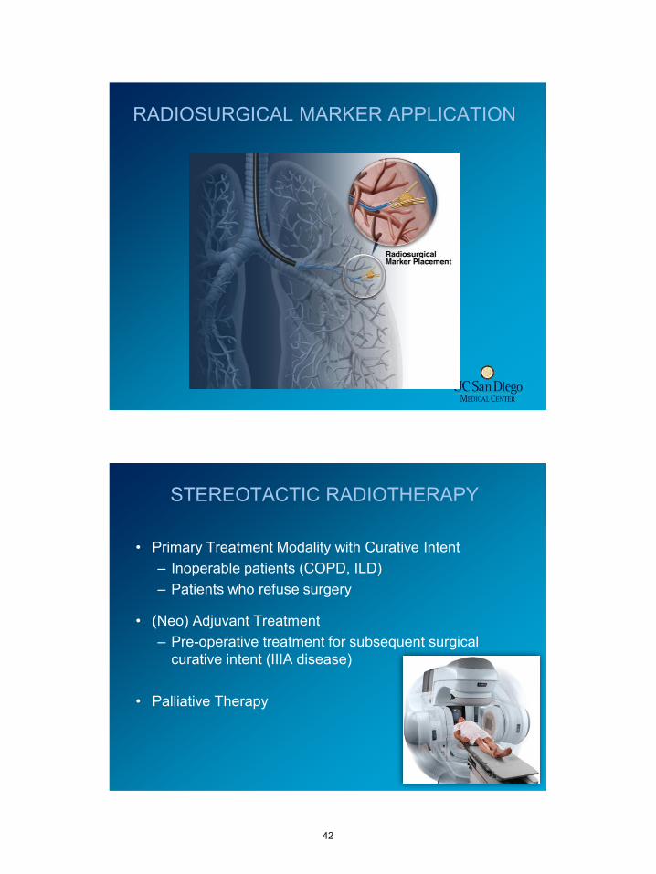
RADIOSURGICAL MARKER APPLICATION
STEREOTACTIC RADIOTHERAPY
• Primary Treatment Modality with Curative Intent – Inoperable patients (COPD, ILD) – Patients who refuse surgery
• (Neo) Adjuvant Treatment – Pre-operative treatment for subsequent surgical
curative intent (IIIA disease)
• Palliative Therapy
42

STEREOTACTIC RADIOTHERAPY
• Current Challenges
– Lung tumor movement occurs with respiratory cycle • Unpredictable baseline shifts and variable amplitude of
respiratory rates • Change in tumor morphology or position relative to other
structures
– Accurate alignment during stereotactic planning and delivery is required
• maximize radiation to target • minimize “collateral damage”
• EMN radiosurgical marker placement provides enhanced localization of tumor during respiratory variation to guide radiotherapy with minimal risk – Pneumothorax – Migration
1. Eberhardt, et al., 2007
2. Wilson, et al., JOB 2007
STEREOTACTIC RADIOTHERAPY Planning Phase
• Target selected lesions with planning software
Navigation
• EMN navigation to lesion
Marker Placement – Several Markers and
Methods
• Angiocatheter with backloading (labeled) • Wang/Conmed 19ga/21ga TBNA • Endobronchial brush
Stereotactic radiotherapy treatment
43

STEREOTACTIC RADIOTHERAPY Study N Statistics and Efficacy
McLemore et al. WCB 2006 20 •Accurate and safe approach
•No complications
•Markers were very stable
Anantham et al. Chest 2007 9 •Lesion size: 35.8 ± 16.7mm
•Markers successfully placed in 89% (8/9 patients)
•90% of markers still in place after 7-10 days
•No bronchoscopic complications observed
McGuire et al. JOB 2007 - •Angulated catheter method provides most accurate and safe
method
•Angulated catheter allows the placement of markers in multiple
planes without withdrawing the catheter
Kupelian et al. Int J Rad Oncol Biol Phys 2007 8 •Average lesion size was 2.6mm
•No pneumothorax
•Implanted markers were stable within tumor throughout treatment
duration regardless of implantation method
Quinn, et al. ACRO 2009 and CyberKnife 2009 44 •23 transcutaneous placement; 10 developed pneumothorax (43%)
•21 transbronchial placement; 3 developed pneumothorax (14%)
•3 patients had marker migration with transbronchial; no migration
with transcutaneous
Harley, et al. Ann Thorac Surg 2010 43 •ENB was used in 12 patients with peripheral lesions
•Average of 3.7 markers per patient were deployed
•86.7% of markers identified radiologically at SRS planning CT, 2
weeks after deployment
•1 pneumothorax (pigtail catheter placement)
•All patients were able to undergo SRS without additional marker
placement or other procedures
LIMITATIONS
• Not every patient is a good candidate
• “ The Bronchus Sign”
44

“ The Bronchus Sign”
• Visible airway on CT imaging leading to SPN
• Prior reports demonstrate increased diagnostic yield (60% vs. 25%)
• Seijo et al -Evaluation of bronchus sign with EMN
– Results: 67% total diagnostic yield (34/51) • With bronchus sign – 79% (30/38) • Without bronchus sign – 13% (4/13)
Seijo et al CHEST 2010
OTHER PERCEIVED LIMITATIONS
• Reimbursement
– CPT code now available with increased value in 2013
– Down Stream Revenue Generation • Radiology • CT Surgery • Oncology • Radiation Oncology • Pathology • Recovery Area / PACU
45

CASE PRESENTATIONS
• 63 y/o female with severe COPD with enlarging PET negative pulmonary nodule
• Best method of diagnosis? 1) Surveillance imaging 2) CT guided biopsy 3) Bronchoscopy with navigation and trans bronchial
biopsy 4) Open lung biopsy
46

Best method of diagnosis?
1. Surveillance imaging 2. CT guided biopsy 3. Bronchoscopy with navigation
and trans bronchial biopsy 4. Open lung biopsy
1. 2. 3. 4.
7%0%
80%
13%
47

Diagnosis
CASE PRESENTATION
• 73 y/o male with a newly noted left lower lobe lung nodule
48

49

CASE PRESENTATION
• 67 y/o male recently heard about lung cancer screening. 50 pack year history of smoking
50

CASE PRESENTATION
• 64 y/o female with a 40 pack year history of smoking. Recently released from jail
51

52

What Next?
• All cultures negative • Stains negative • Serologies negative
• Any further diagnostic testing necessary? 1) None needed 2) CT guided biopsy 3) Open lung biopsy
WHAT NEXT?
All cultures negative Stains negative Serologies negative Any further diagnostic testing necessary?
1. None needed 2. CT guided biopsy 3. Open lung biopsy
1. 2. 3.
91%
5%5%
53

AFB IHC Positive – Dx : Tuberculosis
54

Who should Navigational Bronchoscopy and EBUS Bronchoscopy be performed by?
1) Only by trained Interventional Pulmonologists in tertiary referral centers 2) Any Pulmonary physician with the resources
and expertise to perform the procedure safely and accurately
Who should Navigational Bronchoscopy and EBUS Bronchoscopy be performed by?
1. Only by trained Interventional Pulmonologists in tertiary referral centers
2. Any Pulmonary physician with the resources and expertise to perform the procedure safely and accurately
1. 2.
67%
33%
55

FUTURE APPLICATIONS
Slide Courtesy M. Simoff MD
Thank You!
Samir Makani, MD Interventional Pulmonology
University of California, San Diego
O:619-543-2793
56