Embed Size (px)
Citation preview

Sam Powdrill PA-C
University of Kentucky PA program

Learning Objectives
At the end of this session, participants should be able to: • Evaluate clinical vignettes for pertinent positive and
negative ocular information • Recognize sight-threatening historical and clinical
findings • Differentiate between common, urgent and non-
urgent ocular diagnoses • Formulate management plans for both urgent and
non-urgent ocular diagnoses

Leading Eye diseases in the U.S.
age-related macular degenerationCataractsglaucoma diabetic retinopathy,

The Globe Anterior
chamber aqueous
Iris
Lens
Posterior chamber vitreous
Optic nerve

Angle of the Eye

Trauma waiting to happen…
Prevention
is better
than
Treatment


History
Visual disturbance – blurred vision, visual change, diplopia, halos, floaters, flashing lights
Discharge – tears, mucus, blood or pus Which eye – OS OD OU Pain – sharp, dull, ache, sand sensation, burning,
foreign body sensation, photophobia Swelling, tumors, Trauma , foreign bodies, irritants, Glasses or contacts

Eye history Was there an injury? Time of injury or onset of problem What was the mechanism of injury? Is there pain – where and how severe? Previously was vision good and has the vision
changed and how? Has the patient used any treatment Are they up on tetanus immunization Has the patient eaten recently - NPO Determine if there are legal implications

History (cont.)
Systemic illnesses – hypertension, diabetes, cardiovascular, inflammatory, infections.
Eye disease in family – glaucoma, macular degeneration, cataract or other
Eye protective wear

Instruments needed to examine the eye
1. Visual acuity chart2. Flashlight3. Ophthalmoscope4. Tonometer5. Simple loupe

Pathways of Blindness
Corneal Anterior chamber Lens Vitreous Retina Optic nerve Occipital Functional

Physical exam Visual acuity Inspection - external structures, redness,
pupil, extra ocular movements Ophthalmoscope - lids, anterior chamber,
iris shadow( indirect light) , red reflex and lens (+4), magnified view of cornea and anterior chamber (+15), retinal and optic nerve (0 or adjusted to pt. refraction)

Visual acuity:
The vital sign of the eye

Rosenbaum near chart
tested at 14 inches

Near visionRosenbaum chart
Test for reading distance vision
Test for presbyopia – loss of lens accommodation
Changes with age

Physical exam - special tests
Slit lamp exam – if available Fluorescein stain Intraocular pressure – never check if
lacerated or ulcerated eye. Visual field Dilated retinal exam if safe to do so. Imaging – X-ray, CT, MRI

Slit lamp view of the eye


Visual fields by confrontation

Peripheral visionTesting the visual field
Confrontation method – compares the examiner’s visual field with the patient’s
Automated – done in optometrist’s or ophthalmologist’s office
Abnormal in glaucoma

Central vision
Test for loss of central vision - macula
Amsler grid
Abnormal in macular degeneration

Amsler grid

Tonometry
Measurement of the intraocular pressure
• Schiotz• Applanation • Air puff• Tonopen
• Normal is 10 to 20 mmHg

Tonometry
Can be done in the emergency room or urgent care setting
Eye must be anesthetized with topical tetracaine drops
Fluorescene is often used Should only be learned under supervision
and care taken not to scratch the cornea and cause a corneal abrasion

Extraocular Muscles
Cardinal eye movements (big H ) Corneal light reflex position Nystagmus Cover - uncover test Convergence

Eyelids
Inspect Trauma Opening and closing completely Eyelash position Ptosis, ectropion or entropion Swelling or infection
Injury and dryness from non contact are the greatest eyelid threats to sight

Evert the eyelid

CorneaInspect – shine the flashlight from the
side at first
Arcus senilus Clarity – scars obscure part of the iris. Light reflex – reflex is scattered in edema Corneal sensitivity to light with inflammation
infection or trauma Depth of anterior chamber – iris shadow Abrasions or foreign bodies – use fluorescein
stain with blue light to examine the cornea for defects or foreign bodies.

Iris and pupil
Inspect Iris Color Angle lesions
Inspect Pupil Size Shape Reaction to light Reaction to near Consensual response PERRLA

Conjunctivitis Very common –
many causes Usually viral If bacterial there is
usually some sort of trauma involved
Treatment is warm or cool compresses, treat the cause and avoid spread

tearing

Red Eye: overview
• Allergic Conjunctivitis • Infectious Conjunctivitis• Infectious Keratitis• Orbital cellulitis• Measles • Vitamin A deficiency • Inflammatory causes• Tumors• Traumatic causes• Glaucoma• Contact Lens complications• Eyelid Margin disease
infectious
inflammatorynutritional
neoplastic
mechanical
allergic

Infectious Keratitis
Corneal Ulcer– This is differentiated from conjunctivitis
because the cornea is opaque Viral
– HSV, HZV Bacterial
– Staphylococcus, Streptococcus, Pseudomonas Fungal
– This is usually keratitis that does not improve with treatment

Corneal ulcer
Treat aggressively with topical antibiotics

blindingor
non-blinding
If both eyes are red treat them
If one eye is red and painful refer

Use extreme caution with steroids
in a red eye
You should probably consult an Ophthalmologist first

Orbital cellulitis
Hospitalize and treat with IV antibiotics

cataract Ophthlmoscope
set on +4 Shadows against
Red reflex

Traumatic Cataract

Angle Closure
Glaucoma
Sudden rise in intraocular pressure Often presents with nausea Treated with pupil constriction and surgery Can use acetazolamide or manitol

Iris shadow

ACG or Iritis

Cell and flare in Iritis
https://timroot.com/cell-and-flare-in-the-eye-video/

Keratic precipitates in Iritis

Mutton fat KPs

anterior synechiae

Treatment
ACG– Treated with pupil constriction and surgery– Can use ac
Iritis– dilate the pupil – Anti-inflammatory drops - prednisone

Other non trauma eye emergencies
Sudden drop in vision Central artery occlusion Central vein oclusion Retinal detachment

Epidemiology 85% of eye trauma is accidental 15% caused by assault Men are 4 times more likely to have eye trauma
than women Average age of trama is 30 years Blunt trauma is most common Most occur at home Highest industrial cause is construction Airbags and seat belts have reduced eye trauma

Mechanisms of Eye Trauma
Foreign body
Blunt
Sharp
Burns
Miscellaneous

Foreign body trauma
Low velocity– Dust, grass seeds
High velocity– Metal-on-metal contact (eg hammering nails)
Signs:– Red eye, foreign body sensation, linear
fluorescein pattern with blue (cobalt) light

“Something’s in my eye!”
Fluorescein test shows vertical linear streaks, but no corneal FB is seen. Where could it be?

Invert the upper eyelid

Upper eyelid foreign body
Common presentation: “something in the eye” •look under the lower lid & evert the upper lid. •Avoid using a topical anesthetic – it masks the symptoms
Photo credit :WHO

Grass seed stuck to cornea
Removal: topical anesthetic & cotton tip applicator; or 25ga needle, bevel up and parallel with corneal surface. Treatment: Antibiotic drops & cycloplegics.
Photo credit :WHO

Corneal metal foreign body
These are removed the same way as before. A rust ring (brown stain) may form & should be removed, if possible.
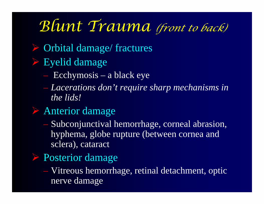
Blunt Trauma (front to back)
Orbital damage/ fractures Eyelid damage
– Ecchymosis – a black eye– Lacerations don’t require sharp mechanisms in
the lids! Anterior damage
– Subconjunctival hemorrhage, corneal abrasion, hyphema, globe rupture (between cornea and sclera), cataract
Posterior damage– Vitreous hemorrhage, retinal detachment, optic
nerve damage

Ecchymosis
Usually not sight threatening and typically no treatment

Subconjunctival hemorrhage
This is not a serious
condition, but appears
threatening.
Check for deeper injury - usually no Treatment needed.

Subconjunctival Hemorrhage
Note: this is usually not from trauma

Corneal abrasion
Treatment: antibiotic drops or ointments, cycloplegics (eg cyclopentolate or homatropine) for comfort; NO STEROIDS,NO TOPICAL ANESTHETICS. Patching is optional.
Photo credit :WHO

Cornea staining with fluoroscein dye

Status post football (soccer) injury
What sort of damage has occurred to the eye?

Which would be the most useful physical exam sign to assist in a diagnosis of this
case?
A. Have the patient look laterally on the injured side
B. Observe for ptosisC. Palpate for edemaD. Have the patient look upwardE. Palpate for a fracture

Ptosis and restriction in superior gaze
This requires extensive surgery. Give po Antibiotics for nasal flora, and no nose blowing (makes swelling worse). Refer to eye surgeon.

Before surgery Two weeks post-op
Orbital floor (“blowout”) fracture with tissue entrapment (and periorbital swelling
causing ptosis)

Blunt trauma iridodialysis
Usually no treatment needed

Partial hyphema
Treatment: atropine (careful!), bedrest, eye pressure drops (if needed).
Photo credit :WHO

Total hyphema
Same treatment as partial hyphema (atropine, bedrest, IOP drops prn).
Photo credit :WHO

Cataract and subluxed lens
dilation is needed for Diagnosis. Refer to an eye surgeon.
Photo credit :WHO

Common results of trauma
Corneal abrasion or laceration
Hyphema Iris tear Ciliary bodyshut down Cataract Vitreous loss or hemorrhage Retinal hemorrhage or
detachment Optic nerve damage

Sharp Trauma Lacerations
– Knives, scissors, chopping firewood Perforating injuries
– Metal-on-metal contact (eg hammering nails or pipes), sticks, thorns, pellets/bullets
– This means something has entered the eye and remains there
– Signs: red eye, corneal laceration, cataract, iris transillumination. Requires high degree of suspicion

Left eyelid laceration
This can be repaired in the field with basic instruments.

Corneal puncture and Iris prolapse
Remember: this can happen from blunt or sharp trauma. This is an open globe. Cover with a shield & refer to an eye surgeon.
Photo credit :WHO

Corneal laceration
This needs to be repaired as soon as possible to avoid
infection and recover optimum vision

Penetrating injury
Note: scattered light reflex, irregular pupil, cataract
Photo credit :WHO

Referral of a corneal laceration
•Do not remove an intraocular foreign body
•Use a cup rather than an eye pad to avoid further injury to the eye.
•Cover for tetanus
•Keep the patient NPO

Late complications of trauma
Secondary infections– Corneal ulcers or scars from abrasions– Orbital cellulitis from skin abrasions or
lacerations Poor vision (refer to eye surgeons)
– Traumatic cataracts– Endophthalmitis (infection of the whole globe)– Glaucoma or low eye pressure– Sympathetic ophthalmia

Referral
Take a visual acuity as a baseline Shield not pressure dressing Antibiotic drops – avoid neomycin PO fluoroquinalones (cipro) Cyclopentolate not amethocaine for pain If you dilate the eye – record it. PO pain control

Burns of EENT areas
Thermal– Fire, hot liquid, unprotected welding
Chemical– alkalines are worse than acids; both are bad
Electrical Radiation

Alkaline chemical burn
Copious irrigation immediately is required stat, especially for alkaline injuries. Then, Cycloplegics for pain and Antibiotic ointment, Patch as needed.
Photo credit :WHO

Parts of the ophthalmoscope exam
1. Assess the red reflex +4 at 10 inches distance 2. examine the cornea lids and AC with magnified
view +20 at 5 cm distance 3. examine the inside the eye starting on 0 then
adjusting the focus to get a clear view of the retina. 4. Optic disc All 4 quadrants and vessels 5. macula

Ophthalmoscope exam The exam is done starting at
a distance and at 15 degrees away from the patient’s central gaze.
Position the patient to improve your view. Adjust for patient’s height
Patient should look straight ahead at a distant point over your shoulder (don’t block their view)
15degrees

Focusing the ophthalmoscope

Here is how to find the optic disk on retinal
exam
Watch the next slide animation
Find the optic disk by finding a vessel on retinal exam and follow it to
where it converges at the optic disk

Retina

Retina

Glaucoma cupping

What do you see ?

• Nicking
• Hollenhorst plaque
• Drusen
• Small hemorrhage
• Early cotton wool spot

Retinopathy• Non prolipherative
– Exudates– Hemorrhages– Micro aneurisms
• Proliferative– New vessel growth

Maculopathy Have patient
look directly into the light
Changes in the macular area

Copper and Silver wiring

Retinal detachment


references Triaging Ocular Emergencies http://www.bsmcpss.com/resources/study-guides/OPH%2003%2014-117.pdf
Prevention and Treatment of Common Eye Injuries in Sportshttp://www.aafp.org/afp/2003/0401/p1481.html
Women at Higher-Risk than Men for Sight-Threatening Eye Diseases And Conditions
https://www.preventblindness.org/women-higher-risk-men-sight-threatening-eye-diseases-and-conditions-1

The End
Kwaheri