Embed Size (px)
Citation preview

DEPARTMENT OF DENTAL MEDICINE
Karolinska Institutet, Stockholm, Sweden
SALIVARY BIOMARKERS IN CHRONIC MUSCLE PAIN
Hajer Jasim
Stockholm 2020

The cover illustrates the protein profile signature in stimulated whole saliva of one female
patient diagnosed with myalgia associated with temporomandibular disorder.
Picture to the left is reprinted with permission from www.shutterstock.com.
All previously published papers were reproduced with permission from the publishers,
Nature Publishing Group, Wiley, and MDPI.
Published by Karolinska Institutet.
Printed by E-print AB, 2020
© Hajer Jasim, 2020
ISBN 978-91-7831-820-9
Salivary biomarkers in chronic muscle pain
THESIS FOR DOCTORAL DEGREE (Ph.D.)
PUBLIC DEFENCE
Friday 29th of May 2020, 9.00 am
Department of Dental Medicine lecture hall 4U
Alfred Nobels allé 8, Huddinge
By
Hajer Jasim Principal Supervisor:
Professor Malin Ernberg
Karolinska Institutet
Department of Dental Medicine
Division of Oral Diagnostics and Rehabilitation
Co-supervisor(s):
Associate Professor Bijar Ghafouri
Linköping University
Department of Medical and Health Sciences
Division of Pain and Rehabilitation Centre
Associate Professor Britt Hedenberg-Magnusson
Folktandvården Stockholms län AB
Eastman Institutet
Orofacial Pain and Jaw function
Professor Jochen M Schwenk
KTH Royal Institute of Technology
Department of Protein Science
Division of Affinity Proteomics
Opponent:
Associate Professor, Consultant Mads U. Werner
Rigshospitalet, Copenhagen University Hospitals
Multidisciplinary Pain Center
Neuroscience Center
Examination Board:
Professor EwaCarin Ekberg
Malmö University
Faculty of Odontology
Orofacial Pain and Jaw Function
Associate Professor Margaret Sällberg Chen
Karolinska Institutet
Department of Dental Medicine
Unit of Oral Diagnostics and Surgery
Associate Professor, Senior Consultant Lars Ståhle
Karolinska Institutet
Danderyds Hospital
Pain Clinic


“The knowledge of anything, since all things have causes,
is not acquired or complete unless it is known by its causes”
Abu Ali Al-Hussein Ibn Abdullah Ibn Sina (980-1037 A.D.)
Dedicated to my beloved mother and father

ABSTRACT
Background: Muscle related temporomandibular disorders (TMD myalgia), one of the most
common orofacial pain conditions, is characterized by facial pain and often accompanied by
jaw movement limitations. Although the underlying biological mechanisms are still unclear, a
cluster of proteins and peptides is assumed may mirror the pathophysiology. These proteins
and peptides may be measured in a simple non-invasive saliva sample. However, the variability
in saliva sample collections and analyses should be kept to a minimum to ensure that
reproducibility testing can accurately assess changes between health and disease state.
Aims: This thesis investigated whether saliva can be used to sample algogenic substances that
can serve as molecular biomarkers for TMD myalgia. The specific aims of the methodological
section were to compare saliva collection methods and to evaluate the daily variation of pain-
related mediators. The specific aims of the clinical section were to evaluate algesic mediators
and the protein profile in saliva of TMD myalgia for potential diagnostic salivary biomarkers.
Material and methods: Saliva and blood samples were collected from healthy individuals
(n=69) and patients diagnosed with TMD myalgia (n=39) according to the Diagnostic Criteria
for TMD. Unstimulated and stimulated whole, parotid, and sublingual saliva were analysed.
The protein profiles were investigated using two-dimensional gel electrophoresis followed by
identification with liquid chromatography tandem mass spectrometry. Levels of nerve growth
factor (NGF), calcitonin gene-related peptide (CGRP), and brain derived neuro-tropic factor
(BDNF) were determined using western blotting based technology and multiplex electro-
chemiluminescence assay panel. Glutamate, serotonin, and substance P (SP) were determined
using commercially available methods.
Results: The results showed that different saliva collection approaches resulted in significant
differences in the protein profile as well as in the expression of NGF, BDNF, CGRP, SP, and
glutamate. Stimulated whole saliva showed least variability in protein concentration (35%) and
was correlated to plasma levels of glutamate (rs = 0.56; P = 0.011). Unlike SP and glutamate,
NGF and BDNF expressed a rhythmic variation in salivary expression with higher levels in the
morning (P < 0.05). Patients with a diagnosis of TMD myalgia had significantly higher levels
of salivary glutamate but lower salivary NGF and BDNF compared to controls (P < 0.05); in
addition, the lower NGF and BDNF levels correlated to psychological dysfunction (rs > -0.462;
P < 0.001). The quantitative proteomics data revealed 20 proteins that were significantly altered
in patients compared to controls. The identified proteins are involved in metabolic processes,
immune response, and stress response. Dissimilarities in protein profile and clinical variables
were observed between TMD myalgia and myofascial pain.
Conclusion: The thesis highlights the importance of consistency in saliva collection app-
roaches, including the timing of the collection. It displayed significant changes in pain specific
mediators and protein profile in TMD myalgia and furthermore dissimilarities between
subclasses indicating different pathophysiology. After extensive validation, potential salivary
biomarkers can be combined with clinical features to better understand and diagnose TMD
myalgia.
عن الأطروحة نبذه مختصره
لم العضلي، المرتبط بإضطرابات مفصل الفك الصدغي، من أكثر حالات آلام الفم والوجه شيوعا. الأيعتبر الفكرة السابقة:
حيث أنه يتميزعادة بألم في الوجه والفك ويصاحبه قيودا في حركة الفك في أغلب الحالات. و بالرغم من أن المسببات
الإفتراض أن تجمعات من المؤشرات والوسائط البيولوجية المصاحبة لهذا الإضطراب ليست واضحة، إلى أنه من الممكن
عينة من لعاب المرضى أخذ البروتينيه تعكس الفيزيولوجيا المرضية لهذا الإضطراب والتي من الممكن قياسها بواسطة
المهم الحفاظ على التباين في جمع عينات اللعاب حتى نتمكن من كما أنه منبطريقة بسيطة وغير مؤلمة بغرض تحليلها.
ييم التغيرات بين الحالة الصحية وحالة المرض بشكل موحد ومتكرر.تق
إن الهدف العام من هذه الأطروحة هوالتحقق من إمكانية أستخدام اللعاب لإيجاد مؤشرات الألم الهدف من الدراسة:
لوجه. وقد حددت الأهداف لآلام في عضلات ااالموجودة فيه والتي يمكن أن تكون مؤشرا حيويا جزيئيا لمسببات البروتينية
الخاصة بمنهجية هذا البحث لغرض مقارنة الطرق المختلفة المتعلقة بجمع اللعاب بالإضافة الى تقييم الإختلاف في تواجد
البروتينات الوسيطة في اللعاب خلال اليوم الواحد. حددت أهداف القسم الإكلينيكي لغرض تقييم البروتينات الوسيطة
لبروتينات الموجودة في لعاب مرضى الآم عضلات الوجه المتعلقة بإضطرابات مفصل الفك الصدغي ل مظهرالكليوال
لتحديد المؤشرات الحيوية التشخيصية المحتملة.
من المرضى الذين تم تشخيصهم 39من الأفراد الأصحاء وعدد 69: تم جمع عينات اللعاب والدم بعدد المواد والطرق
وفقا لمعايير التشخيص الـخاصه بإضطرابات مفصل الفك الصدغي. حيث تم تجميع اللعاب ) بألم في عضلات الوجه
محفز وغير محفز( من الغدد المختلفة في الفم. و تم تحليل المظهرالكلي للبروتينات باستخدام الفصل الكهربائي للهلام ثنائي
تم تحديد مستويات بعض البروتينات والعوامل وقد اللون. الأبعاد متبوعا بالتحديد باستخدام مطياف الكتلة السائل مترادف
و عامل التغذية (CGRP)(، البيبتيدات المرتبطة بالجين الكالسيتونيني NGFالعصبية وهي : عامل النمو العصبي )
بالاضافة بإستخدام تقنية الصبغة الغربية ولوحة الفحص الكهروميكانيكية المتعددة. (BDNF)العصبية المستمد من الدماغ
باستخدام (SP) والمادة ب (5HT)والسيروتونين (GLU) الي أنه تم تحديد المؤشرات البروتينية وهي: الغلوتامات
الطرق المتاحة تجاريا.
أظهرت النتائج أن طرق جمع اللعاب المختلفة أدت إلى اختلافات كبيرة في المظهرالكلي للبروتينات نتائج الأطروحة:
. أظهر اللعاب الكامل المحفز (GLU)و (SP)، (BDNF)، (CGRP)(، NGFالبروتينات التالية: )وكذلك في
في في البلازما و (GLU)٪(. بالإضافة الى وجود إرتباط لمستوى الغلوتامات 35اختلاف في تركيز البروتينات بنسبة )
باين إيقاعي خلال اليوم حيث سجل أعلى ت BDNFو NGFظهر أاللعاب الكامل المحفز. عند تحليل اللعاب الكامل،
. سجل المرضى الذين (GLU)و (SP) في حين لم يوجد هذا التباين عند تحليل (P <0.05مستويات له في الصباح )
يعانون من الآم عضلات الوجه الخاصة بإضطرابات مفصل الفك الصدغي بمستويات أعلى من الغلوتامات في اللعاب
رتبط ارتباطا هذه المستويات تو BDNF)و (NGFمستويات نخفاضوإ الكامل المحفز -< rsبالمشاكل النفسية ) وثيقا
0.462 p< ،0.001 مقارنة بالأشخاص الأصحاء، سجلت بيانات المرضى الذين يعانون من الآلام عضلات الوجه .)
في بروتينا. الجدير بالذكرأن هذه البروتينات المكتشفه تكون 20الخاصة بإضطرابات مفصل الفك الصدغي اختلافا
ختلافات في خصائص البروتين إلتوتر. وقد لوحظت متواجده طبيعيا في عمليات الأيض والمناعة والعمليات المتعلقة با
أكثر من مكان.المنتشرة في الكلية والمتغيرات الاكلينيكية بين الآلام المحددة بمكان واحدة والآلآم
تسلط الأطروحة الضوء على أهمية التناسق في النهج المتبع لجمع اللعاب متضمنة الوقت الأنسب لجمع تلك الخلاصة:
أظهرت تغيرات ملحوظة في البروتينات الوسيطة الخاصة بالألم والمظهرالكلي للبروتينات المتعلقة بالآمالعينات. حيث
عضلات الوجه الخاصة بإضطرابات مفصل الفك الصدغي بالإضافه الي أنها أوجدت إختلافا بين فئات فرعية متعلقة بالآم
تشير إلى اختلاف في فيزيولوجيا المرض. بعد التحقق عضلات الوجه الخاصة بإضطرابات مفصل الفك الصدغي والتي قد
الشامل المكثف، يمكن الجمع بين المؤشرات البروتينية المحتملة الموجودة في اللعاب والخصائص الاكلينيكية لفهم
وتشخيص الآم عضلات الوجه المتعلقة بإضطرابات مفصل الفك الصدغي في العيادة.

LIST OF SCIENTIFIC PAPERS
I. The proteomic profile of whole and glandular saliva in healthy pain-free
subjects
H. Jasim, P. Olausson, B. Hedenberg-Magnusson, M. Ernberg, B.Ghafouri.
Scientfic Reports. 2016 Dec 15;6:39073
II. Saliva as a medium to detect and measure biomarkers related to pain
H. Jasim, A. Carlsson, B. Hedenberg-Magnusson, B. Ghafouri, M. Ernberg.
Scientfic Reports. 2018 Feb 19;8:3220
III. Daytime changes of salivary biomarkers involved in pain
H. Jasim, B. Ghafouri, A. Carlsson, B. Hedenberg-Magnusson, M. Ernberg.
Journal of Oral Rehabilitation. 2020 Apr 11 00:1–8
IV. Altered levels of salivary and plasma pain related markers in temporo-
mandibular disorders
H. Jasim, B. Ghafouri, B. Gerdle, B. Hedenberg-Magnusson, M. Ernberg.
Submitted manuscript
V. Protein signature in saliva of temporomandibular disorders myalgia
H. Jasim, M. Ernberg, A. Carlsson, B. Gerdle, B. Ghafouri.
International Journal of Molecular Sciences. 2020 Apr 7;21(7)


LIST OF ABBREVIATIONS
2DE Two-dimensional gel electrophoresis
BDNF Brain-derived neurotropic factor
CGRP Calcitonin gene-related peptide
CNS Central nervous system
CPI Characteristic pain intensity
CRISP-3 Cysteine-rich secretory protein 3
CV-ANOVA Cross validated analysis of variance
DC/TMD Diagnostic criteria for temporomandibular disorders
ELISA Enzyme-linked immunosorbent assay
FABP Fatty-acid binding protein
GAD-7 Generalized anxiety disorder-7
GAPDH Glyceraldehyde-3-phosphate dehydrogenase
GCPS Graded chronic pain scale
ICHD International classification of headache disorders
ICOP International classification of orofacial pain
Ig Immunoglobulin
IPG Immobilized pH gradient
ISI Insomnia severity index
JFLS Jaw functional limitation scale
MVA Multivariate analysis
NGF Nerve growth factor
NRS Numeric rating scale
OHIP Oral health impact profile
OPLS-DA Orthogonal projections to latent structures discriminant
analysis
PCA Principal component analysis
PCS Pain catastrophizing scale
PGK1 Phosphoglycerate Kinase 1
PHQ Patient health questionnaire
PPT Pressure pain threshold
PSS-10 Perceived stress scale-10
RDC/TMD Research diagnostic criteria for temporomandibular
disorders
SAA Salivary alpha-amylase
SDS-PAGE Sodium dodecyl sulphate polyacrylamide gel
electrophoresis
SP Substance P
TMD Temporomandibular disorders
VIP Variable influence on projection

CONTENTS
Introduction ............................................................................................................................. 1
Pain perception ................................................................................................................ 1
Orofacial pain .................................................................................................................. 2
Objective pain measures ................................................................................................. 5
Saliva as a diagnostic tool ............................................................................................... 8
Aims ...................................................................................................................................... 11
Material And Methods .......................................................................................................... 13
Healthy participants ....................................................................................................... 13
Patients ........................................................................................................................... 13
Exclusion criteria and examination ............................................................................... 13
Saliva collection ............................................................................................................ 14
Blood collection............................................................................................................. 16
Subjective measures ...................................................................................................... 16
Pain assessment ................................................................................................... 16
Questionnaires ..................................................................................................... 17
Biochemical analysis ..................................................................................................... 19
Proteomic profiling .............................................................................................. 19
Capillary isoelectric focusing immunoassay ...................................................... 20
Kinetic enzymatic analysis .................................................................................. 21
Multiplex electrochemiluminescence assay panel .............................................. 21
Enzyme-linked immunosorbent assay ................................................................ 21
Statistical analysis ......................................................................................................... 21
Results ................................................................................................................................... 23
Methodological studies ................................................................................................. 23
Descriptive data ................................................................................................... 23
Proteomic profile of different saliva collection methods ................................... 23
Pain biomarkers between different saliva collection methods ........................... 24
Daily variation of pain biomarkers ..................................................................... 25
Clinical studies .............................................................................................................. 27
Descriptive data ................................................................................................... 27
Proteomic profile of TMD myalgia .................................................................... 28
Pain biomarkers in TMD myalgia ....................................................................... 29
Discussion ............................................................................................................................. 31
Methodological studies ................................................................................................. 31
Clinical studies .............................................................................................................. 34
Implications and further prospect ......................................................................................... 39
Populärvetenskaplig sammanfattning .................................................................................. 43
Acknowledgments ................................................................................................................ 47
References ............................................................................................................................. 49

1
INTRODUCTION
Pain is an individual and subjective sensation that could be described in many ways. The
International Association for the Study of Pain describes pain as “an unpleasant sensory and
emotional experience associated with actual or potential tissue damage or described in terms
of such damage”; however, there are no precise techniques to objectively measure pain, which
makes it impossible to compare the sensation between individuals [1].
PAIN PERCEPTION
Nociception is the sensory process that provides the signals that lead to pain. This process
occurs through nociceptors, specialized peripheral sensory neurons that are activated by
physical or potential harmful stimuli. The nociceptors mediate the signals from the activated
receptor in the periphery through afferent fibres that transmit the signals to the brain via the
spinal cord. The pain perception occurs when the signals are interpreted by central areas of the
brain [175].
The ability to perceive pain is extremely essential and may be the strongest drive to survival.
Typically, pain is classified from a temporal perspective as acute and chronic, but it can also
be categorized based on its aetiology as nociceptive, neuropathic, nociplastic, or idiopathic
[175; 184].
Acute pain is an important warning signal that indicates a threat to the body and aims to protect
the body by activating reflexes that lead to withdrawal or immobilization of body parts. The
pain is provoked by a specific disease or damage to tissue and typically has a sudden onset and
limited duration [175]. During their life, most people experience acute pain, for example,
headache, back pain, burns, toothache, or post-surgical pain. Usually, acute pain resolves with
the healing of its underlying cause. In some cases, the pain persists beyond the expected normal
healing time or arises without any history of disease or damage (e.g., chronic pain) [182].
Unlike acute pain, chronic pain lacks a protective value or obvious function for survival.
Usually, pain is regarded as chronic when it lasts or recurs for more than three months.
Compared to acute pain, chronic pain is poorly understood and is more complex [182]. In
chronic pain, the nervous system is not hardwired, which implies that the exact same noxious
stimulus each time elicits a different nervous system response. Melzack and Wall suggest that
repeated stimulation of nociceptors results in a progressive accumulation of electrical response
in the CNS, winding up the CNS and eventually intensifying activity in secondary nerve fibres
[124]. This phenomenon, called wind-up or central sensitization, is responsible for pain
continuing long after expected recovery time for an injury.
Patients suffering from chronic pain may not show the behaviours associated with acute pain.
Chronic pain can affect physiological systems such as immunological, endocrine, autonomic,
and motoric functions. Other problems usually accompany the pain, such as fatigue, sleep
disturbance, mood changes, and cognitive functions. Together, these factors can lead to social
isolation and impaired quality of life [182]. In addition to suffering, the annual cost to society
related to chronic pain is relatively high, including health care service, loss of work, decreased
productivity, and disability compensation [14; 86]. In the United States, the costs associated
with chronic pain are estimated to be approximately $560-635 billion per year [59] and exceed
the costs estimated for public health disease such as cardiovascular disease, cancer, and
diabetes. In Sweden, socioeconomic costs and national healthcare of conditions associated with
chronic pain run into €32 billion every year and represent a significant part of the gross
domestic product [14]. A recent population-based survey shows that between 20-35% of the
adult population suffer from chronic pain [13; 14; 29; 49]. The spread in prevalence between
studies may reflect differences in definition of chronic pain, pain intensity, and selection of
subjects. Nevertheless, the most common sites for pain are back, joints, head, and neck [13].
OROFACIAL PAIN
Orofacial pain involves pain perceived from the area of the fifth cranial nerve, trigeminal nerve.
The trigeminal nerve consists of three branches on either side that innervate the skin of the face,
oral mucosa, parts of the tongue, teeth, nasal cavity, paranasal sinuses, salivary glands, ear, and
head. The trigeminal nerve is primary sensory, but it also has motor branches that innervate the
muscles of mastication.
Orofacial pain usually starts as acute pain; however, if not treated, this pain develops into
chronic pain. These pain conditions present a recurrent, persistent, or disabling pattern because
of the particular complex anatomy of the orofacial area and difficulties in the diagnostics and
management of chronic pain. These pain conditions are often associated with psychosocial co-
morbidities such as anxiety, depression, and somatization. Chronic pain in the orofacial region
is most commonly due temporomandibular disorders (TMD).
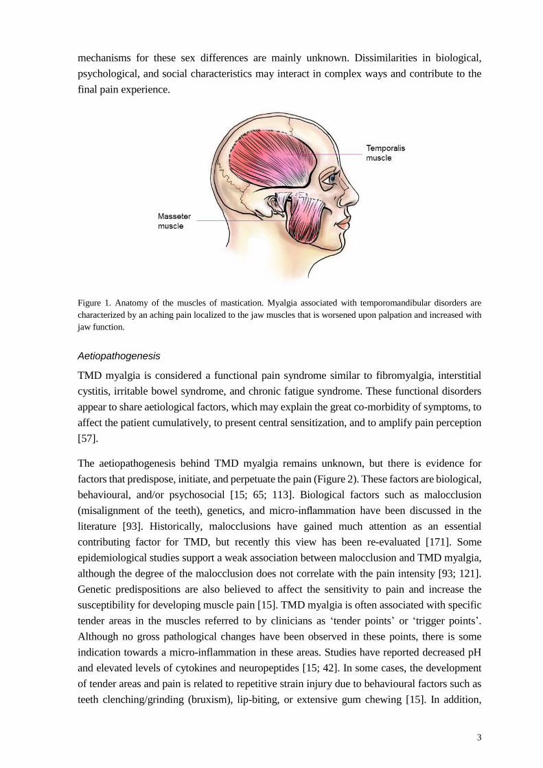
Temporomandibular disorders
TMD is a group of related conditions in the masticatory muscles, temporomandibular joint, and
associated surrounding structures (e.g., ligaments and connective tissues). The disorders are
characterized by a triad of clinical features involving muscle and/or joint pain, limited jaw
movements, joint noises, and alteration in the mandibular movements (Figure 1) [50; 150].
TMD causes a great deal of suffering in the community and is a widespread problem in clinical
practices. It affects 10-15% of the adult population with an incidence rate between 2-4% and
TMD myalgia seems to be the most frequent diagnosed TMD pain condition [37; 110; 135]
with a frequency of 42% [50]. The reported prevalence of TMD in different age groups varies
widely due to differences in study cohorts, diagnostic criteria, and examination methods.
Several studies have demonstrated a rather low prevalence of TMD in childhood. However,
TMD becomes more prevalent during adolescence and early adulthood and appears to peak
during midlife and to decrease in the elderly [85; 110]. Women are more susceptible to TMD;
according to epidemiological studies, two of every three patients with TMD signs are female
[85]. Several studies have reported greater evoked pain, lower pain threshold and pain tolerance
in women compared to men [51; 76]. However, women also show a greater adaptation to
sustained stimuli and habituation to repeated stimuli [76]. The underlying pathophysiological
3
mechanisms for these sex differences are mainly unknown. Dissimilarities in biological,
psychological, and social characteristics may interact in complex ways and contribute to the
final pain experience.
Figure 1. Anatomy of the muscles of mastication. Myalgia associated with temporomandibular disorders are
characterized by an aching pain localized to the jaw muscles that is worsened upon palpation and increased with
jaw function.
Aetiopathogenesis
TMD myalgia is considered a functional pain syndrome similar to fibromyalgia, interstitial
cystitis, irritable bowel syndrome, and chronic fatigue syndrome. These functional disorders
appear to share aetiological factors, which may explain the great co-morbidity of symptoms, to
affect the patient cumulatively, to present central sensitization, and to amplify pain perception
[57].
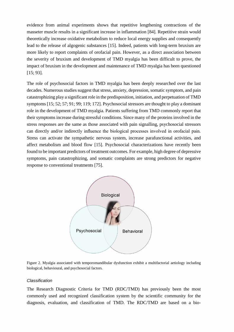
The aetiopathogenesis behind TMD myalgia remains unknown, but there is evidence for
factors that predispose, initiate, and perpetuate the pain (Figure 2). These factors are biological,
behavioural, and/or psychosocial [15; 65; 113]. Biological factors such as malocclusion
(misalignment of the teeth), genetics, and micro-inflammation have been discussed in the
literature [93]. Historically, malocclusions have gained much attention as an essential
contributing factor for TMD, but recently this view has been re-evaluated [171]. Some
epidemiological studies support a weak association between malocclusion and TMD myalgia,
although the degree of the malocclusion does not correlate with the pain intensity [93; 121].
Genetic predispositions are also believed to affect the sensitivity to pain and increase the
susceptibility for developing muscle pain [15]. TMD myalgia is often associated with specific
tender areas in the muscles referred to by clinicians as ‘tender points’ or ‘trigger points’.
Although no gross pathological changes have been observed in these points, there is some
indication towards a micro-inflammation in these areas. Studies have reported decreased pH
and elevated levels of cytokines and neuropeptides [15; 42]. In some cases, the development
of tender areas and pain is related to repetitive strain injury due to behavioural factors such as
teeth clenching/grinding (bruxism), lip-biting, or extensive gum chewing [15]. In addition,
evidence from animal experiments shows that repetitive lengthening contractions of the
masseter muscle results in a significant increase in inflammation [84]. Repetitive strain would
theoretically increase oxidative metabolism to reduce local energy supplies and consequently
lead to the release of algogenic substances [15]. Indeed, patients with long-term bruxism are
more likely to report complaints of orofacial pain. However, as a direct association between
the severity of bruxism and development of TMD myalgia has been difficult to prove, the
impact of bruxism in the development and maintenance of TMD myalgia has been questioned
[15; 93].
The role of psychosocial factors in TMD myalgia has been deeply researched over the last
decades. Numerous studies suggest that stress, anxiety, depression, somatic symptom, and pain
catastrophizing play a significant role in the predisposition, initiation, and perpetuation of TMD
symptoms [15; 52; 57; 91; 99; 119; 172]. Psychosocial stressors are thought to play a dominant
role in the development of TMD myalgia. Patients suffering from TMD commonly report that
their symptoms increase during stressful conditions. Since many of the proteins involved in the
stress responses are the same as those associated with pain signalling, psychosocial stressors
can directly and/or indirectly influence the biological processes involved in orofacial pain.
Stress can activate the sympathetic nervous system, increase parafunctional activities, and
affect metabolism and blood flow [15]. Psychosocial characterizations have recently been
found to be important predictors of treatment outcomes. For example, high degree of depressive
symptoms, pain catastrophizing, and somatic complaints are strong predictors for negative
response to conventional treatments [75].
Figure 2. Myalgia associated with temporomandibular dysfunction exhibit a multifactorial aetiology including
biological, behavioural, and psychosocial factors.
Classification
The Research Diagnostic Criteria for TMD (RDC/TMD) has previously been the most
commonly used and recognized classification system by the scientific community for the
diagnosis, evaluation, and classification of TMD. The RDC/TMD are based on a bio-

5
behavioural model of pain: axis I includes physical signs and symptoms and axis II includes
psychological and disability factors [38; 39; 65].
In 2014, the International Research Diagnostic Criteria for Temporomandibular Dysfunction
Consortium Network published an updated classification structure for TMD – Diagnostic
Criteria for TMD (DC/TMD). The new criteria aimed to improve the sensibility and specificity
of the previous RDC/TMD through improvement of axis and axis II, which can be used not
only in research but also in clinical settings [160]. As DC/TMD is used worldwide, it has been
translated into several languages. Axis I contains an effective screener for detecting any pain-
related TMD and effective diagnostic criteria for discriminating between the most common
pain-related TMD with a sensitivity ≥ 0.86 and a specificity ≥ 0.98. In addition, axis I also
exhibits a strong inter-examiner reliability for pain-related TMD with a kappa ≥ 0.85. The axis
II protocol includes both a screening and a self-assessment instrument. The screening
instruments assess pain intensity, disability related to pain, psychological distress, limitations
in jaw function, parafunctional behaviours, and a pain drawing to assess the location of pain.
The self-assessment instruments assess in more detail limitations in jaw function, anxiety,
psychological distress, and presence of comorbid pain conditions [160].
The DC/TMD includes tests for diagnosing myalgia such as pain with jaw movements and
palpation of the masseter and temporalis muscles (Figure 1). Pain from the clinical
examinations must resemble the patient’s pain complaint from the previous month. Myalgia is
further subclassified into three types: local myalgia, defined as pain localized to the site of
palpation; myofascial pain, defined as pain spreading beyond the site of palpation but within
the boundary of the muscle being palpated; and myofascial pain with referral, defined as pain
at a site beyond the boundary of the muscle being palpated [47]. Myalgia as a class exhibited a
sensitivity of 0.90 and specificity of 0.99, and the subtype myofascial pain with referral
exhibited slightly reduced sensitivity (0.86) and specificity (0.98). The sensitivity and
specificity for myofascial pain have yet to be established [160].
Myofascial pain has also been included in the newly published International Classification of
Orofacial Pain (ICOP), a document that is aligned with the International Classification of
Diseases (11th revision) and the International Classification of Headache Disorders (3rd edition)
(ICHD-3). ICOP aims to create an instrument that will improve research as well as clinical
management of orofacial pain and increase collaboration between professionals working on
pain in the orofacial area. The classification committee has adopted the DC/TMD criteria for
ICOP, but it only included TMD diagnosis associated with pain and modified the presentation
style to that of ICHD-3 based on its frequency [1].
OBJECTIVE PAIN MEASURES
Since the nociceptive mechanisms that underlie TMD pain are still not fully understood, the
clinician must rely on subjective measures such as patients’ anamnesis, questionnaires, and
semi-objective findings such as muscle palpation or assessment or pressure pain threshold
(PPT). According to the DC/TMD, the three subclasses of myalgia differ only regarding the
presence of pain spread upon palpation as described earlier, but the pathogenesis underlying
these diagnoses may not be the same. As pain is a subjective experience, semi-objective
methods have limited sensitivity and correlate weakly with subjective pain scores [42].
Consequently, objective and sensitive tools are needed, a situation that has led to a growing
interest in molecular biomarkers
Molecular biomarkers
Biological markers (biomarkers) are specific molecules that provide either diagnostic,
prognostic, predictive, or therapeutic information and can be measured objectively in human
tissues, cells, or body fluids [123]. In muscle pain, biomarkers can be separated into algesic
biomarkers, tissue metabolites, and inflammatory mediators [42].
Sampling and analysing biomarkers
In TMD myalgia, clinically valuable biomarkers have to be easy to measure and correlate to
pain ratings. An ideal biomarker should be measurable in samples that are easy and non-
invasive to collect and handle, such as blood, urine, and saliva. However, a common limitation
for all sampling methods are potential diurnal variations of the biomarker concentration. These
variations need to be properly evaluated or ruled out before collection. Furthermore, potential
biomarkers may be influenced by various factors such as gender, age, body mass, and general
health [60; 127].
The rapid development of molecular biology and laboratory technology has led to innovative
techniques that can identify biomarkers. New sensitive methods have the possibility to combine
and analyse several biomarkers simultaneously, a development that saves time, sample
material, and expenses. Multiplex assays such as antibody microarray, Luminex®, and Meso
Scale Discovery combine the efficiencies of multiplexing with the sensitivity, precision,
reproducibility, and simplicity of enzyme-linked immunosorbent assay (ELISA). Multiplex
assay requires knowledge of which analytes to be measured in a specific sample. Proteomic
analyses, on the other hand, usually eliminate this requirement and therefore enable the use of
blind studies of all the proteins expressed in a sample (e.g., the proteome) and accurately and
reliably quantify changes in protein abundance in health and disease to identify potential
biomarkers. There are several proteomic methods available such as gel-based methods,
including one-dimensional and two-dimensional polyacrylamide gel electrophoresis (2DE) or
gel-free high throughput liquid chromatography coupled with tandem mass spectrometry
(shotgun proteomic). Shotgun proteomics, 2DE, and protein microarrays can be used to obtain
overviews of differences in protein abundance in a sample at a given time or under particular
conditions [20].
2DE followed by mass spectrometry is considered a powerful tool for proteomics work [62] as
it separates and quantifies different protein isoforms. In 2DE, proteins are separated on two
dimensions. In the first dimension, the proteins are separated according to their net charge, also
known as isoelectric points. In the second dimension, sodium dodecyl sulphate polyacrylamide
gel electrophoresis (SDS-PAGE) separates proteins by their mass. Therefore, complex mix-
7
tures consisting of hundreds to thousands of proteins can be separated and the relative amount
of each protein can be determined. Changes in charge and mass can also easily be detected by
this approach as it is very unusual that two different proteins or protein isoforms resolves to the
same place in both dimensions. Each protein spot can then be removed from the gel, digested
with tryptic enzymes and identified using mass spectrometry [4].
Several proteomic studies have been performed in different painful conditions (e.g.,
neuropathic pain, trapezius myalgia, fibromyalgia, rheumatoid arthritis, and burning mouth
syndrome) by analysing proteins in plasma, saliva, cerebrospinal fluid, synovial fluid,
interstitial fluid, or biopsies [6; 7; 26; 67; 72; 94; 103; 141-143]. Saliva contains a complex
mixture of proteins, peptides, and other molecules that may yield information about the
pathophysiology behind TMD myalgia and can be used to identify new biomarkers for the
disorder. Proteomic analysis of saliva in patients with TMD myalgia represents a new field of
research as the proteomic techniques constantly improve [90; 92]. Only two studies have
investigated widespread myalgia using the salivary proteome. These studies, using gel-based
proteomics applied to saliva samples from patients with fibromyalgia, reported altered protein
expression between patients and controls with an over-expression of transaldolase,
phosphoglycerate mutase I, serotransferrin, and alpha-enolase [7; 26].
Potential biomarkers for TMD myalgia
The masticatory muscles are innervated by Aδ and C afferent fibres with free nerve endings.
These trigeminal afferent nerve fibres are activated by noxious mechanical and ⁄or chemical
stimuli and express receptors for algogenic substances such as glutamate, serotonin, capsaicin,
and ATP. Consistent with this role, they also contain neuropeptides [15; 60].
The most studied algogenic substance in TMD myalgia is the excitatory amino acid glutamate.
Injection of pharmacological dose of glutamate in healthy masseter muscles is associated with
pain symptoms and altered pain sensitivity [179]. Studies have shown that interstitial glutamate
is elevated in the masseter muscles of patients with TMD myalgia compared to healthy controls
[19; 31; 179]. These findings imply that glutamate may be related to ongoing pain and mech-
anical sensitivity in TMD myalgia [60].
Serotonin is another potential algogenic substance discussed in the pathogenesis of TMD
myalgia [15]. Biopsies from human masseter muscles have revealed the presence of serotonin
receptors on sensory nerves in the muscle, and patients with TMD myalgia expressed more of
these receptors compared to healthy controls [24]. In addition, the interstitial concentration of
serotonin in the masseter muscle of patients appears to be higher compared to the levels found
in healthy controls [43; 117], and injection of the serotonin-receptor antagonist granisetron into
the masseter muscle seems to alleviate myalgia associated with TMD [25].
Few studies have investigated interstitial and circulatory levels of serotonin or glutamate in
TMD myalgia and the findings are inconsistent. Increased interstitial muscle levels of serotonin
and glutamate are reported in patients compared to controls [19; 43], but the levels in

plasma/serum showed no differences [31; 44]. The inconsistent results may be due to
methodological and diagnostic dissimilarities and the low number of subjects in certain studies.
Nerve growth factor (NGF) is a neuropeptide that facilitates neuronal regeneration and acts as
a protective factor for neurons. It has been suggested that NGF plays an important role in
hyperalgesia as the concentration of the molecule has been found to be increased after
inflammatory injury and up-regulated in response to noxious stimuli [130]. Calcitonin gene-
related peptide (CGRP), brain derived neurotropic factor (BDNF), and substance P (SP) are
other examples of abundant neuropeptides in nervous tissue. These play important roles in the
development of pain and hyperalgesia. CGRP and BDNF may play a role in migraine and
headaches based on its increased saliva and plasma concentration during active pain periods
[5; 54; 89; 196]. Some evidence suggests that salivary SP levels increase with noxious
stimulation, indicating that SP may play a role in central sensitization associated with chronic
pain [71; 89]. These substances have also been discussed in TMD myalgia, but their
significance in clinical pain has yet to be established [50; 60].
Usually, all potential biomarkers are measured in plasma, cerebrospinal fluid, or intestinal
fluid; however, some studies have measured these mediators in saliva [11; 54; 89; 98; 129; 130;
196]. Although salivary glands are integrated in the neuroendocrine system, the sampling and
processing techniques are limited and in need of further improvement. Clearly, measuring
salivary algogenic mediators and neuropeptides could provide a valuable diagnostic and
prognostic tool for chronic painful conditions and provide an objective approach to the study
of pain. However, collection methods need to be evaluated and more sensitive techniques for
the subsequent analysis need to be developed.
SALIVA AS A DIAGNOSTIC TOOL
Saliva performs many biological functions essential for the maintenance of oral health such as
lubrication, cleansing, buffering, and digestion. However, the functions of saliva are not only
restricted to the oral cavity [4; 35; 46] as saliva contains many classes of proteins and peptides
that represent several significant biological functions that may mirror both oral and systemic
health conditions [114]. With the advancements made in analytical technologies for saliva over
the last several decades, saliva has gained increased attention also for clinical diagnostics [17;
30; 114].
The salivary glands
Healthy adults produce on average 0.3-0.7 ml of saliva per minute, producing a range of 0.5 up
to 1.5 litres daily. Secretion of saliva is an active and continuous process under sympathetic
and parasympathetic stimulation [194]. Saliva is not a single fluid but rather a complex mixture
derived predominantly from three pairs of major salivary glands (parotid, sublingual, and
submandibular gland), which contribute about 90% of the total fluid secretion, and from the
minor salivary glands in the oral mucosa, which contribute about 10% of the total volume. In
addition, whole saliva also contains gingival crevicular fluid, nasal and bronchial secretions,
oral mucosal cells, serum filtrate, microbiota, and food debris [125; 194]. Although the parotid

9
glands are the largest (Figure 3), they produce only about 20% of the total saliva in the
unstimulated resting state, and the minor and sublingual glands together contribute to about an
additional 15%. The submandibular glands are by far the most active in the unstimulated resting
state, and they are estimated to produce about 65% of the total resting volume. However, when
salivary glands are stimulated, the parotid can account for more than 50% of the whole saliva
volume in the mouth [83; 133]. Some of the salivary glands are purely serous (parotid gland),
others are mucous (minor palatine glands), and some are mixed (submandibular, sublingual,
and minor buccal glands).
Figure 3. The major salivary glands are
the parotid, the submandibular, and
sublingual glands. The saliva from the
parotid glands is secreted through the
“Stensen´s duct”, which opens into the
mouth in the buccal mucosa near the
parotid papilla opposite the second
maxillary molar. The saliva from the
submandibular and sublingual glands
enters the mouth through the “Wharton
duct” and several smaller sublingual
ducts, which open into the floor of the
mouth.
The salivary glands are composed of secretory units called acini, which are built-up of acinar
cells that can be either serous or mucous. The acini cells are surrounded by myoepithelial cells,
which contract the acini to secrete saliva (Figure 4). Each salivary gland is surrounded by blood
vessels and is highly permeable, an arrangement that allows for the free exchange of blood-
borne molecules into the acinus [194]. The saliva is composed of 99% water, 0.3% proteins,
and 0.2% of inorganic substances. The formation of saliva occurs in a two-stage process. The
first stage involves the secretion of an isotonic plasma-like primary saliva by the acinar cells
into the luminal terminal pieces of the gland parenchyma. In the second stage, the saliva
changes to a hypotonic saliva as it passes though the ducts into the oral cavity. Salivary proteins
are continuously secreted by exocytosis of granules from acinar cell. Some proteins are secreted
into saliva by other mechanism such as vesicular transport or transcytosis. The majority of all
saliva proteins are synthesized by the salivary glands (e.g., glycoproteins, proline-rich proteins,
histatins, and statherin), but a small amount of the proteins originates from the plasma [151;
194].
Mirror of bodily functions
Saliva can provide information about diseases and offers distinctive advantages over blood.
Salivary diagnostic approaches have been developed to monitor cancer [77] as well as
Figure 4. The salivary gland is very permeable and surrounded by blood vessels, which allows for diffusion of
blood-derived molecules into the saliva. The protein content in saliva is mostly synthetized and secreted by salivary
acinar cells, but a small amount is secreted from the blood.
autoimmune [81], viral [40; 120; 137], and bacterial diseases [2]. However, the full potential
of saliva in medicine was recently recognized [109].
Many substances enter saliva from the blood by passing through the intercellular spaces by
transcellular or paracellular diffusion [36; 73; 97; 194]. As a result, most of the abundant
substances found in blood are also commonly present in saliva. Approximately 27% of the
proteome in plasma and nearly 40% of the proteins that have been suggested to be candidate
markers for diseases can be found in whole saliva [115]. Therefore, saliva can be regarded as
functionally equivalent to plasma with respect to its ability to reflect the physiological state of
the body. In addition, saliva collection provides some advantages over blood. The collection is
a simple procedure and non-invasive, so it dramatically diminishes any discomfort associated
with blood, cerebrospinal fluid, or interstitial collection, methods often used in pain research
[53]. For the patient, non-invasive collection reduces anxiety and discomfort. Moreover, non-
invasive collection simplifies procurement of repeated samples for longitudinal monitoring. In
addition, saliva collection also has many advantages in terms of sampling, storage, and
shipping. For the clinician, saliva is safer than venipuncture, which could expose healthcare
providers to blood-borne and infectious diseases. Consequently, saliva has the potential to be
used as a diagnostic and prognostic specimen in pain research [109; 163].
11
AIMS
This thesis investigates whether saliva can be used to sample algogenic substances that can
serve as molecular biomarkers for TMD myalgia.
The methods section evaluates several saliva sampling methods and analysis techniques. The
clinical section evaluates methods applied to patients. The specific objectives are as follows:
Identify the best method for saliva sampling and standardize collection procedure for
studying serotonin, glutamate, SP, CGRP, BDNF, and NGF.
Evaluate if a diurnal variation in saliva exists for these biomarkers.
Compare salivary and plasma levels of the above biomarkers in patients with a
diagnosis of TMD myalgia and healthy pain-free controls.
Develop a proteomic workflow for a comprehensive identification of proteins in saliva,
and study the differences in the proteome expression between different types of saliva
(whole and glandular saliva).
Apply the above proteomic approach to study the proteomic signature in patients with
TMD myalgia and pain-free controls.

13
MATERIAL AND METHODS
The methods and selection of participants were approved by the Regional Ethical Review
Board in Stockholm, Sweden (2014/17-31/3).
All participants were recruited through advertisement and from patients referred to the
specialist clinic for orofacial pain and jaw function at the University Dental Clinic at Karolinska
Institute (Huddinge, Sweden). The studies were all conducted at Department of Dental
Medicine at Karolinska Institute (Huddinge, Sweden). All participants received careful
information regarding the objectives and procedures of the study and signed an informed
written consent form prior to participation. The study protocols followed good clinical practice
and the guidelines according to the Declaration of Helsinki.
HEALTHY PARTICIPANTS
In total, 69 healthy participants were included, 47 women and 22 men. The participants in
Studies I-III were matched according to age and gender. Note that samples from participants
in Study I were also included in Study II. The distribution of the participants included in Studies
I-V are presented in Table 1.
Inclusion criteria were age above 18 years old, good general health, and a body mass index <
30 kg/m2. In addition, participants had to be free of fever/or cold and maintain exceptional oral
hygiene on the day of collection.
PATIENTS
In total, 39 patients (32 women and 7 men) with chronic masticatory muscle pain (TMD
myalgia) were included in Studies IV-V (Table 1). Note that samples from participants in Study
IV were also included in Study V.
The inclusion criteria were age above 18 years old and a diagnosis of myalgia or myofascial
pain with or without referral according to the DC/TMD [160].
EXCLUSION CRITERIA AND EXAMINATION
For both groups, the following exclusion criteria were used: smoking; diagnosed systemic
muscular or joint diseases such as fibromyalgia and rheumatoid arthritis; whiplash-associated
disorder; migraine; neurological or neuropsychiatric disorders; diseases of salivary glands such
as sialadenitis and salivary gland tumours; pregnancy or lactation; obesity; regular use of
medications; use of analgesics during the last 24 hours; oral complaints, such as oral dryness
or mucosal lesions; participants with less than 22 teeth and extensive prosthodontics
rehabilitations; and poor oral hygiene, hyposalivation, oral diseases (severe periodontal
diseases and mucosal pain or ulcerations), or extensive dental abrasion.
Whether a patient should be excluded was determined using information gathered from patient
questionnaires, medical histories, and dental examinations. Participants were also asked about
factors influencing saliva secretion and composition such as level of physical activity. During
the clinical examination, participants were checked for attrition, decayed teeth, periodontal
diseases, mucosal lesions, oral hygiene, jaw movements, and occlusal contacts.
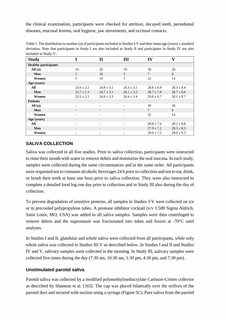
Table 1. The distribution in number (n) of participants included in Studies I-V and their mean age (years) ± standard
deviation. Note that participants in Study I are also included in Study II and participates in Study IV are also
included in Study V.
Study I II III IV V
Healthy participants
All (n) 10 20 10 39 20
Men 5 10 5 7 6
Women 5 10 5 32 14
Age (years)
All 23.6 ± 2.2 24.8 ± 3.1 26.3 ± 3.1 28.8 ± 6.9 28.3 ± 8.4
Men 23.7 ± 2.3 24.7 ± 3.1 26.1 ± 3.2 28.7 ± 7.9 28.7 ± 8.6
Women 23.5 ± 2.1 24.9 ± 3.3 26.4 ± 3.4 29.0 ± 6.7 28.1 ± 8.7
Patients
All (n) - - - 39 20
Men - - - 7 6
Women - - - 32 14
Age (years)
All - - - 28.8 ± 7.4 28.1 ± 8.8
Men - - - 27.9 ± 7.2 28.5 ± 8.3
Women - - - 29.0 ± 7.3 28.0 ± 9.3
SALIVA COLLECTION
Saliva was collected in all five studies. Prior to saliva collection, participants were instructed
to rinse their mouth with water to remove debris and moisturize the oral mucosa. In each study,
samples were collected during the same circumstances and in the same order. All participants
were requested not to consume alcoholic beverages 24 h prior to collection and not to eat, drink,
or brush their teeth at least one hour prior to saliva collection. They were also instructed to
complete a detailed food log one day prior to collection and in Study III also during the day of
collection.
To prevent degradation of sensitive proteins, all samples in Studies I-V were collected on ice
or in precooled polypropylene tubes. A protease inhibitor cocktail (v/v 1:500 Sigma Aldrich,
Saint Louis, MO, USA) was added to all saliva samples. Samples were then centrifuged to
remove debris and the supernatant was fractionated into tubes and frozen at -70°C until
analyses.
In Studies I and II, glandular and whole saliva were collected from all participants, while only
whole saliva was collected in Studies III-V as described below. In Studies I and II and Studies
IV and V, salivary samples were collected in the morning. In Study III, salivary samples were
collected five times during the day (7:30 am, 10:30 am, 1:30 pm, 4:30 pm, and 7:30 pm).
Unstimulated parotid saliva
Parotid saliva was collected by a modified polymethylmethacrylate Carlsson-Critten collector
as described by Shannon et al. [165]. The cup was placed bilaterally over the orifices of the
parotid duct and secured with suction using a syringe (Figure 5C). Pure saliva from the parotid

15
gland was collected through a 25-cm plastic tubing placed in a precooled polypropylene tube.
To reduce probable contamination, the first drops of saliva were discarded. The tubes were then
weighed, and the salivary flow rate was calculated assuming a saliva density of 1.0 g/ml.
Stimulated parotid saliva
To collect stimulated parotid saliva, the Carlsson-Critten collector was used as described above.
To stimulate salivary flow, aqueous 2% citric acid solution was applied bilaterally on the sides
of the tongue with a cotton swab every 30 seconds. The tubes were then weighed, and salivary
flow rate was calculated assuming a saliva density of 1.0 g/ml.
Unstimulated sublingual saliva
While blocking the orifices of the parotid duct with the Carlsson-Critten collector, sublingual
saliva from the submandibular and sublingual gland could be collected simultaneously. Saliva
was collected every second minute from the floor of the mouth with a sterile syringe into
precooled tubes (Figure 5B). Similar to collection of unstimulated parotid saliva, the initial
drops were discarded to neutralize salivary flow. The tubes were then weighed, and salivary
flow rate was calculated assuming a saliva density of 1.0 g/ml.
Stimulated sublingual saliva
Saliva Bio Oral Swab® (Salimetrics LCC Carlsbad, CA, US) was used to collect stimulated
sublingual saliva. The absorbent pad made by synthetic material was placed under the tongue
for around two minutes while stimulating saliva flow with aqueous 2% citric acid solution on
the sides of the tongue until the pad was fully soaked with saliva. Stimulated sublingual saliva
was extracted by centrifugation (1500xg, 15 min, 4°C) of the Saliva Bio Oral Swab®.
Unstimulated whole saliva
Unstimulated whole saliva was collected while the participants were seated comfortably with
eyes open and head slightly tilted forward (Figure 5A). Participants were instructed to allow
saliva to accumulate on the floor of the mouth without stimulation and passively drool into
precooled 5-ml polypropylene tube. Total drooling time was documented, and salivary flow
calculated.
Stimulated whole saliva
Whole saliva was mechanically stimulated using sterile paraffin gum (Orion Diagnostica,
Esbo, Finland) (Figure 5E). First, the participants were instructed to chew the gum until it was
smooth and flexible. After about 60 seconds, the participants were asked to swallow the
produced saliva and then start to chew and expectorate the secreted saliva into a precooled
graded polypropylene tube until sufficient volume of saliva was collected. Total spitting time
was documented and salivary flow calculated.
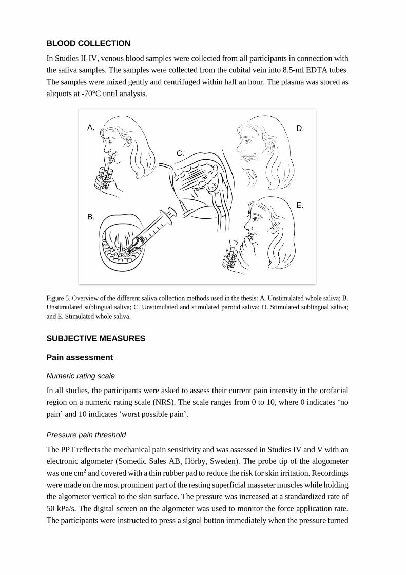
BLOOD COLLECTION
In Studies II-IV, venous blood samples were collected from all participants in connection with
the saliva samples. The samples were collected from the cubital vein into 8.5-ml EDTA tubes.
The samples were mixed gently and centrifuged within half an hour. The plasma was stored as
aliquots at -70°C until analysis.
Figure 5. Overview of the different saliva collection methods used in the thesis: A. Unstimulated whole saliva; B.
Unstimulated sublingual saliva; C. Unstimulated and stimulated parotid saliva; D. Stimulated sublingual saliva;
and E. Stimulated whole saliva.
SUBJECTIVE MEASURES
Pain assessment
Numeric rating scale
In all studies, the participants were asked to assess their current pain intensity in the orofacial
region on a numeric rating scale (NRS). The scale ranges from 0 to 10, where 0 indicates ‘no
pain’ and 10 indicates ‘worst possible pain’.
Pressure pain threshold
The PPT reflects the mechanical pain sensitivity and was assessed in Studies IV and V with an
electronic algometer (Somedic Sales AB, Hörby, Sweden). The probe tip of the alogometer
was one cm2 and covered with a thin rubber pad to reduce the risk for skin irritation. Recordings
were made on the most prominent part of the resting superficial masseter muscles while holding
the algometer vertical to the skin surface. The pressure was increased at a standardized rate of
50 kPa/s. The digital screen on the algometer was used to monitor the force application rate.
The participants were instructed to press a signal button immediately when the pressure turned
17
into a pain sensation. PPT was also recorded over the tip of the index finger as a reference point
to register any possible systemic sensibility. Therefore, a pinch handle was attached to the
algometer, which allows the fingertip to be pressed against the handle by the probe during the
recording.
The participants were given a verbal and illustrated description of the procedure. PPT was then
recorded three times on each location and the average threshold of the three recordings was
registered.
Questionnaires
In all studies, questionnaires were used to assess psychological predispositions and conditions.
The following instruments included in the DC/TMD axis II questionnaire were used to assess
symptoms of depression, somatic symptoms, anxiety, psychological stress, jaw function, oral
health, sleep disturbance, and pain catastrophizing: the Patient Health Questionnaire (PHQ-9
and PHQ-15), the Generalized Anxiety Disorder scale (GAD-7), the Perceived Stress Scale-10
(PSS-10), the Jaw Functional Limitation Scale (JFLS), the Oral Health Impact Profile (OHIP),
the Insomnia Severity Index (ISI), and the Pain Catastrophizing scale (PCS).
The Patient Health Questionnaire
The PHQ, a validated instrument for mental health disorders, is part of a family of related
measures, including the PHQ-9, which is the depression module from the PHQ, and the PHQ-
15, which is the somatic symptom module from the PHQ. Studies have found good correlation
between PHQ diagnoses and those of independent mental health professionals [105; 106].
The PHQ-9 includes nine symptoms of depression and assesses the level of the depression by
the frequency of the symptoms within the last two weeks. Scores ranged between 0 and 27, and
scores of 5, 10, 15, and 20 are considered cut-off values for mild, moderate, moderately severe,
and severe depression, respectively. Studies of the reliability and validity in adults shows that
the PHQ-9 has a 61% sensitivity and 94% specificity [105; 122].
The PHQ-15 includes 15 somatic symptoms or symptom clusters that account for more than
90% of the physical complaints. In determining the PHQ-15 score, each symptom is coded as
0, 1, or 2, and the total score ranges from 0 to 30. Scores of 5, 10, and 15 are considered cut-
off values for mild, moderate, and severe somatic symptoms, respectively [104; 173].
Generalized Anxiety Disorder scale
The GAD-7, a frequently used diagnostic self-administered scale for assessing generalized
anxiety disorder symptoms, measures anxiety based on seven items scored from 0 to 3. The
whole scale score can range from 0 to 21, and scores of 5, 10, and 15 are considered cut-off
values for mild, moderate, and severe anxiety, respectively [96]. The scale has been validated
within a large sample of patients in a primary care setting [174] as well as within the general
population [118] and is a reliable measure of anxiety. A meta-analysis shows that the GAD-7
has acceptable properties for identifying generalized anxiety disorder at cut-off scores 7-10
[149]. The scale is statistically associated with age and gender and shows high comorbidity
with depressive and somatic symptoms [174].
Perceived Stress Scale
The PSS-10 is a self-reported instrument that assesses how unpredictable, uncontrollable, and
overloaded participants find their lives during the previous month. The PSS-10 is rated on a
five-point Likert scale (0-4). Of the ten items of the PSS-10, four are considered negative and
six are considered positive. The total score is calculated after reversing the positive item scores
and summing all scores and ranges from 0 to 40, where a higher score indicates greater
perception of stress [45]. In some studies, high PSS scores have been correlated to high
biomarker levels of stress, such as cortisol [154].
Jaw Function Limitation Scale
JFLS is designed to assess jaw function disability. The scale assesses the function of the
masticatory system in three dimensions: mastication, vertical jaw mobility, and emotional and
verbal expression [139; 160]. The scale consists of 20 items with each item is rated on a NRS,
where 0 corresponds to no limitation and 10 to severe limitation. Calculation of a global score
(0-10) as the average of the ratings for eight of the items is recommended [160]; a higher score
indicates insufficient jaw function. Another suggestion is to sum all 20 items, resulting in a
score that ranges 0-200 [123]. Norms have not been yet established for this scale. A recent
study suggests a cut-off value of 28 or more out of 200 to define a limitation in jaw function
[138]. The instrument exhibits very good psychometric properties and displays strong internal
reliability for items and individual responses [139].
Oral Health Impact Profile
OHIP is a reliable and valid instrument for assessing the impact of oral health on masticatory
ability and psychosocial function. All studies used a shortened (14-item and 5-item) version of
the OHIP consisting of statements that have been rephrased as questions. The participants were
asked to rate on a five-point Likert scale how often they experienced each problem within a
period of one month. The option ‘do not know/not applicable’ was also included among the
answers. Higher scores indicate a poorer oral health-related quality of life [107; 128].
Insomnia severity index
The ISI is a short screening instrument used to measure the symptoms of insomnia. The ISI
consists of seven items measuring self-reported problems with sleep such as trouble falling or
staying asleep, early awakenings, satisfaction with sleep pattern, distress caused by lack of
sleep, and impacts on daily functioning. Each item is scored from 0 to 4, resulting in a total
possible score of 28, with a score above 15 indicating clinical insomnia. ISI is a reliable and
valid instrument for detecting individuals with insomnia and clinically evaluating the response
to treatment [126].

19
Pain catastrophizing scale
Pain catastrophizing is characterized by feelings of helplessness, active reflection, and
magnification of thoughts and feelings towards the painful stimulation [111]. The PCS consists
of thirteen items. Each item is scored between 0 and 4, resulting in a total possible score of 52
points. Higher scores indicate higher presence of catastrophizing thoughts [176]. Previous
studies have reported that a cut-off value of more than 30 points is associated with pain
catastrophizing of clinical relevance. PCS exhibits good psychometric properties with high
reliability and internal consistency [111; 145]. The PCS score also seems to correlate to pain
intensity, pain-related disability, fear avoidance, and psychological distress [111].
BIOCHEMICAL ANALYSIS
Proteomic profiling
The protein concentration was determined using the Bio-Rad protein assay according to
Bradford (Bio-Rad, Hercules, CA, USA). Saliva was desalted into 12mM ammonium
bicarbonate using Amicon® Ultra centrifugal filters (Merck Millipore, Billericia, MA, USA).
Proteins were then lyophilized and dissolved with 0.20 ml urea solution according to Görg et
al. [70].
The denatured proteins in each sample (containing 50 ug of protein in Study I and 300 ug of
protein in Study V) were separated according to isoelectric point in the first dimension by in-
gel rehydration according to the manufacturer’s instructions for 12 h in Study I and 10 h in
Study V using low voltage (30 V) in pH 3-10 non-linear 18 cm (Study I) and 24 cm (Study IV)
IPGs (GE Healthcare, Stockholm, Sweden). The proteins were then focused for up to 32 000
Vhs in Study I and up to 40 000 Vhs in Study V at a maximum voltage of 8000 V to assure a
steady state. IPGs were then immediately stored at -70°C until analysed.
All the IPG gel strips were then equilibrated in SDS equilibration buffer (urea 6 M, SDS 4%
(w/v), glycerol 30.5% (w/v), and Trizma-HCl 50 mM) and DTT 1% (w/v) for 15 minutes and
then with iodacetamide 4.5% (w/v) for additional 15 minutes.
The second dimension (SDS-PAGE) was run horizontally in Study I and vertically in Study V.
In Study I, the horizontal run was carried out by transferring the proteins to gradient gels
(ExcelGel XL 245x180x0.5 mm, 12–14%T, 3%C) running at 20-40 mA, up to 1000 V for
about 5 h using Multiphor (GE Healthcare). In Study V, SDS-PAGE was carried out using a
vertical 2DE setup (ETTAN™ DALTsix Electrophoresis system, Amersham, Pharmacia
Biotech, Uppsala, Sweden) as previously described by Bäckryd et al. [6]. Briefly, the IPGs
were mounted on precast homogenous polyacrylamide gels (DALT gel 260 × 200 × 1.0 mm,
12.5 %) and run according to protocol for about 7-8 h ( 2.5 W per gel, 600 V, 400 mA for 30
minutes, followed by an additional 5 hours at 15 W per gel until the blue front reached the
bottom of the gel) at a constant temperature of 25°C.
In addition, the staining differed between studies. In Study I, the analytical gels were stained
with silver according to Shevchenko et al. [166] with a detection limit of 5ng/spot [178] using
a Stainer Shaker (Hoefer Processor Plus, Amersham Bioscience, UK). In Study V, the gels
were fluorescently stained with One-Step Lumitein™ (Biotium, Hayward, CA, USA)
according to the manufacturer’s protocol.
The 2DE protein patterns of all the gels were visualized as digitized images using a charged
coupled device camera system, Versa Doc (Bio-Rad Hercules, CA, USA), in combination with
a computerized imaging 16-bit system designed for evaluation of 2DE patterns (PDQuest V
8.0.1; Bio-Rad). Protein spots were detected and matched among different samples, and the
amount of protein in each individual spot was assessed as background-corrected optical density
integrated over all pixels in the spot and expressed as integrated optical density.
In Study I, the protein spots were identified by comparing previously identified saliva proteins
from a local database available at the laboratory [62]. However, protein spots of interest in
Study V were excised from the gel and digested with trypsin (Promega Corporation, Madison,
WI, USA) as previously described by Ghafouri et al. [62]. The trypsinated peptides were
analysed using a nano liquid chromatography system (EASY-nLC, Thermo Scientific,
Waltham, MA, USA) coupled to an LTQ Orbitrap Velos Pro MS (Thermo Scientific). Database
searching was performed using software MaxQuant (version 1.5.8.3) and the findings were
compared with the human Swissprot/UniProt database [27].
Capillary isoelectric focusing immunoassay
BDNF, CGRP, and NGF in Study II and III were analysed with a capillary isoelectric focusing
(IEF) immunoassay. Saliva samples were thawed before the analysis and centrifuged to remove
debris, and the supernatants were extracted to a new tube. The samples were then diluted with
Bicince and concentrated and desalted using Amicon® Ultra centrifugal filters (Merck
Millipore, Billericia, MA, USA). Total saliva protein was measured with 2D-Quant kit
according to the manufacturer’s instructions (GE Healthcare, Little Chalfont, UK). Plasma
samples were subjected to albumin and IgG removal kit (GE Healthcare) and then concentrated
and desalted using Amicon® Ultra centrifugal filters.
The saliva samples were analysed using a charge-based assay in Study II and size-based assay
in Study III. The latter method was used to analyse the plasma samples. All samples were
analysed using capillary isoelectric focusing with Peggy system (ProteinSimple, Santa Clara,
CA, USA) per manufacturer’s protocols. A protein concentration of 0.5 mg/ml was used to
analyse BDNF, CGRP, and NGF. The proteins were detected using antibodies against BDNF
(Mouse monoclonal, ab10505, Cambridge, UK), CGRP (Rabbit polycloncal, ab189786,
Cambridge, UK), and NGF (Rabbit polyclonal, ab6199, Cambridge, UK). The signal was
detected with Luminol and Peroxide and scanned with a charged coupled device camera.
Higher chemiluminescence equalled to higher expression. The data generated were analysed
in compass software version 2.7.1 (ProteinSimple, Santa Clara, CA, USA).
21
Kinetic enzymatic analysis
The concentration of glutamate in Studies II, III, and IV was determined as previously
described by Gerdle et al. [61]. To remove debris, saliva and plasma samples were centrifuged
at 4°C for five minutes at 12 000 × g. The supernatant was collected and transferred to a new
tube and 5 µl was immediately analysed using ISCUSS analyser (CMA Microdialysis). The
detection limit was 1.0 to 150 µmol/l.
Multiplex electrochemiluminescence assay panel
In Study IV, BDNF and NGF concentrations were analysed with the multiplex electrochemi-
luminescence assay panel from Meso Scale Discovery (MSD, Rockville, MD, USA) per
manufacturer’s instructions. The detection limits were 0.373 pg/ml and 0.036 pg/ml for BDNF
and NGF, respectively.
Enzyme-linked immunosorbent assay
Commercially-available enzyme kits were used to quantify the levels of SP and serotonin.
For detection of SP, the enzyme-linked immunosorbent assay kit (ADI-900-018) was used;
for detection of serotonin, the colorimetric competitive enzyme immunoassay kit (ADI-900-
175) from Enzo Life Sciences (Farmingdale, NY, USA) was used. The detection limit for SP
was 8.04 pg/ml and the detection limit for serotonin was 0.293 ng/ml. All kits were used
according to the manufacturer’s instructions using 96 well plates.
STATISTICAL ANALYSIS
Univariate statistics
The Shapiro-Wilks test was used to test for normality. For continuous variables with normal
distribution, independent t-test was used to study differences between two independent groups
or repeated measures analysis of variance (ANOVA) for repeated observations with Bonferroni
as post-hoc test. Only substances that were detected in more than half of the samples were
included in the statistical analysis.
For categorical variables or variables that were non-normal distributed, the Mann-Whitney U-
test was applied to study differences between two groups or Friedman’s ANOVA for repeated
observations. When significant, post-hoc analysis with Wilcoxon matched pair-test was applied
with Bonferroni correction.
The Pearson’s correlation test was used to test for significant correlations for normally
distributed data. Otherwise, correlations between variables were tested for statistical
significance using the Spearman correlation test adjusted for multiple comparisons according
to Bonferroni.
Descriptive data are presented as mean and standard deviation (SD) or median and interquartile
range (IQR). For all analyses, the significance level was set at P < 0.05. Statistical analyses
were performed using Statistica version 13 (StatSoft, Tulsa, OK, USA).

Multivariate statistics
Multivariate analysis (MVA) is the statistical analysis of several variables simultaneously. In
proteomics, each sample can generate hundreds to thousands of potentially equally interacting
proteins. MVA has the ability to find interactions between several supposedly independent
variables and to determine the contribution of each variable to the measured gains. Multivariate
techniques provide a powerful tool to test for significance compared to common univariate
techniques [6; 41; 189]. MVA was performed in Study I and Study V using SIMCA-P+ v.15.0
(UMETRICS, Umeå, Sweden).
In the first step, when little is known about the data, a simple overview of the information in
the data is needed. This overview can be obtained using principal component analysis (PCA).
PCA produces a summary and has the ability to uncover time trends and sudden changes in the
data, displaying how the observations are related and if there are any outliers or deviating
groups of observations in the data. PCA requires the definition of a few latent variables – i.e.,
principal components – that describe the principal structure in the data. The principal
components are uncorrelated to one another, but they simplify and summarize the data and
facilitate the discovery of important patterns in the data. The PCA analysis produces a score
plot and a loading plot: the score plot describes the relationship between the participants and
the loading plot describes the relationships between variables.
In the second step, orthogonal projections to latent structures discriminant analysis (OPLS-DA)
were used to investigate the multivariate correlations between the proteins and to identify the
variables responsible for discriminating between patients and controls. Therefore, the outcome
variable (Y) nominally represented patient or control, and the predictor variable (X)
numerically represented each protein spot measured by integrated optical density. The
variable’s influence on projection (VIP) indicates the relevance of each X variable pooled over
all dimensions and the Y variables the group of variables that best explains Y. Variables with
VIP ≥ 1.0 and with a 95% confidence interval are usually considered significant in MVA.
However, in Study I and Study V, VIP > 1.2 and 1.5, respectively, were considered significant.
The sign of the corresponding loading was used to determine whether the relationship was
positive or negative.
The R2 value describes the goodness of fit – the fraction of sum of squares of all the variables
explained by a principal component. R2 ranges between 0 and 1, with higher value indicating
higher predictive accuracy. The Q2 describes the goodness of prediction – the fraction of the
total variation of the variables that can be predicted by a principal component using cross
validation methods. Q2 ranges between 0 and 1, with the higher the value indicating better
predictability of the model. If R2 is considerably higher than Q2, the strength of the model is
poor; therefore, R2 should not exceed Q2 by too many units. That is, the difference between R2
and Q2 should not exceed 0.3. To validate the obtained model, cross validated analysis of
variance (CV-ANOVA) was used. The OPLS-DA model was considered significant if the CV-
ANOVA showed P < 0.05.
23
RESULTS
METHODOLOGICAL STUDIES
These studies aimed to analyse and compare different saliva collection methods by analysing
the general protein profile as well as specific proteins participating in pathways of pain and the
daily variation of these proteins.
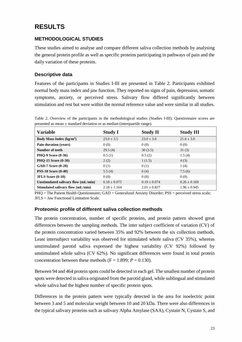
Descriptive data
Features of the participants in Studies I-III are presented in Table 2. Participants exhibited
normal body mass index and jaw function. They reported no signs of pain, depression, somatic
symptoms, anxiety, or perceived stress. Salivary flow differed significantly between
stimulation and rest but were within the normal reference value and were similar in all studies.
Table 2. Overview of the participants in the methodological studies (Studies I-III). Questionnaire scores are
presented as mean ± standard deviation or as median (interquartile range).
Variable Study I Study II Study III
Body Mass Index (kg/m2) 23.0 ± 3.5 23.0 ± 3.0 21.6 ± 3.0
Pain duration (years) 0 (0) 0 (0) 0 (0)
Number of teeth 29.5 (4) 30 (3.5) 31 (3)
PHQ-9 Score (0-36) 0.5 (1) 0.5 (2) 1.5 (4)
PHQ-15 Score (0-30) 2 (2) 1 (1.5) 4 (3)
GAD-7 Score (0-28) 0 (1) 0 (1) 1 (4)
PSS-10 Score (0-40) 5.5 (4) 6 (4) 7.5 (6)
JFLS Score (0-10) 0 (0) 0 (0) 0 (0)
Unstimulated salivary flow (mL/min) 0.18 ± 0.075 0.19 ± 0.074 0.26 ± 0.169
Stimulated salivary flow (mL/min) 2.18 ± 1.164 2.01 ± 0.827 1.96 ± 0.945
PHQ = The Patient Health Questionnaire; GAD = Generalized Anxiety Disorder; PSS = perceived stress scale;
JFLS = Jaw Functional Limitation Scale.
Proteomic profile of different saliva collection methods
The protein concentration, number of specific proteins, and protein pattern showed great
differences between the sampling methods. The inter subject coefficient of variation (CV) of
the protein concentration varied between 35% and 92% between the six collection methods.
Least intersubject variability was observed for stimulated whole saliva (CV 35%), whereas
unstimulated parotid saliva expressed the highest variability (CV 92%) followed by
unstimulated whole saliva (CV 62%). No significant differences were found in total protein
concentration between these methods (F = 1.899; P = 0.130).
Between 94 and 464 protein spots could be detected in each gel. The smallest number of protein
spots were detected in saliva originated from the parotid gland, while sublingual and stimulated
whole saliva had the highest number of specific protein spots.
Differences in the protein pattern were typically detected in the area for isoelectric point
between 3 and 5 and molecular weight between 10 and 20 kDa. There were also differences in
the typical salivary proteins such as salivary Alpha Amylase (SAA), Cystain N, Cystain S, and
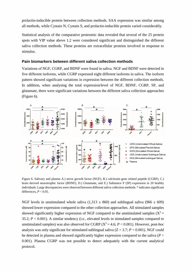
prolactin-inducible protein between collection methods. SAA expression was similar among
all methods, while Cystain N, Cystain S, and prolactin-inducible protein varied considerably.
Statistical analysis of the comparative proteomic data revealed that several of the 25 protein
spots with VIP value above 1.2 were considered significant and distinguished the different
saliva collection methods. These proteins are extracellular proteins involved in response to
stimulus.
Pain biomarkers between different saliva collection methods
Variations of NGF, CGRP, and BDNF were found in saliva. NGF and BDNF were detected in
five different isoforms, while CGRP expressed eight different isoforms in saliva. The isoform
pattern showed significant variations in expression between the different collection methods.
In addition, when analysing the total expression/level of NGF, BDNF, CGRP, SP, and
glutamate, there were significant variations between the different saliva collection approaches
(Figure 6).
Figure 6. Salivary and plasma A.) nerve growth factor (NGF), B.) calcitonin gene related peptide (CGRP), C.)
brain derived neurotrophic factor (BDNF), D.) Glutamate, and E.) Substance P (SP) expression in 20 healthy
individuals. Large discrepancies were observed between different saliva collection methods. * indicates significant
differences, P < 0.05.
NGF levels in unstimulated whole saliva (1,313 ± 860) and sublingual saliva (966 ± 609)
showed lower expression compared to the other collection approaches. All stimulated samples
showed significantly higher expression of NGF compared to the unstimulated samples (X2 =
35.2; P < 0.001). A similar tendency (i.e., elevated levels in stimulated samples compared to
unstimulated samples) was also observed for CGRP (X2 = 4.6; P < 0.001). However, post-hoc
analysis was only significant for stimulated sublingual saliva (Z = 3.7; P < 0.001). NGF could
be detected in plasma and showed significantly higher expression compared to the saliva (P <
0.001). Plasma CGRP was not possible to detect adequately with the current analytical
protocol.
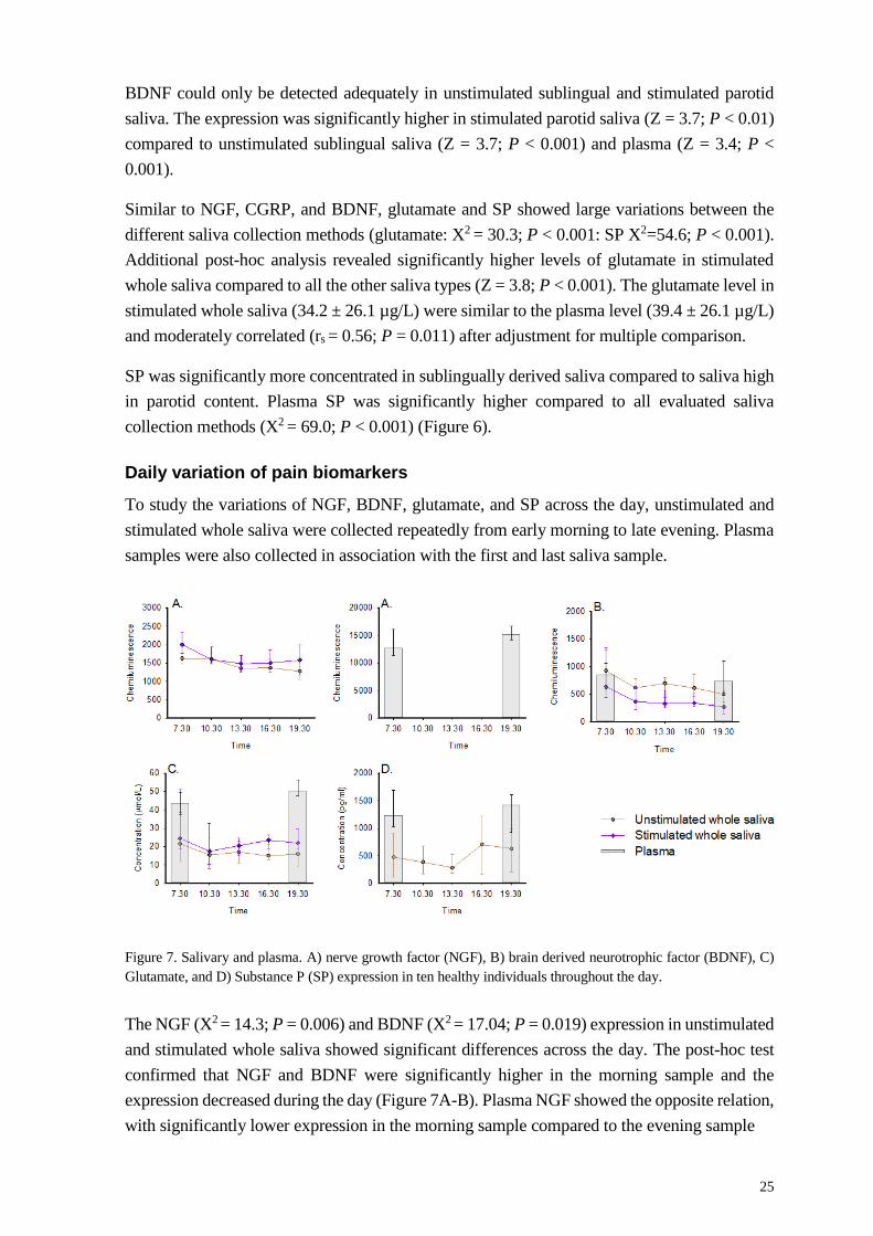
25
BDNF could only be detected adequately in unstimulated sublingual and stimulated parotid
saliva. The expression was significantly higher in stimulated parotid saliva (Z = 3.7; P < 0.01)
compared to unstimulated sublingual saliva (Z = 3.7; P < 0.001) and plasma (Z = 3.4; P <
0.001).
Similar to NGF, CGRP, and BDNF, glutamate and SP showed large variations between the
different saliva collection methods (glutamate: X2 = 30.3; P < 0.001: SP X2=54.6; P < 0.001).
Additional post-hoc analysis revealed significantly higher levels of glutamate in stimulated
whole saliva compared to all the other saliva types (Z = 3.8; P < 0.001). The glutamate level in
stimulated whole saliva (34.2 ± 26.1 µg/L) were similar to the plasma level (39.4 ± 26.1 µg/L)
and moderately correlated (rs = 0.56; P = 0.011) after adjustment for multiple comparison.
SP was significantly more concentrated in sublingually derived saliva compared to saliva high
in parotid content. Plasma SP was significantly higher compared to all evaluated saliva
collection methods (X2 = 69.0; P < 0.001) (Figure 6).
Daily variation of pain biomarkers
To study the variations of NGF, BDNF, glutamate, and SP across the day, unstimulated and
stimulated whole saliva were collected repeatedly from early morning to late evening. Plasma
samples were also collected in association with the first and last saliva sample.
Figure 7. Salivary and plasma. A) nerve growth factor (NGF), B) brain derived neurotrophic factor (BDNF), C)
Glutamate, and D) Substance P (SP) expression in ten healthy individuals throughout the day.
The NGF (X2 = 14.3; P = 0.006) and BDNF (X2 = 17.04; P = 0.019) expression in unstimulated
and stimulated whole saliva showed significant differences across the day. The post-hoc test
confirmed that NGF and BDNF were significantly higher in the morning sample and the
expression decreased during the day (Figure 7A-B). Plasma NGF showed the opposite relation,
with significantly lower expression in the morning sample compared to the evening sample

(P = 0.028). Glutamate concentration did not express any significant changes throughout the
day (X2 = 7.289; P = 0.121). SP expression in unstimulated whole saliva and plasma did not
change significantly over time. In stimulated whole saliva, SP could not be analysed adequately
in a minimum of 50% of all samples and according to the criteria.
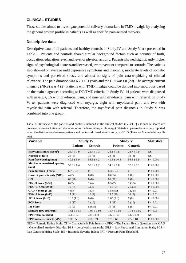
27
CLINICAL STUDIES
These studies aimed to investigate potential salivary biomarkers in TMD myalgia by analysing
the general protein profile in patients as well as specific pain-related markers.
Descriptive data
Descriptive data of all patients and healthy controls in Study IV and Study V are presented in
Table 3. Patients and controls shared similar background factors such as country of birth,
occupation, education level, and level of physical activity. Patients showed significantly higher
signs of psychological distress and decreased jaw movement compared to controls. The patients
also showed on average mild depressive symptoms and insomnia, moderate levels of somatic
symptoms and perceived stress, and almost no signs of pain catastrophizing of clinical
relevance. The pain duration was 6.7 ± 6.3 years and the CPI was 60 (20). The average current
intensity (NRS) was 4 (2). Patients with TMD myalgia could be divided into subgroups based
on the main diagnoses according to DC/TMD criteria. In Study IV, 14 patients were diagnosed
with myalgia, 16 with myofascial pain, and nine with myofascial pain with referral. In Study
V, ten patients were diagnosed with myalgia, eight with myofascial pain, and two with
myofascial pain with referral. Therefore, the myofascial pain diagnosis in Study V was
combined into one group.
Table 3. Overview of the patients and controls included in the clinical studies (IV-V). Questionnaire scores are
presented as mean ± standard deviation or as median (interquartile range). Statistical parameters are only reported
when the distributions between patients and controls differed significantly, P < 0.05 (T-test or Mann–Whitney U-
test).
Variable Study IV
Patients Controls
Study V
Patients Controls
Statistics
Body Mass Index (kg/m2) 23.7 ± 3.9 22.7 ± 3.3 24.4 ± 3.8 22.7 ± 3.0 NS
Number of teeth 28 (3) 30 (3) 28 (2) 30 (3) NS
Pain-free opening (mm) 40.6 ± 9.9 56.5 ± 6.2 41.4 ± 10.8 56.4 ± 5.9 P < 0.001
Maximum unassisted opening
(mm) 52.5 ± 6.4 57.9 ± 6.2 54.0 ± 6.0 57.7 ± 6.1 P < 0.001
Pain duration (Years) 6.7 ± 6.3 0 6.3 ± 6.3 0 P < 0.001
Current pain intensity (NRS) 4 (2) 0 (0) 6 (2.5) 0 (0) P < 0.001
CPI 60 (20) 0 (0) 65 (27) 0 (0) P < 0.001
PHQ-9 Score (0-36) 6 (7) 1 (4) 6.5 (7) 1 (3.5) P < 0.001
PHQ-15 Score (0-30) 10 (7) 3 (4) 11.5 (9) 2.5 (4) P < 0.001
GAD-7 Score (0-28) 4 (5) 1 (3) 3.5 (8.5) 1 (2.5) P < 0.01
PSS-10 Score (0-40) 17 (11) 10 (9) 15.5 (10) 10 (8) P < 0.01
JFLS Score (0-10) 1.15 (1.8) 0 (0) 1.65 (2.0) 0 (0) P < 0.001
PCS Score 14 (17) 3 (10) 15 (18) 5 (10) P < 0.01
ISI Score 10 (9) 5 (5.5) 10 (15) 5 (5) P < 0.01
Salivary flow (mL/min) 1.61 ± 0.56 1.98 ± 0.93 1.57 ± 0.50 1.74 ± 1.05 P < 0.05
PPT reference (kPa) 356 ± 121 439 ±119 382 ± 127 437 ±130 NS
PPT masseter muscle (kPa) 180 ± 56 268 ± 71 179 ± 63 272 ± 81 P < 0.001
NRS = Numeric Rating Scale; CPI = Characteristic Pain Intensity; PHQ = The Patient Health Questionnaire; GAD
= Generalized Anxiety Disorder; PSS = perceived stress scale; JFLS = Jaw Functional Limitation Scale; PCS =
Pain Catastrophizing Scale; ISI = Insomnia Severity Index; PPT = Pressure Pain Threshold.
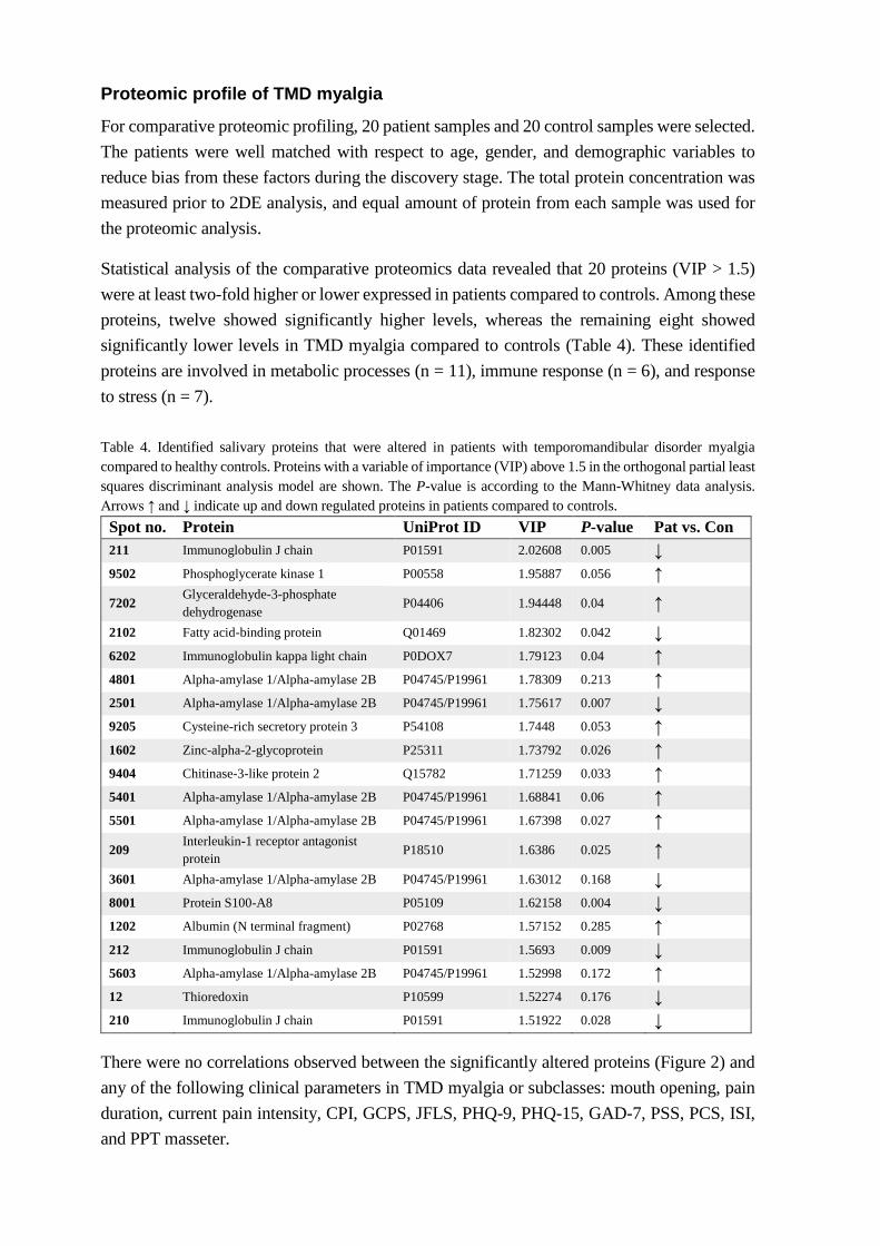
Proteomic profile of TMD myalgia
For comparative proteomic profiling, 20 patient samples and 20 control samples were selected.
The patients were well matched with respect to age, gender, and demographic variables to
reduce bias from these factors during the discovery stage. The total protein concentration was
measured prior to 2DE analysis, and equal amount of protein from each sample was used for
the proteomic analysis.
Statistical analysis of the comparative proteomics data revealed that 20 proteins (VIP > 1.5)
were at least two-fold higher or lower expressed in patients compared to controls. Among these
proteins, twelve showed significantly higher levels, whereas the remaining eight showed
significantly lower levels in TMD myalgia compared to controls (Table 4). These identified
proteins are involved in metabolic processes (n = 11), immune response (n = 6), and response
to stress (n = 7).
Table 4. Identified salivary proteins that were altered in patients with temporomandibular disorder myalgia
compared to healthy controls. Proteins with a variable of importance (VIP) above 1.5 in the orthogonal partial least
squares discriminant analysis model are shown. The P-value is according to the Mann-Whitney data analysis.
Arrows ↑ and ↓ indicate up and down regulated proteins in patients compared to controls.
Spot no. Protein UniProt ID VIP P-value Pat vs. Con
211 Immunoglobulin J chain P01591 2.02608 0.005 ↓
9502 Phosphoglycerate kinase 1 P00558 1.95887 0.056 ↑
7202 Glyceraldehyde-3-phosphate
dehydrogenase P04406 1.94448 0.04 ↑
2102 Fatty acid-binding protein Q01469 1.82302 0.042 ↓
6202 Immunoglobulin kappa light chain P0DOX7 1.79123 0.04 ↑
4801 Alpha-amylase 1/Alpha-amylase 2B P04745/P19961 1.78309 0.213 ↑
2501 Alpha-amylase 1/Alpha-amylase 2B P04745/P19961 1.75617 0.007 ↓
9205 Cysteine-rich secretory protein 3 P54108 1.7448 0.053 ↑
1602 Zinc-alpha-2-glycoprotein P25311 1.73792 0.026 ↑
9404 Chitinase-3-like protein 2 Q15782 1.71259 0.033 ↑
5401 Alpha-amylase 1/Alpha-amylase 2B P04745/P19961 1.68841 0.06 ↑
5501 Alpha-amylase 1/Alpha-amylase 2B P04745/P19961 1.67398 0.027 ↑
209 Interleukin-1 receptor antagonist
protein P18510 1.6386 0.025 ↑
3601 Alpha-amylase 1/Alpha-amylase 2B P04745/P19961 1.63012 0.168 ↓
8001 Protein S100-A8 P05109 1.62158 0.004 ↓
1202 Albumin (N terminal fragment) P02768 1.57152 0.285 ↑
212 Immunoglobulin J chain P01591 1.5693 0.009 ↓
5603 Alpha-amylase 1/Alpha-amylase 2B P04745/P19961 1.52998 0.172 ↑
12 Thioredoxin P10599 1.52274 0.176 ↓
210 Immunoglobulin J chain P01591 1.51922 0.028 ↓
There were no correlations observed between the significantly altered proteins (Figure 2) and
any of the following clinical parameters in TMD myalgia or subclasses: mouth opening, pain
duration, current pain intensity, CPI, GCPS, JFLS, PHQ-9, PHQ-15, GAD-7, PSS, PCS, ISI,
and PPT masseter.
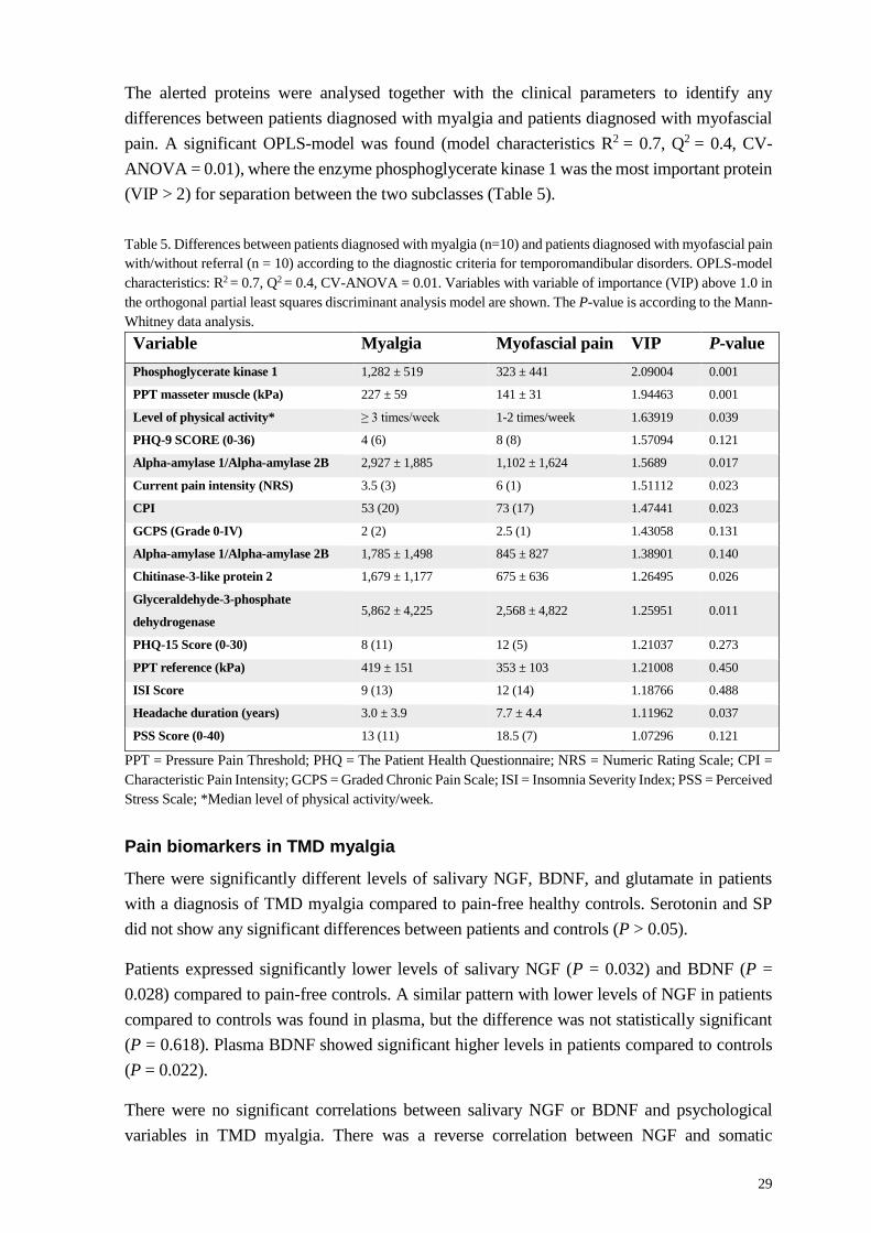
29
The alerted proteins were analysed together with the clinical parameters to identify any
differences between patients diagnosed with myalgia and patients diagnosed with myofascial
pain. A significant OPLS-model was found (model characteristics R2 = 0.7, Q2 = 0.4, CV-
ANOVA = 0.01), where the enzyme phosphoglycerate kinase 1 was the most important protein
(VIP > 2) for separation between the two subclasses (Table 5).
Table 5. Differences between patients diagnosed with myalgia (n=10) and patients diagnosed with myofascial pain
with/without referral (n = 10) according to the diagnostic criteria for temporomandibular disorders. OPLS-model
characteristics: R2 = 0.7, Q2 = 0.4, CV-ANOVA = 0.01. Variables with variable of importance (VIP) above 1.0 in
the orthogonal partial least squares discriminant analysis model are shown. The P-value is according to the Mann-
Whitney data analysis.
Variable Myalgia Myofascial pain VIP P-value
Phosphoglycerate kinase 1 1,282 ± 519 323 ± 441 2.09004 0.001
PPT masseter muscle (kPa) 227 ± 59 141 ± 31 1.94463 0.001
Level of physical activity* ≥ 3 times/week 1-2 times/week 1.63919 0.039
PHQ-9 SCORE (0-36) 4 (6) 8 (8) 1.57094 0.121
Alpha-amylase 1/Alpha-amylase 2B 2,927 ± 1,885 1,102 ± 1,624 1.5689 0.017
Current pain intensity (NRS) 3.5 (3) 6 (1) 1.51112 0.023
CPI 53 (20) 73 (17) 1.47441 0.023
GCPS (Grade 0-IV) 2 (2) 2.5 (1) 1.43058 0.131
Alpha-amylase 1/Alpha-amylase 2B 1,785 ± 1,498 845 ± 827 1.38901 0.140
Chitinase-3-like protein 2 1,679 ± 1,177 675 ± 636 1.26495 0.026
Glyceraldehyde-3-phosphate
dehydrogenase 5,862 ± 4,225 2,568 ± 4,822 1.25951 0.011
PHQ-15 Score (0-30) 8 (11) 12 (5) 1.21037 0.273
PPT reference (kPa) 419 ± 151 353 ± 103 1.21008 0.450
ISI Score 9 (13) 12 (14) 1.18766 0.488
Headache duration (years) 3.0 ± 3.9 7.7 ± 4.4 1.11962 0.037
PSS Score (0-40) 13 (11) 18.5 (7) 1.07296 0.121
PPT = Pressure Pain Threshold; PHQ = The Patient Health Questionnaire; NRS = Numeric Rating Scale; CPI =
Characteristic Pain Intensity; GCPS = Graded Chronic Pain Scale; ISI = Insomnia Severity Index; PSS = Perceived
Stress Scale; *Median level of physical activity/week.
Pain biomarkers in TMD myalgia
There were significantly different levels of salivary NGF, BDNF, and glutamate in patients
with a diagnosis of TMD myalgia compared to pain-free healthy controls. Serotonin and SP
did not show any significant differences between patients and controls (P > 0.05).
Patients expressed significantly lower levels of salivary NGF (P = 0.032) and BDNF (P =
0.028) compared to pain-free controls. A similar pattern with lower levels of NGF in patients
compared to controls was found in plasma, but the difference was not statistically significant
(P = 0.618). Plasma BDNF showed significant higher levels in patients compared to controls
(P = 0.022).
There were no significant correlations between salivary NGF or BDNF and psychological
variables in TMD myalgia. There was a reverse correlation between NGF and somatic
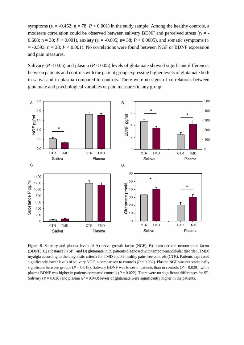
symptoms (rs = -0.462; n = 78; P < 0.001) in the study sample. Among the healthy controls, a
moderate correlation could be observed between salivary BDNF and perceived stress (rs = -
0.608; n = 38; P < 0.001), anxiety (rs = -0.605; n= 38; P < 0.0005), and somatic symptoms (rs
= -0.593; n = 38; P < 0.001). No correlations were found between NGF or BDNF expression
and pain measures.
Salivary (P < 0.05) and plasma (P < 0.05) levels of glutamate showed significant differences
between patients and controls with the patient group expressing higher levels of glutamate both
in saliva and in plasma compared to controls. There were no signs of correlations between
glutamate and psychological variables or pain measures in any group.
Figure 8. Salivary and plasma levels of A) nerve growth factor (NGF), B) brain derived neurotrophic factor
(BDNF), C) substance P (SP), and D) glutamate in 39 patients diagnosed with temporomandibular disorder (TMD)
myalgia according to the diagnostic criteria for TMD and 39 healthy pain-free controls (CTR). Patients expressed
significantly lower levels of salivary NGF in comparison to controls (P = 0.032). Plasma NGF was not statistically
significant between groups (P = 0.618). Salivary BDNF was lower in patients than in controls (P = 0.028), while
plasma BDNF was higher in patients compared controls (P = 0.022). There were no significant differences for SP.
Salivary (P = 0.026) and plasma (P = 0.043) levels of glutamate were significantly higher in the patients.
31
DISCUSSION
METHODOLOGICAL STUDIES
Saliva is usually neglected as a body fluid with diagnostic or prognostic value even though
sampling in general is well accepted by the patients. This neglect is due to lack of standardized
collection procedure and reliable analytical protocols. The methodological section of the thesis
aimed to compare different saliva collection methods on specific pain-related mediators as well
as the total protein profile and further to study the periodicity of pain-related markers. The main
findings were that different saliva collection approaches resulted in clear differences in the
protein profile as well as in the expression of several pain-related mediators. Furthermore, some
neuropeptides expressed a rhythmic variation in their salivary expression (NGF and BDNF),
but other neurotransmitters showed no variation throughout the day (SP and glutamate).
The variable nature of saliva secretion and composition suggests that different approaches may
have to be adopted when studying its possible role as a diagnostic and/or prognostic tool. To
date, there is extensive literature on the diagnostic possibilities of saliva, but there are still no
standardized techniques for saliva sample collection. Different studies apply different sampling
methods, and countless studies fail to describe the sample collection procedure such that the
procedure can be reproduced [4; 53; 80]. Additionally, the description of the enrolled
participants is often insufficient, and a proper clinical examination is often lacking [192]. Taken
together, these failures make it difficult to compare results from different studies. A majority
of the studies focus exclusively on whole saliva [4; 62; 69; 79; 89; 101; 129; 146; 181; 196]
because it can be easily obtained by simply spitting into a test tube, chewing on a sponge, or
passively dribbling from the mouth. Only a few studies have focused on ductal saliva such as
parotid, sublingual, or submandibular saliva obtained separately from the major glands in the
oral cavity [34; 78; 185; 192]. As far as we know, these are the first attempts to structurally
compare whole and glandular saliva in a carefully characterized and clinically examined
cohort.
Some studies have compared the proteome of different saliva types [34; 185; 192]. Using
similar approaches, Waltz et al. describe differences in the protein expression in whole, parotid,
and sublingual-submandibular saliva using samples from four participants [185]. They reported
less specific protein spots in glandular saliva compared to our findings. The loss of proteins
can be because of the additional saliva filtration these authors applied to remove insoluble
material and bacterial contamination. In another small poorly described cohort, Wu et al. found
significantly diverse protein profiles resolved in parotid and submandibular/sublingual saliva
[192]. The salivary proteome depends on the contribution from each salivary gland [17].
Samples collected by the same method display closer correlation than those collected by
different methods. Close correlation was observed between the protein pattern of unstimulated
and stimulated parotid saliva as well as unstimulated sublingual and whole saliva.
When investigating specific pain-related peptides, significant differences could also be
observed between different collection methods and between times of sample collection. When

quantifying NGF, BDNF, and CGRP, automatized western-blot was applied based on charge
and size. This technique has the benefit of identifying different isoforms based on protein
charge. NGF, BDNF, and CGRP were consequently found in different isoforms in saliva and
showed great variation between different collection approaches. A few studies of healthy
subjects have been performed on these mediators in saliva. Nam et al., measuring NGF in three
sources of saliva, found similar levels to our study [130]. We could also demonstrate a
significant association between increased salivary flow by stimulation and increased NGF
expression. Although some previous studies have investigated salivary concentration of NGF,
no studies have investigated the daytime periodicity of the neuropeptide in saliva. We were
able to show daily fluctuation of salivary NGF similar to those described in plasma by Bersani
et al. The elevated NGF levels in the morning suggest an increased NGF production during the
night similar to what has been observed for other endocrine molecules [10].
Similar to NGF, BDNF expression increases by saliva stimulation (Study III). It is noteworthy
that charge-based methods could only detect BDNF in glandular saliva. Nevertheless, when a
size-based approach was applied, BDNF could also be detected in whole saliva and similar to
NGF expressed a daily variation throughout the day. Other studies have confirmed a circadian
variation of BDNF secretion with significant decrease throughout the day [8; 23; 148; 181]. In
addition, the amplitude of the fluctuation varies across the menstrual cycle [148].
The levels of SP and glutamate in the methodological studies were analysed using
commercially available methods. SP levels were significantly more pronounced in saliva high
in sublingual/submandibular secretion, such as unstimulated whole saliva and sublingual
saliva. This finding may be explained by SP containing sensory fibres in the submandibular
glands [68]. However, SP and glutamate levels did not show any variation throughout day.
Interestingly, glutamate levels in stimulated whole saliva showed association to circulatory
levels in plasma.
When searching for the best saliva collection methods to study proteins and peptides related to
pain, there are strengths and limitations with all sampling methods that need to be properly
addressed before choosing a method. Parotid saliva has the advantage of being highly viscous
and pure, which makes it easy to process during analysis. The collection, however, is time-
consuming and requires special devices [108] and blockage from the other unrequired glands.
Because of the slow flow-rate in the unstimulated state, it was not possible to acquire enough
material for the analysis in Study II. Therefore, parotid saliva is not suitable when larger sample
volumes are needed and not practical in large cross-sectional or clinical studies involving
individuals with impaired health states.
The two glands on the floor of the mouth (i.e., submandibular and sublingual glands) are
anatomically closely situated and can sometimes share the same ducts [78]. Therefore, it is
difficult to separate the saliva from these glands safely and without accidently traumatizing the
duct and cause bleeding. This is why saliva was collected from both glands simultaneously in
the methodological studies even though they show different protein expressions [78].
Furthermore, the use of absorbent materials to collect saliva is common in saliva research but
33
could be problematic when the expected volume of saliva is small and can negatively affect the
validity of the assays when the saliva is filtered through a cotton pad [17].
Contrary to glandular saliva, whole saliva is easy to control and access and express high levels
of proteins and pain-related markers. The collection of unstimulated whole saliva by simply
allowing saliva to accumulate in the mouth and then allowing it to dribble out through a funnel
or straw into a tube is subject to considerable variation. For example, to varying extents the
participants may try to speed up the collection by moving their tongue and cheeks or by mental
exertion. These factors could partly explain the large variability in saliva secretion and protein
concentration (CV 62 %). Compared to unstimulated saliva, whole stimulated saliva is also
subject to some concerns because of the higher possibility of contamination by debris and
bacteria in the oral cavity while chewing and spitting. In our studies, all participants were
instructed to not eat/drink prior to collection and all exhibited good oral health, which
diminishes the risk of contamination. Furthermore, all samples were collected in chilled tubes,
treated with a protease inhibitor, and immediately centrifuged to reduce the risk for bacterial
overgrowth and protein degradation. Whole stimulated saliva had the highest volume obtained,
least variability in protein expression (CV 35%), low levels of glycoproteins, and high levels
in all analytes except BDNF and SP.
A strength of the study methodology was that saliva samples were collected in the same
structured manner, at the same time, and with the same surroundings conditions. To reduce the
effect of external factors on salivary flow and protein expression, the inclusion criteria for the
participants enrolled in the studies were very rigid. The health of the participants was ensured
via their anamnesis and careful oral and dental examination. According to some findings, age
may affect protein expression and saliva quality [28; 56; 127], but the participants were within
a limited age interval to reduce the influence of such a confounding factor.
However, some limitations need to be considered. One study has shown the possible influence
of sex hormones on BDNF secretion [148]. However, since it is not clear how the menstrual
cycle affects the saliva compositions, we did not control for the phase of the menstrual cycle.
Another limitation is that the strict criteria in combination with the limited age interval reduced
the external validity of the findings.
Based on these results, it can be concluded that there are great variations between different
collection methods both on the protein profile and specific pain-related peptides. It could be
suggested that stimulated whole saliva may be a preferable collection method based on its
simplicity, low variability, and significantly higher expression of several biomarkers. The
samples should be collected at the same time of day to minimize the influence of the daily
variation. The methodological studies stress the importance of consistency in the saliva
collection approach rather than the method or collection time itself.

CLINICAL STUDIES
Saliva contains a complex mixture of proteins, peptides, and other substance that may yield
information about the pathophysiology behind TMD myalgia and be used to identify new
disease-specific biomarkers. The clinical section of the thesis aimed to evaluate specific pain-
related mediators as well as the total protein profile in saliva of TMD myalgia to identify
potential diagnostic or prognostic salivary biomarkers and to investigate the relationship
between these proteins and clinical features. The main findings were significantly altered levels
of salivary NGF, BDNF, and glutamate as well as altered levels of 20 proteins involved in
metabolic processes, responses to stress, and immune response in TMD myalgia. Patients
showed elevated plasma levels of BDNF and glutamate. However, there were no correlations
between any of these proteins and pain measures.
Previous studies of conditions associated with chronic pain have suggested the involvement of
serotonin, glutamate, NGF, SP, and BDNF [9; 54; 88; 101; 134; 146; 170]. Increased local and
circulatory levels of NGF have been reported in chronic migraine [89], burning mouth
syndrome [11], interstitial cystitis [95], and rheumatoid arthritis [156; 164]. Only two studies
have explored salivary NGF in chronic pain. Both showed higher levels of salivary and
circulatory NGF in chronic pain, a finding that contrasts with our study [11; 89]. However, the
lower levels of salivary NGF in TMD myalgia and comparable plasma levels in both of our
groups are in line with a recently published article on fibromyalgia [88]. Reduced levels of
NGF have previously been reported in patients with depressive disorders [22], and a possible
explanation for the lower levels of NGF in TMD myalgia may be related to the psychological
maladjustments of these patients [88]. In addition, patients with TMD in general exhibit greater
psychological distress compared to pain-free individuals [42; 52; 91; 172]. This well-
established finding is also confirmed in our study cohort where patients reported significantly
higher levels of depressive and somatic symptoms, anxiety, stress, pain catastrophizing, and
sleep disturbances compared to pain-free controls. In addition, we found a moderate correlation
between salivary NGF and somatic symptoms after adjustment for multiple comparison. This
finding may indicate that the altered salivary NGF levels in patients are not caused by the pain
itself but reflect a psychological maladjustment. Similar to salivary NGF, salivary BDNF was
significantly lower in TMD myalgia. There was also an inverse correlation between salivary
BDNF and perceived stress, anxiety, and somatic symptoms in the controls. One may speculate
that salivary BDNF, like salivary NGF, may reflect psychological maladjustment. However,
further studies are needed to confirm such an interpretation. Circulatory BDNF, on the other
hand, was higher in patients compared to controls. This is in line with several studies of
different chronic pain conditions [9; 54; 88; 170]. Some studies have found decreased BDNF
levels in individuals with depressive symptoms or insomnia [18; 64]. The patients in Study IV
reported a higher degree of insomnia and depressive symptoms compared to the controls.
However, we found no relationship between circulatory BDNF levels and psychological
factors. Therefore, the increase of circulatory BDNF might be interpreted as a general reaction
to nociception [88].
35
Salivary SP decreased in chronic pain patients compared to pain-free controls [146] and similar
findings were also observed for burning mouth syndrome [11]; however, studies on salivary
levels of SP in chronic pain are inconsistent and show diverse findings. Jang et al. reported
increased plasma and salivary SP levels in migraine and observed an increase in SP with higher
pain intensity [89]. On the contrary, a recent study by Källman et al. reported no differences in
SP levels in chronic neuropathic pain compared to pain-free controls [98]. Our findings support
the latter study [98]. The plasma levels of SP in Study IV were in line with previous reported
concentrations [90; 98], whereas saliva showed lower concentrations of SP compared to earlier
studies [89; 146]. These differences are most probably due to different collection approaches,
since salivary SP concentrations seem to decrease with increased flow rate [89]. Therefore, the
low levels of SP in whole stimulated saliva are most probably due to the collection approach
as this saliva contains 20% of the levels detected in resting whole saliva [90]. This collection
approach may also have affected the traceability in our saliva samples, resulting in measurable
levels in only half of the samples.
Salivary serotonin was excluded because it was difficult to detect in sufficient amount. As
serotonin occurs in platelet-rich plasma to a higher extent than saliva, measurable levels were
detected in most of the plasma samples. The levels were higher in healthy controls compared
to patients but within normal reference values [12; 32]. The non-significant differences in TMD
myalgia and healthy controls agree with earlier studies in serum and plasma [32; 44].
Glutamate has previously been studied in the interstitial fluid of the masticatory muscles and
in plasma of patients with TMD myalgia, but to date not in saliva [19; 31; 60; 117; 167].
Interstitial muscle levels of glutamate were shown to be elevated in TMD myalgia compared
to pain-free individuals [19; 31], but there were no significant differences in circulatory levels
[19]. Based on the close vicinity of the salivary glands to the masticatory muscles, one may
hypothesize that glutamate and other local biomarkers may diffuse into the saliva and therefore
saliva levels reflect muscle levels. The concentration of the biomarker may also increase with
salivary flow due to the reduced modification in the ducts. The patients in Study IV displayed
significantly higher levels of salivary as well as circulatory glutamate compared to healthy
individuals, suggesting that elevated salivary glutamate may be an indicator of TMD myalgia.
This finding is also in line with a recent study showing that patients with chronic migraine had
significantly higher levels of salivary glutamate compared to episodic migraine and healthy
controls [129].
By applying proteomic approaches to saliva samples, we were able to show alteration in several
proteins associated with metabolic processes, stress, and immunity in TMD myalgia, and
interestingly also in subclasses of TMD myalgia (myalgia and myofascial pain). The most
important proteins for separation between patients and controls and further between subclasses
of TMD myalgia were PGK1, GAPDH, CHI3L2, and SAA. PGK1 expression has previously
been described in various malignancies [177] and high expression of PGK1 has been observed
in synovial tissue and blood of patients with rheumatoid arthritis [195]. Over-expression of
salivary PGK1 in TMD myalgia has to date never been described. Therefore, the significance
of the enzyme in myalgia remains unclear and needs further investigation. GAPDH, similar to
PGK1, is also overexpressed in various malignancies and positively correlates with tumour
progression [183; 188]. Glycolytic enzymes such as PGK1 and GAPDH are usually found in
the cytoplasm of the cell and released in the general circulation during pathological states that
are associated with cell damage or apoptosis [183]. In this context, it may be assumed that
conditions of oxidative stress, which lately has been discussed in relation to myalgia, may
increase the need of PGK1 and GAPDH and lead to the observed elevated levels in saliva [48;
159].
The levels of SAA were significantly altered in TMD myalgia compared to controls and further
significantly increased in patients with myalgia compared to myofascial pain. The interest in
this digestive and antimicrobial enzyme has gradually increased because of its role as a
potential marker for sympatho-adrenal medullary activity [131; 132; 187]. Studies have shown
that SAA levels increase in individuals during physical and psychological stress [21; 66; 132;
136; 190], so the enzyme has been suggested as a marker for psychological and physical stress
[16; 131; 180]. The patients in our study sample reported significantly higher levels of
perceived stress, but no significant correlation could be observed between SAA and perceived
stress level. Therefore, the validity of SAA as a stress marker remains debatable. Nonetheless,
orofacial pain can act as a potential stressor and affect psychological as well as physiological
systems [91]. Recent studies have proposed SAA as a biomarker for an objective assessment
of pain intensity [3; 169]. One study reported a significant decrease in SAA level after pain
reduction in chronic back pain and similar findings were also found in odontogenic pain [3;
169]. On the other hand, several studies have failed to observe a significant correlation between
ongoing pain and SAA levels [16; 55; 186].
Inflammatory proteins and peptides associated with myalgia have been debated [60] and some
attempts have been made to study cytokines and other inflammatory markers in TMD myalgia
[116]. The proteomic analysis revealed significant alterations in several proteins related to
inflammation and immunity such as immunoglobulin j chain (JCHAIN), immunoglobulin
kappa light chain (P0DOX7), and interleukin-1 receptor antagonist protein (IL-1RA). The
JCHAIN is expressed by mucosal and glandular plasma cells and regulates polymer formation
of immunoglobulin (Ig)A and IgM. P0DOX7 is also a subunit of an antibody. The secretary
protein IL1RA was elevated in patients. This protein is a member of the IL‐1 cytokine family
and is secreted by various types of cells to inhibit the pro-inflammatory effect of IL1α and
IL1β. It also modulates different IL-1-related immune and inflammatory responses [144].
Significantly altered expression of some more specific immunity-related proteins was also
observed. Cysteine-rich secretory protein 3 (CRISP 3), for example, has recently been
suggested as a potential biomarker for prostate cancer [63] and was found up-regulated in TMD
myalgia, while the fatty-acid binding protein (FABP) was significantly lower in patients
compared to controls. This protein family has recently been discussed as a novel marker for
diagnosis of diseases associated with oxidative stress, such as heart diseases, renal failure,
Sjögren´s Syndrome, and Alzheimer’s disease [58; 140; 157; 168; 193]. FABPs are intra-
cellular lipid-binding proteins that exhibit a variety of expressions depending on the specific
37
organ or cell type. To date, at least nine different isoforms have been identified. FABP1 has
been suggested as a marker for renal failure, FABP3 for myocardial infarction, and FABP5 has
lately been debated as a diagnostic marker for Sjögren´s Syndrome [140; 157; 168; 193].
FABP5 has also been shown to be highly expressed in nociceptive dorsal root ganglia neurons
and FABP inhibitors exert analgesic properties on a peripheral and supraspinal level. These
findings indicate that peripheral FABP inhibitors may be used therapeutically to reduce pain
and inflammation [147]. Another interesting observation was the significantly lower level of
S100-A8, also known as MRP8. S100A8 is not only increased locally in sites of inflammation
in rheumatoid arthritis but also in the general circulation. Moreover, the concentration of the
protein seems to be strongly associated with disease activity [153].
The clinical studies have some limitations that need to be considered. The studies were
performed in adults between 18 and 45 years old representing the peak of TMD prevalence;
other age groups were not considered because of the possibility of age variability [152]. Sub-
categories of myofascial pain could not be properly addressed in Study V due to the limited
number of patients in each subclass. Moreover, female participants represented a majority of
the study population to reflect the distribution in the clinic where women to a higher extent
seek care for TMD myalgia [85]. Consequently, sex differences in proteins and mediators could
not be properly addressed in any of the clinical studies. The female sex hormones can alter pain
levels and therefore another limitation is that the female participants were not screened for
menstrual cycle phase. However, it is most likely the women included were in different phases
of their menstrual cycle, which would remove such a confounding factor.
In these studies, there are also some strengths that should be raised. Patients and controls
enrolled in the studies were matched and they were all properly examined by the author to
exclude systemic or oral conditions that may affect the salivary composition and/or biomarker
levels. The author was calibrated to a reference standard researcher according to the most recent
DC/TMD criteria to ensure accurate diagnosis of TMD myalgia. Patients with pain conditions
other than TMD were excluded from the study. In both studies, the diurnal variation of salivary
proteins were considered and a majority of the samples were collected between 9 and 11 am.
The saliva collection was also standardized and followed a specific protocol to decrease the
inter- and intra-individual variability.
To conclude, the clinical studies displayed significant changes in salivary pain-specific
mediators as well as proteins related to metabolic processes, stress, and immunity in TMD
myalgia compared to healthy controls. Significant differences in some proteins and clinical
parameters could be observed between subclasses of TMD myalgia, indicating that they display
different aetiopathogenesis. Patients showed significantly higher levels of salivary and plasma
glutamate as well as plasma BDNF compared to healthy pain-free individuals, but lower levels
of salivary NGF and BDNF, which further correlated to psychological variables.
Future larger studies are needed to validate these potential biomarkers in a subset of TMD
patients and evaluate any potential clinical correlation between the candidate biomarkers and
clinical features.

39
IMPLICATIONS AND FURTHER PROSPECT
Saliva has considerable potential to mirror local and systemic health conditions. Nearly 40%
of all the proteins that have been suggested to be candidate biomarkers for different diseases
can be found in saliva. These comparisons together with the growing advancement in
technology have made the analysis of saliva more attractive in pain research, whereas other
sampling techniques such as microdialysis, biopsies, and cerebrospinal fluid present
complications [17; 60; 115]. However, saliva is not a homogenous and stable body fluid as it
is constantly changing. For example, the composition of saliva is affected by sampling
methodology, circadian rhythms, environment, age, gender, oral hygiene, physical activity,
medications, psychological status, and general health [17; 33; 87; 112; 127; 151; 152]. This
characteristic of saliva limits its ability to be used as a diagnostic medium is the inter- and intra-
individual variability, which was also obvious in this thesis, making the comparison between
studies and patients challenging. For saliva-based diagnostics and techniques to be useful, there
is a need for proper evaluation and standardization. Most studies using saliva as a diagnostic
or prognostic medium do not provide proper description of participant preparation, time of
sampling, sampling procedure, or the subsequent management of the sample [4; 53]. Since
many studies also apply different collection approaches and sometimes do not describe their
methods adequately, it is difficult to compare findings across studies. To our knowledge, the
methodological studies included in this thesis are the first to properly address these aspects by
evaluating different saliva collection approaches in healthy individuals within a limited age
interval.
Significant differences in the protein profile signature and the relative amount of specific pain-
related molecules were observed between saliva collection approaches. These results support
previous studies in saliva [34; 185; 192]; however, there are substantial differences. That is, we
investigated up to six saliva collection methods and carefully considered factors known to
affect saliva composition. In addition, using new sensitive methods, we observed for the first
time several isoforms for NGF, BDNF, and CGRP and showed that plasma glutamate can be
reflected by the concentration in simulated whole saliva. Another relevant observation that
emerged from the thesis was the daily variation in salivary NGF and BDNF, a finding that
resembled previous studies of plasma [8; 10; 23; 181]. Our findings have led us to conclude
that independent of the method chosen several conditions should be standardized. The
differences between saliva collection approaches for specific molecules as well as for the
protein profile made it clear that the collection method is a key factor for successful detection
of peptides and proteins. What kind of saliva collection protocol is recommended in pain
research? As previously described, whole saliva is a mixture of the saliva from the major and
minor salivary glands as well as other fluids present in the oral cavity [17]. Consequently,
gland-specific saliva should be used for gland-specific pathologies, and whole saliva should be
used for local and systemic diseases. A drawback against the collection of whole saliva is the
contamination of exogenous compounds such as microbiota, nasal secretions, blood
contamination, and food debris. However, a majority of saliva research studies used whole
instead of glandular saliva [17]. The findings in this thesis suggest that the collection of gland-
specific saliva is somehow invasive, requires special device, time-consuming, and results in
small volumes. As in other studies, we also witnessed greater variability and less protein spots
in the gland-specific saliva [34; 69; 185]. Based on findings in this thesis and in the literature,
whole saliva shows compelling advantages. However, the study of whole saliva can be done
using different collection procedures. Collection of unstimulated whole saliva by passive
drooling seems to be the most frequently used and is regarded by researchers as a gold standard
[3; 4; 7; 17; 26; 62; 82; 103; 129; 162; 181]. However, in clinical practice, this method is subject
to some concerns. It is difficult to maintain the individual steady for several minutes without
affecting and stimulating the saliva secretion, which most probably is the reason for the great
inter-individual variability. However, unstimulated whole saliva is preferable for substances
such as SP, whose concentration and therefore detection is considerably affected by the flow
rate. Given the simplicity of the method, high volume, and low variability in stimulation, this
thesis used stimulated whole saliva to study proteins and peptides in chronic pain. To improve
reproducibly, the sample should be collected in the morning two to three hours after awaking
to reduce the influence of the circadian rhythm and the participants should rinse their mouth
with water prior to collection.
The collection protocol as described was applied and stimulated whole saliva samples were
collected in the morning from patients with a diagnosis of TMD myalgia to detect diagnostic-
specific biomarkers. Saliva can potentially be used as a specimen for diagnosis in TMD because
it can exchange substances with blood. A thin layer of epithelial cells separating the salivary
ducts from the systemic circulation enables the transfer of substances from the saliva by means
of active carriage, diffusion through cell membrane, or passive diffusion. Nevertheless, there
are also disease-specific biomarkers that are only present in saliva but not in blood, including
some biomarkers for oral cancer [30].
This first study of the saliva proteome in TMD myalgia reveals that proteins related to
metabolism, stress, and immunity were altered in patients and supports some of the
pathological aspects discussed in the literature [42; 50; 65; 75; 113]. The present technique
combining 2DE analysis with LC-MS/MS allows for an explorative approach, not focusing on
predetermined proteins, to understand the mechanisms and discover novel biomarkers. Using
this technique, we observed significant differences between myogenic TMD diagnoses in
biological as well as clinical parameters, suggesting distinctive pathological mechanisms.
Patients diagnosed with TMD myofascial pain with referral expressed decreased levels in the
glycolytic enzymes PGK-1 and GAPDH and the digestive enzyme SAA compared to patients
with myalgia. These findings suggest that pathological mechanism associated to oxidative and
psychological stress are more pronounced in TMD myalgia or may indicate a depletion in
myofascial pain with referral following prolonged or excessive secretion as described for
cortisol under conditions of chronic stress [74]. Interestingly, patients with myofascial pain
with referral reported higher pain intensity, depressive and somatic symptoms, perceived stress,
and co-morbid headache. In addition, these patients had significantly lower pressure pain
threshold of the masseter muscle than patients with myalgia. This finding in combination with
the presence of pain spreading in myofascial pain correspond to central sensitization, whereas
41
changes in the properties of neurons in the CNS leads to increased pain hypersensitivity [191].
Thus, patients with myofascial pain seem to have an altered nociceptive function that is unlike
what patients with myalgia experience. The results from the thesis support that myofascial pain
should be regarded as nociplastic pain condition and treated as such. The term nociplastic pain
was recently introduced by the International Association for the Study of Pain Taxonomy to
describe pain states that arises from altered nociception even though no clear evidence exists
of tissue damage causing the activation of nociceptors or evidence for disease or lesion to the
CNS triggering the chronic pain. Nociplastic, pain in contrast to nociceptive pain, usually
responds better to centrally-targeted therapies [102; 184].
When investigating specific pain-related proteins in saliva, patients surprisingly showed lower
levels of NGF and BDNF, a finding that also correlated to psychological dysfunction. These
findings are supported by meta-analysis showing that circulatory NGF and BDNF are
indicative biomarkers for depressive disorders [22; 100]. One may speculate that salivary NGF
and BDNF mirror psychological maladjustment usually associated with TMD myalgia.
Although patients with myofascial pain reported higher pain intensity and scored higher in
psychological malfunction, there were no statistically significant differences between
subclasses.
Glutamate was increased in patients, and these findings were in line with other studies showing
that glutamate levels positively correlate with perceived pain sensitivity [19; 60; 158; 161;
167]. Increased glutamate levels contribute to the nociceptive process by lowering neural
threshold and/or increasing the pain response creating central sensitization [42; 161]. This
could explain the increased salivary and plasma levels in TMD myalgia compared to pain-free
controls. Elevated glutamate levels have also been reported in other pain conditions such as
migraine, headache, and chronic widespread pain; consequently, glutamate is suggested as a
potential biomarker for pain [60; 61; 129; 155; 161]. However, our studies are the first to
support stimulated whole saliva as an alternative diagnostic medium to measure glutamate and
to show that glutamate in saliva, as with interstitial fluid, is increased in TMD myalgia [19; 42;
60].
The greatest challenges for saliva diagnostics it to positively translating the identified
biomarkers and results from the laboratory bench to clinical practice. Candidate biomarkers
need to be verified and validated on a larger study group with appropriate clinical classification
system. Proteome analysis provided significant insight, but 2DE shows only a certain part of
the proteome. Therefore, there is a need for complement analysis of proteins and peptides not
detectable by this technique. Furthermore, there is a need to validate potential biomarkers and
their clinical value in larger patient cohorts. We may be able to combine these potential
biomarkers with other clinical features to better understand and diagnose TMD myalgia as well
as subclasses and evaluate therapeutic outcomes.

43
POPULÄRVETENSKAPLIG SAMMANFATTNING
En stor del av den vuxna befolkningen i världen lever idag med långvarig muskelsmärta, hos
drygt varannan person är smärtan lokaliserad till ansiktet och käkar. Detta innebär att drygt 10-
15% av den vuxna befolkningen är drabbad. Att leva med långvarig käkmuskelsmärta påverkar
många aspekter av livet och har därför stor betydelse för livskvalitén hos den drabbade
individen, men även för de närstående. Symptomen är vanligtvis lokal smärta, trötthet och
ömhet i käkmusklerna ofta i kombination med nedsatt käkfunktion och huvudvärk. Förutom
det individuella lidandet är långvarig käkmuskelsmärta ett stort problem för samhället med
ökad sjukfrånvaro och sjukvårdskostnader som följd.
Individer som lider av långvarig käkmuskelsmärta har ofta träffat många olika vårdgivare. De
har vanligtvis genomgått ett flertal olika undersökningar inom hälso- och sjukvården, och
prövat en mängd olika behandlingar, men ofta utan framgång. Långvarig käkmuskelsmärta
anses idag av många kliniker som en diagnostisk utmaning som kräver stora vårdresurser och
i flera fall specialistkompetens. Processen är ofta lång innan den drabbade individen erhåller
korrekt diagnos och behandling, och resan från de första symptomen till adekvat
omhändertagande tar ofta många år, vilket tyvärr ökar risken för en smärtöverkänslighet i
nervsystemet s.k. central sensitisering. Den långa processen är ofta även förknippad med
psykisk ohälsa.
De bakomliggande sjukdomsmekanismerna vid långvarig käkmuskelsmärta är till stor del
okända, varför diagnostiken bygger på uppgifter från patientens sjukdomshistoria och den
kliniska undersökningen. Liksom för andra långvariga smärttillstånd är forskarna dock överens
om att de innefattar flera faktorer som tillsammans bidrar till, underhåller och förvärrar
tillståndet. Psykisk ohälsa i kombination med stressrelaterad tandgnissling och tandpressning
är viktiga bidragande faktorer. Dessutom har biologiska och genetiska faktorer
uppmärksammas på senare tid som andra viktiga faktorer. Nya forskningsrön har visat att flera
substanser som har samband med smärta och inflammation, till exempel signalsubstanserna
glutamat, serotonin och nervtillväxtfaktor, är förhöja i den smärtande käkmuskeln, vilket
antyder att de kan bidra till sjukdomsutvecklingen. I olika studier har dessa substanser
analyserats i muskel, blod och ryggmärgsvätska; mätmetoder som ofta är förenade med smärta
och obehag. Munhålans saliv, som är en mycket informationsrik kroppsvätska, förbises ofta
som ett prov av diagnostiskt värde, troligen på grund av avsaknad av standardiserade rutiner
för provtagning. Saliven är en komplex vätska som främst består av sekret från tre stora parade
körtlar och ett hundratal små körtlar samt vätska från tandköttsfickan och annat sekret. Saliven
är ett filtrat av blodplasma och många substanser når därför salivkörtlarna från blodbanan.
Analyser har visat att drygt en tredjedel av blodets proteiner kan återfinnas i saliven. Därför
finns det goda skäl för att använda saliv som ett diagnostiskt redskap i forskningen kring
långvarig käkmuskelsmärta.
En metod som kan användas för att urskilja markörer för ett tillstånd är att studera alla de
proteiner som finns i ett prov, t.ex. ett salivprov, från en sjuk patient och jämföra det mot en
frisk kontrollperson. Proteomik, det vill säga läran om vilka proteiner som finns i kroppen, är
en mycket ung och växande vetenskap. Det så kallade proteomet, det vill säga de proteiner som
uttrycks av en cell, kroppsvävnad eller organism vid en specifik tidpunkt, är en komplex
funktion av de speciella förhållanden som cellen/vävnaden/organismen utsätts för. Proteinerna
är inte bara av stor vikt för att upprätthålla en god hälsa utan förändringar i uttryck och
koncentrationen av dessa kan även avslöja sjukdom. Ökad förståelse för vilka proteiner som
uttrycks hos patienter med långvarig käkmuskelsmärta i jämförelse med friska individer kan
ge oss värdefull kunskap om de biologiska mekanismerna bakom långvarig käkmuskelsmärta.
Vi kan genom omfattande analys av hela proteomet även urskilja karaktäristiska för
patienterna, och på detta sätt förbättra diagnostiken. Det kan på sikt leda till effektivare
behandlingar som riktar sig direkt mot sjukdomsmekanismerna. Det övergripande målet med
avhandlingen var att öka kunskapen avseende orsaksmekanismerna vid långvarig käkmuskel-
smärta för att därigenom förbättra diagnostiken och ge ett mer rationellt omhändertagande.
Men för att kunna använda saliven som diagnostiskt redskap behövs först omfattande arbete
med att skapa standardiserade rutiner för salivprovtagning. Därför syftade avhandlingens första
del till att studera skillnader mellan olika salivprovtagningsmetoder i avseende att hitta den
bästa provtagningsmetoden för att studera proteiner och andra substanser hos individer med
långvarig käkmuskelsmärta. Dessutom behöver man undersöka om det finns dygnsvariationer
av dessa substanser i saliven och bestämma den optimala provtagningstiden.
I denna avhandling samlades först sex olika typer av saliv in från friska individer, både saliv
från de olika salivkörtlarna och helsaliv, det vill säga den blandade saliven som finns i
munhålan. För att studera sammansättningen samlades proverna in både i vila och genom att
stimulera salivutsöndringen med citronsyra eller mekaniskt genom tuggning. Prover togs även
upprepade gånger under dagen från ett antal friska individer för att följa dygnsvariationen.
Resultaten från analyserna visade att det finns mycket stora skillnader mellan de olika
salivtyperna, både avseende hela proteomet och vissa specifika smärtrelaterade substanser.
Tuggstimulerad helsaliv uppvisade bättre stabilitet och fler proteiner jämfört med de övriga
salivtyperna. Man kunde även se en stor variation i mängden av de smärtrelaterade
substanserna i de olika salivtyperna, och att vissa substanser ökade med högre salivflödet.
Utsöndringen av signalsubstanserna nervtillväxtfaktor och neurotrofisk faktor visade sig vara
högst på morgonen och mängden minskade sedan under dagen.
Från dessa experiment kunde man dra slutsatsen att det finns mycket stora skillnader mellan
olika salivtyper. Det viktigaste lärdomen var att metoden spelade mindre roll, utan det
viktigaste är att vara konsekvent och ta provet vid samma tidpunkt och på exakt samma sätt
varje gång. Emellertid hade tuggstimulerad helsaliv flera fördelar, varför den metoden valdes
för att i kliniska studier samla in saliv från ett fyrtiotal patienter med långvarig
käkmuskelsmärta och lika många friska kontrollpersoner.
Resultaten visade att uttrycket av ett tjugotal proteiner skiljde sig åt mellan patienterna och de
friska kontrollpersonerna. Elva av dessa är proteiner involverade i ämnesomsättningen, sex
deltar i immunförsvaret och de resterande sju är främst involverade i stressreaktioner. Det visar
att dessa system sannolikt är av stor betydelse för sjukdomsprocessen. Dessutom så obser-

45
verades skillnader i proteinuttryck hos patienterna beroende på om smärtan var lokaliserad
enbart i käkmusklerna eller om den var spridd även till angränsande vävnader i ansiktet. De
patienter som hade en lokal smärta hade högre nivåer av proteiner men rapporterade lägre
smärtintensitet och psykisk ohälsa. Motsatsen kunde ses hos de patienter som hade en mer
spridd smärta. Dessa skillnader antyder på att mekanismerna mellan dessa patientkategorier
kan skilja sig åt och att det föreligger tecken på central sensitisering bland patienter med en
spridd smärta.
Vidare analys av vissa specifika smärtrelaterade substanser visade att nivåerna av glutamat var
betydligt högre hos patienterna jämfört med de friska individerna. Detta är intressant eftersom
tidigare studier har visat förhöja muskelnivåer av glutamat hos patienter med långvarig
käkmuskelsmärta. Denna studie är dock den första som visar att dessa skillnader även speglas
i tuggstimulerad helsaliv. Signalsubstanserna nervtillväxtfaktor och neurotrofisk faktor var
däremot lägre hos patienterna jämfört med de friska individerna och det fanns även ett samband
mellan nivåerna av dessa substanser och psykisk ohälsa.
Avslutningsvis antyder resultaten från denna avhandling att det finns stor potential för saliv
som ett diagnostiskt hjälpmedel för att studera mekanismerna bakom långvarig
käkmuskelsmärta. Potentiella markörer behöver utredas ytterligare och verifieras i en större
patientgrupp för att i framtiden kunna användas i kliniken. Dessa markörer kan förhoppningsvis
i framtiden kopplas samman med kliniska fynd för att bättre förstå, diagnostisera och behandla
långvarig käkmuskelsmärta.

47
ACKNOWLEDGMENTS
First and foremost, I would like to express my sincerest gratitude to my main supervisor, Malin
Ernberg. Thank you for taking me under your wings during the first year as a dental candidate
and nourishing my scientific curiosity and believing in me. There is no doubt that you have
played a key role in my development as a researcher and person. Thank you for your support,
persistence, and generosity. You are truly a wonderful person and inspiring mentor. I will
forever be grateful for having you as my supervisor during these years and look forward to
upcoming projects.
I would also like to thank my co-supervisor, Bijar Ghafouri. Thank you for your scientific
guidance, cheerful talks, and support through these years. Thank you for your invaluable
contribution to the study design and for warmly introducing me to your research group and
your family.
I also wish to express my gratitude towards my co-supervisors, Britt Hedenberg-Magnusson
and Jochen Schwenk. Thank you for your support and the expertise you have shared during
these years.
My appreciation and gratitude to all co-authors who have contributed with valuable feedback
and expertise, and special thanks to Björn Gerdle. Thank you for your advice concerning the
multivariate statistics and remarks on the manuscripts.
THANK YOU to all participants that have contributed to this thesis. Without you, there would
have been no studies.
A special gratitude to Nikolaos Christidis. Thank you for always listening and offering
encouragement and invaluable support.
Lars Fredriksson, my mentor, thank you for your guidance and assistance during these years.
Dan Ericson, thank you for kindly lending me the Carlson Crittenden cup for saliva collection.
And a special thanks to all former and present members of our research group and the
wonderful colleagues and friends at the Department of Dental Medicine for always providing
an encouraging scientific and social work environment. Special thanks to Sofia Louca
Jounger for always offering great encouragement, advice, and making our trips more
entertaining. Abdelrahman Alhilou and Golnaz Barjandi, thank you for your valuable
remarks on the thesis. Amal Al-Khotani thank you for your assistance with the translation of
the abstract into Arabic.
To my former and present wonderful colleagues at the University Dental Clinic at Karolinska
Institutet, thank you for your endless support and kindness. Special thanks to my wonderful
and caring dental nurse Stina Nyström, who always takes care of me. And gratitude towards
the head of the clinic, Joannis Grigoriadis. Thank you for your guidance and easing my
combination of research and the specialist training in orofacial pain.

The brilliant PAINOMICS® research group, especially Anders Carlsson, Karin Wåhlen, and
Patrik Olausson (R.I.P), thank you for patiently training me in laboratory work.
All the delightful colleagues at the National Clinical Research School in Odontology. It was
fun and enlightening undertaking courses with you in different universities and cities all over
the country.
Thank you to my former colleagues and friends at Folktandvården Stockholms län AB. My
deepest appreciation to Amar Dzabic, Head of the Folktandvården Karlaplan and admin-
istrator Gunnel Enoksson. Thank you for simplifying my combination of clinical work and
research.
All my wonderful, incredible, and strong friends for always being there and giving me joy and
perspective. I am grateful every day for having you in my life. Special gratitude to you Sewit
Msghina who always tirelessly have been there through thick and thin, always having my best
interest in mind.
Thank you Mahmood Majeed for supporting me though this journey.
My wonderful sister Sarah and brother Adam, thank you for your limitless support and all the
joy you bring.
And most of all my beloved mama Azhar and baba Salam. There are no words that can
describe how deeply grateful I am to you. Without your endless couching and support nothing
of this would have been possible. I love you from the bottom of my heart!
I wish to express my gratitude towards everyone else who have contributed to and supported
this work, but I unintentionally forgot to acknowledge, thank you!
The research was financially supported by grants from the Swedish Research Council, the
Stockholm County Council (ALF and SOF grant), the Swedish Rheumatism Association, the
Swedish Dental Society, Folktandvården Stockholms län AB, Östergötland County Council
(ALF grant), Karolinska Institutet, and American Dental Society in Sweden.
49
REFERENCES
[1] International Classification of Orofacial Pain, 1st edition (ICOP). Cephalalgia : an
international journal of headache 2020;40(2):129-221.
[2] Adam DJ, Milne AA, Evans SM, Roulston JE, Lee AJ, Ruckley CV, Bradbury AW. Serum
amylase isoenzymes in patients undergoing operation for ruptured and non-ruptured abdominal
aortic aneurysm. Journal of vascular surgery 1999;30(2):229-235.
[3] Ahmadi-Motamayel F, Shahriari S, Goodarzi MT, Moghimbeigi A, Jazaeri M, Babaei P.
The relationship between the level of salivary alpha amylase activity and pain severity in
patients with symptomatic irreversible pulpitis. Restorative dentistry & endodontics
2013;38(3):141-145.
[4] Al Kawas S, Rahim ZH, Ferguson DB. Potential uses of human salivary protein and peptide
analysis in the diagnosis of disease. Archives of oral biology 2012;57(1):1-9.
[5] Ashina M, Bendtsen L, Jensen R, Schifter S, Olesen J. Evidence for increased plasma levels
of calcitonin gene-related peptide in migraine outside of attacks. Pain 2000;86(1-2):133-138.
[6] Backryd E, Ghafouri B, Carlsson AK, Olausson P, Gerdle B. Multivariate proteomic
analysis of the cerebrospinal fluid of patients with peripheral neuropathic pain and healthy
controls - a hypothesis-generating pilot study. Journal of pain research 2015;8:321-333.
[7] Bazzichi L, Ciregia F, Giusti L, Baldini C, Giannaccini G, Giacomelli C, Sernissi F,
Bombardieri S, Lucacchini A. Detection of potential markers of primary fibromyalgia
syndrome in human saliva. Proteomics Clinical applications 2009;3(11):1296-1304.
[8] Begliuomini S, Lenzi E, Ninni F, Casarosa E, Merlini S, Pluchino N, Valentino V, Luisi S,
Luisi M, Genazzani AR. Plasma brain-derived neurotrophic factor daily variations in men:
correlation with cortisol circadian rhythm. The Journal of endocrinology 2008;197(2):429-435.
[9] Berniger Romariz JA, Nonnemacher C, Abreu M, Dickel Segabinazi J, Bandeira JS, Beltran
G, Souza A, Torres IL, Caumo W. The Fear of Pain Questionnaire: psychometric properties of
a Brazilian version for adolescents and its relationship with brain-derived neurotrophic factor
(BDNF). Journal of pain research 2019;12:2487-2502.
[10] Bersani G, Iannitelli A, Massoni E, Garavini A, Grilli A, Di Giannantonio M, Conti CM,
Pancheri P. Ultradian variation of nerve growth factor plasma levels in healthy and
schizophrenic subjects. International journal of immunopathology and pharmacology
2004;17(3):367-372.
[11] Borelli V, Marchioli A, Di Taranto R, Romano M, Chiandussi S, Di Lenarda R, Biasotto
M, Zabucchi G. Neuropeptides in saliva of subjects with burning mouth syndrome: a pilot
study. Oral diseases 2010;16(4):365-374.
[12] Brand T, Anderson GM. The measurement of platelet-poor plasma serotonin: a systematic
review of prior reports and recommendations for improved analysis. Clinical chemistry
2011;57(10):1376-1386.
[13] Breivik H, Collett B, Ventafridda V, Cohen R, Gallacher D. Survey of chronic pain in
Europe: prevalence, impact on daily life, and treatment. European journal of pain (London,
England) 2006;10(4):287-333.
[14] Breivik H, Eisenberg E, O'Brien T. The individual and societal burden of chronic pain in
Europe: the case for strategic prioritisation and action to improve knowledge and availability
of appropriate care. BMC public health 2013;13:1229.
[15] Cairns BE. Pathophysiology of TMD pain--basic mechanisms and their implications for
pharmacotherapy. Journal of oral rehabilitation 2010;37(6):391-410.
[16] Campos MJ, Raposo NR, Ferreira AP, Vitral RW. Salivary alpha-amylase activity: a
possible indicator of pain-induced stress in orthodontic patients. Pain medicine (Malden, Mass)
2011;12(8):1162-1166.
[17] Capelo-Martínez J-L. Emerging Sample Treatments in Proteomics. Switzerland: Springer,
2019.
[18] Castren E, Kojima M. Brain-derived neurotrophic factor in mood disorders and
antidepressant treatments. Neurobiology of disease 2017;97(Pt B):119-126.
[19] Castrillon EE, Ernberg M, Cairns BE, Wang K, Sessle BJ, Arendt-Nielsen L, Svensson P.
Interstitial glutamate concentration is elevated in the masseter muscle of myofascial
temporomandibular disorder patients. Journal of orofacial pain 2010;24(4):350-360.
[20] Chandramouli K, Qian PY. Proteomics: challenges, techniques and possibilities to
overcome biological sample complexity. Human genomics and proteomics : HGP 2009;2009.
[21] Chatterton RT, Jr., Vogelsong KM, Lu YC, Hudgens GA. Hormonal responses to
psychological stress in men preparing for skydiving. The Journal of clinical endocrinology and
metabolism 1997;82(8):2503-2509.
[22] Chen Y-W, Lin P-Y, Tu K-Y, Cheng Y-S, Wu C-K, Tseng P-T. Significantly lower nerve
growth factor levels in patients with major depressive disorder than in healthy subjects: a meta-
analysis and systematic review. Neuropsychiatr Dis Treat 2015;11:925-933.
[23] Choi SW, Bhang S, Ahn JH. Diurnal variation and gender differences of plasma brain-
derived neurotrophic factor in healthy human subjects. Psychiatry research 2011;186(2-3):427-
430.
[24] Christidis N, Kang I, Cairns BE, Kumar U, Dong X, Rosen A, Kopp S, Ernberg M.
Expression of 5-HT3 receptors and TTX resistant sodium channels (Na(V)1.8) on muscle nerve
51
fibers in pain-free humans and patients with chronic myofascial temporomandibular disorders.
The journal of headache and pain 2014;15:63.
[25] Christidis N, Omrani S, Fredriksson L, Gjelset M, Louca S, Hedenberg-Magnusson B,
Ernberg M. Repeated tender point injections of granisetron alleviate chronic myofascial pain-
-a randomized, controlled, double-blinded trial. The journal of headache and pain 2015;16:104.
[26] Ciregia F, Giacomelli C, Giusti L, Boldrini C, Piga I, Pepe P, Consensi A, Gori S,
Lucacchini A, Mazzoni MR, Bazzichi L. Putative salivary biomarkers useful to differentiate
patients with fibromyalgia. Journal of proteomics 2019;190:44-54.
[27] Consortium TU. UniProt: a worldwide hub of protein knowledge. Nucleic Acids Research
2018;47(D1):D506-D515.
[28] Cruz-Almeida Y, Aguirre M, Sorenson H, Tighe P, Wallet SM, Riley JL, 3rd. Age
differences in salivary markers of inflammation in response to experimental pain: does
venipuncture matter? Journal of pain research 2017;10:2365-2372.
[29] Dahlhamer J, Lucas J, Zelaya C, Nahin R, Mackey S, DeBar L, Kerns R, Von Korff M,
Porter L, Helmick C. Prevalence of Chronic Pain and High-Impact Chronic Pain Among Adults
- United States, 2016. MMWR Morbidity and mortality weekly report 2018;67(36):1001-1006.
[30] Dawes C, Wong DTW. Role of Saliva and Salivary Diagnostics in the Advancement of
Oral Health. Journal of dental research 2019;98(2):133-141.
[31] Dawson A, Ghafouri B, Gerdle B, List T, Svensson P, Ernberg M. Effects of experimental
tooth clenching on pain and intramuscular release of 5-HT and glutamate in patients with
myofascial TMD. The Clinical journal of pain 2015;31(8):740-749.
[32] Dawson A, Stensson N, Ghafouri B, Gerdle B, List T, Svensson P, Ernberg M. Dopamine
in plasma - a biomarker for myofascial TMD pain? The journal of headache and pain
2016;17(1):65.
[33] de Almeida Pdel V, Gregio AM, Machado MA, de Lima AA, Azevedo LR. Saliva
composition and functions: a comprehensive review. The journal of contemporary dental
practice 2008;9(3):72-80.
[34] Denny P, Hagen FK, Hardt M, Liao L, Yan W, Arellanno M, Bassilian S, Bedi GS, et al.
The proteomes of human parotid and submandibular/sublingual gland salivas collected as the
ductal secretions. Journal of proteome research 2008;7(5):1994-2006.
[35] Dodds M, Roland S, Edgar M, Thornhill M. Saliva A review of its role in maintaining oral
health and preventing dental disease. Bdj Team 2015;2:15123.
[36] Drobitch RK, Svensson CK. Therapeutic drug monitoring in saliva. An update. Clinical
pharmacokinetics 1992;23(5):365-379.
[37] Duckro PN, Tait RC, Margolis RB, Deshields TL. Prevalence of temporomandibular
symptoms in a large United States metropolitan area. Cranio : the journal of craniomandibular
practice 1990;8(2):131-138.
[38] Dworkin SF, LeResche L. Research diagnostic criteria for temporomandibular disorders:
review, criteria, examinations and specifications, critique. Journal of craniomandibular
disorders : facial & oral pain 1992;6(4):301-355.
[39] Dworkin SF, Sherman J, Mancl L, Ohrbach R, LeResche L, Truelove E. Reliability,
validity, and clinical utility of the research diagnostic criteria for Temporomandibular
Disorders Axis II Scales: depression, non-specific physical symptoms, and graded chronic
pain. Journal of orofacial pain 2002;16(3):207-220.
[40] Emmons W. Accuracy of oral specimen testing for human immunodeficiency virus. The
American journal of medicine 1997;102(4a):15-20.
[41] Eriksson L, Byrne T, Johansson E, Trygg J, Vikström C. Multi- and Megavariate Data
Analysis - Basic Principles and Applications. Malmö, Sweden: Umetrics Academy, 2013.
[42] Ernberg M. Masticatory Muscle Pain Biomarkers. In: J Goulet, A Velly, editors. Orofacial
Pain Biomarkers. Berlin, Heidelberg: Springer, 2017. pp. 79-93.
[43] Ernberg M, Hedenberg-Magnusson B, Alstergren P, Kopp S. The level of serotonin in the
superficial masseter muscle in relation to local pain and allodynia. Life sciences
1999;65(3):313-325.
[44] Ernberg M, Hedenberg-Magnusson B, Alstergren P, Lundeberg T, Kopp S. Pain,
allodynia, and serum serotonin level in orofacial pain of muscular origin. Journal of orofacial
pain 1999;13(1):56-62.
[45] Eskin M, Parr D. Introducing a Swedish version of an instrument measuring mental stress.
Stockholm: Dept. of Psychology, Stockholm University, 1996.
[46] Fabian TK, Hermann P, Beck A, Fejerdy P, Fabian G. Salivary defense proteins: their
network and role in innate and acquired oral immunity. International journal of molecular
sciences 2012;13(4):4295-4320.
[47] Farah CS, Balasubramaniam R, McCullough MJ. Contemporary oral medicine : a
comprehensive approach to clinical practice. 2019.
[48] Fatima G, Das SK, Mahdi AA. Oxidative stress and antioxidative parameters and metal
ion content in patients with fibromyalgia syndrome: implications in the pathogenesis of the
disease. Clinical and experimental rheumatology 2013;31(6 Suppl 79):S128-133.
[49] Fayaz A, Croft P, Langford RM, Donaldson LJ, Jones GT. Prevalence of chronic pain in
the UK: a systematic review and meta-analysis of population studies. BMJ open
2016;6(6):e010364.

53
[50] Fernandez-de-las-Penas C, Svensson P. Myofascial Temporomandibular Disorder.
Current rheumatology reviews 2016;12(1):40-54.
[51] Fillingim RB. Sex, gender, and pain: women and men really are different. Current review
of pain 2000;4(1):24-30.
[52] Fillingim RB, Ohrbach R, Greenspan JD, Knott C, Diatchenko L, Dubner R, Bair E,
Baraian C, Mack N, Slade GD, Maixner W. Psychological factors associated with development
of TMD: the OPPERA prospective cohort study. The journal of pain : official journal of the
American Pain Society 2013;14(12 Suppl):T75-90.
[53] Fischer HP, Eich W, Russell IJ. A possible role for saliva as a diagnostic fluid in patients
with chronic pain. Seminars in arthritis and rheumatism 1998;27(6):348-359.
[54] Fischer M, Wille G, Klien S, Shanib H, Holle D, Gaul C, Broessner G. Brain-derived
neurotrophic factor in primary headaches. The journal of headache and pain 2012;13(6):469-
475.
[55] Fischer S, Doerr JM, Strahler J, Mewes R, Thieme K, Nater UM. Stress exacerbates pain
in the everyday lives of women with fibromyalgia syndrome--The role of cortisol and alpha-
amylase. Psychoneuroendocrinology 2016;63:68-77.
[56] Fleissig Y, Reichenberg E, Redlich M, Zaks B, Deutsch O, Aframian DJ, Palmon A.
Comparative proteomic analysis of human oral fluids according to gender and age. Oral
diseases 2010;16(8):831-838.
[57] Furquim BDA, Flamengui LMSP, Conti PCR. TMD and chronic pain: a current view.
Dental Press J Orthod 2015;20(1):127-133.
[58] Furuhashi M, Hotamisligil GS. Fatty acid-binding proteins: role in metabolic diseases and
potential as drug targets. Nature reviews Drug discovery 2008;7(6):489-503.
[59] Gaskin DJ, Richard P. The economic costs of pain in the United States. The journal of
pain : official journal of the American Pain Society 2012;13(8):715-724.
[60] Gerdle B, Ghafouri B, Ernberg M, Larsson B. Chronic musculoskeletal pain: review of
mechanisms and biochemical biomarkers as assessed by the microdialysis technique. Journal
of pain research 2014;7:313-326.
[61] Gerdle B, Larsson B, Forsberg F, Ghafouri N, Karlsson L, Stensson N, Ghafouri B.
Chronic widespread pain: increased glutamate and lactate concentrations in the trapezius
muscle and plasma. The Clinical journal of pain 2014;30(5):409-420.
[62] Ghafouri B, Tagesson C, Lindahl M. Mapping of proteins in human saliva using two-
dimensional gel electrophoresis and peptide mass fingerprinting. Proteomics 2003;3(6):1003-
1015.

[63] Gibbs GM, Roelants K, O'Bryan MK. The CAP superfamily: cysteine-rich secretory
proteins, antigen 5, and pathogenesis-related 1 proteins--roles in reproduction, cancer, and
immune defense. Endocrine reviews 2008;29(7):865-897.
[64] Giese M, Unternahrer E, Huttig H, Beck J, Brand S, Calabrese P, Holsboer-Trachsler E,
Eckert A. BDNF: an indicator of insomnia? Molecular psychiatry 2014;19(2):151-152.
[65] Gil-Martínez A, Paris-Alemany A, López-de-Uralde-Villanueva I, La Touche R.
Management of pain in patients with temporomandibular disorder (TMD): challenges and
solutions. Journal of pain research 2018;11:571-587.
[66] Gilman S, Thornton R, Miller D, Biersner R. Effects of exercise stress on parotid gland
secretion. Hormone and metabolic research = Hormon- und Stoffwechselforschung =
Hormones et metabolisme 1979;11(7):454.
[67] Giusti L, Baldini C, Bazzichi L, Ciregia F, Tonazzini I, Mascia G, Giannaccini G,
Bombardieri S, Lucacchini A. Proteome analysis of whole saliva: a new tool for rheumatic
diseases--the example of Sjogren's syndrome. Proteomics 2007;7(10):1634-1643.
[68] Goedert M, Nagy JI, Emson PC. The origin of substance P in the rat submandibular gland
and its major duct. Brain research 1982;252(2):327-333.
[69] Golatowski C, Salazar MG, Dhople VM, Hammer E, Kocher T, Jehmlich N, Volker U.
Comparative evaluation of saliva collection methods for proteome analysis. Clinica chimica
acta; international journal of clinical chemistry 2013;419:42-46.
[70] Gorg A, Obermaier C, Boguth G, Harder A, Scheibe B, Wildgruber R, Weiss W. The
current state of two-dimensional electrophoresis with immobilized pH gradients.
Electrophoresis 2000;21(6):1037-1053.
[71] Greco R, Tassorelli C, Sandrini G, Di Bella P, Buscone S, Nappi G. Role of calcitonin
gene-related peptide and substance P in different models of pain. Cephalalgia : an international
journal of headache 2008;28(2):114-126.
[72] Hadrevi J, Ghafouri B, Larsson B, Gerdle B, Hellstrom F. Multivariate modeling of
proteins related to trapezius myalgia, a comparative study of female cleaners with or without
pain. PloS one 2013;8(9):e73285.
[73] Haeckel R, Hanecke P. The application of saliva, sweat and tear fluid for diagnostic
purposes. Annales de biologie clinique 1993;51(10-11):903-910.
[74] Hannibal KE, Bishop MD. Chronic stress, cortisol dysfunction, and pain: a
psychoneuroendocrine rationale for stress management in pain rehabilitation. Phys Ther
2014;94(12):1816-1825.
[75] Harper DE, Schrepf A, Clauw DJ. Pain Mechanisms and Centralized Pain in
Temporomandibular Disorders. Journal of dental research 2016;95(10):1102-1108.
55
[76] Hashmi JA, Davis KD. Deconstructing sex differences in pain sensitivity. Pain
2014;155(1):10-13.
[77] Hu S, Arellano M, Boontheung P, Wang J, Zhou H, Jiang J, Elashoff D, Wei R, Loo JA,
Wong DT. Salivary proteomics for oral cancer biomarker discovery. Clinical cancer research :
an official journal of the American Association for Cancer Research 2008;14(19):6246-6252.
[78] Hu S, Denny P, Denny P, Xie Y, Loo JA, Wolinsky LE, Li Y, McBride J, Ogorzalek Loo
RR, Navazesh M, Wong DT. Differentially expressed protein markers in human
submandibular and sublingual secretions. International journal of oncology 2004;25(5):1423-
1430.
[79] Hu S, Loo JA, Wong DT. Human saliva proteome analysis. Annals of the New York
Academy of Sciences 2007;1098:323-329.
[80] Hu S, Loo JA, Wong DT. Human saliva proteome analysis and disease biomarker
discovery. Expert review of proteomics 2007;4(4):531-538.
[81] Hu S, Wang J, Meijer J, Ieong S, Xie Y, Yu T, Zhou H, Henry S, Vissink A, Pijpe J,
Kallenberg C, Elashoff D, Loo JA, Wong DT. Salivary proteomic and genomic biomarkers for
primary Sjogren's syndrome. Arthritis and rheumatism 2007;56(11):3588-3600.
[82] Huang CM. Comparative proteomic analysis of human whole saliva. Archives of oral
biology 2004;49(12):951-962.
[83] Humphrey SP, Williamson RT. A review of saliva: normal composition, flow, and
function. The Journal of prosthetic dentistry 2001;85(2):162-169.
[84] Hutchins MO, Skjonsby HS. Microtrauma to rat superficial masseter muscles following
lengthening contractions. Journal of dental research 1990;69(9):1580-1585.
[85] Isong U, Gansky SA, Plesh O. Temporomandibular joint and muscle disorder-type pain
in U.S. adults: the National Health Interview Survey. Journal of orofacial pain 2008;22(4):317-
322.
[86] Israel HA, Scrivani SJ. The interdisciplinary approach to oral, facial and head pain. Journal
of the American Dental Association (1939) 2000;131(7):919-926.
[87] Izawa S, Miki K, Liu X, Ogawa N. The diurnal patterns of salivary interleukin-6 and C-
reactive protein in healthy young adults. Brain, behavior, and immunity 2013;27(1):38-41.
[88] Jablochkova A, Backryd E, Kosek E, Mannerkorpi K, Ernberg M, Gerdle B, Ghafouri B.
Unaltered low nerve growth factor and high brain-derived neurotrophic factor levels in plasma
from patients with fibromyalgia after a 15-week progressive resistance exercise. Journal of
rehabilitation medicine 2019;51(10):779-787.
[89] Jang MU, Park JW, Kho HS, Chung SC, Chung JW. Plasma and saliva levels of nerve
growth factor and neuropeptides in chronic migraine patients. Oral diseases 2011;17(2):187-
193.
[90] Jasim H, Carlsson A, Hedenberg-Magnusson B, Ghafouri B, Ernberg M. Saliva as a
medium to detect and measure biomarkers related to pain. Scientific reports 2018;8(1):3220.
[91] Jasim H, Louca S, Christidis N, Ernberg M. Salivary cortisol and psychological factors in
women with chronic and acute oro-facial pain. Journal of oral rehabilitation 2014;41(2):122-
132.
[92] Jasim H, Olausson P, Hedenberg-Magnusson B, Ernberg M, Ghafouri B. The proteomic
profile of whole and glandular saliva in healthy pain-free subjects. Scientific reports
2016;6:39073.
[93] Jeffrey PO, DMD. Management of Temporomandibular Disorders and Occlusion: Mosby,
2019.
[94] Ji EH, Diep C, Liu T, Li H, Merrill R, Messadi D, Hu S. Potential protein biomarkers for
burning mouth syndrome discovered by quantitative proteomics. Molecular pain
2017;13:1744806916686796.
[95] Jiang YH, Peng CH, Liu HT, Kuo HC. Increased pro-inflammatory cytokines, C-reactive
protein and nerve growth factor expressions in serum of patients with interstitial
cystitis/bladder pain syndrome. PloS one 2013;8(10):e76779.
[96] Jordan P, Shedden-Mora MC, Löwe B. Psychometric analysis of the Generalized Anxiety
Disorder scale (GAD-7) in primary care using modern item response theory. PloS one
2017;12(8):e0182162.
[97] Jusko WJ, Milsap RL. Pharmacokinetic principles of drug distribution in saliva. Annals
of the New York Academy of Sciences 1993;694:36-47.
[98] Kallman TF, Ghafouri B, Backryd E. Salivary beta-endorphin and substance P are not
biomarkers of neuropathic chronic pain propensity. Heliyon 2018;4(8):e00718.
[99] Kindler S, Samietz S, Houshmand M, Grabe HJ, Bernhardt O, Biffar R, Kocher T, Meyer
G, Volzke H, Metelmann HR, Schwahn C. Depressive and anxiety symptoms as risk factors
for temporomandibular joint pain: a prospective cohort study in the general population. The
journal of pain : official journal of the American Pain Society 2012;13(12):1188-1197.
[100] Kishi T, Yoshimura R, Ikuta T, Iwata N. Brain-Derived Neurotrophic Factor and Major
Depressive Disorder: Evidence from Meta-Analyses. Frontiers in psychiatry 2017;8:308.
[101] Komatsu K, Hasegawa H, Honda T, Yabashi A, Kawasaki T. Nerve growth factor in
saliva stimulated by mastication. Oral Science International 2008;5(2):78-84
57
[102] Kosek E, Cohen M, Baron R, Gebhart GF, Mico JA, Rice AS, Rief W, Sluka AK. Do we
need a third mechanistic descriptor for chronic pain states? Pain 2016;157(7):1382-1386.
[103] Krief G, Haviv Y, Deutsch O, Keshet N, Almoznino G, Zacks B, Palmon A, Aframian
DJ. Proteomic profiling of whole-saliva reveals correlation between Burning Mouth Syndrome
and the neurotrophin signaling pathway. Scientific reports 2019;9(1):4794.
[104] Kroenke K. Physical symptom disorder: a simpler diagnostic category for somatization-
spectrum conditions. Journal of psychosomatic research 2006;60(4):335-339.
[105] Kroenke K, Spitzer RL, Williams JB. The PHQ-9: validity of a brief depression severity
measure. Journal of general internal medicine 2001;16(9):606-613.
[106] Kroenke K, Spitzer RL, Williams JB. The PHQ-15: validity of a new measure for
evaluating the severity of somatic symptoms. Psychosomatic medicine 2002;64(2):258-266.
[107] Larsson P, List T, Lundstrom I, Marcusson A, Ohrbach R. Reliability and validity of a
Swedish version of the Oral Health Impact Profile (OHIP-S). Acta odontologica Scandinavica
2004;62(3):147-152.
[108] Lashley KS. Reflex secretion of the human parotid gland. Journal of Experimental
Psychology 1916;1(6):461-493.
[109] Lee YH, Wong DT. Saliva: an emerging biofluid for early detection of diseases.
American journal of dentistry 2009;22(4):241-248.
[110] LeResche L. Epidemiology of temporomandibular disorders: implications for the
investigation of etiologic factors. Critical reviews in oral biology and medicine : an official
publication of the American Association of Oral Biologists 1997;8(3):291-305.
[111] Leung L. Pain catastrophizing: an updated review. Indian journal of psychological
medicine 2012;34(3):204-217.
[112] Li-Hui W, Chuan-Quan L, Long Y, Ru-Liu L, Long-Hui C, Wei-Wen C. Gender
differences in the saliva of young healthy subjects before and after citric acid stimulation.
Clinica chimica acta; international journal of clinical chemistry 2016;460:142-145.
[113] List T, Jensen RH. Temporomandibular disorders: Old ideas and new concepts.
Cephalalgia : an international journal of headache 2017;37(7):692-704.
[114] Liu J, Duan Y. Saliva: a potential media for disease diagnostics and monitoring. Oral
oncology 2012;48(7):569-577.
[115] Loo JA, Yan W, Ramachandran P, Wong DT. Comparative human salivary and plasma
proteomes. Journal of dental research 2010;89(10):1016-1023.
[116] Louca Jounger S, Christidis N, Svensson P, List T, Ernberg M. Increased levels of
intramuscular cytokines in patients with jaw muscle pain. The journal of headache and pain
2017;18(1):30-30.
[117] Louca S, Christidis N, Ghafouri B, Gerdle B, Svensson P, List T, Ernberg M. Serotonin,
glutamate and glycerol are released after the injection of hypertonic saline into human masseter
muscles - a microdialysis study. The journal of headache and pain 2014;15:89.
[118] Lowe B, Decker O, Muller S, Brahler E, Schellberg D, Herzog W, Herzberg PY.
Validation and standardization of the Generalized Anxiety Disorder Screener (GAD-7) in the
general population. Medical care 2008;46(3):266-274.
[119] Maixner W, Diatchenko L, Dubner R, Fillingim RB, Greenspan JD, Knott C, Ohrbach
R, Weir B, Slade GD. Orofacial pain prospective evaluation and risk assessment study--the
OPPERA study. The journal of pain : official journal of the American Pain Society 2011;12(11
Suppl):T4-11.e112.
[120] Malamud D. Oral diagnostic testing for detecting human immunodeficiency virus-1
antibodies: a technology whose time has come. The American journal of medicine
1997;102(4a):9-14.
[121] Manfredini D, Perinetti G, Stellini E, Di Leonardo B, Guarda-Nardini L. Prevalence of
static and dynamic dental malocclusion features in subgroups of temporomandibular disorder
patients: Implications for the epidemiology of the TMD-occlusion association. Quintessence
international (Berlin, Germany : 1985) 2015;46(4):341-349.
[122] Maurer DM. Screening for depression. American family physician 2012;85(2):139-144.
[123] Mayeux R. Biomarkers: potential uses and limitations. NeuroRx 2004;1(2):182-188.
[124] Melzack R, Wall PD. Pain mechanisms: a new theory. Science (New York, NY)
1965;150(3699):971-979.
[125] Miles T, Nauntofte B, Svensson P. Clinical Oral Physiology: Quintessence Publishing
Company, 2004.
[126] Morin CM, Belleville G, Belanger L, Ivers H. The Insomnia Severity Index:
psychometric indicators to detect insomnia cases and evaluate treatment response. Sleep
2011;34(5):601-608.
[127] Murr A, Pink C, Hammer E, Michalik S, Dhople VM, Holtfreter B, Volker U, Kocher T,
Gesell Salazar M. Cross-Sectional Association of Salivary Proteins with Age, Sex, Body Mass
Index, Smoking, and Education. Journal of proteome research 2017;16(6):2273-2281.
[128] Naik A, John MT, Kohli N, Self K, Flynn P. Validation of the English-language version
of 5-item Oral Health Impact Profile. Journal of prosthodontic research 2016;60(2):85-91.

59
[129] Nam JH, Lee HS, Kim J, Kim J, Chu MK. Salivary glutamate is elevated in individuals
with chronic migraine. Cephalalgia : an international journal of headache
2017:333102417742366.
[130] Nam JW, Chung JW, Kho HS, Chung SC, Kim YK. Nerve growth factor concentration
in human saliva. Oral diseases 2007;13(2):187-192.
[131] Nater UM, Rohleder N. Salivary alpha-amylase as a non-invasive biomarker for the
sympathetic nervous system: current state of research. Psychoneuroendocrinology
2009;34(4):486-496.
[132] Nater UM, Rohleder N, Gaab J, Berger S, Jud A, Kirschbaum C, Ehlert U. Human
salivary alpha-amylase reactivity in a psychosocial stress paradigm. International journal of
psychophysiology : official journal of the International Organization of Psychophysiology
2005;55(3):333-342.
[133] Nieuw Amerongen AV, Ligtenberg AJ, Veerman EC. Implications for diagnostics in the
biochemistry and physiology of saliva. Annals of the New York Academy of Sciences
2007;1098:1-6.
[134] Nijs J, Meeus M, Versijpt J, Moens M, Bos I, Knaepen K, Meeusen R. Brain-derived
neurotrophic factor as a driving force behind neuroplasticity in neuropathic and central
sensitization pain: a new therapeutic target? Expert opinion on therapeutic targets
2015;19(4):565-576.
[135] Nilsson IM, List T, Drangsholt M. Incidence and temporal patterns of
temporomandibular disorder pain among Swedish adolescents. Journal of orofacial pain
2007;21(2):127-132.
[136] Noto Y, Sato T, Kudo M, Kurata K, Hirota K. The relationship between salivary
biomarkers and state-trait anxiety inventory score under mental arithmetic stress: a pilot study.
Anesthesia and analgesia 2005;101(6):1873-1876.
[137] Ochnio JJ, Scheifele DW, Ho M, Mitchell LA. New, ultrasensitive enzyme immunoassay
for detecting vaccine- and disease-induced hepatitis A virus-specific immunoglobulin G in
saliva. Journal of clinical microbiology 1997;35(1):98-101.
[138] Oghli I, List T, John MT, Haggman-Henrikson B, Larsson P. Prevalence and normative
values for jaw functional limitations in the general population in Sweden. Oral diseases
2019;25(2):580-587.
[139] Ohrbach R, Larsson P, List T. The jaw functional limitation scale: development,
reliability, and validity of 8-item and 20-item versions. Journal of orofacial pain
2008;22(3):219-230.
[140] Ohrfelt A, Andreasson U, Simon A, Zetterberg H, Edman A, Potter W, Holder D,
Devanarayan V, Seeburger J, Smith AD, Blennow K, Wallin A. Screening for new biomarkers
for subcortical vascular dementia and Alzheimer's disease. Dementia and geriatric cognitive
disorders extra 2011;1(1):31-42.
[141] Olausson P, Gerdle B, Ghafouri N, Larsson B, Ghafouri B. Identification of proteins
from interstitium of trapezius muscle in women with chronic myalgia using microdialysis in
combination with proteomics. PloS one 2012;7(12):e52560.
[142] Olausson P, Gerdle B, Ghafouri N, Sjostrom D, Blixt E, Ghafouri B. Protein alterations
in women with chronic widespread pain--An explorative proteomic study of the trapezius
muscle. Scientific reports 2015;5:11894.
[143] Olausson P, Ghafouri B, Backryd E, Gerdle B. Clear differences in cerebrospinal fluid
proteome between women with chronic widespread pain and healthy women - a multivariate
explorative cross-sectional study. Journal of pain research 2017;10:575-590.
[144] Opal SM, DePalo VA. Anti-inflammatory cytokines. Chest 2000;117(4):1162-1172.
[145] Osman A, Barrios FX, Kopper BA, Hauptmann W, Jones J, O'Neill E. Factor structure,
reliability, and validity of the Pain Catastrophizing Scale. Journal of behavioral medicine
1997;20(6):589-605.
[146] Parris WC, Kambam JR, Naukam RJ, Rama Sastry BV. Immunoreactive substance P is
decreased in saliva of patients with chronic back pain syndromes. Anesthesia and analgesia
1990;70(1):63-67.
[147] Peng X, Studholme K, Kanjiya MP, Luk J, Bogdan D, Elmes MW, Carbonetti G, Tong
S, Gary Teng YH, Rizzo RC, Li H, Deutsch DG, Ojima I, Rebecchi MJ, Puopolo M, Kaczocha
M. Fatty-acid-binding protein inhibition produces analgesic effects through peripheral and
central mechanisms. Molecular pain 2017;13:1744806917697007.
[148] Pluchino N, Cubeddu A, Begliuomini S, Merlini S, Giannini A, Bucci F, Casarosa E,
Luisi M, Cela V, Genazzani AR. Daily variation of brain-derived neurotrophic factor and
cortisol in women with normal menstrual cycles, undergoing oral contraception and in
postmenopause. Human reproduction (Oxford, England) 2009;24(9):2303-2309.
[149] Plummer F, Manea L, Trepel D, McMillan D. Screening for anxiety disorders with the
GAD-7 and GAD-2: a systematic review and diagnostic metaanalysis. General hospital
psychiatry 2016;39:24-31.
[150] Poveda Roda R, Diaz Fernandez JM, Hernandez Bazan S, Jimenez Soriano Y, Margaix
M, Sarrion G. A review of temporomandibular joint disease (TMJD). Part II: Clinical and
radiological semiology. Morbidity processes. Medicina oral, patologia oral y cirugia bucal
2008;13(2):E102-109.
[151] Proctor GB. The physiology of salivary secretion. Periodontology 2000 2016;70(1):11-
25.

61
[152] Prodan A, Brand HS, Ligtenberg AJ, Imangaliyev S, Tsivtsivadze E, van der Weijden F,
Crielaard W, Keijser BJ, Veerman EC. Interindividual variation, correlations, and sex-related
differences in the salivary biochemistry of young healthy adults. European journal of oral
sciences 2015;123(3):149-157.
[153] Pruenster M, Vogl T, Roth J, Sperandio M. S100A8/A9: From basic science to clinical
application. Pharmacology & therapeutics 2016;167:120-131.
[154] Pruessner JC, Hellhammer DH, Kirschbaum C. Burnout, perceived stress, and cortisol
responses to awakening. Psychosomatic medicine 1999;61(2):197-204.
[155] Pyke TL, Osmotherly PG, Baines S. Measuring Glutamate Levels in the Brains of
Fibromyalgia Patients and a Potential Role for Glutamate in the Pathophysiology of
Fibromyalgia Symptoms: A Systematic Review. The Clinical journal of pain 2017;33(10):944-
954.
[156] Raychaudhuri SP, Raychaudhuri SK, Atkuri KR, Herzenberg LA, Herzenberg LA. Nerve
growth factor: A key local regulator in the pathogenesis of inflammatory arthritis. Arthritis and
rheumatism 2011;63(11):3243-3252.
[157] Rezar R, Jirak P, Gschwandtner M, Derler R, Felder TK, Haslinger M, Kopp K,
Seelmaier C, Granitz C, Hoppe UC, Lichtenauer M. Heart-Type Fatty Acid-Binding Protein
(H-FABP) and its Role as a Biomarker in Heart Failure: What Do We Know So Far? Journal
of clinical medicine 2020;9(1).
[158] Rosendal L, Larsson B, Kristiansen J, Peolsson M, Sogaard K, Kjaer M, Sorensen J,
Gerdle B. Increase in muscle nociceptive substances and anaerobic metabolism in patients with
trapezius myalgia: microdialysis in rest and during exercise. Pain 2004;112(3):324-334.
[159] Ruggiero L, Manganelli F, Santoro L. Muscle pain syndromes and fibromyalgia: the role
of muscle biopsy. Current opinion in supportive and palliative care 2018;12(3):382-387.
[160] Schiffman E, Ohrbach R, Truelove E, Look J, Anderson G, Goulet JP, List T, Svensson
P, Gonzalez Y, Lobbezoo F, Michelotti A, Brooks SL, Ceusters W, Drangsholt M, Ettlin D,
Gaul C, Goldberg LJ, Haythornthwaite JA, Hollender L, Jensen R, John MT, De Laat A, de
Leeuw R, Maixner W, van der Meulen M, Murray GM, Nixdorf DR, Palla S, Petersson A,
Pionchon P, Smith B, Visscher CM, Zakrzewska J, Dworkin SF. Diagnostic Criteria for
Temporomandibular Disorders (DC/TMD) for Clinical and Research Applications:
recommendations of the International RDC/TMD Consortium Network* and Orofacial Pain
Special Interest Groupdagger. Journal of oral & facial pain and headache 2014;28(1):6-27.
[161] Schultz J, Uddin Z, Singh G, Howlader MMR. Glutamate sensing in biofluids: recent
advances and research challenges of electrochemical sensors. The Analyst 2020;145(2):321-
347.
[162] Schulz BL, Cooper-White J, Punyadeera CK. Saliva proteome research: current status
and future outlook. Critical reviews in biotechnology 2013;33(3):246-259.
[163] Segal A, Wong DT. Salivary diagnostics: enhancing disease detection and making
medicine better. European journal of dental education : official journal of the Association for
Dental Education in Europe 2008;12 Suppl 1:22-29.
[164] Seidel MF, Herguijuela M, Forkert R, Otten U. Nerve growth factor in rheumatic
diseases. Seminars in arthritis and rheumatism 2010;40(2):109-126.
[165] Shannon IL, Prigmore JR, Chauncey HH. Modified Carlson-Crittenden device for the
collection of parotid fluid. Journal of dental research 1962;41:778-783.
[166] Shevchenko A, Wilm M, Vorm O, Mann M. Mass spectrometric sequencing of proteins
silver-stained polyacrylamide gels. Analytical chemistry 1996;68(5):850-858.
[167] Shimada A, Castrillon EE, Baad-Hansen L, Ghafouri B, Gerdle B, Wahlen K, Ernberg
M, Cairns BE, Svensson P. Increased pain and muscle glutamate concentration after single
ingestion of monosodium glutamate by myofascial temporomandibular disorders patients.
European journal of pain (London, England) 2016.
[168] Shinzawa M, Dogru M, Den S, Ichijima T, Higa K, Kojima T, Seta N, Nomura T,
Tsubota K, Shimazaki J. Epidermal Fatty Acid-Binding Protein: A Novel Marker in the
Diagnosis of Dry Eye Disease in Sjögren Syndrome. International journal of molecular
sciences 2018;19(11):3463.
[169] Shirasaki S, Fujii H, Takahashi M, Sato T, Ebina M, Noto Y, Hirota K. Correlation
between salivary alpha-amylase activity and pain scale in patients with chronic pain. Regional
anesthesia and pain medicine 2007;32(2):120-123.
[170] Simao AP, Mendonca VA, de Oliveira Almeida TM, Santos SA, Gomes WF, Coimbra
CC, Lacerda AC. Involvement of BDNF in knee osteoarthritis: the relationship with
inflammation and clinical parameters. Rheumatology international 2014;34(8):1153-1157.
[171] Skarmeta NP, Pesce MC, Saldivia J, Espinoza-Mellado P, Montini F, Sotomayor C.
Changes in understanding of painful temporomandibular disorders: the history of a
transformation. Quintessence international (Berlin, Germany : 1985) 2019;50(8):662-669.
[172] Slade GD, Ohrbach R, Greenspan JD, Fillingim RB, Bair E, Sanders AE, Dubner R,
Diatchenko L, Meloto CB, Smith S, Maixner W. Painful Temporomandibular Disorder:
Decade of Discovery from OPPERA Studies. Journal of dental research 2016;95(10):1084-
1092.
[173] Spitzer RL, Kroenke K, Williams JB. Validation and utility of a self-report version of
PRIME-MD: the PHQ primary care study. Primary Care Evaluation of Mental Disorders.
Patient Health Questionnaire. Jama 1999;282(18):1737-1744.
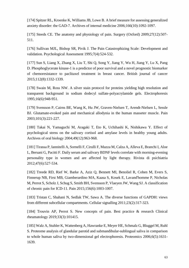
63
[174] Spitzer RL, Kroenke K, Williams JB, Lowe B. A brief measure for assessing generalized
anxiety disorder: the GAD-7. Archives of internal medicine 2006;166(10):1092-1097.
[175] Steeds CE. The anatomy and physiology of pain. Surgery (Oxford) 2009;27(12):507-
511.
[176] Sullivan MJL, Bishop SR, Pivik J. The Pain Catastrophizing Scale: Development and
validation. Psychological Assessment 1995;7(4):524-532.
[177] Sun S, Liang X, Zhang X, Liu T, Shi Q, Song Y, Jiang Y, Wu H, Jiang Y, Lu X, Pang
D. Phosphoglycerate kinase-1 is a predictor of poor survival and a novel prognostic biomarker
of chemoresistance to paclitaxel treatment in breast cancer. British journal of cancer
2015;112(8):1332-1339.
[178] Swain M, Ross NW. A silver stain protocol for proteins yielding high resolution and
transparent background in sodium dodecyl sulfate-polyacrylamide gels. Electrophoresis
1995;16(6):948-951.
[179] Svensson P, Cairns BE, Wang K, Hu JW, Graven-Nielsen T, Arendt-Nielsen L, Sessle
BJ. Glutamate-evoked pain and mechanical allodynia in the human masseter muscle. Pain
2003;101(3):221-227.
[180] Takai N, Yamaguchi M, Aragaki T, Eto K, Uchihashi K, Nishikawa Y. Effect of
psychological stress on the salivary cortisol and amylase levels in healthy young adults.
Archives of oral biology 2004;49(12):963-968.
[181] Tirassa P, Iannitelli A, Sornelli F, Cirulli F, Mazza M, Calza A, Alleva E, Branchi I, Aloe
L, Bersani G, Pacitti F. Daily serum and salivary BDNF levels correlate with morning-evening
personality type in women and are affected by light therapy. Rivista di psichiatria
2012;47(6):527-534.
[182] Treede RD, Rief W, Barke A, Aziz Q, Bennett MI, Benoliel R, Cohen M, Evers S,
Finnerup NB, First MB, Giamberardino MA, Kaasa S, Kosek E, Lavand'homme P, Nicholas
M, Perrot S, Scholz J, Schug S, Smith BH, Svensson P, Vlaeyen JW, Wang SJ. A classification
of chronic pain for ICD-11. Pain 2015;156(6):1003-1007.
[183] Tristan C, Shahani N, Sedlak TW, Sawa A. The diverse functions of GAPDH: views
from different subcellular compartments. Cellular signalling 2011;23(2):317-323.
[184] Trouvin AP, Perrot S. New concepts of pain. Best practice & research Clinical
rheumatology 2019;33(3):101415.
[185] Walz A, Stuhler K, Wattenberg A, Hawranke E, Meyer HE, Schmalz G, Bluggel M, Ruhl
S. Proteome analysis of glandular parotid and submandibular-sublingual saliva in comparison
to whole human saliva by two-dimensional gel electrophoresis. Proteomics 2006;6(5):1631-
1639.
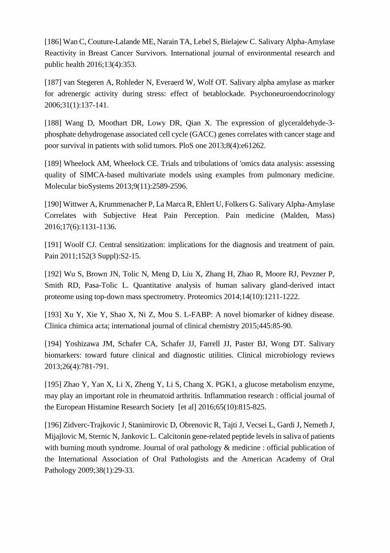
[186] Wan C, Couture-Lalande ME, Narain TA, Lebel S, Bielajew C. Salivary Alpha-Amylase
Reactivity in Breast Cancer Survivors. International journal of environmental research and
public health 2016;13(4):353.
[187] van Stegeren A, Rohleder N, Everaerd W, Wolf OT. Salivary alpha amylase as marker
for adrenergic activity during stress: effect of betablockade. Psychoneuroendocrinology
2006;31(1):137-141.
[188] Wang D, Moothart DR, Lowy DR, Qian X. The expression of glyceraldehyde-3-
phosphate dehydrogenase associated cell cycle (GACC) genes correlates with cancer stage and
poor survival in patients with solid tumors. PloS one 2013;8(4):e61262.
[189] Wheelock AM, Wheelock CE. Trials and tribulations of 'omics data analysis: assessing
quality of SIMCA-based multivariate models using examples from pulmonary medicine.
Molecular bioSystems 2013;9(11):2589-2596.
[190] Wittwer A, Krummenacher P, La Marca R, Ehlert U, Folkers G. Salivary Alpha-Amylase
Correlates with Subjective Heat Pain Perception. Pain medicine (Malden, Mass)
2016;17(6):1131-1136.
[191] Woolf CJ. Central sensitization: implications for the diagnosis and treatment of pain.
Pain 2011;152(3 Suppl):S2-15.
[192] Wu S, Brown JN, Tolic N, Meng D, Liu X, Zhang H, Zhao R, Moore RJ, Pevzner P,
Smith RD, Pasa-Tolic L. Quantitative analysis of human salivary gland-derived intact
proteome using top-down mass spectrometry. Proteomics 2014;14(10):1211-1222.
[193] Xu Y, Xie Y, Shao X, Ni Z, Mou S. L-FABP: A novel biomarker of kidney disease.
Clinica chimica acta; international journal of clinical chemistry 2015;445:85-90.
[194] Yoshizawa JM, Schafer CA, Schafer JJ, Farrell JJ, Paster BJ, Wong DT. Salivary
biomarkers: toward future clinical and diagnostic utilities. Clinical microbiology reviews
2013;26(4):781-791.
[195] Zhao Y, Yan X, Li X, Zheng Y, Li S, Chang X. PGK1, a glucose metabolism enzyme,
may play an important role in rheumatoid arthritis. Inflammation research : official journal of
the European Histamine Research Society [et al] 2016;65(10):815-825.
[196] Zidverc-Trajkovic J, Stanimirovic D, Obrenovic R, Tajti J, Vecsei L, Gardi J, Nemeth J,
Mijajlovic M, Sternic N, Jankovic L. Calcitonin gene-related peptide levels in saliva of patients
with burning mouth syndrome. Journal of oral pathology & medicine : official publication of
the International Association of Oral Pathologists and the American Academy of Oral
Pathology 2009;38(1):29-33.