Embed Size (px)
Citation preview

GREEN
Safety Assessment of Phytosterols
as Used in Cosmetics
Status: Draft Report for Panel Review Release Date: August 16, 2013 Panel Meeting Date: September 9-10, 2013
The 2013 Cosmetic Ingredient Review Expert Panel members are: Chairman, Wilma F. Bergfeld, M.D., F.A.C.P.; Donald V. Belsito, M.D.; Curtis D. Klaassen, Ph.D.; Daniel C. Liebler, Ph.D.; Ronald A Hill, Ph.D. James G. Marks, Jr., M.D.; Ronald C. Shank, Ph.D.; Thomas J. Slaga, Ph.D.; and Paul W. Snyder, D.V.M., Ph.D. The CIR Director is Lillian J. Gill, D.P.A. This report was prepared by Lillian C. Becker, Scientific Analyst/Writer.
© Cosmetic Ingredient Review 1101 17th Street, NW, Suite 412 Washington, DC 20036-4702 ph 202.331.0651 fax 202.331.0088 [email protected]

__________________________________________________________________________________________ 1101 17th Street, NW Suite 412, Washington, DC 20036
(Main) 202-331-0651 (Fax) 202-331-0088 (Email) [email protected] (Website) www.cir-safety.org
Commitment & Credibility since 1976
MEMORANDUM
To: CIR Expert Panel and Liaisons
From: Lillian C. Becker, MS
Scientific Analyst and Writer
Date August 16, 2013
Subject: Draft Report of the Safety Assessment of Phytosterols as Used in Cosmetics
Attached, please find the Draft Report of Phytosterols as used in Cosmetics. The Scientific Literature Review was posted for public comment in May of 2013. Comments from the Personal Care Products Council have been addressed. No other comments were submitted. Since phytosterols are ubiquitous in plants and are a normal part of the diet, this report concentrates on the potential for reproductive toxicity, genotoxicity, carcinogenicity, irritation, and sensitization. Oral toxicity was not addressed because exposure to phytosterols through food far exceeds any exposure through cosmetic use. Relevant information from the PEG soy sterols 2004 safety assessment is summarized and the report is included in this package. The Panel is to review the report and decide if there is sufficient data to come to a safety conclusion. If not, then the Panel is to issue an insufficient data conclusion and indicate the additional data that are needed. If the data are sufficient, the Panel is to develop language for the Discussion and issue a Tentative Report.

Distrbuted for comment only -- do not cite or quote

History of Phytosterols May, 2013 – SLR was posted for public comment. September, 2013 – Panel examines Draft Report.
Distrbuted for comment only -- do not cite or quote

Phytosterol Data Profile for September, 2013. Writer - Lillian Becker
ADME Acute toxicity Repeated dose toxicity Irritation Sensitizatio
n
Derm
al P
enetration
Log Kow
Use
Oral
Derm
al
Inhale
Oral
Derm
al
Inhale
Ocular
Irritation
Derm
al Irr. A
nimal
Derm
al Irr H
uman
Sensitization
Anim
al
Sensitization
Hum
an
Repro/D
evel toxicity
Genotoxicity
Carcinogenicity
Phototoxicity
Brassica campestris (rapeseed) sterols
X
Canola Sterols C10-40 isoalkyl acid phytosterol esters
Dihydrophyto-steryl octyldecanoate
Diosgenin Euterpe oleracea sterols X
Glycine soja (soybean) sterols X X X X
Persea gratissima (avocado) sterols X
Phytosterols X X X X X X X X Phytosteryl butyrate
Phytosteryl canolate X
Phytosteryl caprylate/caprate
Phytosteryl hydroxystearate
Phytosteryl isostearate X
Phytosteryl linoleate
Phytosteryl linoleate/ linolenate
Phytosteryl macadamiate X
Phytosteryl nonanoate
Phytosteryl oleate X Phytosteryl rice branate X
Phytosteryl ricinoleate
Phytosteryl sunflowerseedate
Punica granatum sterols X X X X X X
Beta-sitosterol X Beta-sitosteryl acetate
Soy sterol acetate X Tall oil sterol X OTHER phytosterols X X X
Distrbuted for comment only -- do not cite or quote

Search Strategy for Phytosterols
SciFinder – Searched “phytosterols” for 35 possible hits. 15 were useful. Internet search – Phytosterols. Located European scientific opinions.
Distrbuted for comment only -- do not cite or quote
Safety Assessment of Phytosterols
as Used in Cosmetics
Status: Draft Report for Panel Review Release Date: August 16, 2013 Panel Meeting Date: September 9-10, 2013
The 2013 Cosmetic Ingredient Review Expert Panel members are: Chairman, Wilma F. Bergfeld, M.D., F.A.C.P.; Donald V. Belsito, M.D.; Curtis D. Klaassen, Ph.D.; Daniel C. Liebler, Ph.D.; Ronald A Hill, Ph.D. James G. Marks, Jr., M.D.; Ronald C. Shank, Ph.D.; Thomas J. Slaga, Ph.D.; and Paul W. Snyder, D.V.M., Ph.D. The CIR Director is Lillian J. Gill, D.P.A. This report was prepared by Lillian C. Becker, Scientific Analyst/Writer.
© Cosmetic Ingredient Review 1101 17th Street, NW, Suite 412 Washington, DC 20036-4702 ph 202.331.0651 fax 202.331.0088 [email protected]
Distrbuted for comment only -- do not cite or quote

TABLE OF CONTENTS TABLE OF CONTENTS ................................................................................................................................................................................. ii
INTRODUCTION ............................................................................................................................................................................................ 1
CHEMISTRY ................................................................................................................................................................................................... 1
Definition, Structure, and Composition ....................................................................................................................................................... 1
Physical and Chemical Properties ................................................................................................................................................................ 2
Method of Manufacture ............................................................................................................................................................................... 2
Impurities ..................................................................................................................................................................................................... 3
USE .................................................................................................................................................................................................................. 3
Cosmetic ...................................................................................................................................................................................................... 3
Non-Cosmetic .............................................................................................................................................................................................. 4
TOXICOKINETICS ......................................................................................................................................................................................... 2
Absorption, Distribution, Metabolism, and Excretion ................................................................................................................................. 2
Oral ......................................................................................................................................................................................................... 2
Cytotoxicity ................................................................................................................................................................................................. 2
ANIMAL TOXICOLOGY ............................................................................................................................................................................... 2
Dermal - Non-Human ............................................................................................................................................................................. 2
Peg Soy Sterol Report ............................................................................................................................................................................. 2
REPRODUCTIVE AND DEVELOPMENTAL tOXICITY............................................................................................................................. 3
ESTROGENIC EFFECTS ................................................................................................................................................................................ 3
In Vitro ........................................................................................................................................................................................................ 3
In Vivo ......................................................................................................................................................................................................... 4
GENOTOXICITY ............................................................................................................................................................................................ 4
CARCINOGENICITY ..................................................................................................................................................................................... 4
IRRITATION AND SENSITIZATION ........................................................................................................................................................... 5
Irritation ....................................................................................................................................................................................................... 5
Dermal – Non-Human ............................................................................................................................................................................. 5
Dermal – Human ..................................................................................................................................................................................... 5
In-Vitro ................................................................................................................................................................................................... 5
Ocular ..................................................................................................................................................................................................... 5
Sensitization ................................................................................................................................................................................................. 5
Non-Human ............................................................................................................................................................................................ 5
Human ..................................................................................................................................................................................................... 5
In Vitro .................................................................................................................................................................................................... 6
CLINICAL USE ............................................................................................................................................................................................... 6
Case Studies ................................................................................................................................................................................................. 6
SUMMARY ..................................................................................................................................................................................................... 6
DISCUSSION ................................................................................................................................................................................................... 7
CONCLUSION ................................................................................................................................................................................................ 7
TABLES ........................................................................................................................................................................................................... 8
REFERENCES ............................................................................................................................................................................................... 15
Distrbuted for comment only -- do not cite or quote

INTRODUCTION This report reviews the available scientific information relevant to the safety of a group of 27 phytosterols and steryl
alkanoates as used in cosmetics. The functions of these ingredients include: skin-conditioning agents, hair conditioning agents, viscosity increasing agents, skin protectants, antioxidants, and fragrances (Table 1).1 The ingredients in this report are:
• brassica campestris (rapeseed) sterols • canola sterols • C10-40 isoalkyl acid phytosterol esters • dihydrophytosteryl octyldecanoate • diosgenin • euterpe oleracea sterols • glycine soja (soybean) sterols • persea gratissima (avocado) sterols • phytosterols • phytosteryl butyrate • phytosteryl canolate • phytosteryl caprylate/caprate • phytosteryl hydroxystearate • phytosteryl isostearate
• phytosteryl linoleate • phytosteryl linoleate/linolenate • phytosteryl macadamiate • phytosteryl nonanoate • phytosteryl oleate • phytosteryl rice branate • phytosteryl ricinoleate • phytosteryl sunflowerseedate • punica granatum sterols • beta-sitosterol • beta-sitosteryl acetate • soy sterol acetate • tall oil sterol
Plant sterols, or phytosterols, occur naturally as free alcohols and as fatty acid esters (i.e., naturally occurring steryl
alkanoates). The ingredients in this report are sterol alcohols or esters (in some cases mixtures of both) extracted from plants, some of which have been saponified to the free alcohols and then esterified with plant-derived fatty acids. These resultant ester-derivatized phytosterols (i.e., steryl alkanoates) share a great deal of structural overlap with the naturally occurring phytosterol esters. Indeed, most of these derived esters are likely to be exact synthetic copies of the components of the naturally occurring phytosterol esters. Accordingly, since there is expected to be a great deal of component overlap between the ingredients in this group, and reviewing them separately would involve a large amount of redundancy, it is more efficient to review them as an ingredient family and employ read-across. The similar chemical structures/components, physicochemical properties, and functions and concentrations in cosmetics enable grouping these ingredients and reading across the available toxicological data to support the safety assessment of the entire group. Table 2 lists these component chemicals and notes whether they are cosmetic ingredients, have been reviewed by the Cosmetic Ingredient Review (CIR) Expert Panel (Panel), and, if so, the Panel’s conclusions. All of the reviewed component cosmetic ingredients were found to be safe as used. Butyric acid, caprylic acid/capric acid, and linoleic acid/linolenic acid have not been reviewed. Octyldecanoic acid is not a cosmetic ingredient.
In 2000, the Panel found the data on PEG-5, -10, -16, -25, -30, and -40 soy sterols to be insufficient to support the safety of these ingrdients.2 In 2004, the Panel found these PEG soy sterols to be safe as used in an amended safety assessment that included data on phytosterols and phytosterol esters.3 The Panel’s approach in these safety assessments was to review the safety of PEGs and phytosterols/soy sterols, as well as the conjugated polyethers, and assessed the safety of the PEG phytosterols from those data. Because the data on the phytosterols/soy sterols are relevant for this safety assessment, summaries of the data from these two safety asessments are provided below in the appropriate sections below.
In 2004, the Panel concluded that dioscorea villosa (wild yam) root extract (with a diosgenin, an ingredient in this report, content of 3.5%) was safe as used.4 The maximum reported concentration of use was 15%.
Many of the phytosterols in this study are from edible plant sources. Exposure to these phytosterols from consuming foods results in much greater systemic doses than could result from the use of cosmetic products. It was noted in the PEG soy sterol reports that phytosterols and phytosterol esters are not significantly absorbed after oral exposure, and thus, did not result in systemic exposure.2,3 Therefore, acute and repeated dose oral toxicity potential of these phytosterols as cosmetic ingredients will not be addressed again in depth in this report. The focus of this report is on other end points: reproductive toxicity, genotoxicity, carcinogenicity, irritation, and sensitization. Pertinent data from the PEG soy sterol safety assessment are summarized below in the appropriate sections.
CHEMISTRY
Definition, Structure, and Composition The definitions and functions of the ingredients in this report are presented in Table 1. The phytosterol ingredient group is comprised of the plant-derived, free sterols and their esters, the steryl alkanoates. β-Sitosterol is an example of a discreet, free phytosterol ingredient. To generate a steryl alkanoate with an ester at the 3-position of the sterol, the hydroxyl group at the 3-position of the cyclopentenophenanthrene backbone is esterified, with an alkyl acid or acid chloride (Figure 1). β-Sitosteryl Acetate is an
Distrbuted for comment only -- do not cite or quote

example of a steryl alkanoate (Figure 2).
H3C
H3C
CH3
CH3
CH3
HO
H3C
12
34
5 6 7
89
1112
13
14 1516
1719
1820
21
22 23 24
25
27
26
β-Sitosterol
10
Figure 1. β-Sitosterol
H3C
H3C
CH3
CH3
CH3
O
H3C
3
β-Sitosteryl Acetate
H3C
O
Figure 2. β-Sitosteryl Acetate
Phytosterols occur in plants in the free alcohol, steryl alkanoate, or glycoside forms (e.g., diosgenin). The free phytosterols are characteristic components of the non-saponifiable fractions of plant oils.5 The steryl alkanoate and glycoside forms, however, are broken down to the free phytosterol form (and respective acid or sugar) under saponification conditions. The majority of the ingredients in this report are mixtures of either sterols or steryl alkanoates, with component concentrations that vary with growth and extraction conditions.
Soybean oil that had been alkali-refined typically contained 0.446 mg/100 mg oil total sterols and 0.287 mg/100 mg oil free sterol. The ratio of esterified to free sterol was 0.55.6
Refined plant sterols are reported to contain ~ 88% total sterol content. Of that percentage, 56% is γ-sitosterol, 28% is campesterol, and 4% is stigmasterol. Other compounds isolated from the phytosterols are 4% - 6% sterol hydrocarbons and cholesterol, and 4% - 6% triterpene alcohols, keto-steroids, and other steroid-like substances.7 PEGS SOY STEROL REPORT The chemical characterization of a plant sterol material is provided in Table 3. The distribution of phytosterols in common vegetable oils are provided in Table 4.
In an analysis of another source of phytosterols (source not provided), it was reported that the principal phytosterols were present as follows: β-sitosterol, 47.9%; campesterol, 28.8%; and stigmasterol, 23.3%. No impurities were found. In an analysis of phytosterol esters, it was reported that the principal phytosterols were present as fatty acid esters: β-sitosterol, 47.3%; campesterol, 28.1 %; and stigmasterol, 24.5%. The distribution of the fatty acid chain lengths was consistent with fatty acids derived from sunflower oil.8,9
Physical and Chemical Properties Physical and chemical properties of representative phytosterols in the form of vegetable oil sterols and tall oil sterols/stanols are provided in Table 5. Phytosterols and their fatty acid esters are thermally stable and only degrade at high temperatures (>100°C) in the presence of oxygen.10
Method of Manufacture Free phytosterol alcohols and phytosterol alkanoates are characteristic components of plant oils; saponification of
these oils is the primary means of producting free phytosterol alcohols for commercial use.5 Soy sterol is isolated from soybean oil distillates in a saponification process in which the phytosterol alcohols are
separated from the fatty acids by extraction with a fat solvent.2 The phytosterols in the resulting extract are separated from
Distrbuted for comment only -- do not cite or quote

the tocopherols in the mother liquor, and then purified and/or separated into constituent sterols. Tall oil sterol, an example of a phytosterol mixture, is obtained from tall oil soap in a multi-step process.5 The
production process involves fractional distillation of the tall oil soap to remove volatile compounds. The resulting residue (tall oil pitch) containing esterified sterols (i.e., steryl alkanoates) is treated with alkali (saponified) to release the free sterol alcohols. After neutralization, the material is subjected to a two-stage distillation process. The distillate is then dissolved in methanol/methylethylketone solvent and the sterols crystallizing from this solution are obtained by filtration, washed with solvent and dried. This procedure results in a lower stanol and a higher sterol content of the phytosterol mixture. Conifers that have naturally lower stanol content are now used as the primary source of the tall oil soap. Stanols (obtained by catalytical hydrogenation of the phytosterol mixture) are added before the crystallization step in order to maintain the original stanol/sterol ratio. The phytosterol composition of the tall oils produced from the two processes is provided in Table 6.
Steryl alkanoates are produced from free sterols by classical esterification methods, via free acids or acid chlorides. Sterol alkanoates may be derived from neutralized, refined, bleached and deodorized (N/RBD) soybean distillates.11,12 Crude soybean oil is degummed, neutralized, bleached and deodorized to yield N/RBD soybean oil and distillates. The deodorized distillate undergoes further processing (crystallization and/or distillation), resulting in a sterol mixture. This sterol mixture is then crystallized and esterified with fatty acids (from food grade vegetable oils such as rapeseed or sunflower oil), washed, bleached and deodorized to give the final plant steryl alkanoates.
A manufacturer reported that for the manufacture of glycine soja (soybean) sterols, and punica granatum sterols, the raw materials are tested for acceptable qualifications (not specified) before they are cold pressed for oil.13,14 The oil is then tested for quality (not specified) before the oil is fractionated to isolate the sterols. Pomegranate sterols are heat sterilized at 100°C before fractionation.15
Impurities In assessing the data on soybean oil sterols, the Scientific Panel on Dietetic Products, Nutrition and Allergies noted
that there are limited analytical data of sufficient sensitivity and reliability regarding the possible residual allergen (protein) content of phytosterols.11 The limited analytical data regarding the protein (allergen) content of N/RBD soybean oil-derived plant stanol esters were insufficient to predict the likelihood of adverse reactions in soybean allergic individuals. This Panel concluded that since the starting material is refined soybean oil and there is an adequate subsequent production process, it is not very likely that this product will cause a severe allergic reaction in the majority of soybean allergic individuals.
When selected phytosterol samples (a phytosterol blend and a phytosterol blend spiked with reference protein) were analyzed for residual soybean protein using ELISA (enzyme-linked immunosorbent assay), soy protein was not detectable at or above the 10-20 μg/g detection limit.16 Tall oil sterols/stanols was reported to contain < 0.1 mg/kg lead.10 Vegetable oil sterols, in general, are reported to have < 2.0 mg/kg impurities (mercury, < 0.1%; lead, < 0.1%; cadmium, < 0.1%; and arsenic, < 0.1%). Both contain < 2 ppb PAHs and < 1.5 ng-TEQ/kg dioxins and dioxin-like PCBs. No pesticides were detected.
In an analysis of euterpe oleracea sterols, glycine soja (soybean) sterols, and punica granatum sterols, none of these ingredients contained detectable levels of multiple allergens, including: amyl cinnamal, benzyl alcohol, citronellol, coumarin, linalool, and farnesol (Table 7). Another analysis did not detect several pesticides, including: DDT (detection level 1.00 mg/kg), methidathion (0.20 mg/kg), and pyrethrins (3.00 mg/kg).17-19 PEGS SOY STEROL REPORT
Analyses of various lots of soy sterols for pesticide residues were negative for a number of pesticides, including PCB, DDE, DDT, malathion, and β-hexachloride.20
PRECURSORS
The final protein content of N/RBD soybean oils (the source of soy phytosterols) depends on the quality and efficiency of purification steps.11 The protein content of N/RBD oils may be reduced to low levels within the 0.02-0.44 μg/kg range.21
When two samples of edible soy oil (crude virgin and deodorized) were analyzed for proteins (by heat-extracted with PBS and BCA assay), 1.89 µg/mL and 0.32 µg/mL of proteins were present, respectively. 22
USE Cosmetic
Data on ingredient usage are provided to the Food and Drug Administration (FDA) Voluntary Cosmetic Registration Program (VCRP; Table 8).23 A survey was conducted by the Personal Care Products Council (Council) of the maximum use concentrations for ingredients in this group.24
Data were available from both the VCRP and the Council for the following ingredients: • Brassica campestris (rapeseed) sterols was reported to be used in 50 leave-on products up to 7% (the highest
amount in lipstick) and 7 rinse-off products up to 0.13%. • Glycine soja (soybean) sterols was reported to be used in 194 leave-on products (mostly skin care and makeup
Distrbuted for comment only -- do not cite or quote

products) up to 1% (the highest concentration in eye lotion, cuticle softeners, and other skin preparations) 45 rinse-off products up to 4.1% (the highest concentration in skin cleansing products) and one bath product. It is used in tonics, dressings and other hair grooming aids, including an aerosol and a pump at 0.000001%.
• Phytosterols was reported to be used in 177 leave-on products up to 5% including lipsticks (up to 5%) deodorants (up to 0.06%), and eye makeup (up to 0.006%). It is also used in 215 rinse-off products up to 0.5% including hair products (up to 0.5%), bath soaps and detergents (up to 0.005%), and indoor tanning preparations (up to 0.0001%). It is reported to be used in face powders up to 0.05%.
• Phytosteryl isostearate was reported to be used in 15 leave-on products up to 3% and one rinse-off product up to 0.5%. It is used in lipsticks up to 3% and in eye makeup up to 0.5%.
• Phytosteryl [phytosterol] macadamiate was reported to be used in 181 leave-on products up to 8%. It is reported to be used in two rinse-off products up to 1%. It is used in 100 lipsticks up to 7% and in moisturizing products up to 8%.
• Phytosteryl oleate was reported to be used in 20 leave-on products up to 3%. It was reported to be used in 6 paste masks/mud packs (no concentrations of use reported).
• Phytosteryl rice branate was reported to be used in an eye makeup and a moisturizing product. The Council reported that it was used in eye lotions up to 1%, foundations up to 0.5%, and face and neck products up to 0.5%.
• Punica granatum sterols was reported to be used in 29 rinse-off products up to 5% (including 15 lipsticks). It was also reported to be used in two rinse-off products (no concentration of use reported).
• Beta-sitosteryl was reported to be used in 46 leave-on products up to 0.06% and in two rinse-off products (no concentration of use reported).
• Tall oil sterol was reported to be used in 7 leave-on products up to 0.0046%. It is also reported to be used in skin cleansing products up to 0.0006%.
Data were only available on the frequency of use (VCRP) for the following ingredients: • Euterpe oleracea sterols was reported to be used in one lipstick and one foundation. • Soy sterol acetate was reported to be used in one moisturizing product. Data were only available on use concentration (Council) for the following ingredients: • Persea gratissima (avocado) sterols was reported to be used in eye lotion up to 1%, lipstick up to 0.65%, and face
and neck products up to 0.1%. • Phytosteryl canolate was reported to be used in eye shadow up to 0.06%. There were no use or concentration of use data reported for: • Canola sterols • C10-40 isoalkyl acid phytosterol esters • Dihydrophytosteryl octyldecanoate • Diosgenin • Phytosteryl butyrate • Phytosteryl caprylate/caprate • Phytosteryl hydroxystearate
• Phytosteryl linoleate • Phytosteryl linoleate/linolenate • Phytosteryl nonanoate • Phytosteryl ricinoleate • Phytosteryl sunflowerseedate • Punica granatum sterols • Beta-sitosteryl acetate
As noted above, glycine soja (soybean) sterols was reported to be used in propellant and pump spray tonics, dressings
and other hair grooming aids up to 0.000001% and phytosterols are reported to be used in face powders up to 0.05%. In practice, 95% to 99% of the droplets/particles released from cosmetic sprays have aerodynamic equivalent diameters >10 µm; with propellant sprays yielding a greater fraction of droplets/particles below 10 µm compared with pump sprays. Therefore, most droplets/particles incidentally inhaled from cosmetic sprays would be deposited in the nasopharyngeal and bronchial regions and would not be respirable (i.e., they would not enter the lungs) to any appreciable amount.25-30
Non-Cosmetic Phytosterols (stigmasterol-rich plant sterols: stigmasterol, >85%; brassicasterol, 1.7%; β-sitosterol, 3%; campesterol, 1.7%) are used in ready-to-freeze alcoholic beverages as a stabilizer.31 Phytosterols and phytostanols are commonly used in food products for their properties that reduce absorption of cholesterol in the gut and lower cholesterol in food products.10 The optimal daily dose for this purpose is 2 - 3 g. For example, in Europe, phytosterol esters are added to margarines and low fat spreads (3.4 g/30 g), yogurts (1.25 g/125 mL), yogurt drinks (3.4 g/100mL), and milk (5 g/L).
The Scientific Committee on Food (SCF) and European Food Safety Authority (EFSA) concluded that phytosterols, phytostanols and their esters are approved for use in various foods (i.e., yellow fat spreads, soya drinks, salad dressings, rye
Distrbuted for comment only -- do not cite or quote

bread) within the EU at levels resulting in intake of up to 3 g/day.11,12,16,32-43
TOXICOKINETICS Absorption, Distribution, Metabolism, and Excretion
No published dermal or inhalation ADME studies were discovered and no unpublished data were submitted. Oral
The Western diet consists of ~160-360 mg/d phytosterols consisting of ~80% β-sitosterol. The diet also includes some campesterol and stigmasterol with small amounts of brassicasterol and trace amounts of δ-5-saturated plant stanols.44
Less than 5% of dietary phytosterols, phytostanols, and their esters are absorbed in the gastrointestinal tracts of rats and humans.31 Following absorption, phytosterols/phytostanols are transported in the serum via HDLs in rats and LDLs in humans to various organs and tissues, mostly to the liver. In the liver, phytosterols may be converted to bile acids. Absorbed phytosterols/phytostanols are predominantly excreted as such or as bile acids by the biliary route into the feces. The metabolic fate of phytosterols, phytostanols, and their esters is similar between rats and humans. The individual plant sterols are metabolized in a similar manner to each other. The phytosterols that are not absorbed in the gastrointestinal tract enter the colon intact and are rapidly excreted in the feces.44-47
In an oral study (n = 10 healthy men) the intestinal absorption of phytosterols were: campesterol, 9.6%; stigmasterol, 4.8%; and sitosterol, 4.2%.48 The authors noted that these results were consistent with the results of animal studies showing that increasing the side chain length of cholesterol reduced the absorbability of the sterol with the exception of campersterol. The 5α-campersterol-saturated had greater absorbability than campersterol. Absorption was measured by an intestinal perfusion technique over a 50-cm segment of the upper jejunum.
In male subjects, the biliary secretion rate of β-sitosterol was faster (1.23 mg/h) than that of campesterol (0.76 mg/h).49
Plant sterols, including stigmasterol and stanols (34 g/kg in feed), were able to cross the blood-brain barrier in a 90-day feeding study of Watanabe heritable hyperlipidemic rabbits.50
Cytotoxicity β-sitosterol (200 μg/mL in ethanol) and β-sitosterol/campesterol (50%/40%; 200 μg/mL in ethanol) were cytotoxic to mouse macrophages (strain C57BL/6).51 Cytotoxicity was demonstrated through cell viability, lipid uptake, lactate dehydrogenase (LDH) leakage, cellular protein content, and a 3’-[1-(phenylaminocarbonyl)-3,4-tetrazolium]bis(4-methoxy-6-nitro)benzene sulfonic acid hydrate (XTT) assay. Phytosterols (0.01 – 40 mM; derived from pomegranate) were not cytotoxic in a Neutral Red Cytotoxicity assay.52 PEG SOY STEROL REPORT
β-Sitosterol (l00 µg/ml; 5% in DMSO and saline) was cytotoxic to seven cancer cell lines.
ANIMAL TOXICOLOGY Many of the phytosterols in this report are from edible sources and exposure to these phytosterols from food would result in a much larger systemic dose than that resulting from use in cosmetic products. A summary of toxicity data on phytosterols, including oral data, from the PEG soy sterol report is presented below for information purposes. However, this report does not address their oral toxicity potential but is focused on the potential for reproductive toxicity, genotoxicity, carcinogenicity, irritation and sensitization. A summary of toxicity data on phytosterols, including oral data, from the PEG soy sterol report is presented below for background information on toxicity. Dermal - Non-Human
The dermal LD50 of two mixtures of phytosterol esters was reported to be > 2000 mg/kg.53 A wood-derived mixture (a stanol composition of ~94% β-sitostanol and ~6% campestanol in corn oil; WDPSE) and a vegetable oil–derived mixture of phytostanol esters (~68% β-sitostanol and ~32% campestanol in corn oil; VODPSE) were administered dermally to rats (n = 5/sex) for 24 h according to the Organization for Economic Co-operation and Development (OECD) Test Guideline 404. No deaths or clinical signs of toxicity were observed after application of WDPSE. One male rat in the VODPSE group died of unrelated causes during the 14-day observation period. Peg Soy Sterol Report
Wistar rats administered a basal diet supplemented with cholesterol and maize phytosterols (72.5% β-sitosterol, 0.5% campesterol, and 7% stigmasterol) had decreased hepatic cholesterol concentrations.54 Rats given the high dose of cholesterol and phytosterols had decreased malic enzyme and acetylCoA carboxylase activities, and had hypotriglyceridemia.
Wistar rats administered subcutaneous injections of 250 to 500 µg/100 g β-sitosterol for 60 days had no gross or microscopic lesions of the liver or kidneys.55 Rats administered 1000 µg/100 g had mild fibroblastic proliferation around the
Distrbuted for comment only -- do not cite or quote

hepatic lobules and mild microscopic lesions of the kidney. Serum cholesterol was reduced in a dose-dependent manner, and serum protein was markedly reduced in rats of the high dose group.
In a 90-day oral toxicity study in female Wistar rats (n = 4), diets containing plant phytosterol esters up to 8.1 % were well tolerated.56 Some small hematology and blood chemistry variations from the controls were observed. No treatment related effects were observed with organ weights and histological examination and there was no evidence of systemic toxicity. Absent any organ effects, the small hematology and blood chemistry variations were not considered of toxicological significance.
Thirteen dogs fed a basic diet supplemented with 0.5 to 1.0 g/kg/day of β-sitosterol had no gross or microscopic changes after 8 to 22 months of treatment. Weight gains and clinical parameters did not differ from controls.20
No adverse effects or gross or microscopic abnormalities were observed in six New Zealand white rabbits of both sexes that were given feed containing 3% cottonseed sterols and 4% soy sterols for 70-212 days.20
REPRODUCTIVE AND DEVELOPMENTAL TOXICITY In a two-generation feeding study, the no observed adverse effect level (NOAEL) for phytosterol esters was 8.1% in the diet.57,58 Wistar rats, F0 generation, (n = 28/sex) were administered phytosterol esters (0, 1.6%, 3.2%, 8.1%) in feed for 10 weeks before mating, and continuing through gestation and weaning. The F1 generation (n = 28/sex) were fed the same diet as their F0 parents and mated after 10 weeks. The analysis of the phytosterols revealed the following breakdown: brassicasterol (2.9%), campesterol (26.7%), stigmasterol (17.7%), β-sitosterol (51.0%), cholesterol (0.2%), and unknowns (1.5%). There were no maternal or teratogenic effects attributed to the test substance. There were no effects on fertility and reproductive parameters, including sexual maturity, estrous cycle length, precoital time, and the histopathology of reproductive tissues in either generation. There were no developmental or reproductive effects observed in either generation. Necropsies were unremarkable.
The NOEL (8.1%) is equivalent to 3.3-6.5 g phytosterol esters/kg/d during the 10-week pre-mating period (~ 2.1-4.1 g phytosterols/kg/d or 400-900 mg stigmasterol/kg/d) and 2.5-9.1 g phytosterol esters/kg/d during gestation (~1.4-5.7 g/kg/d or 300-1200 mg stigmasterol/kg/d). The authors concluded that 2.5-9.1 g phytosterol esters/kg/d and 1.54-5.62 g phytosterols/kg/d (~ 335-1219 mg stigmasterol/kg/d), dependent on the phase of the study, was the NOAEL of daily oral administration of phytosterol esters for two successive generations.57,58
There were no signs of reproductive toxicity to American minks (n = 70/sex) orally administered β-sitosterol (at 0, 5, 10 or 50 mg/kg/d) for 10 months.59 In the second part of the study, after 7 months of exposure, males (n = 10–11) were mated with 4–5 females each. There were no differences in number of pregnant females, litter and kit numbers, postnatal mortality and development and there were no treatment-related changes. After 3 months of exposure, 15 males/group were killed and investigated for organ weights and hematological and clinical chemistry parameters. Males exhibiting low quality fur were selected for this part of the study. There were differences in body fat masses (omental, mesenteric, retroperitoneal, intra-abdominal fat) reported, but increases in fat masses were not dose dependent. There were increased blood hemoglobin and serum high-density lipoprotein cholesterol concentrations observed.
Subcutaneous injections of β-sitosterol (5 mg/kg/d) for 16 to 48 days reduced sperm concentrations and fertility, and decreased testis and accessory sex tissue weights in a time-dependent manner in male Wistar rats.60 Rats administered 0.5 mg/kg/d had a decrease in sperm concentration of the caput epididymis after 48 days of treatment, but no reduction in fertility. The observed decreases in sperm concentration persisted after termination of treatment, and appeared to be due to a reduction in the rate of spermatogenesis.
ESTROGENIC EFFECTS In Vitro
There were no signs of estrogenic activity of phytosterols and phytosterol esters in an in vitro competitive estrogen receptor binding assay (up to 1 x 10⁻⁴ mol/L) and a recombinant yeast assay (2 x 10⁻⁴ mol/L).61 The phytosterols tested consisted of a mixture of β-sitosterol (47.9%), campesterol (28.8%), and stigmasterol (23.3%) and were sourced from a variety of edible vegetable oil distillates (e.g., sunflower, soya bean and rapeseed oils). The esters were phytosterols esterified with fatty acids from sunflower oil. The competitive estrogen receptor binding assay used a preparation of estrogen receptors isolated from 10-week-old Wistar rat uteri and measured the concentration-dependent substitution of [2,4,5,6-3H]estradiol at the estrogen receptor.
The hormonal activity of the pure substances β-sitosterol, stigmasterol, and their purified chlorine dioxide oxidation products showed estrogenic activity in an estrogen receptor binding assay.62 In an androgen receptor binding assay, the phytosterols and their oxidation products showed a small but measurable activity.
Four phytostanol mixtures (0, 1, 10 or 100 μmol/L) showed no estrogenic activity in human mammary adenocarcinoma (MCF-7) cells.53 Estrogenic activity was measured as the ability to induce proliferation of these cells. Proliferation was measured by staining the cells with the protein stain sulforhodamine B and measuring optical density. The MCF-7 cells were cultured for 6 days. 17β-Estradiol was used as a positive control. The percentage of β-sitostanol in the phytostanols, derived from vegetable oil, ranged from 58% - 67%, and campestanol ranged from 29% - 32%. The
Distrbuted for comment only -- do not cite or quote
phytosterol content was < 4%. Precipitation and slight cytotoxicity were observed at the highest test concentration with all mixtures. No cell proliferation was observable in cells treated with phytostanols. Under the conditions of this study, the phytostanol mixtures tested showed no estrogenic activity.
In Vivo
Neither WDPSE nor VODPSE administered in feed (0, 8.3%) for 4 days influenced the uterine weights of female Wistar rats (n = 10; 17-day-old) in a Teicco assay.53 Diethylstilbestrol (5, 10 or 20 µg/kg) in the diet was used as positive control. Uterine weight was used as an indicator of estrogenic activity. No treatment-related effects on general condition, body weight or food consumption were observed.
β-Sitosterol, stigmasterol, and their oxidation products were inactive in a 28-day mosquito fish masculinization assay at concentrations up to 100 μg/L.62
There were no signs of estrogenic activity for phytosterols and phytosterol esters tested in an in vivo immature rat uterotrophic assay (n = 10; up to 500 mg/kg).61 The phytosterols tested consisted of a mixture of β-sitosterol (47.9%), campesterol (28.8%), and stigmasterol (23.3%) and were sourced from a variety of edible vegetable oil distillates (e.g. sunflower, soya bean and rapeseed oils). The phytosterol esters were prepared by esterifying these phytosterols with fatty acids from sunflower oil.
Absolute and relative uterine weights were unaffected in an immature rat uterotropic assay of a mixture of phytosterols and phytostanols (0, 500, 1000, 2500 mg/kg) administered twice daily for 4 days when compared with the negative control.53 The mixture of phytosterols and phytostanols used in this study was derived by solvent extraction (~40– 55% β-sitosterol, 16–31% β-sitostanol, 11–15% campesterol and 2–11% campestanol; MPSS-SE) was assessed using female, Crl:CD (SD)IGS BR VAF/Plus, 19-day-old rats (n = 10). Ethinyl estradiol was used as a positive control. Body weight gains of animals in the 2000 and 5000 mg/kg groups were reduced. PEG SOY STEROL REPORT
Dose-dependent uterotrophic effects of β-sitosterol in ovariectomized rats and its synergism with estradiol could be due to the phytosterol's intrinsic estrogenic properties, and that the effects of β-sitosterol could be inhibited by progesterone.55
β-Sitosterol was an effective estrogen-like agonist in exerting vaginal cornification and caused uterine weight gain in adult, ovariectomized Wistar rats.63 Subcutaneous injections of the sterol caused dose-related increases in uterine glycogen concentration after 10 days.
Progesterone treatment partially suppressed the phytosterol-induced elevation of glycogen concentration when administered in combination with the median and high phytosterol doses. β-Sitosterol also stimulated glucose-6-phosphate dehydrogenase, phosphohexose isomerase, and total lactate dehydrogenase activities.
In a related study, uterine RNA, DNA, and protein concentrations were increased by treatment with β-sitosterol.55 Other studies of well-characterized phytosterols and phytosterol esters demonstrated no effect in an estrogen-binding
study, a recombinant yeast assay for estrogen or estrogen-like activity, or a juvenile rat uterotrophic assay for estrogen or estrogen-like activity.55,61,64
Sulfates of β-sitosterol act as abortifacients in female rats and Dutch-belted rabbits via estrogenic effects. They also exhibit spermicidal effects. β-Sitosterol itself had anti-estrogenic, anti-progestational, gonadotrophic, anti-gonadotrophic, and anti-androgenic effects.44,65,66
GENOTOXICITY
In multiple in vitro (up to 5000 µg/plate) and in vivo (up to 2000 mg/kg) assays, phytosterols and phytosterol esters were negative for genotoxicity (Table 8). These tests included reverse mutation, chromosomal aberrations, gene mutation, clastogenicity, sister chromatid exchange (mice), micronucleus induction (rats and mice), and unscheduled DNA synthesis assays (rats).67-70 PEG SOY STEROL REPORT
Phytosterols and phytosterol esters were not genotoxic, with or without metabolic activation, in the Ames assay, a human lymphocyte chromosome damage assay, an unscheduled DNA synthesis assay, or a rat bone marrow micronucleus assay.55,71-77
CARCINOGENICITY No new published carcinogenicity studies were discovered and no unpublished data were submitted.
PEG SOY STEROL REPORT
Sitosterol inhibited the tumor-promoting activity of TPA in the skin of female ICR mice after initiation with DMBA. The percent reduction in the average number of tumors at week 18 was 40% in mice given TPA, DMBA, and sitosterol. Sitosterol applied topically before treatment with TPA inhibited TPA-induced epidermal ODC activity; ODC induction can
Distrbuted for comment only -- do not cite or quote

be representative of the effects of phorbol esters with strong tumor promoting activity. Additionally, dermal inflammation caused by a single application of TPA was slightly inhibited by sitosterol and stigmasterol.44,78
Male Fischer CD rats coadministered the direct-acting carcinogen N -methylnitrosourea (by cannulation on days 1, 4, 7, 10) and β-sitosterol (95% pure, with 4% campesterol and 1 % stigmasterol; 0.2% in feed for 28 weeks) had significantly fewer colonic tumors (benign or benign and malignant) compared to rats given the carcinogen alone after 28 weeks.79 Of rats given the carcinogen alone, 54% had tumors. Of rats given both the carcinogen and sitosterol, 33% had tumors. The incidence of rats with malignant colonic neoplasms increased after coadministration of the phytosterols; 15% (7/48) had invasive carcinomas in the sterol plus carcinogen group compared to 7% (5171) of rats given the carcinogen alone.
The phytosterols decreased epithelial cell proliferation of the colon in mice (0.1 % in feed) and rats (0.2% in feed after induction with N-methyl-N-nitrosourea), and were cytotoxic for human epidermoid carcinoma of the nasopharynx (> 20 μg/ml).80,81
IRRITATION AND SENSITIZATION
Irritation Dermal – Non-Human WDPSE (2000 mg/kg) administered to the clipped skin of male albino rabbits (n = 3) for 4 h under semi-occlusion was not irritating.53 VODPSE caused very slight erythema after 1 h of treatment, which was completely reversed within 24 h after treatment. Skin irritation/corrosion was tested with rabbits in according to OECD Test Guideline 404. Dermal – Human
Phytosterols (100%; 1 mL; derived from pomegranate) were not irritating to scarified skin in a repeat irritation assay (n = 10).82 The test site was scratched with a 30-guage needle. The test material was administered to the same scarified location on the forearm, using a chamber, for 24 h for three consecutive days. The site was examined 30 min after removal and before the next treatment.
In-Vitro
In an EpiDerm™ assay, phytosterols (100%) from three sources (derived from pomegranate, soybean, and acai) were not predicted to be dermal irritants.83-85 Ocular There was no irritation potential revealed for WDPSE and VODPSE in a chicken enucleated eye assay.53 WDPSE and VODPSE (concentration not provided; assumed 100%) were considered minimally irritating in a Draize assay using albino rabbits (n not provided).53 The assay was conducted in accordance to OECD Test Guideline 405. WDPSE and VODPSE (concentration not provided; assumed 100%) caused slight and slight or moderate discharge, respectively, which was reversible within 24 h after treatment.
In an EpiOcular™ assay, phytosterols (100%) from three sources (derived from pomegranate, soybean, and acai) were not predicted to be ocular irritants.83-85
Sensitization Non-Human Neither WDPSE nor VODPSE (concentration not provided) caused signs of skin sensitization after administration to male guinea pigs (n = 10) in a maximization assay conducted in accordance with OECD Test Guideline 406.53 Human There were no signs of irritation or sensitization in a human repeat insult patch test (HRIPT; n = 50) of sterols (100%; 0.2 mL; 0.2 g; derived from pomegranate).86 The test material was heated to liquefy it, then it was applied to an occlusive, hypoallergenic patch. The patch was applied to the infrascapular regions of the back for nine treatments. The same concentration and amount of the test substance used in the challenge phase. None of the subjects with confirmed soy allergies (n = 29) had a positive reaction to a skin prick test of plant stanol ester.12 An open challenge with plant stanol ester within four weeks of the HRIPT (cumulative dose 5.55g) was negative in 26 of 33 (the original 29 + 4 more) subjects. Positive reactions consisted of itching of the throat in three participants, cutaneous symptoms in three, and loose stools in one subject. The reactions were observed after the final cumulative dose of plant stanol ester; all symptoms resolved without treatment.
A follow-up double-blind placebo controlled food challenge (DBPCFC) study with plant stanol ester performed on 6 of the subjects with positive reactions in the skin prick test had negative results. The DBPCFC with plant stanol ester in the remaining seventh subject (female) was interpreted as negative, although she reported loose stools the morning after the last challenge, which contained plant stanol ester. In view of the cumulative oil intake, a nonimmune-mediated reaction may be considered.12
Distrbuted for comment only -- do not cite or quote
Of 22 subjects that had positive reactions to a commercial soy extract in a skin prick test, 16 had a positive reaction to soy isolate and 6 to soy.16 None had a reaction to phytosterols.
In Vitro CONSTITUENTS
In an immunoblotting assay for soybean proteins using polyclonal, soybean-specific antiserum from rabbits (RBiopharm) and sera from nine soybean-allergic subjects, no soy protein or other protein was detected.16 Oleosin was added as a control; the oleosin fraction was shown to be a minor IgE-binding constituent of the total soybean protein. The limit of detection was 50 ng of the reference soybean extract and 100 ng of oleosin.
All hydrophilic extracts of vegetable oil deodorized distillate (VOD) samples (n = 9) analyzed by immunoblotting with soy-specific antiserum from rabbits and by IgE-immunoblotting with a pooled human serum detected no soy protein or other protein. There was no IgE binding with the VOD or the phytosterol samples using either the pooled human serum or the serum of one subject who had experienced mild oral allergy syndrome after a DBPCFC with phytosterols. The authors concluded that no IgE-binding proteins were present in the VOD and phytosterol samples at or above 1 and 10 μg/g, respectively.16
Refined soybean oils exhibited no detectable IgE binding activity using immunoblotting and enzyme allergosorbent test (EAST) inhibition assays.87
CLINICAL USE Case Studies
A female subject excreted increasing amounts of β-sitosterol, campesterol and stigmasterol through the skin as oral intake of phytosterols increased over sustained periods of time.88 When phytosterols were removed from the diet, the amount of β-sitosterol in the skin decreased from 6 mg/d to 0.08 mg/d within 83 days and finally became undetectable. Similar results were reported for the other two phytosterols. Twenty days after the administration of 30 g/d phytosterols, β-sitosterol, as well as campesterol and stigmasterol, reappeared in the skin and was excreted at 5 mg/d by 6 weeks.
SUMMARY A total of 27 phytosterols and steryl alkanoates are described for use in cosmetics. These ingredients are sterols
derived from plants, many of which are then esterified with plant-derived fatty acids. These ingredients are reported to function as skin-conditioning agents, hair conditioning agents, viscosity increasing agents, skin protectants, antioxidants, drug astringents, and fragrances.
The Panel concluded that PEG-5, -10, -16, -25, -30, and -40 soy sterols to be safe as used in a prior amended safety assessment. The component chemicals that are cosmetic ingredients that have been reviewed by the Panel were all found to be safe as used. Butyric acid, caprylic acid/capric acid, and linoleic acid/linolenic acid have not been reviewed. Octyldecanoic acid is not a cosmetic ingredient.
Phytosterols are from edible plant sources and exposure to these phytosterols in food results in a much greater systemic exposure than that resulting from use in cosmetic products containing these ingredients. It was noted in the PEG soy sterol report that phytosterols and phytosterol esters are not significantly absorbed after oral exposure. Therefore, acute and repeated dose oral toxicity potential of these phytosterols was not be addressed in this report and the focus is on the potential for reproduction toxicity, genotoxicity, carcinogenicity, irritation, and sensitization.
Protein content of phytosterol blends was not detectable at the detection limits of 10-20 μg/g. The phytosterols are used in all cosmetic categories except for baby products. They are used at maximum
concentrations ranging from 0.000001% - 8%. Phytosterols are used in food products at up to 5 g/L. The Western diet contains ~160-360 mg/d phytosterols
consisting of ~80% β-sitosterol. Less than 5% of dietary phytosterols, phytostanols, and their esters are absorbed in the gastrointestinal tract of rats
and humans. β-sitosterol (200 μg/mL in ethanol) and β-sitosterol/campesterol (50%/40%; 200 μg/mL in ethanol) were cytotoxic
to mouse macrophages in vitro. The LD50 of two mixtures of phytosterol esters was reported to be > 2000 mg/kg. There were no maternal or teratogenic effects attributed to phytosterol esters administered in the feed of rats in a two
generation study. The NOAEL was ≥8.1%, the highest concentration tested. There were no signs of reproductive toxicity to male and female American minks orally administered β-sitosterol up to 50 mg/kg/d for 10 months.
Subcutaneous injections of β-sitosterol at 5 mg/kg/d for 16 to 48 days reduced sperm concentrations and fertility, and decreased testis and accessory sex tissue weights in a time-dependent manner in male rats.
In multiple in vitro (up to 5000 µg/plate) and in vivo (up to 2000 mg/kg) genotoxicity assays, phytosterols and phytosterol esters were negative. These tests included reverse mutation, chromosomal aberration, gene mutation, clastogenicity, micronucleus induction, and unscheduled DNA synthesis assays.
A phytosterol mixture was not irritating to albino rabbits at 2000 mg/kg.
Distrbuted for comment only -- do not cite or quote

Two phytosterol mixtures were minimally irritating to albino rabbits. Phytosterols derived from pomegranate at 100% were not irritating to scarified skin in a human repeat irritation
assay. Two phytosterol mixtures were not sensitizing to guinea pigs. Phytosterols derived from pomegranate were not
sensitizing in and HRIPT at 100%. None of 29 subjects with confirmed soy allergies had a positive reaction to a skin prick test with plant stanol ester. Of 22 subjects that had positive reactions to a commercial soy extract in a skin prick test, none had a reaction to phytosterols.
There were no IgE-binding proteins detected in multiple hydrophilic extracts of vegetable oils samples using immunoblotting or an EAST inhibition assays.
There was little or no estrogenic activity detected in phytosterols using in vitro estrogen binding assays. Two phytosterol ester mixes administered in feed at 8.3% for 4 days did not affect the uterus weights of 17-day-old rats in a Teicco assay.
There were no signs of estrogenic activity in phytosterol mixtures up to 2500 mg/kg in immature rat uterotrophic assays.
DISCUSSION
The CIR Expert Panel will develop the Discussion at the September Panel meeting.
CONCLUSION The CIR Expert Panel will develop the Conclusion at the September Panel meeting.
Distrbuted for comment only -- do not cite or quote
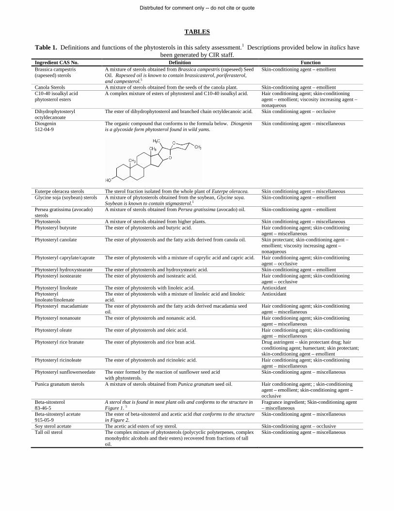
TABLES Table 1. Definitions and functions of the phytosterols in this safety assessment.1 Descriptions provided below in italics have
been generated by CIR staff. Ingredient CAS No. Definition Function Brassica campestris (rapeseed) sterols
A mixture of sterols obtained from Brassica campestris (rapeseed) Seed Oil. Rapeseed oil is known to contain brassicasterol, poriferasterol, and campesterol.5
Skin-conditioning agent – emollient
Canola Sterols A mixture of sterols obtained from the seeds of the canola plant. Skin-conditioning agent – emollient C10-40 isoalkyl acid phytosterol esters
A complex mixture of esters of phytosterol and C10-40 isoalkyl acid. Hair conditioning agent; skin-conditioning agent – emollient; viscosity increasing agent – nonaqueous
Dihydrophytosteryl octyldecanoate
The ester of dihydrophytosterol and branched chain octyldecanoic acid. Skin conditioning agent – occlusive
Diosgenin 512-04-9
The organic compound that conforms to the formula below. Diosgenin is a glycoside form phytosterol found in wild yams.
Skin conditioning agent – miscellaneous
Euterpe oleracea sterols The sterol fraction isolated from the whole plant of Euterpe oleracea. Skin conditioning agent – miscellaneous Glycine soja (soybean) sterols A mixture of phytosterols obtained from the soybean, Glycine soya.
Soybean is known to contain stigmasterol.5 Skin-conditioning agent – emollient
Persea gratissima (avocado) sterols
A mixture of sterols obtained from Persea gratissima (avocado) oil. Skin-conditioning agent – emollient
Phytosterols A mixture of sterols obtained from higher plants. Skin conditioning agent – miscellaneous Phytosteryl butyrate The ester of phytosterols and butyric acid. Hair conditioning agent; skin-conditioning
agent – miscellaneous Phytosteryl canolate The ester of phytosterols and the fatty acids derived from canola oil. Skin protectant; skin-conditioning agent –
emollient; viscosity increasing agent – nonaqueous
Phytosteryl caprylate/caprate The ester of phytosterols with a mixture of caprylic acid and capric acid. Hair conditioning agent; skin-conditioning agent – occlusive
Phytosteryl hydroxystearate The ester of phytosterols and hydroxystearic acid. Skin-conditioning agent – emollient Phytosteryl isostearate The ester of phytosterols and isostearic acid. Hair conditioning agent; skin-conditioning
agent – occlusive Phytosteryl linoleate The ester of phytosterols with linoleic acid. Antioxidant Phytosteryl linoleate/linolenate
The ester of phytosterols with a mixture of linoleic acid and linoleic acid.
Antioxidant
Phytosteryl macadamiate The ester of phytosterols and the fatty acids derived macadamia seed oil.
Hair conditioning agent; skin-conditioning agent – miscellaneous
Phytosteryl nonanoate The ester of phytosterols and nonanoic acid. Hair conditioning agent; skin-conditioning agent – miscellaneous
Phytosteryl oleate The ester of phytosterols and oleic acid. Hair conditioning agent; skin-conditioning agent – miscellaneous
Phytosteryl rice branate The ester of phytosterols and rice bran acid. Drug astringent – skin protectant drug; hair conditioning agent; humectant; skin protectant; skin-conditioning agent – emollient
Phytosteryl ricinoleate The ester of phytosterols and ricinoleic acid. Hair conditioning agent; skin-conditioning agent – miscellaneous
Phytosteryl sunflowerseedate The ester formed by the reaction of sunflower seed acid with phytosterols.
Skin-conditioning agent – miscellaneous
Punica granatum sterols A mixture of sterols obtained from Punica granatum seed oil. Hair conditioning agent; ; skin-conditioning agent – emollient; skin-conditioning agent – occlusive
Beta-sitosterol 83-46-5
A sterol that is found in most plant oils and conforms to the structure in Figure 1. 5
Fragrance ingredient; Skin-conditioning agent – miscellaneous
Beta-sitosteryl acetate 915-05-9
The ester of beta-sitosterol and acetic acid that conforms to the structure in Figure 2.
Skin-conditioning agent – miscellaneous
Soy sterol acetate The acetic acid esters of soy sterol. Skin-conditioning agent – occlusive Tall oil sterol The complex mixture of phytosterols (polycyclic polyterpenes, complex
monohydric alcohols and their esters) recovered from fractions of tall oil.
Skin-conditioning agent – miscellaneous
Distrbuted for comment only -- do not cite or quote

Table 2. CIR safety assessments of constituents of phytosterol ingredients.
Constituent Conclusion
Maximum concentration of
use reported Reference PEG-5, -10, -16, -25, -30, and -40 soy sterol
Insufficient; Safe as used. 2% 2,3
Diosgenin Safe as used. As a constituent of dioscorea villosa (wild yam) root extract (3.5%).
0.525% 4
Plant-derived fatty acid oils Safe as used. 100% 89 C10-40 isoalkyl acid As C10-40 isoalkyl acid octylodecanol esters, C4-5 isoalkyl cocoate, C32-
36 isoalkyl stearate, and ethylhexyl C10-40 isoalkyl acidate. Safe in the present practices of use and concentration described in this safety assessment when formulated to be non-irritating.
78% 90
Octyldecanoic acid Not a cosmetic ingredient. - Butyric acid Not reviewed. - Caprylic acid/capric acid Not reviewed. - Hydroxystearic acid Safe as used. 10% 91 Isostearic acid Safe as used. 26% 92,93 Linoleic acid/linoleic acid Not reviewed. - Nonanoic acid As pelargonic acid. Safe as used. 74% 94 Oleic acid Safe as used. 43% 95,96 Rice bran acid Safe as used. 100% 89,97 Ricinoleic acid Safe as used. 69% 98 Sunflower seed acid Safe as used. 100% 89 Acetic acid Safe as used. 0.4% 99 Tall oil acid Safe as used. 8% 100
Table 3. Chemical characterization of a single sample and multiple samples of plant sterol material (source plant not provided).3,8,9
Phytosterol Distribution of phytosterols (%)
Single sample Five samples from five batches Brassicasterol 1.1 2.7-3.1 Campesterol 25.8 26.5-27.0 Stigmasterol 21.6 17.4-18.1 Β-Sitosterol 48.7 50.8-51.2 Β-Sitostanal 1.8 Not provided Cholesterol 0.4 0.2-0.3 Other sterols 0.8 1.2-1.7
Table 4. Percent distribution of phytosterols from common vegetable oils.3,6 Oil source Brassicasterol Campesterol Stigmasterol Β-Sitosterol Δ7 Stigmastenol Unknown Cocoa butter 8-11 24-31 59-62 Coconut 2 6-9 18-19 69-75 Corn 10-20 Trace-6 74-89 1 Cottonseed Trace-1 8 89-91 Linseed 2 28 10 53 4 Olive 1-3 2 80-97 18 Palm 20-21 12-13 62-67 Peanut 1 10-19 6-12 70-76 Rapeseed 5-19 22-37 52-62 Rice bran 14-33 3-6 55-63 Safflower 8-13 4-9 52-57 23 Soybean 15-21 10-24 57-72 1 Sunflower 11-12 8-12 62-75 20
Distrbuted for comment only -- do not cite or quote

Table 5. Chemical and physical properties of representative sterols. Property Value Reference
Vegetable oil sterols Physical Form Crystalline waxy powder
or prills Waxy, free-flowing
granular powder
10
16
Color White to off white 10 Odor Vegetable oil-like 16 Melting Point oC 138-158 10 Water Solubility g/L @ < 0.01 10 Other Solubility Fat at ambient temperature Acetone Ethyl acetate Isopropanol
2.5%
Soluble Soluble Soluble
10
Tall oil sterols/stanols Physical Form Crystalline waxy powder
or prills 10
Color White to off white 10 Melting Point oC 138-158 10 Water Solubility g/L @ oC & pH < 0.01 10 Other Solubility Fat at ambient temperature Acetone Ethyl acetate Isopropanol
2.5%
Soluble Soluble Soluble
10
Table 6. Comparison of phytosterol content of tall oil extracted by simpler saponification process and a more complicated, multi-step processes.2,5
Phytosterol Saponification process (%) Multi-step process (%) Total phytosterols 98.1 99.7 Major phytosterols 88.7 92.7 β-Sitosterol 49.1 59.8 β-Sitostanol 19.9 23.2 Campesterol 15.0 6.5 Stigmasterol < 1% < 1% Other phytosterols 9.3 (including stigmasterol) 7.0 (including stigmasterol)
Table 7. Allergens not detected in phytosterols derived from acai, soybean, and pomegranate.17-19 Alpha-isomethyl ionone Amyl cinnamal Anise alcohol Benzyl alcohol Benzyl benzoate Benzyl cinnamate Benzyl salicylate Butylphenyl methylpropional Cinnamal Cinnamyl alcohol Citral Citronellol Coumarin Eugenol Fanesol Geraniol Hexyl Cinnamal Hydroxycetronellal Hydroxymethylpentyl 3-cyclohexene carboxaldehyde
Isoeugenol Limonene
Linalool Methyl 2 octynoate Evernia prunastri Evernia furfuracea Amylcinnamyl alcohol
Distrbuted for comment only -- do not cite or quote

Table 8. Frequency of use according to duration and exposure of phytosterols.23,24
Use type Uses
Maximum Concentration
(%) Uses
Maximum Concentration
(%) Uses
Maximum Concentration
(%) Uses
Maximum Concentration
(%)
Brassica campestris (rapeseed) sterols Euterpe oleracea sterols
Glycine soja (soybean) sterols
Persea gratissima (avocado) sterols
Total/range 57 0.0008-7 2 NR 240 0.000001-4.1 NR 0.1-1 Duration of use
Leave-on 50 0.0008-7 2 NR 194 0.000001-1 NR 0.1-1 Rinse-off 7 0.0055-0.13 NR 45 0.000001-4.1 NR NR
Diluted for (bath) use NR NR NR NR 1 NR NR NR
Exposure type Eye area 3 0.005 NR NR 17 0.001-1 NR 1
Incidental ingestion 2 0.0008-7 1 NR 3 0.1-1 NR 0.65
Incidental Inhalation-sprays 2 NR NR NR 6 0.00000-0.001 NR NR
Incidental inhalation-powders NR NR NR NR 1 0.001-0.1 NR NR
Dermal contact 54 0.0055-0.5 1 NR 193 0.001-4.1 NR 0.1-1 Deodorant (underarm) NR NR NR NR NR NR NR NR
Hair-noncoloring NR 0.13 NR NR 43 0.000001-0.018 NR NR Hair-coloring NR NR NR NR NR NR NR
Nail 1 NR NR NR NR 1 NR NR Mucous
Membrane 4 0.0008-7 1 NR 10 0.01-1 NR 0.65
Baby NR NR NR NR NR NR NR NR
Phytosterols Phytosteryl canolate Phytosteryl isostearate Phytosteryl [phytosterol]
macadamiate Total/range 403 0.0001-5 NR 0.06 16 0.003-3 183 0.001-8
Duration of use Leave-on 177 0.0001-5 NR 0.06 15 0.003-3 181 0.001-8 Rinse-off 215 0.00018-0.5 NR NR 1 0.5 2 0.01-1
Diluted for (bath) use 11 NR NR NR NR NR NR NR
Exposure type Eye area 5 0.00018-2 NR 0.06 4 0.003-0.5 2 0.01-3
Incidental ingestion 63 0.01-5 NR NR 8 2.8-3 100 4.1-7
Incidental Inhalation-sprays 2 0.0001 NR NR NR NR NR NR
Incidental inhalation-powders 1 0.05 NR NR NR NR NR 0.001
Dermal contact 338 0.0001-3.2 NR 0.06 8 0.003-1 82 0.001-8 Deodorant (underarm) NR 0.06 NR NR NR NR NR NR
Hair-noncoloring 2 0.5-2.4 NR NR NR 0.1 1 0.01-1 Hair-coloring NR NR NR NR NR NR NR
Nail NR NR NR NR NR NR NR 0.01 Mucous
Membrane 280 0.0002-5 NR NR 8 2.8-3 100 4.1-7
Baby NR NR NR NR NR NR NR NR
Distrbuted for comment only -- do not cite or quote

Table 8. Frequency of use according to duration and exposure of phytosterols.23,24
Use type Uses
Maximum Concentration
(%) Uses
Maximum Concentration
(%) Uses
Maximum Concentration
(%) Uses
Maximum Concentration
(%) Phytosteryl oleate Phytosteryl rice branate Punica granatum sterols Beta-sitosteryl
Total/range 26 1.5-3 2 0.5-1 31 0.001-5 48 0.00007-0.06 Duration of use
Leave-on 20 1.5-3 NR 0.5-1 29 0.1-5 46 0.00007-0.06 Rinse-off 6 NR NR NR 2 NR 2 NR
Diluted for (bath) use NR NR NR NR NR 0.001 NR NR
Exposure type Eye area 1 NR 1 1 3 NR 3
Incidental ingestion NR 1.5 NR NR 14 0.1-5 1 0.00007-0.0008
Incidental Inhalation-sprays NR NR NR NR NR NR 4 NR
Incidental inhalation-powders NR NR NR NR NR NR NR 0.0021
Dermal contact 26 3 2 0.5-1 15 0.001-0.5 47 0.0004-0.06 Deodorant (underarm) NR NR NR NR NR NR NR NR
Hair-noncoloring NR NR NR NR 2 NR NR NR Hair-coloring NR NR NR NR NR NR NR NR
Nail NR NR NR NR NR NR NR NR Mucous
Membrane NR 1.5 NR NR 15 0.001-5 1 0.00007-0.0008
Baby NR NR NR NR NR NR NR NR
Soy sterol acetate Tall oil sterol Total/range 1 NR 7 0.0006-0.0046
Duration of use Leave-on 1 NR 7 0.0045-0.0046 Rinse-off NR NR NR 0.0006
Diluted for (bath) use NR NR NR NR
Exposure type Eye area NR NR NR NR
Incidental ingestion NR NR NR NR
Incidental Inhalation-sprays NR NR NR NR
Incidental inhalation-powders NR NR NR NR
Dermal contact 1 NR 7 0.0006-0.0046 Deodorant (underarm) NR NR NR NR
Hair-noncoloring NR NR NR NR Hair-coloring NR NR NR NR
Nail NR NR NR NR Mucous
Membrane NR NR NR NR
Baby NR NR NR NR NR = Not Reported; NS = Not Surveyed; Totals = Rinse-off + Leave-on Product Uses. Note: Because each ingredient may be used in cosmetics with multiple exposure types, the sum of all exposure type uses may not equal the sum total uses.
Distrbuted for comment only -- do not cite or quote

Table 8. Genotoxicity assays of phytosterols. Assay Test material(s) (concentration) Results Reference In vitro Reverse mutation Salmonella typhimurium (strains TA98, TA100, TA102)
7-ketositosterol (up to 5% in acetone/tween80, 3:1 v/v), 7β-OH-sitosterol (up to 5%), 7α-OH-sitosterol (up to 1%), 6α-OH-3-keto-/6β-OH-3-ketositosterol (ratio 4:3; up to 2.5%) and a mixture (up to 10%)
Negative with and without metabolic activation
67
Reverse mutation S. typhimurium (TA98, TA100, TA1535 and TA1537)
Phytosterol mixturea (5–5000 µg/plate) Negative with and without metabolic activation
70
Reverse mutation; S. typhimurium (TA98, TA100, TA1535 and TA1537; Escherichia coli WP2 uvrA (pKM101)
Phytosterol estersa (50-5000 µg/plate) Negative with and without metabolic activation
70
Reverse mutation S. typhimurium (TA98, TA100, TA102, TA1535 and TA1537)
Phytosterol oxide concentrate from vegetable oil distillates (1.6-5000 µg/plate)
Negative with and without metabolic activation
68
Reverse mutation S. typhimurium (TA98, TA100, TA1535 and TA1537); E. coli WP2 uvrA
MPSS-SEc (104-1667 µg/plate) Negative with and without metabolic activation
53
Reverse mutation S. typhimurium (TA98, TA100, TA1535, and TA1537); E. coli WP2 uvrA
MPSS-VDd (16-1000 µg/plate) Negative with and without metabolic activation
53
Reverse mutation S. typhimurium (TA98, TA100, TA1535 and TA1537)
WDPSEe (62-5000 µg/plate) Negative with and without metabolic activation
53
Reverse mutation S. typhimurium (TA98, TA100, TA1535 and TA1537)
VODPSEf (62-5000 µg/plate) Negative with and without metabolic activation
53
Reverse mutations histidine-dependent S. typhimurium (TA98, TA100, TA1535, TA1537); tryptophan-dependent E. coli (WP2uvrA)
Pomeganate sterols (50 mg/mL; 0.1 mL) Negative with and without metabolic activation
101
Chromosomal aberration; Human peripheral blood lymphocytes
Phytosterol mixturea (40-160 µg/mL) Negative with and without metabolic activation
70
Chromosomal aberration; Human peripheral blood lymphocytes
Phytosterol estersa (25-200 µg/mL) Negative with and without metabolic activation
70
Chromosomal aberration; Human peripheral blood lymphocytes
Phytosterol oxide concentrateg (131.1-500 µg/mL) Negative with and without metabolic activation
70
Chromosomal aberration; Human peripheral blood lymphocytes
MPSS-SE (100-1200 µg/mL) Negative with and without metabolic activation
53
Chromosomal aberration; Human peripheral blood lymphocytes
MPSS-VD (31.3-1000 µg/mL) Negative with and without metabolic activation
53
Chromosomal aberration; Chinese hamster ovary cells
WDPSE (up to 500 µg/ml) Negative with and without metabolic activation
53
Chromosomal aberration; Chinese hamster ovary cells
VODPSE (up to 2000 µg/ml) Negative with and without metabolic activation
53
Gene mutation; Mouse lymphoma L5178Y cells, Tk+/- locus
Phytosterol estersa (5-80 µg/mL) Negative with and without metabolic activation
68
Gene mutation; Mouse lymphoma L5178Y cells, Tk+/- locus
MPSS-SE (5-167 µg/mL) Negative with and without metabolic activation
53
Gene mutation; Mouse lymphoma L5178Y cells, Tk+/- locus
WDPSE (20-500 µg/ml) Negative with and without metabolic activation
53
Gene mutation; Mouse lymphoma L5178Y cells, Tk+/- locus
VODPSE (125-3000 µg/ml) Negative with and without metabolic activation
53
Clastogenicity (micronucleus induction); Human peripheral blood lymphocytes
Phytosterol oxide concentrateg (up to 625 µg/mL) Negative with and without metabolic activation
68
In vivo Micronucleus induction; male rats, bone marrow
Phytosterol estersb (500-2000 mg/kg/d) for 2 days Negative 70
Micronucleus induction; male and female rats, bone marrow
MPSS-SE (50, 500, 2000 mg/kg) Negative 53
Unscheduled DNA synthesis; male rats, liver Phytosterol estersb (800, 2000 mg/kg) Negative 70 Micronucleus induction; male mice, blood Triols (up to 9.4 mg/kg) and epoxides of a mixture
of β-sitostrol and campesterol (67 mg/kg) Negative 102
Sister chromatid exchange; male NIH mice (8 weeks old)
β-sitostrol (200, 400, 600, 1000 mg/kg) Negative 69
Cellular proliferation kinetics; male NIH mice (8 weeks old)
β-sitostrol (200, 400, 600, 1000 mg/kg) Negative 69
Mitotic index; male NIH mice (8 weeks old) β-sitostrol (200, 400, 600, 1000 mg/kg) Negative 69 Micronucleated polychromatic erythrocytes; male NIH mice (8 weeks old)
β-sitostrol (200, 400, 600, 1000 mg/kg) Negative 69
a Phytosterol composition: campesterol (26.7%), stigmasterol (17.7%), β-sitosterol (51%). b Phytosterol composition: campesterol (28.1%), stigmasterol (18.7%), β-sitosterol (45.5%) c MPSS-SE = Mixture of phytosterols and phytostanols derived from solvent extraction, which consisted of ~40– 55% β-sitosterol, ~16–31% β-
sitostanol, ~11–15% campesterol, and ~2–11% campestanol. d MPSS-VD = Mixture derived from vacuum distillation which consisted of ~63.5% β-sitosterol, ~21.7% β-sitostanol,~ 6.5% campesterol and
~2.8% campestanol.
Distrbuted for comment only -- do not cite or quote

Table 8. Genotoxicity assays of phytosterols. Assay Test material(s) (concentration) Results Reference e WDPSE = A wood-derived stanol mixture which consisted of ~94% β-sitostanol and ~6% campestanol. f VODPSE = A vegetable oil–derived mixture of phytostanol esters which consisted of ~68% β-sitostanol and ~32% campestanol. g Phytosterol oxide concentrate = ~ 30% phytosterol oxides.
Distrbuted for comment only -- do not cite or quote

REFERENCES 1. Gottschalck TE and Breslawec HP. International Cosmetic Ingredient Dictionary and Handbook. 14 ed. Washington, DC: Personal Care Products
Council, 2012.
2. Andersen FA. Final report on the safety assessment of PEG-5, -10, -16, -25, -30, and -40 soy sterol. International Journal of Toxicology. 2000;19(Suppl. 1):29-46.
3. Final report of the amended safety assessment of PEG-5, -10, -16, -25, -30, and -40 soy sterol. International Journal of Toxicology. 2004;23(Suppl. 2):23-47.
4. Andersen FA. Final report of the amended safety assessment of dioscorea villosa (wild yam) root extract. International Journal of Toxicology. 2013;23(Suppl. 2):49-54.
5. DiSalvo RM. Phytosterols. Chapter: III, book 2. Schlossman ML. In: The Chemistry and Manufacture of Cosmetics: Ingredients. 3 ed. Carol Stream, IL: Allured Publising Corporation; 2002:911-914.
6. Bailey's industrial oil and fat products. New York: John Wiley & Sons, 1979.
7. Lundmark L, Chun H, and Melby A. Soya sterols: Functional plant-derived ingredients for toiletries. Soap Cosmetics Chemical Specialties. 1976;52:33-34, 38, 40.
8. Unilever. Phytosterols (Ex Roche): Chemical characterization. 1996. Report No. Study AC960273. pp. 1-24.
9. Unilever. Phytosterol Esters (EX Roche): Chemical characterization. 1996. Report No. AC960274. pp. 1-25.
10. Cantrill R. Phytosterols, phytostanols and their esters: Chemical and technical assessment (CTA). Rome, Italy, Food and Agriculture Organization of the United Nations (FAO). 2008. http://www.fao.org/ag/agn/agns/jecfa/cta/69/Phytosterols_CTA_69.pdf. pp. 1-13.
11. European Food Safety Authority (EFSA). Opinion of the NDA Panel realted to a notification from Raisio Life Sciences on plant stanol esters produced from soybean oil sterols pusuant to Article 6 paragraph 11 of Directive 2000/13/EC. European Food Safety Authority. 2005. Report No. EFSA-Q-2004-131. pp. 1-6.
12. European Food Safety Authority (EFSA). Opinion of the Scientific Panel on Dietetic Products, Nutrition and Allergies on a request from the Commission related to a notification from Raisio Life Sciences on plant stanol esters produced from soybean oil sterols pursuant to Article 6, paragraph 11 of Directive 2000/13/EC - for permanent exemption from labelling. European Food Safety Authority. 2013. http://www.efsa.europa.eu/en/scdocs/scdoc/571.htm. Report No. EFSA-Q-2007-082. pp. 1-6.
13. Active Concepts. 2009. ABS Acai Sterols manufacturing flow chart.
14. Active Concepts. 2013. ABS Soybean Sterols manufacturing flow chart.
15. Active Concepts. 2010. ABS Pomegranate Sterols manufacturing flow chart.
16. European Food Safety Authority (EFSA). Opinion of the Scientific Panel on Dietitic Products, Nutrition and Allergies on a request from the Commission related to a notification from Cognis, ADM and Cargill on vegetable oils-derived phytosterols and phytosterol esters fromn soybean sources pursuant to Article 6 paragraph 11 of Dircetive 2000/13/EC. European Food Safety Authority. 2007. http://www.efsa.europa.eu/fr/efsajournal/pub/486.htm. Report No. EFSA-Q-2006-162. pp. 1-8.
17. Active Concepts. 2006. Compositional breakdown ABS Pomegranate Sterols (Punica Granatum Sterols).
18. Active Concepts. 2013. Compositional breakdown AC Soybean Sterols (Glycine Soja (Soybean) Sterols).
19. Active Concepts. 2013. Compositional breakdown ABS Acai Sterols (Euterpe Oleracea Sterols).
20. General Mills, Inc. Plant sterols, phytosterols, or sitosterols which are comprised of a mixture of the common sterol components. 1979.
21. Nordlee JA, Nienmann LM, Hefle SL, and Taylor SL. Determination of proteins in soybean oil from distinct processing steps. 2002 IFT Annual Meeting and Food Expo. 2002. ift.confex.com/ift/2002/techprogram/paper_13465.htm.
22. Zitouni N, Errahali Y, Metche M, Moutete M, Kanny G, Moneret-Vautrin DA, Nicolas JP, and Fremont S. Soy allergens are detected in some edible soy oils. Journal of Allergy and Clinical Immunology. 2001. 107:(February): pp.188
23. Food and Drug Administration (FDA). Frequency of use of cosmetic ingredients. FDA Database. 2013. Washington, DC: FDA.
24. Personal Care Products Council. 3-21-2013. Concentration of use by FDA category: Planet sterols. 6 pages.
Distrbuted for comment only -- do not cite or quote
25. Bremmer HJ, Prud'homme de Lodder LCH, and van Engelen JGM. Cosmetics Fact Sheet: To assess the risks for the consumer; Updated version for ConsExpo 4. 2006. http://www.rivm.nl/bibliotheek/rapporten/320104001.pdf. Date Accessed 8-24-2011. Report No. RIVM 320104001/2006. pp. 1-77.
26. Bremmer HJ, Prud'homme de Lodder LCH, and van Engelen JGM. General Fact Sheet: Limiting conditions and reliability, ventilation, room size, body surface area; Updated version for ConsExpo 4. 2006. http://www.rivm.nl/bibliotheek/rapporten/320104002.pdf. Date Accessed 8-24-2011. Report No. RIVM 320104002/2006. pp. 1-31.
27. Johnsen MA. The Influence of Particle Size. Spray Technology and Marketing. 2004;24-27.
28. Rothe H, Fautz R, Gerber E, Neumann L, Rettinger K, Schuh W, and Gronewold C. Special aspects of cosmetic spray safety evaluations: Principles on inhalation risk assessment. Toxicol Lett. 8-28-2011;205(2):97-104.
29. Rothe H. Special aspects of cosmetic spray safety evaluation. 2011.
30. Rothe H. Special aspects of powders in decorative cosmetics. 2011.
31. Aguilar, F., Crebelli, R., Dusemund, B., Galtier, P., Gilbert, J., Gott, D. M., Gundert-Remy, U., Konig, J., Lambre, C., Leblanc, J. C., Mortensen, A., Mosesso, P., Parent-Massin, D., Rietjens, I. M. C. M., Stankovic, I., Tobback, P., Waalkens-Berendsen, I., Woutersen, R. A., Wright, M. C., Bemrah, N., Guertler, R., Larsen, J. C., and Tlustos, Ch. Scientific opinion on the safety of stigmasterol-rich plant sterols as food additive. EFSA Journal. 2012;10(5):2659, 38.
32. European Food Safety Authority (EFSA). Opinion of the Scientific Panel on Dietetic products, nutrition and allergies [NDA] related to a Novel Food applicaiton from Forbes Medi-Tech for approval of plant sterol-containing milk-based beverages. European Food Safety Authority. 2003. http://www.efsa.europa.eu/en/scdocs/scdoc/15.htm. Report No. EFSA-Q-2003-075. pp. 1-12.
33. European Food Safety Authority (EFSA). Opinion of the Scientific Panel on Dietic products nutrition and allergies [NDA] related to two scientific publications concerning aspects of serum levles of phtosterols. European Food Safety Authority. 2005. http://www.efsa.europa.eu/en/scdocs/scdoc/211.htm. Report No. EFSA-Q-2004-178. pp. 1-6.
34. European Food Safety Authority (EFSA). Statement on a request from the Commission related to a novel food applicationon rice drinks and added phytosterols by the Scientific Panel on Dietetic Products, Nutrition and Allergies (NDA). European Food Safety Authority. 2006. http://www.efsa.europa.eu/it/efsajournal/pub/201.htm. Report No. EFSA-Q-2005-242. pp. 1-4.
35. European Food Safety Authority (EFSA). Statement of the Scientific Panel on Dietetic Products, Nutrition and Allergies on a request from the Commission related to a novel food application on fruit juices and nectars with added phytosterols. European Food Safety Authority. 2006. http://www.efsa.europa.eu/fr/efsajournal/pub/391.htm. Report No. EFSA-Q-2006-057. pp. 1-4.
36. European Food Safety Authority (EFSA). Plant sterols and blood cholesterol: Scinetific substantiationof a health claim related to plant sterols and lower/reduced blood cholesterol and reduced risk of (coronary) heart disease pursuatn to Article 14 of Regulation (EC) No 1924/2006. European Food Safety Authority. 2008. http://www.efsa.europa.eu/en/efsajournal/doc/781.pdf. Report No. EFSA-Q-2008-085. pp. 1-12.
37. European Food Safety Authority (EFSA). Consumption of food and beverages with added plant sterols in the European Union. European Food Safety Authority. 2008. http://www.efsa.europa.eu/en/efsajournal/doc/133r.pdf. Report No. EFSA/DATEX 03. pp. 1-21.
38. European Food Safety Authority (EFSA). Danacol® and blood cholesterol: Scientific substantiation of a health claim related to a low fat fermented milk product (Danacol®) enriched with plant sterols/stanols and lowering/reducing blood cholesterol and reduced risk of (coronary) heart disease pursuant to Article 14 of Regulation (EC) No 1924/2006. European Food Safety Authority. 2009. http://www.efsa.europa.eu/en/efsajournal/doc/1177.pdf. Report No. EFSA-Q-2008-779. pp. 1-12.
39. European Food Safety Authority (EFSA). Scientific opinion on the substantiation of health claims related to plant sterols and plant stanols and maintenance of normal blood cholesterol concentration (ID 549, 550, 567, 713, 1234, 1235, 1466, 1634, 1984, 2909, 3140), and maintenance of normal prostrate size and normal urination (ID 14, 1467, 1635) pursuant to Article 13(1) of Regulation (EC) No 1924/2006. European Food Safety Authority. 2010. http://www.efsa.europa.eu/en/efsajournal/doc/1813.pdf. pp. 1-22.
40. Scientific Committee on Food (SCF). Opinion of the Scientific Committee on Food on a request for the safety assessment of the use of phytostearol esters in yellow fat spreads. European Commission Health & Consumer Protection Directorate-General. 2000. http://ec.europa.eu/food/fs/sc/scf/out56_en.pdf. Report No. SCF/CS/NF/DOS/1 Final. pp. 1-16.
41. Scientific Committee on Food (SCF). General view of the Scientific Committee on Food on the long-term effects of the intake of elevated levels of phytosterols from multiple dietary sources, with particular attentionto the effects on beta-carotene (expressed on 26 September 2002). Brussels, Belgium, European Commission, Health & Consumer Protection Committees II, Scientific Co-operation and Networks. 2002. http://ec.europa.eu/comm/food/fs/sc/scf/out143_en.pdf. Report No. SCF/CS/NF/DOS/20 ADD 1 Final. pp. 1-23.
42. Scientific Committee on Food (SCF). Opinion of the Scientific Committee on Food on a report on post launch monitoring of "yellow fat spreads with added phytosterol esters" (Expressed on 26 September 2002. Brussels, Belgium, European Commission, Health & Consumer Protection Directorate-General, Directorate C - Scientific Opinions, C2- Management of Scientific Committees II, Scientific Co-
Distrbuted for comment only -- do not cite or quote
operation and Networks. 2002. http://ec.europa.eu/comm/food/fs/sc/scf/out144_en.pdf. Report No. SCF/CS/FN/DOS/21 ADD 2 Final. pp. 1-8.
43. Scientific Committee on Food (SCF). Opinion of the Scientific Committee on Food on applications for approval of a variety of plant sterol-enriched foods (expressed on 5 March 2003). Brussels, Belgium, European Commission, Health & Consumer Protection Directorate-General, Directorate C - Scientific Opinions, C2 - Management of Scientific Committees, Scientific Co-operaion and Networks. 2003. http://ec.europa.eu/comm/food/fs/sc/scf/out174_en.pdf. Report No. SCF/CS/NF/DOS/15 ADD 2 Final. pp. 1-10.
44. Ling, W. H. and Jones, P. J. Dietary phytosterols: a review of metabolism, benefits and side effects. Life Sciences. 1995;57(3):195-206.
45. Sanders DJ, Minter HJ, Howes D, and Hepburn PA. The safety evaluation of phytoesterol esters. Part 6. The comparative absorption and tissue distrubution of phtosterols in the rat. Food and Chemical Toxicology. 2000;38(6):485-491.
46. Hamada T, Goto H, Yamahira T, Sugawara T, Imaizumi K, and Ikeda I. Solubility in and affinity for the bile salt micelle of plant sterols are important determinants of their intestinal absorption in rats. Lipids. 2006;41(6):551-556.
47. Moghadasian MH. Pharmacological properties of plant sterols in vivo and in vitro observations. Life Sciences. 2000;67(6):1340-1346.
48. Heinemann T, Axtmann G, and von Bergmann K. Comparison of intestinal absorption of cholesterol with different plant sterols in man. European Journal of Clinical Investigation. 1993;23(12):827-831.
49. Sudhop T, Sahin Y, Lindenthal B, Hahn C, Luers C, Berthold HK, and von Bergmann K. Comparison of the hepatic clearances of campesterol, sitosterol, and cholesterol in healthy subjects suggest tht efflux transporters controlling intestinal sterol absorption also regulate biliary secretion. Gut. 2002;51(6):860-863.
50. Fricke CB, Schrøder M, Poulsen M, von Bergmann K, Wester I, Ib Knudsen I, Mortensen A, and Dieter Lüjohann D. Increased plant sterol and stanol levels in brain of Watanabe rabbits fed rapeseed oil derived plant sterol or stanol esters. British Journal of Nutrition. 2007;98(5):890-899.
51. Adcox, C, Boyd L, Oehrl L, Allen J, and Fenner G. Comparative efects of phytosterol oxides and cholesterol oxides in cultured macrophage-derived cell lines. Journal of Agriculure and Food Chemistry. 2001;49(4):2090-2095.
52. Active Concepts. 2006. Summary of neutral red cytotoxicity assay for ABS Pomegranate Sterols.
53. Expert Committee on Food Additives (FECFA). Safety evaluation of certain food additives. Geneva, 2009. http://whqlibdoc.who.int/publications/2009/9789241660600_eng.pdf. pp. 1-642.
54. Laraki L, Pelletier X, Mourot J, and Debry G. Effects of dietary phytostrols on liver lipids and lipid metabolism enzymes. Annals of Nutrition and Metabolism. 1993;37:129-133.
55. Malini T and Vanithakumari G. Effect of beta-sitosterol on uterine biochemistry: A comparative study with estradiol and progesterone. Biochmistry & Molecular Biology International. 1993;31(4):659-668.
56. Hepburn PA, Horner SA, and Smith M. Safety evaluation of phytosterol esters. Part 2. Subchronic 90-day oral toxicity study on phytosterol esters--A novel functional food. Food and Chemical Toxicology. 1999;37(5):521-532.
57. Waalkens-Berendsen, D. H., Wolterbeek, A. P. M., Wijnands, M. V. W., Richold, M., and Hepburn, P. A. Safety Evaluation of Phytosterol Esters. Part 3. Two-Generation Reproduction Study in Rats with Phytosterol Esters-a Novel Functional Food. Food and Chemical Toxicology. 1999;37(7):683-696.
58. Waalkens-Berendsen, D. H., Wolterbeek, A. P. M., Wijnands, M. V. W., Richold, M., and Hepburn, P. A. Safety evaluation of phytosterol esters. Part 3. Two-generation reproduction study in rats with phytosterol esters-a novel functional food. [Erratum to document cited in CA131:321734]. Food and Chemical Toxicology. 1999;37(7):683-696.
59. Nieminen P, Pölönen I, Ikonen K, Määttänen M, and Mustonen AM. Evaluation of reproductive safety of β-sitosterol on the American mink (Neovison vison). Chemosphere. 2008;71(3):493-499.
60. Malini T and Vanithakumari G. Antifertility effects of β-sitosterol in male albino rats. Journal of Ethnopharmacology. 1991;35(2):149-153.
61. Baker VA, Hepburn PA, Kennedy SJ, Jones PA, Lea, L. J., Sumpter JP, and Ashby J. Safety evaluation of phytoesters. Part 1. Assessment of oestrogenicity using a combination of in vivo and in vitro assays. Food and Chemical Toxicology. 1999;37(1):13-22.
62. van den Heuvel MR, Leusch FD, Taylor S, Shannon N, and McKague AB. Assessment of the reportductive-endocrine disrupting potential of chlorine dioxide oxidation products of plant sterols. Environmental Science and Technology. 2006;40(8):2594-2600.
63. Malini T and Vanithakumari G. Comparative study of the effects of ß-sitosterol, estradiol and progesterone on selected biochemical parameters of the uterus of ovariectomised rats. Journal of Ethnopharmacology. 1992;36(1):51-55.
Distrbuted for comment only -- do not cite or quote

64. Zava DT, Dollbaum CM, and Blen M. Estrogen and pregestin bioactivity of foods, herbs, and spices. Proceedings of the Society for Experimental Biology and Medicine. 1998;217(3):369-378.
65. Burck PJ, Thakkar AL, and Zimmerman RE. Antifertitily action of a strol sulphate in the rabbit. Journal of Reproduction and Fertility. 1982;66(1):109-112.
66. Malini T and Vanithakumari G. Rat toxicity studies with ß-sitosterol. Journal of Ethnopharmacology. 1990;28(2):221-234.
67. Koschutnig, Karin, Kemmo, Suvi, Lampi, Anna Maija, Piironen, Vieno, Fritz-Ton, Cornelia, and Wagner, Karl Heinz. Separation and isolation of +¦-sitosterol oxides and their non-mutagenic potential in the Salmonella microsome assay. Food Chemistry. 2009;118(1):133-140.
68. Lea, L. J., Hepburn, P. A., Wolfreys, A. M., and Baldrick, P. Safety evaluation of phytosterol esters. Part 8. Lack of genotoxicity and subchronic toxicity with phytosterol oxides. Food and Chemical Toxicology. 2004;42(5):771-783.
69. Paniagua-Pérez R, Madrigal-Bujaidar E, Reyes-Cadena S, Molina-Jasso D, Pérez Gallaga J, Silva-Miranda A, Velazco O, Hernández N, and Chamorro G. Genotoxic and cytotoxic studies of beta-sitosterol and pteropodine in mouse. Journal of Biomedicine and Biotechnology. 2005;2005(3):242-247.
70. Wolfreys AM and Hepburn PA. Safety evaluation of phytosterol esters. Part 7. Assessment of mutagenic activity of phytosterols, phytosterol esters and the cholesterol derivative, 4-cholesten-3-one. Food and Chemical Toxicology. 2002;40(4):461-470.
71. Huntingdon Life Sciences Ltd. Plant sterols: Bacterial mutation assay. 1998. Report No. KA980008. pp. 1-26.
72. Huntingdon Life Sciences Ltd. Phytosterol esters: Metaphase chromosome analysis of human lymphocytes cultured in vitro. 1997. Report No. KC960257. pp. 1-34.
73. Huntingdon Life Sciences Ltd. Phytosterols: Metaphase chromosome analysis of human lymphocyes cultured in vitro. 1997. Report No. KC960255. pp. 1-32.
74. Huntingdon Life Sciences Ltd. Phytosterols: Bacterial mutation assay. 1996. Report No. KA960254. pp. 1-23.
75. Huntingdon Life Sciences Ltd. Phytosterol esters: BActerial mutation assay. 1996. Report No. KA960256. pp. 1-23.
76. Covance Laboratories LTD. Plant sterol ester SSe26698-02: Induction of micronuclei in the bone marrow of treated rats. 1999. Report No. KC990110. pp. 1-32.
77. Covance Laboratories LTD. Plant sterol ester SSE26698-02: Measurement of unscheduled DNA synthesis in rat liver using an iv vivo/in vitro procedure. 1999. Report No. KU990111. pp. 1-37.
78. Yasukawa K, Takido M, Matsumoto T, Takeuchi M, and Nakagawa S. Sterol and triterpene derivatives from plants inhibit the effects of a tumor promoter, and sitosterol and betulinic acid inhibit tumor formation in mouse skin two-stage carcinogenesis. Oncology. 1991;48(1):72-76.
79. Raicht RF, Cohen BI, Fazzini EP, Sarwal AN, and Takahashi M. Protective effect of plant sterols against chemically induced color tumors in rats. Cancer Research. 1980;40(2):403-405.
80. Deschner EE, Cohen BI, and Raicht RF. The kinetics of the protective effect of ß-sitosterol against MNU-induced colonic neoplasia. Journal of Cancer Research and Clinical Oncology. 1982;103(1):49-54.
81. Janezic SA and Rao AV. Dose-dependent effects of dietary phytosterol on epithelial cell proliferation of the murine colon. Food and Chemical Toxicology. 1992;30(7):611-616.
82. Product Investigations Inc. 2006. Evaluation of the skin-irritating propensities of ABS Pomegranate Sterols on scarified skin.
83. Active Concepts. 2012. Summary of dermal and ocular irritation tests of ABS Pomegranate Sterols.
84. Active Concepts. 2013. Summary of dermal and ocular irritation tests of ABS Acai Sterols.
85. Active Concepts. 2013. Summary of dermal and ocular irritation tests of AC Soybean Sterols.
86. AMA Laboratories Inc. 2006. 50 Human subject repeat insult patch test skin irritation/sensitization evaluation (occlusive patch) ABS Pomegranate Sterols.
87. Pachke A, Zunker K, Wigotzki M, and Steinhart H. Determination of the IgE-binding activity of soy lecithin and refined and non-refined soybean oils. Journal of Chromatography B, Biomedical Sciences and Applications. 2001;756(1-2):249-254.
Distrbuted for comment only -- do not cite or quote

88. Bhattachryya AK, Connor WE, and Sin DS. The origin of plant sterols in the skin surface lipids in humans: From diet to plasma to skin. The Journal of Investigative Dermatology. 1983;80(4):294-296.
89. Burnett C, Fiume MM, Bergfeld WF, Belsito DV, Hill RA, Klaassen CD, Liebler DC, Marks Jr.JG, Shank RC, Slaga TJ, and Snyder PW. Plant-derived fatty acid oils as used in cosmetics. Washington, DC, Cosmetic Ingredient Review. 2011. pp. 1-100.
90. Fiume MM, Bergfeld WF, Belsito DV, Hill RA, Klaassen CD, Liebler DC, Marks Jr.JG, Shank RC, Slaga TJ, and Snyder PW. Amended safety assessment of alkyl esters as used in cosmetics. Washington, DC, Cosmentic Ingredient Review. 2013. pp. 1-83.
91. Johnson W, Bergfeld WF, Belsito DV, Hill RA, Klaassen CD, Liebler DC, Marks Jr.JG, Shank RC, and Snyder PW. Amended final report on the safety assessment of hydroxystearic acid. International Journal of Toxicology. 1999;18(Suppl. 1):1-10.
92. Annual review of cosmetic ingredients safety assessments - 2002/2003. International Journal of Toxicology. 2005;24(Suppl. 1):1-102.
93. Elder RL. Final report on the safety assessment of isostearic acid. Journal of the American College of Toxicology. 1983;2(7):61-74.
94. Johnson, Jr W, Heldreth B, Bergfeld WF, Belsito DV, Klaassen CD, Hill RA, Liebler DC, Ma, Shank RC, Slaga TJ, Snyder PW, and Andersen FA. Final report of the Cosmetic Ingredient Review Expert Panel on the safety assessment of pelargonic acid (nonanioic acid) and nonanoate esters. International Journal of Toxicology. 2011;30(Suppl. 3):228S-269S.
95. Annual review of cosmetic ingredient safety assessments - 2004/2005. International Journal of Toxicology. 2006;25(Suppl. 2):1-89.
96. Elder RL. Final report on the safety assessment of oleic acid, lauric acid, palmitic acid, myristic acid, and stearic acid. Journal of the American College of Toxicology. 1987;6(3):321-401.
97. Andersen FA. Amended final report on the safety assessment of oryza sativa (rice) bran oil, oryza sativa (rice) germ oil, rice bran acid, oryza sativa (rice) bran wax, hydrogenated rice bran wax, oryza sativa (rice) bran extact, oryza sativa (rice) extract, oryza sativa (rice) germ powder, oryza sativa (rice) starch, oryza sativa (rice) bran, hydrolyzed rice bran extract , hydrolyzed rice bran protein, hydrolyzed rice extract, and hydrolyzed rice protein. International Journal of Toxicology. 2006;25(Suppl. 2):91-120.
98. Andersen FA. Final report on the safety assessment of ricinus communis (castor) seed oil, hydrogentated castor oil, glyceryl ricinoleate, glyceryl ricinoleate SE, ricinoleic acid, potassium ricinoleate, soldium ricinoleate, zinc ricinoleate, cetyl ricinoleate, ethyl ricinoleate, glycol ricinoleate, isopropyl ricinoleate, methyl ricinoleate, and octyldodecyl ricinoleate. International Journal of Toxicology. 2013;26(Suppl. 3):31-77.
99. Heldreth B, Bergfeld WF, Belsito DV, Hill RA, Klaassen CD, Liebler DC, Marks Jr.JG, Shank RC, Slaga TJ, Snyder PW, and Andersen FA. Final report of the Cosmetic Ingredient Review Expert Panel on the safety assessment of methyl acetate. International Journal of Toxicology. 2012;31(Suppl. 1):112-136.
100. Robinson V, Bergfeld WF, Belsito DV, Klaassen CD, Marks Jr.JG, Shank RC, Slaga TJ, Snyder PW, and Andersen FA. Amended safety assessment of tall oil acid, sodium tallate, potassium tallate, and ammonium tallate. International Journal of Toxicology. 2009;28(Suppl 3):2525-2585.
101. BioScreen Testing Services Inc. 2007. Bacterial reverse mutation tests ABS Pomegranate Sterols.
102. Abramsson-Zetterberg L, Svensson M, and Johnsson L. No evidence of genotoxic effect in vivo of the phytosterol oxidation products triols and epoxides. Toxicology Letters. 2007;173(2):132-139.
Distrbuted for comment only -- do not cite or quote

Commitment & Credibility since 1976
August 16, 2013
MEMORANDUM
To: CIR Expert Panel and Liaisons
From: Lillian C. Becker, MS Scientific Analyst and Writer
Subject: Draft Report of the Safety Assessment of Phytosterols as Used in Cosmetics
Unpublished data were submitted by the Personal Care Products Council. These data include:
1) Concentration of use.
2) Safety studies of phytosterol ingredients. These include:
a. Bacterial reverse mutation assay of pomegranate sterols b. HRIPT of pomegranate sterols c. Human dermal irritation test on scarified skin d. Summary of EpiDermal and EpiOcular assays of pomegranate sterols e. Summary of a neutral red cytotoxicity assay of pomegranate sterols f. Summary of EpiDermal and EpiOcular assays of soybean sterols g. Summary of EpiDermal and EpiOcular assays of acai sterols
3) Impurity and manufacture information on acai, soybean, and pomegranate sterols.
To inform the Panel on data that has already been considered, the 2004 safety assessment of PEG soy sterols is also included.
Distrbuted for comment only -- do not cite or quote

Distrbuted for comment only -- do not cite or quote
Distrbuted for comment only -- do not cite or quote

Distrbuted for comment only -- do not cite or quote

Distrbuted for comment only -- do not cite or quote

Distrbuted for comment only -- do not cite or quote

Distrbuted for comment only -- do not cite or quote

Distrbuted for comment only -- do not cite or quote

Distrbuted for comment only -- do not cite or quote

Distrbuted for comment only -- do not cite or quote
Distrbuted for comment only -- do not cite or quote

Distrbuted for comment only -- do not cite or quote

Distrbuted for comment only -- do not cite or quote

Distrbuted for comment only -- do not cite or quote

Distrbuted for comment only -- do not cite or quote

Distrbuted for comment only -- do not cite or quote

Distrbuted for comment only -- do not cite or quote

Distrbuted for comment only -- do not cite or quote

Distrbuted for comment only -- do not cite or quote

Distrbuted for comment only -- do not cite or quote

Distrbuted for comment only -- do not cite or quote

Distrbuted for comment only -- do not cite or quote

Distrbuted for comment only -- do not cite or quote

Distrbuted for comment only -- do not cite or quote
Distrbuted for comment only -- do not cite or quote
Distrbuted for comment only -- do not cite or quote
Distrbuted for comment only -- do not cite or quote

Distrbuted for comment only -- do not cite or quote

Distrbuted for comment only -- do not cite or quote

Distrbuted for comment only -- do not cite or quote

Distrbuted for comment only -- do not cite or quote

Distrbuted for comment only -- do not cite or quote

Distrbuted for comment only -- do not cite or quote

Distrbuted for comment only -- do not cite or quote

Distrbuted for comment only -- do not cite or quote
Distrbuted for comment only -- do not cite or quote

Distrbuted for comment only -- do not cite or quote

Distrbuted for comment only -- do not cite or quote

Distrbuted for comment only -- do not cite or quote

Distrbuted for comment only -- do not cite or quote

Distrbuted for comment only -- do not cite or quote

Distrbuted for comment only -- do not cite or quote

Distrbuted for comment only -- do not cite or quote

Distrbuted for comment only -- do not cite or quote

Distrbuted for comment only -- do not cite or quote

Distrbuted for comment only -- do not cite or quote

Distrbuted for comment only -- do not cite or quote

Distrbuted for comment only -- do not cite or quote

Distrbuted for comment only -- do not cite or quote

Distrbuted for comment only -- do not cite or quote

Distrbuted for comment only -- do not cite or quote

Distrbuted for comment only -- do not cite or quote

Distrbuted for comment only -- do not cite or quote

Distrbuted for comment only -- do not cite or quote

Distrbuted for comment only -- do not cite or quote

Distrbuted for comment only -- do not cite or quote

Distrbuted for comment only -- do not cite or quote

Distrbuted for comment only -- do not cite or quote

Distrbuted for comment only -- do not cite or quote

Distrbuted for comment only -- do not cite or quote

Distrbuted for comment only -- do not cite or quote

Distrbuted for comment only -- do not cite or quote

Distrbuted for comment only -- do not cite or quote

Distrbuted for comment only -- do not cite or quote

Distrbuted for comment only -- do not cite or quote
Distrbuted for comment only -- do not cite or quote

Distrbuted for comment only -- do not cite or quote

Final Report of the Amended Safety Assessment of PEG-S, -10, -16, '-25, -30, and -40 Soy SteroP
PEGs Soy Sterol are polyethylene glycol (PEG) derivatives of soybean oil sterols used in a variety of cosmetic formulations as surfactants and emulsifying agents, skin-conditioning agents, and cleansing and solubilizing agents. When the safety of these ingredients were first reviewed, the available data were insufficient to support safety. New data have since been received and the safety of these ingredients in cosmetics has been substantiated. Current concentration of use ranges from a low of 0.05 % in makeup preparations to 2% in moisturizers and several. other products. PEGs Soy Sterol are produced by the reaction of the soy sterol hydroxyl with ethylene oxide. In general, ethoxylated fatty acids can contain 1,4-dioxane as a byproduct of ethoxylation. The soy sterols include campesterol, stigmasterol, and ,a-sitosterol. The distribution of sterols found in oils derived from common plants is similar, with ,a-sitosterol comprising a major component. Impurities include sterol hydrocarbons and cholesterol (4 % to 6 % ) and triterpine alcohols, keto-steroids, and other steroid-like substances (4% to 6%). No pesticide residues were detected. PEGS: Because PEGs are an underlying structure in PEGs Soy Sterols, the previous assessment of PEGs was considered. It is generally recognized that the PEG monomer, ethylene glycol, and certain of its monoalkyl ethers are reproductive and developmental toxins. Given the methods of manufacture of PEGs Soy Sterol, there is no likelihood of ethylene glycol or its alkyl ethers being present. Also, the soybean oil sterol ethers in this ingredient are chemically different from the ethylene glycol alkyl ethers of concern. PEGs are not carcinogenic, although sensitization and nephrotoxicity were observed in burn patients treated with a PEG-based cream. No evidence of systemic toxicity or sensitization was found in studies with intact skin. Plant Phytosterols: Intestinal absorption of ingested plant phytosterols is on the order of 5 %, with 95 % ofthe material entering the colon. Absorbed plant phytosterols are transported to the blood. Although there are some data suggesting that sulfates of ,a-sitosterol can act as abortifacients in rats and rabbits, other studies of well-characterized plant phytosterols and phytosterol esters demonstrated no effect in an estrogen-binding study, a recombinant yeast assay for estrogen or estrogen-like activity, or a juvenile rat uterotrophic assay for estrogen or estrogen-like activity. In a two-generation reproduction study using rats, plant phytosterol esters in the diet had no effect on any parameter of reproduction or fertility. Subcutaneous injections of ,a-sitosterol did reduce sperm concentrations and fertility in rats. Sitosterol inhibited tumor promoting activity of l2-0-tetradecanoylphorbol-13-acetate (TPA) in mice after initiation with 7,12-dimethylbenz[a]anthracene (DMBA), and reduced
Received 12 February 2004; accepted 4 June 2004. 1 Reviewed by the Cosmetic Ingredient Review Expert Panel. This report was
prepared by Eric Hooker, Scientific Analyst and Writer. Address correspondence to Eric Hooker, 110 I 17th Street, NW, Suite 310, Washington, DC 20036, USA.
International Journal of Toxicology, 23(Suppl. 2):23--47, 2004 Copyright © Cosmetic Ingredient Review ISSN: 1091-5818 print / 1092-874X online DOl: 10.1080/10915810490499046
the tumors produced by N -methylnitrosourea in rats. Phytosterols were not genotoxic in several bacterial, mammalian, and in vitro assay systems. Phytosterols decreased epithelial cell proliferation in the colon of mice and rats, and were cytotoxic for human epidermoid carcinoma of the nasopharynx. PEGs Soy Sterols: The acute oral LDso in rats of PEG-5-25 Soy Sterol was > 10 glkg. The acute dermal LDso of a liquid eyeliner containing 2% PEG-5 Soy Sterol was > 2 g/kg in rabbits. PEG-5-25 Soy Sterol was not a primary irritant in rabbits when applied undiluted. Undiluted PEG-5 Soy Sterol did not cause sensitization in guinea pigs. PEGs Soy Sterol did not produce ocular toxicity in rabbits. PEG-5 Soy Sterol was negative in the Ames mutagenicity test, with or without metabolic activation. PEG-5 Soy Sterol, at concentrations up to 2% in formulation, did not cause dermal or ocular irritation dermal sensitization, or photosensitization in clinical studies. Be~ cause of the possible presence of l,4-dioxane reaction product and unreacted ethylene oxide residues, it was considered necessary to use appropriate procedures to remove these from PEGs Soy Sterol before blending them into cosmetic formulations. Based on the systemic toxicity and sensitization seen with PEGs applied to damaged skin, it was recommended that PEGs Soy Sterol should not be used in cosmetic products applied to damaged skin. Although no dermal absorption data were available, oral studies demonstrate that phytosterols and phytosterol esters are not significantly absorbed and do not result in significant systemic exposure. Some small amounts did appear in the ovaries, however. This raises a concern about the potential presence of free phytosterols and ,aSitosterol, which could have antiestrogenic, anti progestational, gonadotrophic, antigonadotrophic, and antiandrogenic effects in PEG sterols. These concerns are alleviated by the extensive data showing that well-defined phytosterols and phytosterol esters are not estrogenic and do not pose a hazard to reproduction. Likewise, the absence of impurities in plant phytosterols and phytosterol esters and extensive data demonstrating the absence of any genotoxicity in bacterial and mammalian systems mitigate against the possibility of any carcinogenic effect with those same well-characterized materials. The Cosmetic Ingredient Review (CIR) Expert Panel concluded that the PEGs Soy Sterol are safe as used in cosmetic products.
INTRODUCTION PEG-5, -10, -16-, -25, -30, and -40 Soy Sterol are polyethy
lene glycol (PEG) derivatives of soybean oil sterols that function as non-ionic surfactants and emulsifying agents in cosmetic formulations. The safety of these ingredients was first reviewed by the Cosmetic Ingredient Review (CIR) Expert Panel in a series of meetings leading to a conclusion in 1997 that the
23
Distrbuted for comment only -- do not cite or quote

24 COSMETIC INGREDIENT REVIEW
available data were not sufficient to support the safety of this ingredient in cosmetic products (CIR 1997). One concern was the presence, of free phytosterols in the soy sterol component of the PEGs Soy Sterol group of ingredients and the potential reproductive toxicity of ,8-sitosterol, in particular. The Expert Panel also sought impurities information and genotoxicity data.
New information has been provided that characterizes phytosterols and phytosterol fatty acid esters in general, including analyses of impurities. In addition, results of several genotoxicity assays using the characterized materials were provided, along with absorption, distribution, metabolism, and excretion data. Data from a two-generation reproductive toxicity study were also provided.
The CIR Expert Panel considered that because PEGs Soy Sterol may be broken down to PEGs and Soy Sterols, the safety test data on the PEGs and Soy Sterols are directly relevant to the safety assessment of PEGs Soy Sterol.
Polyethylene Glycol of various chain lengths (PEGs) has been reviewed previously by the CIR Expert Panel and the Final Report has been published. The following conclusion was reached:
PEG -6, -8, -32, -75,150, -14M, and -20M are safe for use at the concentrations reflected in the Cosmetic Use section and in the product formulation safety test data included in the Final Report. The Expert Panel recommends that cosmetic formulations containing these PEGs not be used on damaged skin (Andersen, 1993).
Data on plant phytosterols, to the extent that they are representative of soy phytosterols, are also relevant to this safety assessment and have been included in this amended safety assessment.
It was considered unlikely that there would be any toxicity of PEGs Soy Sterol esters that was not present in the two constituents.
CHEMISTRY
Definition and Structure PEG-n Soy Sterol is a polyethylene glycol derivative of
sterols found in soybean oil where n is the average number of moles of ethylene oxide used in synthesis (Wenninger et al. 2000). These phytosterols (generic term) are structurally similar to cholesterol and mainly consist of sitosterol (C29 HsoO: molecular weight [mw] 414.69), campesterol (C2sH4S O: mw 400.66), and stigmasterol (C29H4S0: mw 412.67) (Applewhite 1985; Budavari 1989; Tyle and Frank 1991).
Campesterol and sitosterol are structurally identical to cholesterol except for side chain substitution of a methyl or ethyl group at the C24 position, respectively. Stigmasterol has an additional double bond at C22 (Heinemann, Axtmann, and von Bergmann 1993). The general structure of cholesterol, campesterol, sitosterol (,8 and y), and stigmasterol are depicted in Figure I.
PEG-n Soy Sterol is also known as PEG-n Soya Sterol (Pepe, Wenninger, and McEwen 2002) or Polyoxyethylene (n) Soya Sterol. A synonym for PEG-16 Soy Sterol is Soyasterole-PEG-16-Ether (Baade and Mueller-Goymann 1994).
Additionally, PEG-I0 and -40 'Soy Sterol go by the names Polyethylene Glycol 500- and -2000 Soya Sterol, respectively (Pepe, Wenninger, and McEwen 2002).
Chemical and Physical Properties Physical and chemical properties of PEG Soy Sterol are sum
marized in Table I. PEG-5 and -10 Soy Sterol are soft, amber-colored, waxy
solids with little or no odor (Lundmark, Chun, and Melby 1976; Nikitakis and McEwen 1990). PEG-16 and -25 Soy Sterol are ivory-colored, hard waxes (Lundmark, Chun, and Melby 1976). The compounds are more hydrophilic as the degree of ethoxylation increases, although the melting points (Tyle and Frank 1991) and interfacial tensions decrease linearly. The polyethylene chains in the PEG Soy Sterols form conical micelles with the base of the cone at the exterior of the micelle (Lundmark, Chun, and Melby 1976).
PEG-5 Soy Sterol is soluble in ethyl alcohol and hot isopropyl myristate. It is also dispersible in water. PEG-5 Soy Sterol melts in the range of 74°C to 88°C, and has a pH of 5.0 to 7.0 in a 1 % aqueous dispersion at 25°C. The compound has an 80 to 110 hydroxyl value (Nikitakis and McEwen 1990).
The apparent hydrophile-lipophile balance (HLB) is 5 (Lundmark, Chun, and Melby 1976). The HLB illustrates the simultaneous relative attraction of the compound for both water and oil, and identifies PEG-5 Soy Sterol as being oil-dispersible (Balsam and Sagarin 1974). The HLB for PEG-1O Soy Sterol is 12 (Lundmark, Chun, and Melby 1976); PEG-1O Soy Sterol is dispersible in mineral and vegetable oils at high temperatures, soluble in isopropyl myristate (Lundmark, Chun, and Melby 1976), and, in water, forms a translucent dispersion (Balsam and Sagarin 1974; Lundmark, Chun, and Melby 1976).
PEG-1O Soy Sterol has reported melting ranges of 73°C to 80°C and 55°C to 58°C, a pH of 4.5 to 7.0 (in 1 % aq. dispersion at standard temperature), and a hydroxyl value of 60 to 90 (Lundmark, Chun, and Melby 1976; Nikitakis and McEwen 1990).
PEG-16 Soy Sterol melts at 46°C to 50°C and has an HLB of 15 (Lundmark et al. 1976). Its hydroxyl value is 70. The critical micelle concentration (CMC) for this compound is 0.22%. PEG-16 Soy Sterol contains enough ethylene oxide ad ducts to be soluble in water (Lundmark, Chun, and Melby 1976).
PEG-25 Soy Sterol melts at 44°C to 48°C (Lundmark, Chun, and Melby 1976; Tyle and Frank 1991) and has an HLB of 17 (Lundmark, Chun, and Melby 1976). The hydroxyl value of the compound is 55. PEG-25 Soy Sterol has a CMC of 0.46%. Like PEG-16 Soy Sterol, this compound is water-soluble (Lundmark, Chun, and Melby 1976).
Distrbuted for comment only -- do not cite or quote

PEGs SOY STEROL 25
HO
Stigmasterol (Budavarl, 1989)
y-Sitosterol (Budavari,1989)
Ciis
HO
CH:l
Canipesterol (Budavari, 1989)
OH
~-Sitosterol
(Lewis~ 1997)
I
CH:! H:!C
Cholesterol (Lundmark et aI., 1976)
OH
FIGURE 1 The general structure of cholesterol, campesterol, sitosterol (fJ and y), and stigmasterol.
Method of Manufacture Phytostero[s
Soy Sterol is isolated from soybean oil distillates in a saponification process in which the phytosterols are separated from the fatty acids by extraction with a fat solvent. The phytosterols in the resulting extract are separated from the tocopherols in the mother liquor, and then purified andlor separated into the constituent sterols.
PEGs Soy Sterol
PEG-n Soy Sterol is formed from the reaction of the soy sterol hydroxyl with n moles of ethylene oxide (Lundmark, Chun, and Melby 1976).
Analytical Methods The PEGs Soy Sterol can be determined by nuclear mag
netic resonance and infrared spectroscopy. In the presence of an amphoteric surfactant, stabilized oil-in-water emulsions are observed, with the association complexes appearing to form liquid crystalline phases at the oil-water interfaces when viewed by polarized light and freeze-fracture electron microscopy (Tyle and Frank 1990). The temperature at which the liquid crystals form was inversely proportional to the degree of hydrophilicity of the phytosterol in question (Tyle and Frank 1991 ).
Baade and Mueller-Goymann (1994) separated PEG-16 Soy Sterol from the surface active local drug lidocaine using gel
Distrbuted for comment only -- do not cite or quote
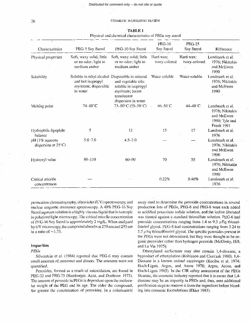
26 COSMETIC INGREDIENT REVIEW
TABLE 1 Physical and chemical characteristics of PEGs soy sterol
PEG-16 PEG-25 Characteristics PEG-5 Soy Sterol PEG-I0 Soy Sterol Soy Sterol Soy Sterol Reference
Physical properties Soft, waxy solid; little Soft, waxy solid; little Hard wax; Hard wax; Lundmark et al. or no odor; light to or no odor; light to ivory-colored ivory-colored 1976; Nikitakis medium amber medium amber and McEwen
1990 Solubility Soluble in ethyl alcohol Dispersible in mineral Water-soluble Water-soluble Lundmark et al.
and hot isopropyl and vegetable oils; 1976; Nikitakis myristate; dispersible soluble in isopropyl and McEwen in water myristate; forms 1990
translucent dispersion in water
Melti'ng point 74-88°C 73-80°C (55-58°C) 46-50°C 44-48°C Lundmark et al.
Hydrophile-lipophile 5 12 balance
pH (1 % aqueous 5.0-7.0 4.5-7.0 dispersion at 25°C)
Hydroxyl value 80-110 60-90
Critical micelle concentration
permeation chromatography, ultraviolet (UV) spectroscopy, and nuclear magnetic resonance spectroscopy. A 40% PEG-16 Soy Sterol aqueous solution is a highly viscous liquid that is isotropic in polarized light microscopy. The critical micelle concentration of PEG-16 Soy Sterol is approximately 2 mg/L. When analyzed by UV microscopy, the compound absorbs at 270 nm and 293 nm in a ratio of ~ 1.73.
Impurities PEGs
Silverstein et al. (1984) reported that PEG-6 may contain small amounts of monomer and dimers. The amounts were not quantified.
Peroxides, formed as a result of autoxidation, are found in PEG-32 and PEG-75 (Hamburger, Azaz, and Donbrow 1975). The amount of peroxide in PEGs is dependent upon the molecular weight of the PEG and its age. The older the compound, the greater the concentration of peroxides. In a colorimetric
1976; Nikitakis and McEwen 1990; Tyle and Frank 1991
15 17 Lundmark et al. 1976
Lundmark et al. 1976; Nikitakis and McEwen 1990
70 55 Lundmark et al. 1976; Nikitakis and McEwen 1990
0.22% 0.46% Lundmark et al. 1976
assay used to determine the peroxide concentrations in several production lots of PEGs, PEG-6 and PEG-8 were each added to acidified potassium iodide solution, and the iodine liberated was titrated against a standard thiosulfate solution. PEG-6 had peroxide concentrations ranging from 1.4 to 9.3 {LEq thiosulfate/ml glycol. PEG-8 had concentrations ranging from 3.24 to 5.7 {LEq thiosulfate/ml glycol. The specific peroxides present in the PEGs were not determined, but they were thought to be organic peroxides rather than hydrogen peroxide (McGinity, Hill, and La Via 1975).
Ethoxylated surfactants may also contain l,4-dioxane, a byproduct of ethoxylation (Robinson and Ciurczak 1980). 1,4-Dioxane is a known animal carcinogen (Kociba et al. 1974; Hoch-Ligeti, Argus, and Arcos 1970; Argus, Arcos, and Hoch-Ligeti 1965). In the CIR safety assessment of the PEGs Stearate, the cosmetic industry reported that it is aware that 1,4-dioxane may be an impurity in PEGs and, thus, uses additional purification steps to remove it from the ingredient before blending into cosmetic formulations (Elder 1983).
Distrbuted for comment only -- do not cite or quote

PEGs SOY STEROL 27
Soy Phytosterols
In a review of the use of soy ster~ls, Lundmark, Chun, and Melby (1976) reported that refined soy sterols contain approximately 88% total sterol. Of that percentage, 56% is sitosterol, 28% is campesterol, and 4% is stigmasterol. Other compounds isolated with the phytosterols are 4% to 6% sterol hydrocarbons and cholesterol and 4% to 6% triterpene alcohols, keto-steroids, and other steroid-like substances (Lundmark, Chun, and Melby 1976).
Swern (1979) summarized the results of analyses of the total (phytosterol esters + free phytosterols), free, and the ester/free ratio in soybean preparations. In 100 mg of alkali-refined soybean oil, there was 0.446 mg total sterol and 0.287 mg free sterol. By subtraction, the phytosterol esters would be 0.159 mg and the ratio of esterified to free sterol was 0.55. In another alkali-refined soybean oil, there was 0.481 mg total sterol and 0.333 mg free sterol. By subtraction, the phytosterol esters would be 0.148 mg and the ratio of esterified to free sterol was 0.44.
Analyses of various lots of soy sterols for pesticide residues were negative for a number of pesticides, including PCB, DDE, DDT, malathion, and J'3-hexachloride (General Mills, Inc. 1979).
Phytosterols Unilever (l998a 1998b) reported on the chemical character
ization and stability of a single sample of plant sterols and from samples from five different batches of a single production process. The plant from which these phytosterols were derived was not stated. High-performance liquid chromatography was used to analyze fatty acid composition, gas chromatography/mass spectrometry for sterols, and Fourier transform infrared analysis as a check that the chemical bonds seen were consistent with the chemical species identified. The results presented in Table 2 show that the distribution of specific phytosterols, etc. are remarkably consistent. Reanalysis indicated the samples to be stable for a period of at least 10 months under refrigerated conditions. The fatty acid content of all samples was around 38% and the phytosterols was 62%.
TABLE 2 Chemical characterization of plant sterol material (Unilever
1998a 1998b)
Distribution of phytosterols (%)
Five samples Phytosterols Single sample from five batches
Brassicasterol 1.1 2.7-3.1 Campesterol 25.8 26.5-27.0 Stigmasterol 21.6 17.4-18.1 J'3 -Sitosterol 48.7 50.8-51.2 J'3-Sitostanal 1.8 Not given Cholesterol 0.4 0.2-0.3 Other sterols 0.8 1.2-1.7
In an analysis of another source of phytosterols using the same techniques described above, Unilever (1996a) reported that the principal phytosterols were present as follows: J'3-sitosterol, 47.9%; campesterol, 28.8%; and stigmasterol, 23.3%. No impurities were found. In an analysis of phytosterol esters, Unilever (1996b) reported that the principal phytosterols were present as fatty acid esters: J'3-sitosterol, 47.3%; campesterol, 28.1 %; and stigmasterol, 24.5%. The distribution of the fatty acid chain lengths was consistent with fatty acids derived from sunflower oil.
Comparison of Phytosterol Compositions from Different Plants
The argument has been made that the available data on phytosterols is relevant to evaluating the safety of the soy phytosterol component of PEGs Soy Sterol (Brock 2000) because of similarity in structure of the phytosterols that have been studied and soy phytosterols. The relevance of phytosterol characterizations as seen above and the safety test data that will follow. to soy sterols is a function of the similarity between phyto-sterols, regardless of source. In one case above, for example, the implication is that the source of the phytosterol esters undergoing analysis was sunflower seed oil and a question should be asked about the relevance of those data to soy sterols. Swern (1979) has compiled data from the literature on the percent distribution of phytosterols from 16 common oils. These data are shown in Table 3.
Clearly, campesterol and stigmasterol are present in significant proportions in most of these oils and J'3-sitosterol is present at a high percentage in all of them. The chemical characterization data in the preceding section shows a distribution of campesterol, stigmasterol, and J'3-sitosterol that is not substantially different from that shown in Table 3 for soy.
In the safety test data that follows, sections will be included that describe results of studies on phytosterols without reference to the plant source. The implication is that these data are likely relevant to the assessment of the campesterol, stigmasterol, and J'3-sitosterol components that principally comprise soy phytosterols based on the comparisons in Table 3.
USE
Cosmetic PEGs 5-40 Soy Sterol serve as smfactants and emulsifying
agents in cosmetic formulations. PEG-5 and -10 Soy Sterol function as skin-conditioning agents and PEG-40 Soy Sterol is used as a cleansing and solubilizing agent (Pepe, Wenninger, and McEwen 2002). These compounds also are used as appearance and consistency modifiers, emollients, viscosity control agents, and pigment dispersion agents (Lundmark, Chun, and Melby 1977).
Table 4 is a summary of the product formulation data submitted to the Food and Drug Administration (FDA) in 1996. PEG-5, -10, -16, -25, and -40 Soya Sterol were used in 41, 35, 15, 5, and
Distrbuted for comment only -- do not cite or quote

28 COSMETIC INGREDIENT REVIEW
TABLE 3 Percent distribution of phytosterols from 16 common vegetable oils (Swem 1979)
Phytosterol composition
Oil source Brassicasterol Campesterol Stigmasterol ,B-Sitosterol t:,. 7 Stigmastenol Unknown
Cocoa butter 8-11 24-31 59-62 Coconut 2 6-9 18-19 69-75 Com 10-20 Trace-6 74-89 Cottonseed Trace-l 8 89-9] Linseed 2 28 10 53 4 Olive 1-3 2 80-97 18 Palm 20-21 12-13 62-67 Peanut ] 10-19 6-12 70-76 Rapeseed 5-19 22-37 52-62 Rice bran 14-33 3-6 55-63 Safflower 8-13 4-9 52-57 23 Soybean 15-21 10-24 57-72 Sunflower 11-12 8-12 62-75 20
1 cosmetic formulations, respectively. PEG-30 Soya Sterol was not reported to be used (FDA 1996).
Concentration of use data are no longer required to be submitted to the FDA by the cosmetics industry, but historical data stated that PEG-5 and -10 Soy Sterol were used at concentrations up to 5%, whereas the maximum concentration of use for
both PEG-16 and -25 was I % (FDA 1984). Current concentration of use information was provided by the industry through the Cosmetic, Toiletry, and Fragrance Association (CTFA). PEG-5 Soy Sterol was used at concentrations of 2% in mascara and eyeliner. In addition, 0.75% PEG-5 Soy Sterol and 1.5% PEG-25 Soy Sterol were reported used in a liquid makeup foundation
TABLE 4 Product formulation data for PEGs Soy Sterol (FDA 1996)
Total no. of Total no. of formulations containing ingredient
formulations in PEG-5 PEG-1O PEG-16 PEG-25 PEG-40 Product category category Soy Sterol Soy Sterol Soy Sterol Soy Sterol Soy Sterol
Eyeliner 533 1 Mascara 218 5 Other eye makeup preparations 136 3 Hair conditioners (noncoloring) 715 3 Shampoos (noncoloring) 972 1 Blushers (all types) 277 Foundations 355 2 Rouges 30 Aftershave lotion 268 I Cleansing 820 1 14 3 Face and neck (excluding shaving) 300 5 Body and hand (excluding shaving) 1012 4 3 Moisturizing 942 8 5 Night 226 1 Paste masks (mud packs) 300 2 3 Other skin care preparations 810 8 Suntan gels, creams, and lotions 196 2 3
1996 totals 41 35 8 5
Distrbuted for comment only -- do not cite or quote

PEGs SOY STEROL 29
TABLES Current concentration of use of PEGs Soy Sterol (CTFA 2000)
Concentration
PEG-5 PEG-I0 PEG-16 PEG-25 Product category Soy Sterol (%) Soy Sterol (%) Soy Sterol (%) Soy Sterol (%)
Eye lotion 2 0.2 Mascara 2 2 Makeup bases 1 Foundations 0.8 2 Other eye makeup preparations 0.1 Hair conditioners (non coloring) 2 Shampoos (noncoloring) 0.5 0.5 Tonics, dressings, and other hair-grooming aids 0.4 0.5 Other makeup preparations 0.05 Shaving cream 2 Other shaving preparation products 2 Face and neck creams, lotions, powders, and sprays 0.5
(excluding shaving preparations), Body and hand creams, lotions, powders, and sprays 0.2-2 2
(excluding shaving preparations) Moisturizing creams, lotions, powders, and sprays
(excluding shaving preparations) Paste masks (mud packs) Skin fresheners
(CTFA 1997). CTFA (2000) updated those data with the information contained in Table 5.
Noncosmetic In 1982, polyoxyethylene adducts of mixed phytosterols (with
5 to 26 moles average polyoxyethylene content) were exempted from tolerance requirements by the Environmental Protection Agency (EPA) when used as surfactants or related surfactant adjuvants in pesticide formulations applied to growing crops (Rothschild 1990; EPA 2002).
GENERAL BIOLOGY Campesterol, sitosterol, and stigmasterol are the most fre
quently occurring plant sterols (Kallianos et aJ. 1963). Approximately 0.25 to 0.5 g of plant sterols are consumed each day in a typical diet (Sabine 1977; Heinemann, Axtmann, and von Bergmann 1993). As a comparison, up to 1.2 g cholesterol, which is derived from animal fat (Tso and Fujimoto 1991), was consumed (Sabine 1977). Plant sterols account for 20% to 25% of total dietary sterols (Heinemann, Axtmann, and von Bergmann ] 993). These sterols are also found in cigarette and tobacco smoke as free sterols (Kallianos et al. 1963).
Phytosterols affect plant membrane structure and water permeability (Hennessey 1992). Membrane fluidity is inversely re-
2 0.2
0.5 2
lated to the amount of sterol found in the membrane (Sabine 1977). Phytosterols are commonly found in animal cell membranes following dietary uptake. In general, sterols intercalate into membrane bilayers and align themselves perpendicularly to the plane of the membrane with the 3' OH facing the water interface. The aliphatic side chain extends into the hydrophobic core to interact with the fatty acid side chains of phospholipids and integral membrane proteins. The phytosterols are less water soluble than cholesterol. Sitosterol and campesterol order bilayer acyl chains most effectively, followed by cholesterol and stigmasterol (Hennessey 1992).
Absorption, Metabolism, Distribution, and Excretion PEGs
Gastrointestinal absorption of PEGs is dependent on the molecular weight of the compound. In general, the greater the molecular weight of the PEG compound, the lesser the absorption that occurs. In both oral and intravenous studies, no metabolism was observed and the PEGs were rapidly eliminated unchanged in the urine and feces. In a study with human bum patients, monomeric ethylene glycol was isolated in the serum following topical exposure to a PEG-based antimicrobial cream, indicating that PEGs are readily absorbed through damaged skin (Andersen 1993).
Distrbuted for comment only -- do not cite or quote

30 COSMETIC INGREDIENT REVIEW
P hytoste rols In general, ingested sterols are emulsified in the stomach,
where lipid material from lipoprotein complexes is released. The coarse emulsion enters the duodenum ofthe small intestine, and the emulsion is solubilized with digestive enzymes and bile. Bile salts become conjugated with fatty acids, monoglycerides, dissolved sterols, and other molecules in the jejunum to form mixed micelles. Sterol ester bonds become hydrolyzed until only free sterol remains. Micellar solutions of lipids are very rapidly absorbed, and represent the major pathway of absorption for sterols and other fats (Sabine 1977). For example, incorporation of sterols into cholic acid micelles was 34%, 30%, 23%, and 15% for campesterol, sitosterol, cholesterol, and stigmasterol, respectively (Hennessey 1992). Micelles release sterols to the cells of the intestinal wall. Absorbed sterols are mixed with cholesterol synthesized within the intestinal cells. Before release from mucosal cells, the sterols are esterified. The resulting esters are transported from the intestine via the lymph (Sabine 1977).
Once consumed, phytosterols only enter the body via intestinal absorption. As the absorption rate for the plant sterols is usually less than 5% of dietary concentrations in humans (Sabine 1977; General Mills, Inc. 1979; Ling and Jones 1995), approximately 95% of dietary phytosterols enters the colon (Ling and Jones 1995). Saturated sterols are virtually not absorbed (Vanhanen and Miettinen 1992).
When cholesterol and phytosterols were simultaneously administered, only cholesterol could be isolated from the lymph duct, demonstrating that the phytosterols had not been significantly absorbed. Phytosterols experimentally injected subcutaneously into dogs were not esterified or metabolized. Instead, the plant sterols were treated like inert, foreign materials. Cholesterol, when similarly injected, was readily esterified (Lange 1950). In other studies, sitosterols did not accumulate or deposit in tissues (Gould 1955; Gould et al. 1969). Freshly absorbed sterols are transported into plasma (Sabine 1977).
Dietary supplementation with phytosterols can increase their serum concentrations until the sterols represent 10% of total serum sterols (Hennessey 1992; Heinemann, Axtmann, and von Bergmann 1993). Ling and Jones (1995) reported that 0.3 to 1.7 mg/dl of phytosterols were found in human serum under normal conditions after daily phytosterol consumption of 160 to 360 mg/day.
During metabolism and excretion, the sterol rings generally remain intact; double bonds, constituent groups, and side chains are often added, removed, or modified. The largest proportion of sterol in the body is converted to bile acids (Sabine 1977). In feeding studies, approximately 20% of absorbed ,B-sitosterol was converted to bile acids (cholic and chenodeoxycholic) in humans (General Mills, Inc. 1979). Boberg et al. (1990b) reported that C21 bile acids were major metabolites of sitosterol in mammals. Sitosterol was apparently not converted into C24 bile acids in humans (Boberg, Einarsson, and Bjorkhem 1990a). Conversion of campesterol into bile acids was reported in rats (Boberg et al. 1990b). Absorbed phytosterols not converted to
normal bile acids were excreted as the free sterol (General Mills, Inc. 1979).
5,6-Epoxides were formed in the liver from ,B-Sitosterol. ,B-Sitosterol was metabolized to cortisol by the adrenal glands and to various steroid hormones by the testes. In r~ts, up to 5% of adrenal gland sterols can be of plant origin, and other tissues can contain large amounts of plant sterols (Sabine 1977).
Phytosterols can act as plant hormone and hormone precursors (Hennessey 1992). Other metabolites include steroid hormones (minor) and vitamin D compounds (Sabine 1977).
Sterols are typically eliminated via feces, urine, milk, and from the skin surface. Skin surface lipids contain 2% to 20% total sterols (Sabine 1977). Ling and Jones (1995) reported that phytosterol elimination via the biliary route appeared to be more rapid than that of cholesterol. The endogenous phytosterol pool size was low compared to cholesterol, due to poor intestinal absorption and faster excretion (Ling and Jones 1995). The excretion rate of sitosterol from bile was ten times greater than that of cholesterol (Gould 1955; American Cyanamid Co. 1957).
Nearly complete recovery of administered phytosterols in mammals was made from the feces (Lange 1950; General Mills, Inc. 1979). Unabsorbed sterols (unspecified) were degraded in the intestinal tract to varying degrees, depending on the species: 5% in rats, 25% in humans, and 65% in monkeys (strain not given) and baboons (Sabine 1977).
Unilever (1997a) traced the fate of radioactively labeled ,B-sitosterol and ,B-sitosterol linoleate administered by gavage to male rats. Twenty young, adult male Charles River CD rats (146 to 172 g) were individually housed. Ten animals received the ,B-sitosterol and 10 received the ester. The vehicle was sunflower oil and delivery was by gavage. Urine and feces were collected and individual CO2 absorbers filtered the air which was drawn through at a rate of 1 Umin. At 4, 8, 24, 72, and 96 h, one rat from each group was weighed and euthanized for whole body autoradiography. At 96 h, heart blood was collected from the remaining animals and they were euthanized for analysis of radioactivity in internal organs.
The absorption, distribution, metabolism, and excretion of ,Bsitosterol and ,B-sitosterol linoleate were similar. Over 90% of the dose was excreted in the feces; less than 0.1 % was excreted in the urine. Absorption from the gut was low. What was absorbed ended up in the intestinal lining, liver, lung, and adrenal gland. The highest concentration as well as persistence of the radioactive label was in the adrenal gland. In the gut, the ,B-sitosterol linoleate was hydrolyzed to release free sterol, but esterification with fatty acids present in the gut with free sterol also occun-ed.
In another study of the fate of radioactively labeled ,B-sitosterol and ,B-sitosterol linoleate, Unilever (1997b) evaluated the impact of a sunflower oil vehicle versus a coconut oil vehicle. In the sunflower oil part of the study, six young male and six young female Charles River CD rats (152 to 174 g) were dosed by gavage. At 24, 48, and 96 h, one animal of each sex was weighed and euthanized for whole body autoradiography. Urine and feces were collected. In the coconut oil portion of the
Distrbuted for comment only -- do not cite or quote

PEGs SOY STEROL 31
study, five young male and five young female Charles River CD rats (163 to 193 g) were also individually housed. At 24 h, the rats were weighed and euthanized for analysis of radioactivity in internal organs.
As in the previous study, ,B-sitosterol and ,B-sitosterollinoleate were poorly absorbed. The radioactive label was slightly higher in females. The adrenal gland showed the greatest amount and retention of radioactive label, but the ovaries, bone marrow, liver, intestinal lining, and spleen were also target organs for what radioactive label was absorbed. The testes in male rats was not a target organ. There was extensive hydrolysis of the ester and there was esterification of which varied as a function of the vehicle; there was more esterification with the sunflower oil than with the coconut oil. f:3-Sitosterol fatty acid esters with fatty acid components found only in coconut oil and not in the diet confirmed that esterification of free f:3-sitosterol.
Many of the results from the above Unilever (1996a, 1996b, 1997a, 1997b) studies were combined with other data on the influence on absorption of specific phytosterols (cholesterol, f:3-sitosterol, f:3-sitostanol, campesterol, or stigmasterol) on absorption, distribution, metabolism, and excretion in the rat in a report by Sanders et al. (2000). Using similar methods, the essential findings from above were repeated: the total absorption of phytosterols is low (cholesterol by contrast was well absorbed); female animals absorb more than males; and the largest amout of radioactivity from labeled phytosterols ends up in the adrenal gland, although the ovaries and intestinal epithelia also have significant levels.
Effects on Cholesterol Absorption and Metabolism The plant sterols are effective inhibitors of cholesterol absorp
tion in the small intestine, producing a hypocholesterolemic effect when the sterols are simultaneously ingested (Sabine 1977; Heinemann et al. 1991, 1993; Tvrzicka et al. 1991) in rabbits, chickens, rats, and humans (Peterson 1951; Laraki et al. 1993; Ling and Jones 1995). Phytosterols also interfere with the absorption of structurally different, unsaturated plant sterols. Plant sterol-induced decreases of sterol absorption may be directly related to the absorption efficiency of sterols (Vanhanen and Miettinen 1992).
Heinemann et al. (1991) reported that 20% to 70% of the 750 to 3000 mgJday total cholesterol (dietary and biliary) that entered the intestinal tract was absorbed after solubilization in mixed micelles containing bile salts, mono- and diglycerides, fatty acids, and lysolecithin. Simultaneous high-dose infusion of cholesterol and sitosterol decreased the overall absorption of cholesterol by 25% to 65% in an intestinal perfusion study using nine subjects. The reduction in cholesterol absorption was due to competition with cholesterol uptake in the micelles (Heinemann et al. 1991).
In a second intestinal perfusion study using 10 subjects, Heinemann, Axtmanri, and von Bergmann (1993) reported that an inverse relationship existed between absorbability of plant sterols and their inhibition of cholesterol absorption. Phytosterol
absorbability was reduced by hydrogenization of the Sa nucleus double bond or by an increase in the side chain length. Sterol absorption rates were 33% for cholesterol, 4.2% for sitosterol, 4.8% for stigmasterol, and 12.5% for campesterol.
A positive con'elation between the absorption rate of cholesterol and campesterol was established; a negative conelation was reported between the ratio of sitosterol to cholesterol and the mass of cholesterol absorption. Generally, the sterol absorption rate depended on micellar solubility. Cholesterol solubility was approximately three times greater than that of sitosterol. In addition, the binding of sitosterol to trihydroxy bile salt micelles was energetically favored over the binding of cholesterol. Hydrogenization of the delta-5-nucleus double bond caused a moderate enhancement of sterol hydrophobicity. The authors suggested that hydrophobic plant sterols with a high affinity and low capacity for micellar binding could have effectively displacyd cholesterol from micellar binding (Heinemann, Axtmann, and von Bergmann 1993).
In a study by Sato et al. (1995), cultured in the presence of plasma lipoproteins, cells acquired cholesterol through recepiormediated endocytosis of low-density lipoprotein (LDL). LDLderived cholesterol esters in lysosome were hydrolyzed, freeing unesterified cholesterol. Free cholesterol crossed the lysosomal membrane and was transported to other intracellular organelles. The rate of esterification of the plant sterols could have been the factor limiting their absorption.
Esterification of endocytosed phytosterols in the endoplasmic reticulum was extremely low. Campesterol esterification was 20% that of cholesterol, and both sitosterol and stigmasterol were not esterified appreciably. When added to cell cultures, sitosterol and stigmasterol did not appear to be transported to intracellular membranes and, therefore, could not substitute for cholesterol to support cell growth. Instead, endocytosed plant sterols accumulated in the phagolysosomes of the cells.
Phytosterol accumulation was not a consequence ofthe cell's inability to esterify sterols in the endoplasmic reticulum as cholesterol did not accumulate when its esterification was blocked. The side chain structure of sterols was critical for the efflux of sterols from lysosomes. Plant sterols were distinguished from cholesterol at the level of the intestinal mucosal cell, but the mechanisms have yet to be determined. The observations made in this study suggested that cultured macrophages were able to differentiate sterols that differed only by a methyl or ethyl group at the C24 position at their lysosomal compartment (Sato et al. 1995).
The conversion of sterols to bile acids is inefficient; phytosterols and other sterols can inhibit the synthesis of bile acids from cholesterol. Unmetabolized sterols secreted into bile were generally less soluble than cholesterol and can precipitate out if bile salt concentrations were reduced (Clayton et al. 1993).
Miscellaneous Effects of Phytosterols Exposure to 0.7 mmolJL (incorporated into liposomes) sitos
terol for 72 h caused contraction of human umbilical vein
Distrbuted for comment only -- do not cite or quote

32 COSMETIC INGREDIENT REVIEW
endothelial cells in vitro and an increased release of intracellular lactate dehydrogenase. At 96 h, partial detachment from the substrate was observed. In addition, 0.35 mmollL of sitosterol caused pertubation of the endothelial cells at 96 h (Boberg, Pettersen, and Prydz 1991).
Results of an in vitro study by Chiang et al. (1991) indicated that ,8-sitosterol at a concentration of 100 {-Lg/ml (5% in DMSO and saline) was cytotoxic against seven cancer cell lines: Co10-205 (colon), Hep-2 (laryngeal epidermoid), HeLa (uterine cervix), KB (nasopharynx), H1477 (melanoma), HA22T (hepatoma), and GBM840llTSGH (glioma). A ,8-sitosterol isolate had antiatherogenic effects through inhibition of platelet aggregation (Pollak 1985). ,8-Sitosterol isolated from piper betle leaves (concentration not given) inhibited human platelet aggregation induced by arachidonic acid, platelet-activating factor, and adenosine diphosphate (ADP) (Saeed et al. 1993). Sitosterol can have potent anti-inflammatory, antibacterial, and antifungal activities (Padmaja, Thankamany, and Hisham 1993).
A concentration of 25 {-Lg/ml of phytosterols slightly decreased beat rates of fetal rat heart cells whereas similar additions of cholesterol increased beat rates (Hennessey 1992). Phytosterols found in human tumors, particularly breast cancers have osteolytic activity, and increase mobilization and excretion of bone calcium (Sabine 1977).
ANIMAL TOXICOLOGY
Acute Toxicity
PEGs Soy Sterol Young, male Sprague-Dawley rats were given 109/kg doses
(50%; in feed) of PEG-5, -10, -16, or -25 Soy Sterol. No adverse effects were reported and the acute oral LD50 for each compound was> 10 g/kg (Warf Institute, Inc. 1974; Henkel Corp. 1995).
North American Science Associates, Inc. (l987a) evaluated the dermal toxicity of a liquid eyeliner containing 2% PEG-5 Soy Sterol using ten New Zealand white rabbits. Each rabbit weighed between 2.5 to 3.1 kg. The test material (2 g/kg) was applied to the clipped upper back of each rabbit, at both intact and abraded skin sites. After application, the trunk of each rabbit was wrapped with polyethylene plastic that was taped in place, thus forming a reservoir over the test site. The rabbits were then fitted with collars and retumed to their respective cages. The wrappings were removed after 24 h, and the collars at test termination. The rabbits were observed for signs of toxicity immediately after treatment, at 4 h, and daily for 14 days. Dermal reactions were scored daily for erythema and edema. On day 14, the skin sites were washed with tap water to remove any remaining residue of the test material. The rabbits were euthanized for necropsy.
No animals died prior to test termination. Three of 10 rabbits had transient diarrhea, and one rabbit "appeared thin" during the last 6 days of the study. Dermal observations were slight redness (9/1 0), swelling (2/1 0) that diminished by day 6, and apparent pustules at the test site (1/10). Four rabbits lost between 0.4 and
0.6 kg of weight. No macroscopic changes of the viscera were observed at necropsy. The acute dermal LD50 was >2 g/kg, and the eyeliner was considered dermally nontoxic for the rabbit (North American Science Associates, Inc. 1987a.).
PEGs
Toxicity studies using rats, rabbits, and dogs indicate that PEGs have low oral and dermal toxicity. In general, the larger molecular weight PEGs appear to be less toxic than the smaller molecular weight PEGs in oral studies. Acute oral LDso values for PEGs in rabbits were 17.3 g/kg (100% PEG-6) and 76 g/kg (100% PEG-75). In acute dermal toxicity studies, no deaths were reported in groups of rabbits dosed with 20 mllkg of undiluted PEG-6 or 40% PEG-20M (Andersen 1993).
Short-Term Toxicity
PEGs
Andersen (1993) reported that there was no evidence of toxicity, with the exception of transient, mild erythema, in rabbits that received daily topical applications of PEG-75 or PEG-20M (0.8 g/kg/day) for 30 days.
Evidence of systemic toxicity was found, however, in a study designed as an animal model to study the effects of PEG-based antimicrobial creams in bum patients. Elevated total serum calcium, elevated osmolality gap, high anion gap metabolic acidosis, and renal failure resulted in rabbits that received repeated dermal applications of an antimicrobial cream containing 63% PEG-6, 5% PEG-20, and 32% PEG-75 to two paravertebral skin excisions (2.5 x 15 cm), followed by wound dressings (changed every 12 h) for 7 days (Andersen 1993).
Phytosterols
Laraki et al. (1993) studied the effects of phytosterols in the diet of rats. Male adult Wistar rats weighing 215 ± 12 g were randomly assigned to eight dietary groups with 12 rats/group. Rats of each group were fed 22 g/day basal diet with or without supplementation by cholesterol or maize phytosterols (72.5% ,8-sitosterol, 20.5% campesterol, and 7% stigmasterol) for 3 weeks (see Table 6).
The basal diet contained 16% casein, 68% comstarch, 8% butter, 4% cellulose, 3% mineral mix, and 1 % vitamin mix. Water was available ad libitum. Feed consumption did not differ between treatment groups; mean consumption was 21.4 ± 0.1 g/day. The rats were euthanized and the livers sampled post mortem, washed, and frozen. Enzymatic activities of acetylcoenzyme A (CoA) carboxylase, malic enzyme, and glucose-6-phosphate dehydrogenase were determined on microsomes purified from 1 g liver homogenized in 10 ml 0.25 M sucrose. Hepatic lipid and fatty acid compositions were determined.
Table 6 presents enzyme activities determined in 12 rats in each treatment group. Acetyl-CoA carboxylase activity was significantly increased in group 4 (12 mg/day cholesterol and 48 mg/day phytosterol) compared to group 1 (12 mg/day cholesterol only); and decreased in groups 6, 7, and 8 (24 mg/day
Distrbuted for comment only -- do not cite or quote

<Jj <Jj
TABLE 6 Diet composition in a rat short-term feeding study (Laraki et al. 1993)
Group
2 3 4 5 6 7 8
Diet Cholesterol (mg/day) 12 12 12 12 24 24 24 24 Phytosterol (mg/day) 12 24 48 24 48 96 Phytosterol: Cholesterol 0 2 4 0 2 4
Ratio Liver enzyme activities (means ± SEM)a
Acetyl-CoA carboxylase 538 ± 109 536 ± 43 522 ± 154 664 ± 123* 741 ± 199 235 ± 59** 225 ± 92*** 233 ± 99*** Malic enzyme 0.39 ± 0.16 0.39 ± 0.07 0.30 ± 0.06 0.32 ± 0.06 0.44 ± 0.13 0.16 ± 0.04* 0.16 ± 0.04* 0.16 ± 0.03* Glucose-6-phosphate 1.33 ± 0.20 1.32 ± 0.20 1.26 ± 0.15 1.48 ± 0.26 2.39 ± 1.23 0.46±0.19 0.57 ± 0.19 0.62 ± 0.15
dehydrogenase Liver fatty acids and sterols (means [wet weight] ± SEM)a
Fatty acids (g/lOO g) 3.1 ± 0.3 3.5 ± 0.3 3.6 ± 0.3 3.0 ± 0.2 7.8 ± 2.8 2.8 ± 0.3*** 2.7 ± 0.3*** 3.0 ± 0.3***
Cholesterol (mg/lOO g) 216.4 ± 1.5 218.7 ± 3.1 210 ± 2.8* 199.9 ± 1.9*** 301.0 ± 13.3 239.4 ± 9.0*** 209.4 ± 6.1 *** 203.4 ± 7.7***
Campesterol (mg/lOO g) 0.81 ± 0.04 1.86 ± 0.17*** 1.96 ± 0.15*** 2.19 ± 0.16*** 0.87 ± 0.07 4.68 ± 0.51 *** 5.03 ± 0.50*** 8.00 ± 0.66
,B-Sitosterol (mg/lOO g) 2.95 ± 0.20 3.96 ± 0.23* 3.74 ± 0.16*** 3.88 ± 0.24*** 2.92 ± 0.06 8.84 ± 0.25*** 10.90 ± 0.27*** 12.86 ± 0.75***
aStudent's t test significance (* p < .05; ** p < .01; *** P < .001) versus the group receiving the same cholesterol supplementation without cholesterol.
Distrbuted for comment only -- do not cite or quote

34 COSMETIC INGREDIENT REVIEW
cholesterol with 24, 48, or 96 m&/day phytosterol, respectively), compared to group 5 (24 rug/day cholesterol only). Malic enzyme activity was decreased in groups 6, 7, and 8 compared to group 5. Glucose-6-phosphate dehydrogenase activity was not decreased significantly in any phytosterol treatment groups.
Analysis of liver fatty acid and sterol contents (see Table 6) demonstrated increases in fatty acids in groups 6, 7, and 8 compared to group 5 (see Table 6). Cholesterol decreases in the liver were seen in groups 3 and 4 compared to group 1; and in groups 6, 7, and 8 compared to group 5. Campesterol incl:eases were seen in groups 2, 3, and 4 compared to group I; and in groups 6 and 7 (but not in group 8, although the value reported in Table 6 would appear to be significant) compared to group 5. ,B-Sitosterol increases were seen in groups 2, 3, and 4 compared to group 1; and in groups 6, 7, and 8 compared to group 5.
The authors noted (see Table 6) that liver fatty acids increased by a factor of2.5 in group 5 (24 mg/day cholesterol) compared to group I (12 mg/day cholesterol) and,that addition of phytosterol to the high cholesterol diet (groups 6, 7, and 8) reduced the liver fatty acid levels to that seen in group I (Laraki et a1. 1993).
Wi star albino rats (10 per sex per group) were given subcutaneous injections of 250,500, or 1000 f,tgll 00 g/day ,B-sitosterol in olive oil for 60 days. No mortality was observed throughout the study. Treated rats given the lower doses had no gross or microscopic lesions of either the liver or kidneys. Mild fibroblastic proliferation around the hepatic lobules and microscopic lesions of the kidney were observed in animals given the high dose. These lesions were of very mild degree and had only a few heterophilic cell infiltrations in the medullary tract. All clinical biochemical parameters (hemoglobin, blood glucose, serum protein, serum bilirubin, aspartate aminotransferase, and alanine transaminase) were in the normal range with the exceptions of serum cholesterol and serum protein. Serum cholesterol was markedly depleted in a dose-dependent manner in both sexes. Serum protein was markedly reduced in the rats treated with 1000 f,tg/100 g/day of ,B-sitosterol (Malini and Vanithakumari 1990).
Subchronic Toxicity PEGs
In subchronic, 90-day oral toxicity studies involving groups of albino rats, the largest (PEG-20M) and smallest (PEG-6) molecular weight PEGs tested did not induce toxicity or death when administered daily at concentrations of 4% or less; PEG-20M was administered in the diet and, PEG-6, in drinking water. In a dermal toxicity study, no evidence of toxicity was observed in a group of rabbits that received daily applications of PEG-6 5 days per week (2 ml/kg/day) for 18 weeks (Andersen 1993).
Phytosterol Esters Hepburn, Horner, and Smith (1999) presented the results
of a 90-day oral toxicity study of phytosterol esters. Groups of 20 male and 20 female Wistar derived rats were fed diets containing phytosterol esters (62% phytosterols and 38%
fatty acids). Composition data were presented earlier in Unilever (1998a) and the mix of phytosterols shown in the first column of Table 2. Fatty acids were linoleic acid (64.6%), oleic acid (21.6%), stearic acid (4.1%), and palmitic acid (9.6%). Five dose groups received 0%, 0.16%, 1.6%, 3.2%, and 8.1 % for 90 consecutive days. Clinical observations, body weights, and food and water consumption were determined during the exposure. At the end of the exposure period, the animals were euth'anized; cardiac blood samples were taken, organs (adrenal gland, brain, epididymides, heart, kidney, liver, spleen, testes, thymus) were weighed, and a standard comprehensive set of tissues taken for histological examination.
Diets containing plant phytosterol esters at all levels were well tolerated. During the exposure, there were no changes in body weight gain, food and water consumption, or clinical signs. There were slightly reduced platelet counts and a small decrease in prothrombin time in female rats at all dose levels compared to controls. There was a small increase in the activated partial thromboplastin time in male rats at the 3.2% and 8.1 % dietary levels. Other small increases in plasma albumin, phosphorus or magnesium levels, and certain serum enzymes were reported for males and/or females at the 1.6%, 3.2%, and 8.1 % dietary levels. No treatment-related effect was seen with organ weights and histological examination revealed no evidence of systemic toxicity. Absent any organ effects, the small hematology and blood chemistry variations were not considered of toxicological significance (Hepburn, Horner, and Smith 1999).
Chronic Toxicity PEGs
Toxic effects also were not observed in groups of dogs that received PEG-8, PEG-32, and PEG-75 at concentrations of 2% in the diet for I year (Andersen 1993).
PhytosteroIs
Thirteen dogs fed a basic diet supplemented with 0.5 to 1.0 g/kg/day of ,B-sitosterol had no gross or microscopic changes after 8 to 22 months of treatment. Weight gains and clinical parameters did not differ from controls (General Mills, Inc. 1979).
No adverse effects or gross or microscopic abnormalities were observed in six New Zealand white rabbits of both sexes that were given feed containing 3% cottonseed sterols and 4% soy sterols for 70-212 days (General Mills, Inc. 1979).
Dermal Irritation and Sensitization PEGs Soy Sterol
Undiluted PEG-5, -10, -16, and -25 Soy Sterol were administered to the clipped back and flanks of albino rabbits (intact and abraded skin), six per group. The treated areas were covered with gauze patches, which were secured with tape. The patches were removed 24 h later, and the treated sites were scored at that time and at 72 h. The primary irritation index (PH) score was
Distrbuted for comment only -- do not cite or quote

PEGs SOY STEROL 35
0.50 for PEG-lO Soy Sterol and 0 fm: PEG-5, -16, and -25 Soy Sterol (WarfInstitute, Inc. 1974; Henkel Corp. 1995).
PEG-5 Soy Sterol at induction and challenge concentration of 100% was a non sensitizer in a study using Pirbright white guinea pigs (number of animals not stated) (Henkel Corp. 1995).
The primary skin irritancy o(a liquid eyeliner containing 2% PEG-5 Soy Sterol was evaluated using six New Zealand white rabbits (North American Science Associates, Inc. 1987b). A 0.5-ml volume of the test sample was applied under a double gauze layer to both intact and abraded skin sites on 1 x I-inch areas of the back. The patches were covered with nonreactive tape and the entire test site was wrapped with a binder. The binders and patches were removed after 24 h, and the skin sites were rinsed with tap water. The sites were evaluated at 24 and 72 h after application using Draize criteria. The PH was 0.96 (barely perceptible), and the investigators concluded that the eyeliner was not a primary skin irritant.
PEGs The PEGs were not irritating to the skin of rabbits or guinea
pigs, and PEG-75 was not a sensitizer. In skin irritation tests, undiluted PEG-6 was applied to the skin of rabbits for 4 hand 50% PEG-75 was applied to guinea pigs for 4 days and to rabbits over a 13-week period. In the guinea pig skin sensitization test, PEG-75 was tested at a concentration of 0.1 % (Andersen 1993).
Ocular Irritation PEGs Soy Sterol
Undiluted PEG-5, -10, -16, and -25 Soy Sterol were instilled into the conjunctival sac of New Zealand white rabbits, six per group. The PEGs Soy Sterol did not induce ocular toxicity in any of the rabbits treated, and the ocular irritation score for each was zero (Warf Institute, Inc. 1974; Henkel Corp. 1995).
A O.I-ml volume of a liquid eyeliner containing 2% PEG-5 Soy Sterol was instilled into the conjunctival sac of six New Zealand white rabbits. Prior to instillation, the eyes were treated with fluorescein stain, flushed with saline, and observed in a darkened room under UV light to detect or confirm preexisting corneal injury. Ocular reactions were evaluated using Draize and Federal Hazardous Substances Act (FHSA) scoring criteria at 24, 48, and 72 h after instillation. Minimal conjunctival redness was observed in 1/6 test eyes, but was not considered significant. The investigators concluded that the liquid eyeliner was not an ocular irritant (North American Science Associates, Inc. 1987c).
PEGs PEG-6 and -75 did not cause corneal injuries when instilled
(undiluted, 0.5 ml) irito the conjunctival sac of rabbits. PEG-8 (35% solution, 0.1 ml) and PEG-32 (melted in water bath, 0.1 ml) induced mild ocular irritation in rabbits (Andersen 1993).
REPRODUCTIVE AND DEVELOPMENTAL TOXICITY
Ethylene Glycol and its Ethers It is generally recognized that the PEG monomer, ethylene
glycol, and certain of its monoalkyl ethers (e.g., methoxyethanol, a.k.a. ethylene glycol monomethyl ether) are reproductive and developmental toxins. The CIR Expert Panel undertook a separate, limited scope review of these compounds in order to assess the possibility that PEG-derived cosmetic ingredients could present similar concerns (CIR 1996). In summary, this report concluded that the ethylene glycol monoalkyl ethers are not themselves toxic, but rather that one or more alcohol or aldehyde dehydrogenase metabolites are toxic. From the available data, the report also concluded that the toxicity of the monoalkyl ethers is inversely proportional to the length of the alkyl chain (methyl is more toxic than ethyl than propyl than butyl, etc.).
Given the methods of manufacture of the PEGs Soy Sterol, there is no likelihood of methoxyethanol, ethoxyethanol, etc. being present as an impurity. Further, the PEGs Soy Sterol are ethers of soybean oil sterols such as stigmasterol, y-sitosterol, and carnpesterol and, as such, are chemically different from alkyl ethers.
Estrogenic Effects of Phytosterols and Phytosterol Esters Malini and Vanithakumari (1992) reported that ,B-sitosterol
acted as an effective estrogen-like agonist in producing vaginal cornification and caused uterine weight gain in adult, ovariectomized Wistar rats. In a short-term study, subcutaneous administration of ,B-sitosterol (50 {Lg/kg/day to 5.0 mg/kg/day) resulted in significant dose-related increases in uterine glycogen concentration after 10 days. At the highest dose, the glycogen concentration increase was equivalent to that produced by 50 {Lg/kg/day of estradiol.
When ,B-sitosterol was given in combination with estradiol, the estradiol-induced glycogen concentration was slightly enhanced. Progesterone partiaIly suppressed the phytosterolinduced elevation of glycogen concentration after the hormone (20 mg/kg/day) was administered in combination with median and high doses of ,B-sitosterol (2.5 and 5.0 mg/kg/day). In addition, treatment with ,B-sitosterol (all doses) stimulated glucose-6-phosphate dehydrogenase, phosphohexose isomerase, and total lactate dehydrogenase activities (Malini and Vanithakumari 1992).
In a later study, Malini and Vanithakumari (1993) administered ,B-sitosterol alone or in combination with estradiol for 10 days caused a marked increase in the uterine weight of ovariectomized animals. When ,B-sitosterol was coadministered with progesterone, a twofold increase in uterine weight was observed. Estradiol administration resulted in a threefold increase. ,B-Sitosterol (50 to 500 {Lg/l 00 g/day) given alone caused a progressive dose-dependent increase in uterine weight with maximal increments occUlTing at the mid- and high-doses (250 and 500 {Lg/1 00 g/day). Other effects reported were increased uterine RNA, DNA, and protein concentrations.
Distrbuted for comment only -- do not cite or quote

36 COSMETIC INGREDIENT REVIEW
These effects varied with coadministration of estradiol or . "
progesterone. Estrogen and ,8-sitosterol induced DNA synthe-sis in the uterine luminal epithelium. Progesterone by itself stimulated a very small increase in DNA concentration; when coadministered with ,8-sitosterol, progesterone inhibited the growth-promoting effect of the phytosterol. Epithelial DNA concentration increased by two-, three-, and sevenfold for the low, mid-, and high doses, respectively; however, the increases were only significant at the two latter doses.
The investigators concluded that the dose-dependent uterotrophic effect of ,8-sitosterol in ovariectomized rats and its synergism with estradiol could be due to the phytosterol's intrinsic estrogenic properties, and that the effects of ,8-sitosterol could be inhibited by progesterone (Malini and Vanithakumari 1993).
In a study on the content and estrogen receptor of phytoestro-. gens in various foods, herbs, and spices, Zava, Dollbaum, and BIen (1998) reported 8 fJ,g of estradiol equivalents per 200 cm3
of soy milk extract in an estrogen receptor binding assay. Activity was extracted from soy milk with ethanol/water (50:50) for 2 days. These authors also report the results of ingestion of 200 cm3 of soy milk on the estrogen receptor binding activity of saliva of four volunteers. Saliva was extracted with diethylether, dried to a precipitate, and reconstituted in growth medium for the binding assay. At least one individual had residual estrogen receptor binding 9 to 10 h after ingestion of the soy milk (all had estrogen receptor binding in saliva I to 4 h after ingestion).
Baker et al. (1999), however, found no evidence of estrogenic activity of phytosterols in a competitive estrogen-binding assay, a recombinant yeast assay, and in a juvenile rat uterotrophic assay. The phytosterols (see Table 2) used were described earlier (Unilever 1996a).
In the estrogen-binding assay, estrogen receptors isolated from 10-week-old female Wi star rats were prepared and incubated with estradiol (positive control) or test substances. Estradiol produced a concentration dependent (from 10- 13 M to 10-6
M) binding, whereas phytosterols had no binding at concentrations of 10-7 M to 10-4 M.
In the recombinant yeast assay, the DNA of the human estrogen receptor and plasm ids with the gene for the ,8-galactocidase enzyme controlled by estrogen responsive sequences are incorporated into yeast cells. In the presence of estrogens, the enzyme is produced by the yeast cells and its activity measured. Coumestrol, a known weak phytoestrogen, was included as a test substance to calibrate the sensitivity of the assay. Estradiol was used as a positive control and the activity of a phytosterol mix (see Table 2) and ,8-sitosterol alone. No enzyme activity was induced by the phytosterol mix (~1O- 10 M to 10-4 M) or ,8-sitosterol (~1 0-7 M to 10-4 M) alone. Both estradiol (maximum induction at ~ I 0-9 M) and coumestrol (maximum induction at ~ I 0-7
M) were active. The juvenile rat uterotrophic assay utilized 21- to 22-day
old Wistar rats. Test materials were dissolved or suspended in arachis oil. Groups of 10 animals received a single dose of 0, 5, 50, or 500 mg/kg in a dosing volume of 10 ml/kg on each of
3 successive days by gavage. Estradiol was the positive control. Daily clinical observations were made over the period of dosing and body weights recorded at the same time. Animals were euthanized 24 h after the last dosing and the absolute uterine weight determined as the utertrophic endpoint. Arachis oil (vehicle), phytosterols, phytosterol esters, cholesterol, and cholesterol palmitate failed to produce any increase in uterine weight. The positive control produced a significant increase when compared to these test materials. Coumestrol was less effective in pr<?ducing increased uterine weight compared to estradiol, but it did produce a clear dose-related increase.
The authors concluded that the well-defined phytosterol materials used in these studies were not estrogenic. They suggested that estrogenic activity seen in other studies may relate to actual phytoestrogens present in crude extracts (Baker et al. 1999).
Reproduction Studies
PEGs Andersen (1993) described an oral toxicity study in which
PEG-75 was given to rats (in drinking water) for 90 days. Doses above 0.23 g/kg/day were associated with testicular tubule degeneration, and scant or degenerated sperm, but historical controls using this strain (strain not given) reportedly had similar effects.
In a chronic oral study, PEG-75 was given to Wistar albino rats in drinking water at doses up to 0.062 g/kg/day and PEG-6-32 was given to a separate group at doses up to 1.69 g/kg/day. Animals were allowed to breed during the study and records . were kept of the FI and F2 generations. No changes or adverse responses to either compound occurred in the three generations (Andersen 1993).
Phytosterols In a study by Malini and Vanithakumari (1991), male Wistar
rats were injected subcutaneously with 0.5 to 5 mg/kg/day ,8-sitosterol for 16, 32, or 48 days. They found reduced sperm concentrations, testis weights, and accessory sex tissue weights in a time-dependent manner. Within 30 days of withdrawal from treatment, the weight of accessory sex tissues were restored to near-normal conditions.
In the same study, half of the long-term-treated rats were mated with unexposed females. Mated females were laparotomised on day 10 of pregnancy and the number of implantation sites was determined. Rats given 5 mg/kg/day of ,8-sitosterol had reduced fertility. Treatment with 0.5 mg/kg/day did not reduce the fertility of male rats, although a small (significant) decrease in sperm concentration of the caput epididymis was observed after 48 days of sitosterol treatment.
Sperm concentration was defined as the sperm count xl 06/ml epididymal plasma. The control sperm concentration was 500 ± 42, and the sperm concentration of rats given the phytosterol was 366 ± 28. This decrease persisted, even after withdrawal of treatment, and appeared to be due to a reduction in the rate
Distrbuted for comment only -- do not cite or quote

PEGs SOY STEROL 37
of speJmatogenesis. Rats of the control groups received 0.5 or 5 mg/kg/day olive oil (Malini and Vanithakumari 1991).
Sulfates of fJ-sitosterol act as abortifacients in female rats and Dutch-belted rabbits via estrogenic and spermicidal effects. ,B-Sitosterol itself had anti estrogenic, antiprogestational, gonadotrophic, antigonadotrophic, and antiandrogenic effects (Malini and Vanithakumari 1990; Burck, Thakkar, and Zimmerman 1982; Ling and Jones 1995).
Phytosterol Esters Noting the estrogenic activity of phytosterols reported by
Malini and Vanithakumari (1993) above, as well as by Mellanen et al. (1996) and Rosenblum et al. (1993), Waalkens-Berendsen et al. (1999) conducted a two-generation reproduction study in rats rec",iving phytosterol esters in their diet. Four groups of Wistar outbred rats (28 animals of each sex in each group) were fed diets with 0%, 1.6%, 3.2%, or 8.1 % phytosterol esters. The composition of the phytosterol esters was 62% phytosterol and 38% fatty acid. The distribution of phytosterols is as shown in the first column of Table 2.
Fatty acids were linoleic acid (64.6%), oleic acid (21.6%), stearic acid (4.1%), and palmitic acid (9.6%). At the end of a 10-week premating period, one male and one female (from the same exposure group) were housed together and the female checked each morning for evidence of mating. Mated Fo females were caged separately. The morning after birth was considered postnatal day 1. On postnatal day 21, F J pups were weaned and 28 males and 28 females selected at random. These animals (~5 weeks old) began a premating period and the mating was again performed as above to produce the F2 generation. Throughout the study, animals were observed daily for clinical signs and abnormal behavior. Body weights were determined weekly. Food consumption was measured weekly.
Reproductive performance included the calculated indicies of mating, fertility, fecundity, gestation, live births, postnatal viability, and lactation, and the sex ratio of pups was determined. Animals not utilized in the ongoing reproductive study were euthanized and subjected to thorough necropsy, including examination of the reproductive organs of the Fo and FJ males that failed to sire. The reproductive organs of the nonmated and nonpregnant females of the low- and mid-dose groups were also examined. At study termination, all surviving parental animals in the Fo and FJ generations were also euthanized and subjected to thorough necropsy.
There were no treatment-related clinical observations. Body weights of male rats of the Fo and FJ generations in highexposure groups were decreased (compared to control males) in weeks 2 to 7 but not consistently at other times. Weight decreases in the low-exposure group occuned at different times. No body weight differences were seen in females. Food consumption was likewise not consistently different between control and exposed groups. There were no treatment-related organ weight changes, nor were there any macroscopic or histological evidence of systemic toxicity in those organs, including reproductive organs.
There was no effect of phytosterol esters on fertility, reproductive performance, sexual maturation, oestrous cycle length, or precoital time. Pup mortality on a per litter basis was not affected by exposure to phytosterol esters in the diet. There were no differences in pre/postimplantation loss, stillborn pups, and litter size and weight. Overall, the authors concluded that dietary phytosterol esters up to 8.1 % had no effect on any parameter of reproduction or fertility (Waalkens-Berendsen et al. 1999).
GENOTOXICITY
PEGs Soy Sterol
PEG-5 Soy Sterol at 8-5000 ltg/plate (in Tween 80lbidist water) was not cytotoxic to Salmonella typhimurium strains TA98, TAlOO, TA1535, TA1537, orTA1538. The test compound, with or without metabolic (S9) activation, did not induce reverse mutations in those bacterial strains (Henkel Corp. 1995).
PEGs
PEG-8 was negative in the Chinese hamster ovary cell mutation test and the sister-chromatid exchange test; the maximum test concentration in both studies was I %. In the unscheduled DNA synthesis assay, a statistically significant increase in radioactive thymidine incorporation into rat hepatocyte nuclei was noted only at the highest concentration tested (0.1 % PEG-8). PEG-I 50 was not mutagenic in the mouse lymphoma forward mutation assay when tested at concentrations up to 150 gIL (Andersen 1993).
Phytosterols and Phytosterol Esters
Huntington Life Sciences Ltd. (1996a, 1996b) evaluated the mutagenicity ofphytosterols in S. typhimurium strains TA1535, TA1537, TA98, and TAlOO with and without metabolic activation. The phytosterol preparation was described earlier (Unilever 1996a). No toxicity at concentrations up to and including 5000 ltg/plate, so that level was used in the mutagenicity assay. S9 liver preparations from Aroclor 1254-induced rats was used for metabolic activation. In the absence of S9 activation, NEthyl-N'-nitro-N-nitrosoguanidine was used as a positive control for TA 1535 and TA I 00, 9-aminoacridine for TA 1537, and 2-nitrofluorene for TA98. With S9 activation, 2-aminoanthracene was used as the positive control. Tetrahydrofuran was used to dissolve the test substances and served as the negative control. No significant increases in mutations with or without metabolic activation was seen with the phytosterol preparation used.
Likewise, no significant increases in mutations with or without metabolic activation was seen with the phytosterol ester preparation (as described by Unilever 1996b).
This same laboratory evaluated the mutagenicity of plant sterols using the same assay described above (Huntington Life Sciences Ltd. 1998). The plant phytosterol preparation was described earlier (Unilever 1998a). No significant increases in mutations with or without metabolic activation was seen with the plant sterols preparation used.
Distrbuted for comment only -- do not cite or quote

3S COSMETIC INGREDIENT REVIEW
A metaphase chromosome apalysis of human lymphocytes in vitro exposed to phytosterols and phytosterol esters was conducted by Huntington Life Sciences Ltd. (l997a, 1997b). The phytosterols and phytosterol esters were characterized earlier (Unilever 1996a, '1996b). Two tests were done with each test material. In the first test, a ~ange of concentrations of test materials were added to one set of duplicate cultures. Acetone was used as a solvent and control. Mitomycin C was added. The second set of cultures received S9 liver fraction and the same concentrations of test materials and control as in the first set (adjusted to accommodate the extra volume of the S9 mix).
Exposure was 3 h for this second set, at which point the cells were washed and resuspended in fresh medium and incubated for IS h. The first set of cultures was incubated for 21 h with the test material. Cells were harvested, Co1cemid was added to arrest mitotic activity, the cells further incubated for 2 h and then washed and treated prior to placement on microscope slides for visual examination. Metaphase figures were examined ("" 1 00 from each culture) at I 000 x magnification. In the second test, the procedure was similar, excep~ that some cultures were harvested at 21 h and others allowed to continue for 45 h. The concentrations of phytosterols and phytosterol esters tested in each regimen with notes regarding formation of precipitate are shown in Table 7.
No significant incidence of chromosome aberrations or change in mitotic index was seen in any of the tests with either phytosterols or phytosterol esters in this test at any concentration, with or without metabolic activation (Huntington Life Sciences Ltd. 1997a, 1997b).
Covance Laboratories Ltd. (1999a) determined unscheduled DNA synthesis in the liver of Han Wistar rats dosed orally with plant sterol esters. In each of two studies, groups of five male rats (196 to 231 g) received com oil (vehicle control), SOO or 2000 mg/kg plant sterol esters, or positive control. In one study 2-acetamidofluorene was the positive control and dimethylnitrosamine in the other. The composition of the plant sterol ester was described earlier (Unilever 1995b). In all cases, the dose volume was 10 ml/kg delivered by oral gavage.
In one study, animals were euthanized 10 to 14 h post dosing and in the other at 2 to 4 h. The liver of each rat was prepared by washing it free of blood and treating with collagenase. The liver was harvested and hepatocytes separated out. Hepatocyte suspensions were placed into wells containing a glass coverslip, incubated at 37°C for at least 90 min to allow adhesion to the coverslip. Adhered cells were washed with serum free medium and then incubated with the same medium with 3H-thymidine added for 4 h. The cells were again washed and incubated overnight in the serum free medium. Incorporation of radioactivity in the cells was determined by counting grains in autoradiographic images of the cells. No evidence of unscheduled DNA synthesis in rat liver hepatocytes exposed in vivo to plant sterol esters was found (Covance Laboratories Ltd. 1999a).
This same laboratory (Covance Laboratories Ltd. 1999b) examined the induction of bone marrow micronuclei in outbred
TABLE 7 'Concentrations of phytosterols and phytosterol esters used in chromosome analysis (Huntington Life Sciences Ltd. 1997a,
1997b)
Concentration (I.,d/ml)
Without metabolic With metabolic activation activation
First test Second test First test Second test
Test agent 21 h 21 h 45 h 21 h 21 h 45 h
Untreated Acetone 10 10 10 10 10 10 Phytosterols 1.3 1.3
2.5 2.5 5 5 10 10 20 20 40a 40b 40b 40 40b 40b
SOb soa soa soa SOb SOb 160b l60a 160a 160a 160a 160a
Phytosterol .7S .7S esters 1.56 1.56
3.13 3.13 6.25 6.25 12.5 12.5 25 25 25 25 50a 50a 50a 50 50 100a 100b 100b 100" 100a 100a
{/ Precipitate on dosing, still apparent at end of treatment. "Precipitate on dosing, not apparent at end of treatment.
Crl:HanWist male rats given plant sterol esters by oral gavage. Groups of eight rats were treated with com oil (vehicle control), 500, 1000, or 2000 mg/kg of plant sterol esters, or cyclophosphamide (positive control). The composition of the plant sterol ester was described earlier (Unilever 1998b). In all cases, the dose volume was 10 ml/kg, given once daily on each of 2 consecutive days. Animals were euthanized 24 h after the second administration. A single femur of each animal was removed, cleaned, prepared, and the bone marrow flushed out with fetal bovine serum (2 ml). Bone marrow cells were concentrated by centrifugation and smeared on microscope slides. The slides were allowed to air dry, then fixed and rinsed. A second fixation and rinsing was done before final drying.
Polychromatic erythrocytes (PCEs) and normochromatic erythrocytes (NCEs) were determined in at least 1000 cells. Continued counting of PCEs was done until a total of 2000 PCEs were counted. All PCEs containing micronuclei were tabulated. Cells from rats dosed with plant sterol esters had the same PCE/NCE ratio as the vehicle control. The frequencies of micronucleated peEs (group means) were similar to controls. The authors concluded that plant sterol esters at doses up to 2000 mg/kg did not
Distrbuted for comment only -- do not cite or quote

PEGs SOY STEROL 39
induce micronuclei in rat bone marrow cells (Covance Laboratories Ltd. 1999b).
CARCINOGENICITY
PEGs PEG-S was used as a solvent control in a number of oral
studies intended to evaluate the carcinogenicity of other chemicals. In those studies, no tumors were observed when the solvent was administered orally to mice (30 weeks of dosing), intraperitoneally to rats (6 months of dosing), subcutaneously (20 weeks of dosing-rats; 1 yr of dosing-mice), or when injected into the gastric antrum of guinea pigs over a period of 6 months (Andersen 1993).
Antitumorigenic Effects Phytosterols
Ling and Jones (1995) reported that evidence is available to suggest that phytosterols inhibit the induction of tumors in animals.
Yasukawa et al. (1991) demonstrated that sitosterol had an inhibitory effect on the tumor-promoting activity of 12-0-tetradecanoylphorbol-13-acetate (TPA) in mouse skin following initiation by 7, 12-dimethylbenz[a janthracene (DMBA). DMBA was administered as a single topical application of 50 fLg to the shaved backs of female, 7-week-old ICR mice (20 per group). One week after initiation, promotion was begun with the twice weekly applications of 2.5 fLg TPA. Thirty to 40 min after each TPA treatment, 5 fLmolll 00 fLl doses of sitosterol or the vehicle, acetone-DMSO (9: 1, 100 fLl) were given. The backs of the treated mice were shaved weekly throughout the IS-week experiment.
The first tumor in mice given TPA and DMSO without sitosterol was observed at week 5; all mice in this treatment group had skin tumors by week 10 (average = 21.1 tumors per mouse at week IS). In the group given sitosterol, SO% had skin tumors (average = 11.2 tumors per mouse at week IS). The percent reduction in the average number of tumors at week IS was 40% in mice given TPA and DMSO plus sitosterol (Yasukawa et al. 1991).
Yasukawa et al. (1991) also hypothesized that epidermal ornithine decarboxylase (ODC) activity induction is a characteristic biochemical alteration elicited by TPA, and can be representative of the effects of phorbol esters with strong tumorpromoting activity. Sitosterol applied topically 30 min prior to treatment with TPA inhibited TPA-induced ODC activity in five female ICR mice. In this study,S fLmol of sitosterol was dissolved in 200 fLl chloroform and applied to the shaved area of the skin. TPA (5 fLg) was dissolved in acetone. Four hours after TPA administration, the mice were euthanized by cervical dislocation. The epidermis of the treated skin was separated by brief heat treatment, and ODC activity was determined in the soluble epidermal supernant by measuring the release of 14C02 from
[1- 14C]ornithine. The results were expressed as nmol C02/30 min/mg protein.
ODC activity was inhibited by 65% compared to the vehicle control (control, 2.S ± 0.23; sitosterol, 1.3 ± O:lS). The researchers also reported that dermal inflammation caused by a single topical application of 1 fLg TPA in mice was slightly reduced by sitosterol and stigmasterol (Yasukawa et al. 1991).
Male Fischer CD rats coadminsitered the direct-acting carcinogen N -methyl-N -nitrosourea and ,B-sitosterol had significantly fewer colon tumors (benign or benign and malignant) compared to rats given the carcinogen alone (Raicht et al. 19S0). The phytosterol was 95% pure, with 4% campesterol and 1 % stigmasterol, and was given at a concentration of 0.2% in feed. The carcinogen (0.5 ml, in 0.9% saline; 2 mg/dose/rat) was administered by cannulation into the colon on days I, 4, 7, and 10. Control rats (10 per group) received either control chow and intracolonic saline or control chow plus ,B-sitosterol and saline.
No deaths occurred during 2S-week experiment, and no adverse effects were observed after feeding of the phytosterol. No tumors were detected in rats of either control group. Of rats given the carcinogen alone, 54% had tumors (1.1 tumors/rat and 2.1 tumors/tumor-bearing rat), and 7S tumors were detected. Thirty-three percent of rats that received the sterol-supplemented diet had tumors (0.44 tumors/rat and 1.3 tumors/tumor-bearing rat), and 21 tumors were found. Most lesions were adenomatous polyps.
The incidence of malignant colonic neoplasms increased in rats given the phytosterol, however; 7 of 4S rats (15%) had invasive carcinomas in the sterol plus carcinogen group compared to 5 of 71 rats (7%) in the carcinogen alone group (Raicht et al. 19S0).
Cell Proliferation Effects
Dietary addition of phytosterols (60% ,B-sitosterol, 30% campesterol, 5% stigmasterol) had a dose-related effect on colonic mucosal cell proliferation in female C57B 1I6J mice (Janezic and Rao 1992). Six mice per group received 0.1 % cholic acid with or without dietary supplementation of 0.1 %,0.3%, or 1.0% phytosterols. The mice were euthanized after two weeks, after being injected with colchicine and [3Hjthymidine to determine the number of cells in the colonic crypts undergoing metaphase and DNA synthesis, respectively. Subsequently, the rate of colonic cell proliferation was determined.
Dietary cholic acid significantly increased colonic epithelial cell proliferation and the highest labeled cell position by 92% and 35%, respectively. The mitotic index was 119%. Al % concentration of dietary phytosterol did not significantly affect cell proliferation compared to the control group, but the mitotic index indicated a significant decrease in the number of cells in mitosis after 0.1 % phytosterol was given (Janezic and Rao 1992).
Phytosterol can decrease epithelial cell proliferation by suppressing the bacterial metabolism of cholesterol and/or secretory bile acids in the colon and by increasing excretion of cholesterol.
Distrbuted for comment only -- do not cite or quote
40 COSMETIC INGREDIENT REVIEW
Dietary phytosterols appear to iphibit colonic cancer development prior to adenoma formation (Ling and Jones 1995).
At concentrations of 10-3 ' to 10-6 M, stigmasterol and campesterol from stinging nettle root extracts inhibited the membrane Na+ /K+ -ATPase activity of benign prostatic hyperplasia by 23% to 67%. This inhibition could have subsequently suppressed prostate cell metabolism and growth (Hirano, Homma, and Oka 1994).
A mixture of ,B-sitosterol, stigmasterol, campesterol, and ducitol isolated from Gymnosporia trilocularis was cytotoxic (EDso 2: 20 jLg/ml) for a human cell line of an epidermoid carcinoma of the nasopharynx (KE) test system (Ling et al. 1981).
Dietary ,B-sitosterol at a concentration of 0.2% decreased the rate of colonic epithelial cell proliferation and compressed the crypt's proliferative compartment, thus suppressing expression of the altered genome, in Fischer rats induced with N -methylN -nitrosourea. Six rats were treated with the phytosterol alone, three were given the carcinogen (by cannulation), and five were given both the sterol and the carcinogen. Six rats served as control and were given stock feed only. Cell proliferation was reduced in rats given N -methyl-N -nitrosourea plus ,B-sitosterol as early as 3 days after initiation of feeding, as compared to rats given the carcinogen alone, and at 28 weeks, when the rats were euthanized. The mean number of radioactive cells per crypt column was 3.3 for rats coadministered ,B-sitosterol and 5.4 in rats given N-methyl-N-nitrosourea (Deschner, Cohen, and Raicht 1982).
CLINICAL ASSESSMENT OF SAFETY
Dermal Irritation and Sensitization
PEGs Soy Sterol Biosearch Incorporated (1992a) patch-tested 12 volunteers,
aged 27 to 54 years, with a mascara containing 2% PEG-5 Soy Sterol in a modified Draize repeat-insult patch test (RIPT) to determine irritation potential. On a Friday, 0.75 x 0.75-inch gauze pads (evenly covered with the test material) were applied to skin sites on the back. The occlusive patches were removed at 24 h. The procedure was repeated on the following Monday, Tuesday, and Wednesday. The skin sites were evaluated 48 h after the final application. All subjects completed the study.
No evidence of skin irritation was observed. Biosearch Incorporated (1992b) performed a different mod
ified Draize RIPT using the same formulation and 84 subjects, 75 of whom completed the study. Approximately 0.01 g of the test material was applied to cover the gauze pad of an occlusive patch. The patch was applied to the back and removed after 24 h. The procedure was repeated 3 alternate days/week until nine applications had been made. Fourteen days after the last induction patching, a challenge patch was applied to an adjacent skin site (untreated). The patch was removed at 24 h, and the site was examined at 24 h, 48 h, and 72 h. No evidence of irritation or sensitization were observed.
PEGs In clinical studies, PEG-6 and PEG-8 induced mild sensiti
zation in 9% and 4% of 23 male subjects tested, respectively. However, later production lots of PEG-6, as well as PEG-75, did not cause reactions in any of the 100 male and 100 female subjects tested. A product formulation containing 3% PEG-8 induced minimal to mild irritation (induction phase) in over 75% of90 volunteers participating in a skin irritation and sensitization study. Responses (not classified) were noted in 22 subjects at the 24 h challenge reading. Cases of systemic toxicity and contact dermatitis in bum patients were attributed to PEG-based topical ointments. The ointment that induced systemic toxicity contained 63% PEG-6, 5% PEG-20, and 32% PEG-75 (Andersen 1993).
Photosensitization PEGs Soy Sterol
Biosearch Incorporated (1987) used a Schwartz-Peck Prophetic Patch Test to determine primary skin irritation of a liquid eyeliner containing 2% PEG-5 Soy Sterol. A group of 101 female subjects, aged 16 to 63 years, was used in this study. Approximately 0.2 g of the test formulation was applied to the upper back using a 0.75-inch-diameter Lintine disc. The patch was then covered with cloth tape and left on the skin for 48 h. Simultaneously, ~0.1 g of the material was applied to the volar forearm as an open patch. The exposed skin sites were examined 48 h after application. Fourteen days later, this procedure was repeated. In addition, the site on the back was exposed to UV light (wave length = 3650 A) at a distance of 12 inches for 1 min. After the second insult application, the closed patch site was irradiated. The application sites were reexamined after 48 h to determine if photosensitization had occurred. No signs of primary skin irritation or photosensitization were observed during this study.
A Draize-Shelanski RIPT was performed using a liquid foundation containing 0.75% PEG-5 Soy Sterol and 1.5% PEG-25 Soy Sterol (Biosearch Incorporated 1991). Eighty-eight subjects (16 to 55 years old) were enrolled in the study, and 77 completed it. A 0.2-ml volume of the foundation was applied (on a 0.75-inch-diameter Lintine disc) to a skin site on the back. The patch was covered with cloth tape. At the same time, 0.2 ml of the test material was applied to the volar forearm as an open patch. At 48 h, the patches were removed and the skin sites were examined. This procedure was repeated three times a week for three and a half weeks. The application site was divided into quadrants such that three were used for induction patching and one for challenge. The first quadrant received the first, fourth, seventh, and tenth applications; the second received the second, fifth, and eighth applications; and the third received the third, sixth, and ninth applications. Fourteen days later, identical open and closed patches were applied and evaluated at 48 h. In addition, the closed patch was irradiated for 1 min using a UV light source (3650 A) after the first, fourth, seventh, tenth, and
Distrbuted for comment only -- do not cite or quote

PEGs SOY STEROL 41
eleventh applications. The subjects were examined 48 h later to determine if photosensitization had occurred. During this study, no signs of prim,ary irritation, sensitization, or photosensitization were observed.
Ten female volunteers (25 to 47 years old) were used in a phototoxicity study of a mascara containing 2% PEG-5 Soy Sterol (Biosearch Incorporated 1992c). A filtered (ISO W) UV solar simulator was used to provide a continuous output in the UVB and UVA region (290 to 400 nm). For exposure to UVA only, a filter was used to eliminate UVB wavelengths (290 to 320 nm). The shutter of the solar simulator was controlled to be closed upon completion of 14 J/cm2 . Prior to the start of the study, each subject's minimum erythemal dose (MED) was determined using skin sites on the back. For the study, the test material (20 f1,1) was applied to two sites on the back (diameter = 1.5 cm).,The third site was untreated and served as an irradiation control site. One treated site and the control site were irradiated 30 to 60 min after application with 0.5 MED of UVB and UVA, and then with 14 J/cm2 of UVA. The sites were evaluated at 24 h, 48 h, and 72 h. No reactions were observed at either of the treated sites.
The photosensitization potential ofthe same formulation was determined in a study using 30 subjects, aged 16 to 50 years (Biosearch Incorporated 1992d). Twenty-nine subjects completed the study. In this study, the shutter closed when 4 J/cm2 was delivered. Approximately 0.1 g of the mascara formulation was applied to the gauze pad of an occlusive patch, which was then affixed to the skin of the back. The patches were applied on Mondays and Wednesdays of three consecutive weeks, and remained in place for 24 h each time. After patch removal, the sites were exposed to 2.0 MEDs ofUVB irradiation and 4 J/cm2 of UVA irradiation. On day 18 after the last induction exposure, the subjects were patched at two separate adjacent untreated sites. The patches were removed at 24 h, and the sites were examined. One site was exposed to 0.5 MED of UVB and 4 J/cm2 of UVA. One untreated site was similarly irradiated. The challenge sites were evaluated at 24 h, 48 h, and 72 h after irradiation. No evidence of photosensitization was observed.
Comedogen icity
PEGs Soy Sterol The Educational & Research Foundation, Inc. (1991 a) used
62 female subjects to evaluate the comedogenicity of SPF 151iquid foundation containing 0.75% PEG-5 Soy Sterol and 1.5% PEG-25 Soy Sterol. Fifty-nine of the panelists completed the study. The subjects were between the ages of 14 and 40. Prior to the study applications, a dermatologist examined each subject for irritation and evaluated all noninflammatory lesions (closed and open comedones) and inflammatory lesions (papules, pustules, and nodules).
Thirty subjects had mild acne (> 10 noninflammatory and inflammatory lesions), and 32 had no acne « 10 lesions). The subjects used the foundation twice a day for 49 consecutive days.
On days 7 and 28, a clinical laboratory technician examined the panelists for tolerance of the product and participant cooperation with testing instructions. The dermatologist examined the subjects at study termination. Several subjects reported stinging and burning sensations, but erythema and scaling were not observed by the dermatologist or technician.
The total scores for subjects with acne decreased from 557 to 397 during the study, and the scores for non-acne subjects remained the same (56 to 55). The foundation was "well tolerated" by the panel, and did not cause comedogenicity in either the acne-prone or non-acne groups (Educational & Research Foundation, Inc. 1991 a).
In a second (45-day) study, the Educational & Research Foundation, Inc. (l99Ib) evaluated an SPF 15 liquid foundation (0.75% PEG-5 Soy Sterol, 1.5% PEG-25 Soy Sterol) using 56 female panelists, 52 of whom completed the study.
Twenty-seven of the panelists had mild acne and 29 had no acne. The subjects used the foundation twice a day (minimum) for 45 consecutive days. On days 28 and 45, the dermatologist examined all subjects for comedones, papules, pustules, and nodules, and a clinical laboratory technician examined them to determine tolerance to the product and participant cooperation with testing instructions. Several ofthe panelists reported itching during the course of the study. One of the subjects had itching and a face rash at the final visit; at subsequent patch testing, no signs of allergenicity were observed.
All subjects had reductions in acne scores, and with the exception of the subject with the rash, none had signs of irritation. No allergic reactions were observed (Educational & Research Foundation, Inc. 199Ib).
Ocular Irritation
PEGs Soy Sterol
North Cliff Consultants, Inc. (1992) evaluated the ocular irritancy of a mascara containing 2% PEG-5 Soy Sterol using 60 female subjects. Each subject was examined by an ophthalmologist prior to the study. Each subject used the mascara twice daily for 4 weeks, and was examined weekly by a trained clinical technician and the ophthalmologist. Four subjects did not complete the study.
Several subjects reported sensations of irritation, but this could not be correlated with the evaluations made by the technician. Eighteen subjects, nine of whom were contact lens wearers, reported itching and/or irritation at least once during the study. Clinical signs of irritation (slight erythema) were observed around the eyes of23 subjects, II of whom were contact wearers (North Cliff Consultants, Inc. 1992).
Biosearch Incorporated (1988) evaluated the cutaneous effects of an eyeliner containing 2% PEG-5 Soy Sterol during a clinical usage study using 60 female subjects between the ages of 18 and 35. Of the subjects, 26 wore contact lenses. Two contact lens wearers and one other subject did not complete the study. The subjects were examined by a dermatologist and
Distrbuted for comment only -- do not cite or quote

42 COSMETIC INGREDIENT REVIEW
ophthalmologist prior to the start of the study. The subjects applied the eyeliner twice daily for 28 consecutive days. They were examined py a clinical technician weekly, and were reexamined by a dermatologist and an ophthalmologist at the conclusion of the study.
Of the non-contact lens wearers, one subject had a moderate amount of mucoid discharge with blue particles on both eyes, but was asymptomatic. Two subjects complained of burning sensations. Five subjects had inert "chunks" of the product in their conjunctiva that were not noticed by the subjects. None of the non-contact lens wearers had visual findings indicative of ocular irritation at the final examination. Several contact lens wearers had signs of ocular irritation, but irritation is not uncommon among individuals wearing contact lenses. Six subjects had changes from their baseline examinations: five subjects had changes that were "exclusively related to their contact lenses," and one had smeary lenses to which adhered numerous fine particles (Biosearch Incorporated 1988).
Miscellaneous Effects PhytosteroL~
The phytosterols are often used to lower cholesterol by blocking its absorption (Pollak 1953; Shipley et al. 1958). Patients with hypercholesteremia given daily doses of sitosterols for periods exceeding 4 years had no signs of adverse effects (Shipley et al. 1958). Upon cessation of phytosterol intake, hemocholesterols returned to the original concentration (Pollak 1953).
Subjects were continuously administered sitosterols in the diet for periods exceeding 4 years, during which the total amount of sitosterol consumed was greater than 50% of the patient's body weight. Kidney and liver function, blood and urine composition, electrocardiogram, and gall bladder visualization were not different from controls. Treatment with sitosterol also did not contribute to the formation or progression of vascular lesions (General Mills, Inc. 1979).
Phytosterolemia is the impaired lipid metabolism characterized by the accumulation of free and esterified plant sterols and cholesterol in blood and tissues (Tvrzick<l et al. 1991) due to excessive intestinal absorption of sterols. Patients with hereditary phytosterolemia develop xanthomata, thrombocytopenia, hemolytic anemia, and premature atherosclerosis (Clayton et al. 1993).
SUMMARY PEG-5, -10, -16, -25, -30, and -40 Soy Sterol are PEG deriva
tives of soybean oil sterols. They function as nonionic surfactants, and as emulsifying, skin-conditioning, cleansing, and solubilizing agents. The PEGs Soy Sterol are also used as appearance and consistency modifiers, emollients, viscosity-control agents, and pigment-dispersion agents. PEGs Soy Sterols are used in a wide variety of cosmetic products. Recent data supplied by industry indicated that PEG-5 Soy Sterol was used at concentrations up to 2%.
Few data were avaliable on the PEGs Soy Sterol. Because these chemicals may easily be hydrolyzed into the component PEGs and soy sterols, data on the PEGs, soy sterols, and phyto sterols in general were include.d because they 'Yere considered relevant to the safety assessment of the PEGs Soy Sterol.
The PEGs Soy Sterol are produced by the reaction of the soy sterol hydroxyl with 11 moles of ethylene oxide. The soy sterols (phytosterols) include campesterol, stigmasterol, and ,B-sitosterol. The distribution of sterols found in oils derived from common plants is similar, with ,B-sitosterol comprising the major component. Analysis of production batches of phytosterols demonstrated consistency among batches and stability over time.
PEGs can contain small amounts of monomer and dimers, as well as peroxides formed during autoxidation. Ethoxylated sUlfactants can contain 1 A-dioxane, which is removed during purification from cosmetic ingredients prior to blending into cosmetic formulations. Refined plant sterols consist of approximately 88% total soy sterol, of which 56% is y-sitosterol, 28% is campesterol, and 4% is stigmasterol. Impurities of the phytosterols are 4% to 6% sterol hydrocarbons and cholesterol, and 4% to 6% triterpene alcohols, keto-steroids, and other steroid-like substances.
The phytosterols are structurally similar to cholesterol. Approximately 0.25 to 0.5 g of plant sterols are consumed in a typical daily diet (20% to 25% of total dietary sterols). The phytosterols affect plant membrane structure and water permeability, and are commonly found in animal cell membranes after dietary intake. Phytosterols are less water soluble than cholesterol, but sitosterol and campesterol order bilayer acyl chains more effectively than cholesterol and stigmasterol.
Gastrointestinal absorption of PEGs was dependent on the molecular weight of the compound. The greater the molecular weight, the less the absorption. No metabolism was observed during oral and intravenous studies, and the PEGs were rapidly eliminated unchanged in the urine and feces. PEGs were readily absorbed through damaged skin.
Once consumed, phytosterols only entered the body via intestinal absorption. The absorption rate for the plant sterols was usually less than 5% of dietary concentrations in humans. Saturated sterols were virtually not absorbed, and approximately 95% of dietary phytosterols entered the colon. During subcutaneous injection studies using dogs, the phytosterols were not esterified or metabolized, and were treated like inert, foreign materials. Cholesterol, when similarly injected, was readily esterified. In other studies, sitosterol did not accumulate in tissues. ,B-Sitosterol and the ester, ,B-Sitosterol linoleate, in particular, have been shown to be poorly absorbed in the gut. A feature of the dynamics in the gut is that the ester may be hydrolyzed and that free sterol may be esterified with linoleate or any other fatty acid present in a dynamic equilibrium.
Freshly absorbed sterols were transported into plasma. The serum concentrations of phytosterols after dietary supplementation increased to 10% of total serum sterols. During metabolism
Distrbuted for comment only -- do not cite or quote

PEGs SOY STEROL 43
and excretion, the sterol rings generally remained intact, whereas double bonds, constituent groups, and side chains were often added, removed, or modified. The largest proportion of sterol (in humans, 20% of absorbed ,B-sitosterol) in the body was converted to bile acids. Absorbed phytosterols not converted to normal bile acids were excreted as free sterol. ,B-Sitosterol can be metabolized to 5,6-epoxides in the liver, cortisol in the adrenal glands, and various steroid hormones in the testes. In general, phytosterols can act as plant hormone and hormone precursors, and can be metabolized to minor steroid hormones and vitamin D compounds.
Sterols were typically eliminated via feces, urine, milk, and from the skin surface. The excretion rate of sitosterol from bile was 10 times greater than that of cholesterol. Nearly complete recovery of administered phytosterols in mammals was made from the'feces. Unabsorbed sterols were degraded in the intestinal tract to varying degrees, depending on the species: 5% in rats, 25% in humans, and 65% in monkeys and baboons.
,B-Sitosterol inhibited human platelet aggregation induced by arachidonic acid, platelet-activating factor, and ADP. A dose of 25 {Lg/ml phytosterols slightly decreased beat rates of fetal rat heart cells, whereas similar doses of cholesterol increased beat rates. Phytosterols in human neoplasms (particularly breast cancers) had osteolytic activity, and increased mobilization and excretion of bone calcium. Human umbilical vein endothelial cells contracted in vitro after being exposed to 0.7 mmollL sitosterol for 72 h, and an increased release of intracellular lactate dehydrogenase was observed. At 96 h, the cells were partially detached from the substrate, and pertubation of the endothelial cells occurred after exposure to 0.35 mmollL. Sitosterol had potent anti-inflammatory, antibacterial, and antifungal activities, and ,B-sitosterol had anti atherogenic effects through the inhibition of platelet aggregation. ,B-Sitosterol (l00 {Lg/ml; 5% in DMSO and saline) was cytotoxic against seven cancer cell lines.
The acute oral LDso in rats of PEG-5-25 Soy Sterol was > 109/kg (50% in feed). The acute dermal LDso of a liquid eyeliner containing 2% PEG-5 Soy Sterol was> 2 g/kg. The acute oral LDso values of the PEGs in rabbits were 17.3 g/kg and 76 g/kg for undiluted PEG-6 and PEG-75, respectively. No deaths were reported when groups of rabbits were treated topically with 20 ml/kg undiluted PEG-6 or 40% PEG-20M.
In an animal model designed to study the potential toxicity of repeated applications of a PEG-based antimicrobial cream to bum patients, renal failure was observed in rabbits that received repeated applications of an antimicrobial cream that contained 63% PEG-6, 5% PEG-20, and 32% PEG-75 to excised skin sites for 7 days. These data were consistent with the clinical picture in which bum patients that received PEG-based topical antimicrobial ointments had renal toxicity and contact dermatitis.
Wistar rats that received a basal diet supplemented with cholesterol and maize phytosterols (72.5% ,B-sitosterol, 20.5% campesterol, and 7% stigmasterol) had decreased hepatic cholesterol concentrations. Rats given the high dose of choles-
terol and phytosterols had decreased malic enzyme and acetylCoA carboxylase activities, and had hypotriglyceridemia.
Wistar rats given subcutaneous injections of 250 to 500 {Lg/ 100 g ,B-sitosterol for 60 days had nq gross or micro:;copic lesions of the liver or kidneys. Rats given 1000 {Lgll 00 g had mild fibroblastic proliferation around the hepatic lobules and mild microscopic lesions of the kidney. Serum cholesterol was depleted in a dose-dependent manner, and serum protein was markedly reduced in rats of the high dose group.
PEG-20M and PEG-6 (:::4% in feed and drinking water, respectively) did not cause death or induce other evidence of toxicity in albino rats dosed daily for 90 days. Rabbits that received daily topical applications ofPEG-6 5 days per week for 18 weeks had no signs of toxicity.
In a 90-day oral toxicity study in rats, diets containing plant phytosterol esters at all levels (maximum 8.1 %) were well tolerated. Some small hematology and blood chemistry variations from the controls were noted. No treatment related effect was seen with organ weights and histological examination revealed no evidence of systemic toxicity. Absent any organ effects, the small hematology and blood chemistry variations were not considered of toxicological significance.
Dogs given PEG-8, PEG-32, and PEG-75 at concentrations of 2% in the diet for 1 year had no signs of toxicity. Dogs given feed containing 0.5 to 1.0 g/kg/day ,B-sitosterol had no gross or microscopic lesions after 8 to 22 months of treatment, and weight gain and clinical parameters did not differ from controls. Rabbits fed a diet containing 3% cottonseed sterols and 4% soy sterols for 70 to 212 days had neither clinical signs of toxicity nor gross and microscopic abnormalities.
PEG-5-25 Soy Sterol were not primary irritants when applied undiluted or up to 2% in formulation to intact and abraded skin of six rabbits per group. Undiluted PEG-5 Soy Sterol did not cause sensitization in a maximization study using Pirbright white guinea pigs. PEG-6 and -8 were not irritating to the skin of rabbits or guinea pigs, and PEG-75 was nonsensitizing to the skin of guinea pigs. Transient, mild erythema was observed when rabbits were given daily topical applications of 0.8 g/kg/day PEG-75 or PEG-20M for 30 days.
In clinical studies, PEG-5 Soy Sterol at concentrations up to 2% in formulation did not cause dermal or ocular irritation, dermal sensitization, or photosensitization. A formulation containing PEG-5 Soy Sterol and PEG-25 Soy Sterol (at concentrations of 0.75% and 1.5%, respectively) was tested for comedogenicity and sensitization; no increase in comedones and no signs of irritation or allergic reactions were observed. PEG-6 and PEG-8 were mild sensitizers during a clinical study, but later production lots ofPEG-6 and PEG-75 did not cause sensitization. A product formulation containing 3% PEG-8 was a minimal to mild skin irritant.
Undiluted PEG-5, -10, -16, and -25 Soy Sterol and 2% PEGS Soy Sterol in formulation did not induce ocular toxicity when instilled into the conjunctival sac of six New Zealand white rabbits. Undiluted PEG-6 and PEG-75 did not cause corneal
Distrbuted for comment only -- do not cite or quote

44 COSMETIC INGREDIENT REVIEW
injuries when instilled into the conjunctival sac of rabbits. A 35% solution of PEG-8 and PEG-32 (melted in a water bath) induced mild ocular irritation in rabbits.
I
No adverse reproductive effects occurred during subchronic (90 days) and chronic (2 years) oral studies of 0.062 to 1.69 g/kg/day PEG-6-32 and PEG-7S.
Although monoalkyl ethers of ethylene glycol are reproductive toxins and teratogenic agents, given the methods of manufacture of the PEGs Soy Sterol, there is no likelihood of methoxyethanol, ethoxyethanol, etc being present as an impurity. Further, the PEGs Soy Sterol are ethers of soybean oil sterols, and as such, are chemically different from alkyl ethers. It is considered unlikely that there would be monoalkyl ethers of ethylene glycol in PEGs Soy Sterol which would cause reproductive or developmental effects based on their structural characte~istics. fJ-Sitosterol was an effective estrogen-like agonist in exerting vaginal cornification and caused uterine weight gain in adult, ovariectomized Wi star rats. Subcutaneous injections of the sterol caused dose-related increases in uterine glycogen concentration after 10 days.
Progesterone treatment partially suppressed the phytosterolinduced elevation of glycogen concentration when administered in combination with the median and high phytosterol doses. fJ-Sitosterol also stimulated glucose-6-phosphate dehydrogenase, phosphohexose isomerase, and total lactate dehydrogenase activities. In a related study, uterine RNA, DNA, and protein concentrations were increased by treatment with fJ-sitosterol. Other studies of well-characterized phytosterols and phytosterol esters demonstrated no effect in an estrogen-binding study, a recombinant yeast assay for estrogen or estrogen-like activity, or a juvenile rat uterotrophic assay for estrogen or estrogen-like activity. Sulfates of fJ-sitosterol acted as abortifacients in female rats and Dutch-belted rabbits via estrogenic and spermicidal effects. fJ-Sitosterol itself had antiestrogenic, anti progestational, gonadotrophic, anti gonadotrophic, and anti androgenic effects.
Subcutaneous injections of 5 mg/kg/day of fJ-sitosterol for 16 to 48 days reduced sperm concentrations and fertility, and decreased testis and accessory sex tissue weights (time-dependent) in male Wi star rats. Rats given 0.5 mg/kg/day had a significant decrease in sperm concentration of the caput epididymis after 48 days of treatment, but no reduction in fertility. The observed decreases in sperm concentration persisted after withdrawal of treatment, and appeared to be due to a reduction of the rate of spermatogenesis. A two-generation reproduction study of phytosterol esters in the diet of rats (maximum 8.1 %) failed to produce any treatment-related adverse effects.
PEG-S Soy Sterol was not cytotoxic or mutagenic to five strains of S. typhimurium, with or without S9 activation. PEG-8 (up to 1 %) was negative in the Chinese hamster ovary cell mutation test and the sister chromatid exchange test. At 0.1 %, the highest concentration tested, a statistically significant increase in radioactive thymidine incorporation into rat hepatocyte nuclei was noted in the unscheduled DNA synthesis assay. PEG-ISO was not mutagenic in the mouse lymphoma forward mutation
assay when tested at concentrations up to 150 gIL. Phytosterols and phytosterol esters were not genotoxic, with or without metabolic activation, in the Ames assay, a human lymphocyte chromosome damage assay, an u~scheduled DNA synthesis assay, or a rat bone marrow micronucleus assay.
PEG-8, as a solvent control in studies of suspected carcinogenic agents, was not carcinogenic when administered orally to mice (30 weeks), intraperitoneally to rats (6 months), subcutaneously to mice (l year) and rats (20 weeks), or when injected into the gastric antrum of guinea pigs over a period of 6 months. Such short duration studies are of utility in assessing the carcinogenicity of only the most potent carcinogens.
Sitosterol inhibited the tumor-promoting activity of TPA in the skin of female ICR mice after initiation with DMBA. The percent reduction in the average number of tumors at week 18 was 40% in mice given TPA, DMBA, and sitosterol. Sitosterol applied topically before treatment with TPA inhibited TPA-induced epidermal ODC activity; ODC induction can be representative of the effects of phorbol esters with strong tumorpromoting activity. Additionally, dermal inflammation caused by a single application of TPA was slightly inhibited by sitosterol and stigmasterol.
Male Fischer CD rats coadministered the direct-acting carcinogen N -methylnitrosourea (by cannulation) and B-sitosterol (95% pure, with 4% campesterol and 1 % stigmasterol; 0.2% in feed) had significantly fewer colonic tumors (benign or benign and malignant) compared to rats given the carcinogen alone after 28 weeks. Of rats given the carcinogen alone, 54% had tumors. Of rats given both the carcinogen and sitosterol, 33% had tumors. The incidence of rats with malignant colonic neoplasms increased after coadministration of the phytosterols; 15% (7/48) had invasive carcinomas in the sterol plus carcinogen group compared to 7% (5171) of rats given the carcinogen alone.
The phytosterols decreased epithelial cell proliferation of the colon in mice (0.1 % in feed) and rats (0.2% in feed after induction with N-methyl-N-nitrosourea), and were cytotoxic for human epidermoid carcinoma of the nasopharynx (>20 {Lg/ml).
DISCUSSION The CIR Expert Panel considers that the safety of PEGs Soy
Sterol ingredients can be evaluated by reviewing the data available on the ingredients themselves, supplemented by the available data in a previous review of PEGs and the available data on soy phytosterols. In addition, because of the similarities between the composition of soy phytosterols and plant phytosterols in general, the available test data on plant phytosterols is also relevant. Also, no particular toxicity, not seen with the two constituents, would be expected from the PEGs Soy Sterol esters.
Although no dermal absorption data were available, oral studies demonstrate that phytosterols and phytosterol esters are not significantly absorbed and do not result in systemic exposure. Some small amounts did appear in the ovaries, however. One of the Panel's concerns in the original safety assessment of
Distrbuted for comment only -- do not cite or quote

PEGs SOY STEROL 45
these ingredients was the potential presence of free phytosterols and f:l-sitosterol, which could have antiestrogenic, antiprogestational, gonadotrophic, anti gonadotrophic, and anti androgenic effects in PEG sterols. These concerns have been alleviated by the extensive data showing that well-defined phytosterols and phytosterol esters are not estrogenic and do not pose a hazard to reproduction. Likewise, the Panel noted the absence of impurities in plant phytosterols and phytosterol esters and extensive data demonstrating the absence of any genotoxicity in bacterial and mammalian systems, and mitigating against the possibility of any carcinogenic effect with those same well-characterized materials.
The Panel noted that, in clinical tests, PEGs Soy Sterol were not irritating, did not sensitize or photosensitize, produced transient ocular irritation, and were not comedogenic. The eIR Expert Panel, however, was concerned about the sensitization potential of the PEG-5, -10, -16, -25, and -40 Soy Sterol when applied to damaged skin. This concern arose because of positive patch tests and incidences of nephrotoxicity in bum patients treated with an antimicrobial cream that contained PEG-6, PEG-20, and PEG-75. PEGs were determined to be the causitive agents in both animal and human studies. No evidence of systemic toxicity or sensitization was found in studies with intact skin. Because PEGs Soy Sterol ingredients may be hydrolyzed to release PEGs, these data were of concern for this safety assessment. The cosmetic industry is advised, therefore, to avoid using PEGs Soy Sterol in cosmetic formulations that may be used on damaged skin.
The Panel members stressed that the cosmetic industry should continue to use the necessary purification procedures to remove possible 1,4-dioxane and ethylene oxide impurities from the ingredients before blending them into cosmetic formulations.
CONCLUSION On the basis of the available information in this report the
elR Expert Panel concludes that the PEG-5, -10, -16, -25, and -40 Soy Sterol are safe as used in cosmetic products.
REFERENCES American Cyanamid Co. 1975. Beta-sitosterol, fats, and arteriosclerosis. Sub
mitted by the Food and Drug Administration (FDA) in response to an FOI request dated 3-15-96.
Andersen, F. A., ed. 1993. Final report on the safety assessment of polyethylene glycols (PEGs) -6, -8, -32, -75, -ISO, -14M. -20M. J. Am. Call. Toxico!. 12:429-457.
Applewhite, T. H., ed. 1985. Bailey's industrial oil and fat products, Vol. 3, 4th ed., 55-58. New York: John Whiley & Sons.
Argus, M. F., J. C. Arcos, and C. Hoch-Ligeti. 1965. Studies on the carcinogenic activity of protein-denaturing agents: hepatocarcinogenicity of dioxane.
J. Natl. Cancer Inst. 35:949-958. Baade, S. T., and C. G. Mueller-Goymann. 1994. Lidocaine and soyasterole
PEG-16-ether-investigations on the interaction between an amphiphilic drug and a non ionic surfactant in aqueous solution. Colloid Polym. Sci. 272:228-
235. Baker, V. A., P. A. Hepburn, S. J. Kennedy, P. A. Jones, L. J. Lea, J. P. Sumpter,
and J. Ashby. 1999. Safety Evaluation of Phytosterol Esters. Part 1. Assess-
ment of oestrogenicity using a combination of in vivo and in vitro assays. Food Chem. Toxicol. 37:13-22.
Balsam, M. S., and E. Sagarin. eds. 1974. Cosmetics: Science and technology, Vol. 3, 582-584. New York: John Wiley & Sons.
Biosearch Incorporated. 1987. Schwartz-Peck Prophetic Patch Test·and DraizeShelanski Repeated Insult Patch Test Conducted With Liquid Eyeliner 2506-130. Unpublished data submitted by Cosmetic, Toiletry, and Fragrance Association (CTFA), 5-1-97 (27 pages)?
Biosearch Incorporated. 1988. Cosmetic Product Usage Study. Unpublished data submitted by CTFA, 5-1-97 (431 pages).2
Biosearch Incorporated. 1991. Draize-Shelanski Repeated Insult Patch Test: SPF Foundation 3038-203 (3079-45). Unpublished data submitted by CTFA, 5-1-97 (14 pages).2
Biosearch Incorporated. 1992a. Irritation Screening StUdy. Unpublished data submitted by CTFA, 5-1-97 (12 pages).2
Biosearch Incorporated. 1992b. Modified Draize Repeated Insult Patch Test Study in Human Subjects. Unpublished data submitted by CTFA, 5-1-97 (II pages).2
Biosearch Incorporated. 1992c. Human Phototoxicity Test. Unpublished data submitted by CTFA, 5-1-97 (8 pages).2
Biosearch Incorporated. 1992d. Human Photoallergy Test. Unpublished data submitted by CTFA, 5-1-97 (9 pages).2
Boberg, K. M., K. Einarsson, and 1. Bjorkhem. 1990a. Apparent lack of conversion of sitosterol into C24-bile acids in humans. J. Lipid Res. 31: 1083-1088.
Boberg, K. M., E. Lund, J. Olund, and I. Bjorkhem. 1990b. Formation of C21 bile acids from plant sterols in the rat. J. Bioi. Chem. 265:7967-7975.
Boberg, K. M., K. S. Pettersen, and H. Prydz. 1991. Toxicity of sitosterol to human umbilical vein endothelial cells in vitro. Scand. J. Clill. Lab. Invest. 51:509-516.
Brock, W. 2000. Phytostero1s and Phytosterol Esters: Toxicity Review. Unpublished data submitted by the Cosmetic, Toiletry, and Fragrance Association. 15 pages.2
Budavari, S., ed. 1989. The Merck Index. An encyclopedia of chemicals, drugs, and biologicals, 11th ed., 261, 1354, 1388. Rahray, NJ: Merck & Co., Inc.
Burck, P. J., A. L. Thakkar, and R. E. Zimmerman. 1982. Antifertility action of a sterol sulphate in the rabbit. J. Reprod. Fert. 66: 109-112.
Chiang, H. c., T. H. Tseng, C. J. Wang, C. F. Chen, and W. S. Kan. 1991. Experimental antitumor agents from Solanum indicum L. Anticancer Res. 11: 1911-1918.
Clayton, P. T., A. Bowron, K. A. Mills, et a!. 1993. Phytosterolemia in children with parenteral nutrition-associated cholestatic liver disease. Gastroenterology 105:1806-1813.
Cosmetic Ingredient Review (CIR). 1996. Special Report on the Reproductive and Developmental Toxicity of Ethylene Glycol and its Ethers. Washington, DC: CIR.2
CIR. 1997. Final Report on the safety assessment of PEG-5, -10, -16, -25, -30, and -40 Soy Sterol. Washington, DC: CIR.
Cosmetic, Toiletry, and Fragrance Association (CTFA). 1997. PEG-5 and -25 Soy Sterol: Safety data summaries, 5-1-97 (2 pages).2
CTFA. (2000) Concentration of use of PEGs Soy Sterol in cosmetic products. Unpublished data submitted by CTFA (I page).2
Covance Laboratories Ltd. 1999a. Plant Sterol Ester SSE26698-02: Measurement of Unscheduled DNA Synthesis in Rat Liver using an in vivo/in vitro Procedure. Study KU990111. Unpublished data submitted by CTFA. 37 pages?
Covance Laboratories Ltd. 1999b. Plant Sterol Ester SSE26698-02: Induction of Micronuclei in the Bone Marrow of Treated Rats. Study KC99011O. Unpublished data submitted by CTFA. 32 pages2
Deschner, E. E., B. 1. Cohen, and R. F. Raicht. 1982. The kinetics of the protective effect of tJ-sitosterol against MNU-induced colonic neoplasia. J. Cancer Res. Clin. Oneo!. 103:49-54.
2Available for review: Director, Cosmetic Ingredient Review, 1101 17th Street, NW, Washington, DC 20036, USA.
Distrbuted for comment only -- do not cite or quote

46 COSMETIC INGREDIENT REVIEW
Educational & Research Foundation, Inc. 1991a. 49-Day Usage Study in Human Subjects-Acnegenicity. Final Report: Liquid Make-Up With Sunscreen Formulas #3079-1 I and #3079-13. Unpublished data submitted by CTFA,
5-1-97 (352 pages).2 Educational & Research Foundation, Inc. 1991 b. 45-Day Usage Study in Human
Subjects-Acnegenicity. Final Report: Liquid Make-Up Foundation #2978-188. Unpublished data submitted by CTFA, 5- I -97 (329 pages).2
Elder, R.L., ed. 1983. Final report on the safety assessment ofPEG-2, -6, -8, -12, -20, -32, -40, -SO, -100, and -ISO Stearates. J. Am. Coli. Toxicol. 2:17-34.
Environmental Protection Agency (EPA). 2002. 40CFR 180.100 1. Tolerances and exemptions from tolerances for pesticide chemicals in food; subpart DExemptions from tolerances.
Food and Drug Administration (FDA). 1984. Cosmetic product formulation and frequency of use data. FDA database. Washington, DC: FDA.
FDA. 1996. Frequency of use of cosmetic ingredients. FDA database. Washington, DC: FDA.
General Mills, Inc. 1979. Plant sterols, phytosterols, or sitosterols which are comprised of a mixture of the common sterol components. Submitted by FDf> in response to an FOI request dated 3-15-96.2
Gould, R. G. 1955. Absorbability of beta-sitosterols. Trans. N. Y. A cad. Sci. 18:129-134.
Gould, R. G., R. J. Jones, G. V. LeRoy, R. W. Wissler, and C. B. Taylor. 1969. Absorbability of tJ-sitosterol in humans. Metabolism 18:652-662.
Hamburger, R., E. Azaz, and M. Donbrow. 1975. Autoxidation of polyoxyethylenic non-ionic surfactants and of polyethylene glycols. Pharm. Acta
He/v. 50:10-17. Heinemann, T., G. Axtmann, and K. von Bergmann. 1993. Comparison of in
testinal absorption of cholesterol with different plant sterols in man. Eur. J. Clin. Invest. 23:827-831.
Heinemann, T., G. A. Kullak-Ublick, B. Pietruck, and K. von Bergmann. 1991. Mechanisms of action of plant sterols on inhibition of cholesterol absorption. Eur. J. Clin. Invest. 40(Suppl. 1):S59-S63.
Henkel Corp. 1995. Data on the PEGs Soya Sterol. Unpublished data submitted by CTFA, 4-22-96 (5 pages).2
Hennessey, T. M. 1992. Effects of membrane plant sterols on excitable cell functions. Compo Biochem. Physiol. 1OIC:I-8.
Hepburn, P. A., S. A. Horner, and M. Smith. 1999. PA. Safety Evaluation ofPhytosterol Esters. Part 2. Subchronic 90-day Oral Toxicity Study on Phytosterol Esters-A Novel Functional Food. Food Chem. Toxicol. 37:521-532.
Hirano, T., M. Homma, and K. Oka. 1994. Effects of stinging nettle root extracts and their steroidal components on the Na+, K+ -ATPase of the benign prostatic hyperplasia. Planta Med. 60:30-33.
Hoch-Ligeti, c., M. F. Argus, and J. C. Arcos. 1970. Induction of carcinomas in the nasal cavity of rats by dioxane. Br. J. Cancer 24: 164-167.
Huntingdon Life Sciences Ltd. 1996a. Phytosterol Esters: Bacterial Mutation Assay. Study KA960256. Unpublished data submitted by CTFA. 23 pages.2
Huntingdon Life Sciences Ltd. 1996b. Phytosterols: Bacterial Mutation Assay. Study KA960254. Unpublished data submitted by CTFA. 23 pages.2
Huntingdon Life Sciences Ltd. 1997a. Phytosterols: Metaphase Chromosome Analysis of Human Lymphocytes Cultured in vitro. Study KC960255. Unpublished data submitted by CTFA. 32 pages.2
Huntingdon Life Sciences Ltd. 1997b. Phytosterol Esters: Metaphase Chromosome Analysis of Human Lymphocytes Cultured in vitro. Study KC960257. Unpublished data submitted by CTFA. 34 pages.2
Huntingdon Life Sciences Ltd. 1998. Plant Sterols: Bacterial Mutation Assay. Study KA980008. Unpublished data submitted by CTFA. 26 pages.2
Janezic, S. A., and A. V. Rao. 1992. Dose-dependent effects of dietary phytosterol on epithelial cell proliferation of the murine colon. Food Chem. Toxieol. 30:611-616.
Kallianos, A. G., F. A. Shelburne, R. E. Means, R. K. Stevens, R. E. Lax, and J. D. Mold. 1963. Indentication of the D-glucosides of stigmasterol, sitosterol, and campesterol in tobacco and cigarette smoke. Biochem. J. 87:596-600.
Kociba, R. J., S. B. McCollister, C. Park, T. R. Torkelson, and P. J. Gehring. 1974. 1,4-Dioxane. 1. Results of a 2-year ingestion study in rats. Toxicol. Appl. Pharmacol. 30:275-286.
Lange, W. 1950. Cholesterol, phytosterol, and tocopherol content of food products and animal tissues. J. Am. Oil Chem. Soc. 27:414-422.
Laraki, L., X. Pelletier, J. Mourot, and G. Debry. 1993. Effects of dietary phytosterols on liver lipids and lipid metabolism enzymes. Ann. Nutr. Metab. 37:129-133.
Lewis, R. J., Sr., ed. 1997. Hawley's condensed chemical dictionary, 1002. New York: John Wiley & Sons.
Ling, H. c., M. L. Ling, M. H. Su, G. L. Chen, and C. T. Wang. 1981. Study on antitumor plant Gyml1osporia trilocularis. J. Chin. Chem. Soc. 28:95-101.
Ling, W. H., and P. J. H. Jones. 1995. Dietary phytosterols: A review of metabolism, benefits, and side effects. Life Sci. 57: 195-206.
Lundmark, L., H. Chun, and A. Melby. 1976. Soya Sterols: Functional PlantDerived Ingredients for Toiletries-Part I. Soap Cosmet. Chem. Spec. 52: 33-4, 38, 40.
Lundmark, L., H. Chun, and A. Melby. 1977. Soya Sterols: Functional PlantDerived Ingredients for Toiletries-Part II. Soap Cosmet. Chem. Spec. 53:33-4,36,66.
Malini, T., and G. Vanithakumari. 1990. Rat toxicity studies with tJ-sitosterol. J. Ethnopharma. 28:221-234.
Malini, T., and G. Vanithakumari. 1991. Antifertility effects of tJ-sitosterol in male albino rats. J. Ethnopharma. 35:149-153.
Malini, T., and G. Vanithakumari. 1992. Comparative study of the effects of tJ-sitosterol, estradiol and progesterone on selected biochemical parameters of the uterus of ovariectomised rats. 1. Ethnopharma. 36:5 ISS.
Malini, T., and G. Vanithakumari. 1993. Effect of beta-sitosterol on uterine biochemistry: A comparative study with estradiol and progesterone. Biochem. Mol. BioI. Int. 31 :659-668.
McGinity, J. w., J. A. Hill, and A. L. La Via. 1975. Influence of peroxide impurities in polyethylene glycols on drug stability. J. Pharm. Sci. 64:356-357.
Mellanen, P., T. Petanen, J. Lehtimaki, S. Makela, G. Bylund, B. Holmbom, E. Mannila, A. Oikari, and R. Santti. 1996. Wood-derived estrogens: Studies in vitro with breast cancer cell lines and in vivo in trout. Toxieol. Appl. Pharmacol. 136:383-388.
Nikitakis, J. M., and G. N. McEwen, Jr., eds. 1990. CTFA compendium of cosmetic ingredient composition-description I. Washington DC: CTFA.
North American Science Associated, Inc. 1987a. Acute Dermal Toxicity Study in the Rabbit of Liquid Eyeliner Formula No. 2506-130. Unpublished data submitted by CTFA, 5-1-97 (6 pages).2
North American Science Associated, Inc. 1987b. Primary Skin Irritation Test in the Rabbit of Liquid Eyeliner Formula No. 2506-130. Unpublished data submitted by CTFA, 5-1-97 (6 pages).2
North American Science Associated, Inc. 1987c. Ocular Irritation Test in the Rabbit of Liquid Eyeliner FormulaNo. 2506-130. Unpublished data submitted by CTFA, 5-1-97 (8 pages).2
North Cliff Consultants, Inc. 1992. Home Use Study of a Mascara Product to Determine the Potential for Irritation. Unpublished data submitted by CTFA, 5-1-97 (271 pages)?
Padmaja, v., V. Thankamany, and A. Hisham.1993. Antibacterial, antifungal, and anthelmintic activities of root barks of Uvaria hookeri and Uvaria narum. J. Ethnopharma. 40:181-186.
Pepe, R. c., J. A. Wenninger, and G. N. McEwen, Jr., eds. 2002. International cosmetic ingredient dictionary and handbook, 9th ed. Washington, DC: CTFA.
Peterson, D. W. 1951. Effet of soybean sterols in the diet on plasma and liver cholesterol in chicks. Proc. Soc. Exp. BioI. Med. 78:143-147.
Pollak, OJ. 1953. Reduction of blood cholesterol in man. Circulation 7:702-706.
Pollak, O. J. 1985. Effect of plant sterols on serum lipids and atheroschlerosis. Pharmac. Ther. 31: 177-208.
Raicht, R. F., B. I. Cohen, E. P. Fazzini, A. N. Sarwal, and M. Takahashi. 1980. Protective effect of plant sterols against chemically induced color tumors in rats. Cancer Res. 40:403-405.
Distrbuted for comment only -- do not cite or quote
PEGs SOY STEROL 47
Robinson, J. J., and E. W. Ciurczak. 1980. Direct gas chromatographic determination of lA-dioxane in ethoxylated surfactants. J. Soc. Cosmet. Chem.
31 :329-337. Rosenblum, E. R., R. E. Stauber, D. H. Van Thiel, I. M. Campbell, and
J. S. Gavaler. 1993. Assessment of the oestrogenic activity of phytoestrogens isolated from bourbon beer. Alcoholism Clill. Exp. Res. 17: 1207-1209.
Rothschild, D. L., Jr. 1990. The Food Che/nicai News Guide to the Current Status of Food and Color Additives, 340-345. Washington, DC: Food Chemical News, Inc.
Sabine, J. R. 1977. Cholesterol. New York: Marcel Dekker, Inc. Saeed, S. A., S. Farnaz, R. U. Simjee, and A. Malik. 1993. Triterpenes and
fl-sitosterol from piper betle: isolation, antiplatelet, and anti-inflammatory effects. Biochem. Soc. Trans. 21 :462S.
Sanders, D. J., H. J. Minter, D. Howes, and P. A. Hepburn. 2000. Safety evaluation of phytosterol esters. Part 6. The comparative absorption and tissue distribution of phytosterols in the rat. Food Chel1l. Toxicol. 38:485-491.
Sato, Y., K. Nishikawa, K. Aikawa, et al. 1995. Side-chain structure is critical for the transport of sterols from lysosomes to cytoplasm. Biochin!. Biophys.
Acta. 1257:38-46. Shipley, R. E., R. R. Pfeiffer, M. M. Marsh, and R. C. Anderson. 1958. Sitosterol
feeding: Chronic animal and clinical toxicity and tissue analysis. Circ. Res.
6:373-382. Silverstein, B. D., P. S. Furcinitti, W. A. Cameron,J. E. Brower, and O. White, Jr.
1984. Biological effects summary report-polyethylene glycol. Government Reports Announcements & Index. Issue 15. NTIS No. DE84007984.
Swern, D. ed. 1979. Bailey's industrial oil and fat products, Vol. I, 55, 62. New York: John Wiley & Sons.
Tso, E, and K. Fujimoto. 1991. The absorption and transport of lipids by the small intestine. Brain Res. Bull. 27:477-482.
Tvrzicka, E., P. Mares, A. Pfsarikovii, J. Novakovic, and P. Hrabak. 1991. Simplified gas chromatographic method for the simultaneous determination of phytosterols and cholesterol. J. Chrol1latogr. 563: 188-192.
Tyle, P., and S. G. Frank. 1990. Phytosterol stabilized emulsions: Interfacial complexation and structural investigations. Drug Dev. Ind. Pharl1l. 16: 1605-1618.
Tyle, P., and S. G. Frank. 1991. Penetration temperatures of aqueous sodium lauriminodipropionate solutions into solid phytosterols. J. Pharm. Sci. 80: 201.
Unilever. 1996a. Phytosterols (Ex Roche): Chemical Characterization. Study AC960273. Unpublished data submitted by CTFA. 24 pages.2
Unilever. 1996b. Phytosterol Esters (Ex Roche): Chemical Characterization. Study AC960274. Unpublished data submitted by CTFA. 25 pages.2
Unilever. 1997a. The Fate in the Male Rat of e4 c]fl-sitosterol and [14Clflsitosterol Linoleate following Gavage Administration. Study AM970460. U\1-published data submitted by CTFA. 51 pages.2
Unilever. 1997b. [14C]fl-sitosterol and [14C]fl-sitosterol Linoleate: The Distribution and Metabolism in the Rat following Gavage Administration. Study AM970152. Unpublished data submitted by CTFA. 64 pages.2
Unilever. 1998a. Plant Sterols: Characterization and Stability. Study AC960458. Unpublished data submitted by CTFA. 22 pages.2
Unilever. 1998b. Plant Sterols: Characterization and Stability. Study AC970209. Unpublished data submitted by CTFA. 24 pages.2
Vaflhanen, H. T, and T A. Miettinen. 1992. Effects of unsaturated and saturated dietary plant sterols on their serum contents. Ciil1. Chil1l. Acta 205:97-107. '
Waalkens-Berendsen, D. H., A. E M. Wolterbeek, M. V. W. Wijnands, M. Richold, and P. A. Hepburn. 1999. Safety evaluation of phytosterol esters. Part 3. Two-generation reproduction study in rats with phytosterol esters-a novel functional food. Food Chem. Toxicol. 37:683-696. .
Warf Institute, Inc. 1974. Oral LD50, skin irritation, and eye irritation of the PEGs Soy Sterol. (Henkel Corp.) Unpublished data submitted by CTFA, 4-22-96. 16 pages.
Yasukawa, K., M. Takido, T Matsumoto, M. Takeuchi, and S. Nakagawa. 1991. Sterol and triterpene derivatives from plants inhibit the effects of a tumor promoter, and sitosterol and betulinic acid inhibit tumor formation in mouse skin two-stage carcinogenesis. Oncology 48:72-76.
Zava, D. T., C. M. Dollbaum, and M. BIen. 1998. Estrogen and pregestin bioactivity of foods, herbs, and spices. Proc. Soc. Exp. BioI. Med. 217:369-378.
Distrbuted for comment only -- do not cite or quote

Distrbuted for comment only -- do not cite or quote

Distrbuted for comment only -- do not cite or quote

Distrbuted for comment only -- do not cite or quote