Embed Size (px)
Citation preview

©Copyright 2020 by Çukurova Anestezi ve Cerrahi Bilimler Dergisi - Available online at http://dergipark.gov.tr/jocass
Abstract Introduction: Ruxolitinib is a Janus kinase (JAK)1/JAK2 inhibitor for the treatment with primary myelofibrosis (PMF), post–polycythemia MF (PPVMF), and post–essential thrombocythemia MF (PETMF) for disease-related splenomegaly or symptoms in adult patients. Ruxolitinib is effective treatment choice for myelofibrosis. But ruxolitinib has some adverse event, hematologic and non-hematologic. In this study, we wanted to present the results of our patients using ruxolitinib. Materials and Methods: Total 40 patient’s data were retrospectively analyzed. Categorical and continuous data were expressed as ratio (%) and median (range). Overall survival (OS) is taken as endpoints of this study. Results: The total number of patients was 40. 4 patients received ruxolitinib for chronic graft versus host disease (cGVHD) after allogeneic stem cell transplantation. The total number of patients who analyzed was 28. The median age of patients was 54 years (35-78). Median ruxolitinib treatment duration was 383 days (37-1596 days). After ruxolitinib, median platelet, hemoglobin, neutrophil nadir durations were 46 days (0-546), 40 days (14-218days) and 112 days (16-546days), respectively. The median nadir hemoglobin and platelet level were 8.3 g/dl (5 g/dl -15 g/dl) and 147.5×10³/µl (29×103/µl -589×103/µl), respectively. The median follows up was 1291 days (40 -8053 days). The 5-year OS rate was 60.6% and 90% in hemoglobin and platelet recovery time <100days and ≥100 days (p=0.9). 7 patients were died, one of them had opportunistic fungal infection. Conclusion: In conclusion, although ruxolitinib has been shown to improve survival in myelofibrosis in the long term, survival may be short due to side effects.
Keywords: Ruxolitinib, adverse events, myelofibrosis.
RUXOLITINIB CLINICAL AND MICROBIOLOGICAL IMPLICATIONS AND POSSIBLE ASSOCIATION WITH B CELL LYMPHOMA
RUKSOLİTİNİB KLİNİK VE MİKROBİYOLOJİK İÇERİKLERİ VE B HÜCRELİ LENFOMA İLE OLASI İLİŞKİSİ
Mufide Okay Ozgeyık1, Arzu Saglam2, Umit Y. Malkan3, Yahya Buyukasık4, Salih Aksu4, Nilgun Sayınalp4, Ibrahim C. Haznedaroglu4
1 Eskisehir City Hospital, Department of Hematology, Eskisehir, Turkey
2 Hacettepe University Medical School, Department of Pathology, Ankara, Turkey.
3 Health Sciences University, Diskapi Yildirim Beyazit Research Hospital, Department of Hematology, Ankara, Turkey.
4 Hacettepe University Medical School, Department of Hematology, Ankara, Turkey.
Sorumlu Yazar/Corresponding Author: Mufide Okay Ozgeyik E-mail: [email protected]
Geliş Tarihi/Received: 08.09.2020 Kabul Tarihi-Accepted: 15.10.2020 Available Online Date/Çevrimiçi Yayın Tarihi: 29.10.2020
Cite this article as: Ozgeyik MO, Saglam A, Malkan UY, Buyukasık Y, Aksu S, Sayınalp N, et al. Ruxolitinib Clinical and Microbiological Implications and Possible Association with
B Cell Lymphoma. J Cukurova Anesth Surg. 2020;3(3):134-41. Doi: 10.36516/jocass.2020.49
0000-0001-5317-0597, 0000-0002-0076-8293, 0000-0001-5444-4895, 0000-0002-2700-295X, 0000-0002-1144-2520, 0000-0002-4782-896X, 0000-0001-8028-9462
134

©Copyright 2020 by Çukurova Anestezi ve Cerrahi Bilimler Dergisi - Available online at http://dergipark.gov.tr/jocass
Introduction
Ruxolitinib is a Janus kinase (JAK)1/JAK2 inhibitor for the treatment with primary myelofibrosis (PMF), post–polycythemia MF (PPVMF), and post–essential thrombocythemia MF (PETMF) for disease related splenomegaly or symptoms in adult patients1. The main pathology of MF is the hyperactivity of JAK-STAT pathway2. The standart risk evaluation for PMF is based on three risk score: The International Prognostic Scoring System (IPSS) at diagnosis, the dynamic IPSS (DIPSS) and DIPSSplus during disease following. All risk scores evaluate five parameters: age >65 years, leukocyte ≥25×103/µl, circulating blast cell ≥1%, hemoglobine <10g/dl, presence of constitutional symptoms (fever, night sweats, loss of weight). Additionally, the cytogenetic results and need for red blood cell transfusion and platelet count are necessary for DIPSSplus3-5. Furthermore, a new risk scoring system (MYSEC-PM) for PPVMF and PETMF myelofibrosis was developed2. Ruxolitinib
is effective treatment choice for myelofibrosis6. But ruxolitinib has some adverse event, hematologic and nonhematologic. Central nervous system, dermatologic, endocrine, and metabolic, gastrointestinal, hepatic system adverse events previously reported. Pneumonia is the second most common adverse event7. Dose-dependent cytopenias were the most common hematologic adverse events7. The JAK-STAT pathway is contributed to the development of malignant lymphoma. In the recent published paper, ruxolitinib can be increased risk for B-cell lymphoma in MF patients8. In this area, it has been reported that lymphoma develops in MPNs under ruxolitinib treatment9,10. Especially if a patient has B cell clone in the bone marrow, the risk is increase8.
Ruxolitinib can use also chronic graft-versus-host disease (cGVHD) not only myelofibrosis. cGHVD is the major complication after allogeneic stem cell transplantation11. T cell reaction is important for GVHD mechanism. Interferon γ (IFNγ) is important for T cell traffic. The JAK/STAT pathway is central to IFNγ - IFNγR signaling, and inhibition of this pathway by ruxolitinib is important
Öz Giriş: Ruksolitinib primer myelofibrozis (PMF), post-polisitemia MF (PPMF) ve post-esansiyel trombositemi MF (PETMF)’de hastalık ilişkili splenomegali ve semptomların kontrolünde kullanılabilen Janus kinaz (JAK)1/ JAK2 inhibitörüdür. Myelofibrozis tedavisinde etkin bir tedavi seçeneğidir. Fakat hematolojik ve hematolojik olmayan yan etkileri mevcuttur. Biz bu çalışma ile ruksolitinib kullanan hastalarımızın sonuçlarını sunmayı amaçladık. Gereç ve Yöntemler: Toplamda 40 hasta verisi geriye yönelik incelendi. Kategorik ve sürekli veriler sırasıyla yüzde (%) ve ortanca (aralık) olarak belirtildi. Genel sağkalım (GS) çalışmada primer sonlanım noktası olarak alındı. Bulgular: Toplam hasta sayısı 40’tı. 4 hasta ruksolitinibi allojenik kök hücre nakli sonrası kronik graft versus host hastalığı (kGVHH) için kullanmaktaydı. Analiz edilen toplam hasta verisi 28’di. Ortanca yaş 54 (35-78) idi. Ortanca ruksolitinib tedavi süresi 383 gündü (37-1596 gün). Ruksolitinibden sonra ortanca trombosit, hemoglobin, nötrofil nadir süresi sırasıyla, 46 gün (0-546 gün), 40 gün (14-218 gün) ve 112 gün (16-546) gün idi. Ortanca nadir hemoglobin ve trombosit seviyesi sırasıyla 8.3 g/dl (5 g/dl -15 g/dl) ve 147.5×10³/µl (29×103/µl -589×103/µl) idi. Ortanca izlem süresi 1291 gün (40 -8053 gün) olarak saptandı. Hemoglobin ve trombosit iyileşme süresi <100 gün ve ≥100 gün olarak alındığında 5-yıllık GS oranı %60,6 ve %90 olarak saptandı (p=0.9). Biri fırsatçı fungal infeksiyondan olmak üzere, toplam yedi hasta hayatını kaybetmişti. Sonuç: Ruksolitinib, uzun dönemde myelofibrozis için sağkalımı uzattığı gösterilse de sağkalım yan etkilerden dolayı kısalabilir.
Anahtar Kelimeler: Ruksolitinib, yan etkiler, myelofibrozis.
135

©Copyright 2020 by Çukurova Anestezi ve Cerrahi Bilimler Dergisi - Available online at http://dergipark.gov.tr/jocass
for GVHD to stabilize. In this study, we wanted to present the results of our patients using ruxolitinib.
Materials and Methods
We evaluated of the patients’ receiving ruxolitinib data retrospectively in our center from June 2015. Total 40 patient’s data were analyzed. The data collection made from our database. The study was approved by local ethic committee (GO 18/1010). All patients gave informed consent about their treatment and information analyses. This study complied with the Declaration of Helsinki. Patients’ age, sex, reason for ruxolitinib treatment, diagnosis, date of diagnosis were analyzed. JAK-V600F mutation status, MPL mutation status, CALR mutation status and cytogenetic analyses results were analyzed. The hemoglobin level, white blood cell counts, platelet counts at diagnosis and HB≥10g/dl, HB<10g/dl, WBC≥25×103/µl, WBC<25×103/µl, platelet≥100×103/µl, platelet<100×103/µl were grouped and analyzed. The symptoms at diagnosis (fever, night sweats, loss of weight, abdominal pain, bone pain) were recorded. IPSS, DIPSS, DIPSS plus score for PMF and MYSEC-PM2 score for PETMF, PPVMF were calculated. Bone marrow biopsy fibrosis grade were recorded, according to the 0-3 scale for reticulin fibrosis. Treatment choices before or with ruxolitinib were recorded. The platelet count, hemoglobin level, neutrophil nadir level and date, also recovery level and date after ruxolitinib therapy were recorded. Patients hepatic transaminases (ALT, AST), creatinine level changes and dizziness, nausea, pruritis were recorded. Serious advers events were recorded. The causes of death of the patients who died were examined.
Statistical analysis
Categorical and continuous data were expressed as ratio (%) and median (range). Overall survival (OS) is taken as endpoints of this study. OS was defined as death by any cause. The patients who did not die and those who did not relapse at last follow-up were censored at this time. Patient and disease related variables and outcomes were summarized by descriptive statistics. Survival analyses were computed via the Kaplan-Meier method. Comparisons of survival rates were done using a log-rank test. Univariate comparisons with a p value <0.2 were included in multivariate analyses in which p < 0.05 was considered statistically significant. Cox regression analysis was used to study the simultaneous effect of selected variables on survival. The analysis was performed with the IBM SPSS Statistics for Windows, version 23.0 (Armonk, NY).
Results
The total number of patients was 40. 4 patients received ruxolitinib for cGVHD after allogeneic stem cell transplantation. 8 patients were excluded the analysis because of lack of the data. The total number of patients who analyzed was 28. The median age of patients was 54 years (35-78). 39.3% (11 patients), 17.9% (5 patients), 39.3% (11 patients) and 3.6% (1 patient) have PMF, PETMF, PPVMF and polycythemia vera, respectively. The median spleen size was 190mm (140-270mm). The median LDH level at diagnosis was 805 (173-1942). The median platelet, hemoglobin and neutrophil levels at diagnosis were
136

©Copyright 2020 by Çukurova Anestezi ve Cerrahi Bilimler Dergisi - Available online at http://dergipark.gov.tr/jocass
211×103/µL (42×103/µL -1000×103/µL), 11.4 g/dL (7 g/dL -18 g/dL) and 12.1×10³/µL (2.7×10³/µL -54.6×10³/µL), respectively. 25/27 patients (92.5%) have JAK positive. There were only 5 and 4 results (as negative) for CALR mutation and MPL mutation. Cytogenetic analysis results (total 20 patients) were 1 patient with trisomy 8, one patient with deletion 20q. The other cytogenetic results were normal karyotype (18 patients). The other characteristics of patients was shown in table 1. The only 1 patient had low MYSEC-PM score, the other patients’ score was not known because of lack of
CALR data. Median ruxolitinib treatment duration was 383 days (37-1596 days). 21.4%, 39.3%, 25%, 14.3% of patients received ruxolitinib 2*5 mg/day, 2*10 mg/day, 2*15 mg/day and 2*20mg/day, respectively. After ruxolitinib, elevation of ALT and AST level was shown 9 and 10 patients, respectively. Only 4 (14.3%), 2 (7.1%) and 3 (10.7%), 4 (14.3%), 4 (14.3%) patients had increase creatinine level, headache, dizziness, muscle spasm and nausea after ruxolitinib, respectively.
Table 1. Characteristics of patients.
Symptoms at diagnosis • Fever 9/28 (32.1%) • Night sweats 7/28 (25%) • Loss of weight 10/28 (35.7%) • Bone pain 4/28 (14.2%) • Abdominal pain 15/28 (53.5%)
DIPSS score (n=28) • Low 4 (14.3%) • Intermediate-1 17 (60.7%) • Intermediate-2 5 (17.9%) • High 2 (7.1%)
IPSS score (n=28) • Low 4 (14.3%) • Intermediate-1 12 (42.9%) • Intermediate-2 6 (21.4%) • High 6 (21.4%)
DIPSS plus score (n=28) • Low 3 (10.7%) • Intermediate-1 12 (42.9%) • Intermediate-2 9 (32.1%) • High 4 (14.3%)
Fibrosis grade at diagnosis (n=27) • Grade 2 6 (22.3%) • Grade 3 21 (77.7%)
Treatment of before ruxo (n=27) • Hidroxyurea (HU) 19 (70.3%) • HU+ interferone 5 (18.5%) • Eritropoetin 2 (7.5%) • Splenectomy 1 (3.7%)
137

©Copyright 2020 by Çukurova Anestezi ve Cerrahi Bilimler Dergisi - Available online at http://dergipark.gov.tr/jocass
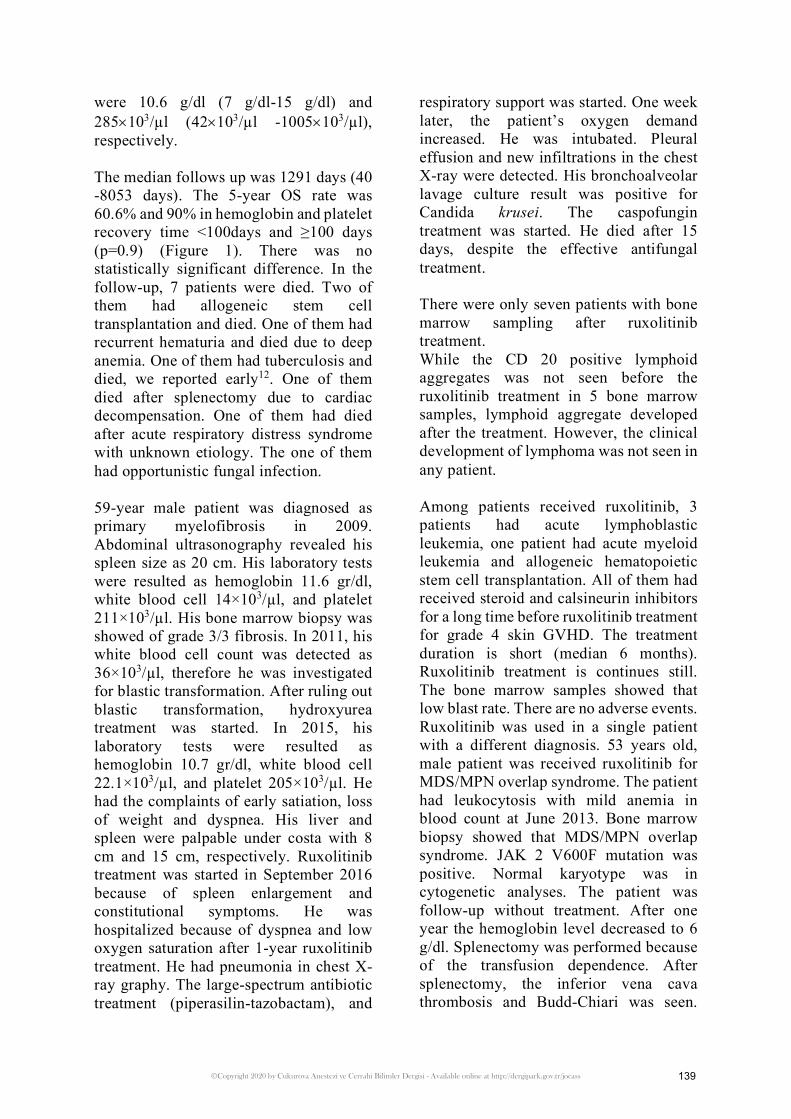
Figure 1. Comparison of OS among groups of time nadir to recovery of hemoglobin.
There is no patient with pruritis, diarrhea and herpes infection. The leukemic transformation was not improving in any patient. The dose modification was made in 8 (28.6%) patients because of adverse events.
After ruxolitinib, median platelet, hemoglobin, neutrophil nadir durations were 46 days (0-546), 40 days (14-218days) and 112 days (16-546days), respectively. The median recovery duration of platelet, hemoglobin, neutrophil durations were 90 days (6-467days), 101 days (6-625days) and 50 days (6-363 days), respectively. When the
median hemoglobin, platelet recovery time was grouped as 100 days; 48.1% (13 patients) and 51.9% (14 patients) of patients have hemoglobin and platelet recovery time <100days, respectively.
67.9% (19 patients) of patients have the nadir hemoglobin level ≤10 g/dl. 32.1% (9 patients) of patients have the nadir platelet ≤100×103/µl. 46.4% (13 patients) of patients have the nadir neutrophil ≤5×10³/µl. The median nadir hemoglobin and platelet level were 8.3 g/dl (5 g/dl -15 g/dl) and 147.5×10³/µl (29×103/µl -589×103/µl), respectively. The median recovery hemoglobin and platelet level
138
©Copyright 2020 by Çukurova Anestezi ve Cerrahi Bilimler Dergisi - Available online at http://dergipark.gov.tr/jocass
were 10.6 g/dl (7 g/dl-15 g/dl) and 285×103/µl (42×103/µl -1005×103/µl), respectively.
The median follows up was 1291 days (40 -8053 days). The 5-year OS rate was 60.6% and 90% in hemoglobin and platelet recovery time <100days and ≥100 days (p=0.9) (Figure 1). There was no statistically significant difference. In the follow-up, 7 patients were died. Two of them had allogeneic stem cell transplantation and died. One of them had recurrent hematuria and died due to deep anemia. One of them had tuberculosis and died, we reported early12. One of them died after splenectomy due to cardiac decompensation. One of them had died after acute respiratory distress syndrome with unknown etiology. The one of them had opportunistic fungal infection.
59-year male patient was diagnosed as primary myelofibrosis in 2009. Abdominal ultrasonography revealed his spleen size as 20 cm. His laboratory tests were resulted as hemoglobin 11.6 gr/dl, white blood cell 14×103/µl, and platelet 211×103/µl. His bone marrow biopsy was showed of grade 3/3 fibrosis. In 2011, his white blood cell count was detected as 36×103/µl, therefore he was investigated for blastic transformation. After ruling out blastic transformation, hydroxyurea treatment was started. In 2015, his laboratory tests were resulted as hemoglobin 10.7 gr/dl, white blood cell 22.1×10³/µl, and platelet 205×103/µl. He had the complaints of early satiation, loss of weight and dyspnea. His liver and spleen were palpable under costa with 8 cm and 15 cm, respectively. Ruxolitinib treatment was started in September 2016 because of spleen enlargement and constitutional symptoms. He was hospitalized because of dyspnea and low oxygen saturation after 1-year ruxolitinib treatment. He had pneumonia in chest X-ray graphy. The large-spectrum antibiotic treatment (piperasilin-tazobactam), and
respiratory support was started. One week later, the patient’s oxygen demand increased. He was intubated. Pleural effusion and new infiltrations in the chest X-ray were detected. His bronchoalveolar lavage culture result was positive for Candida krusei. The caspofungin treatment was started. He died after 15 days, despite the effective antifungal treatment.
There were only seven patients with bone marrow sampling after ruxolitinib treatment. While the CD 20 positive lymphoid aggregates was not seen before the ruxolitinib treatment in 5 bone marrow samples, lymphoid aggregate developed after the treatment. However, the clinical development of lymphoma was not seen in any patient.
Among patients received ruxolitinib, 3 patients had acute lymphoblastic leukemia, one patient had acute myeloid leukemia and allogeneic hematopoietic stem cell transplantation. All of them had received steroid and calsineurin inhibitors for a long time before ruxolitinib treatment for grade 4 skin GVHD. The treatment duration is short (median 6 months). Ruxolitinib treatment is continues still. The bone marrow samples showed that low blast rate. There are no adverse events. Ruxolitinib was used in a single patient with a different diagnosis. 53 years old, male patient was received ruxolitinib for MDS/MPN overlap syndrome. The patient had leukocytosis with mild anemia in blood count at June 2013. Bone marrow biopsy showed that MDS/MPN overlap syndrome. JAK 2 V600F mutation was positive. Normal karyotype was in cytogenetic analyses. The patient was follow-up without treatment. After one year the hemoglobin level decreased to 6 g/dl. Splenectomy was performed because of the transfusion dependence. After splenectomy, the inferior vena cava thrombosis and Budd-Chiari was seen.
139

©Copyright 2020 by Çukurova Anestezi ve Cerrahi Bilimler Dergisi - Available online at http://dergipark.gov.tr/jocass
Ruxolitinib was started because of developing B symptoms and hydroxyurea resistant anemia. After three months ruxolitinib treatment, the hemoglobin level is 10g/dl with no adverse event.
Discussion
In the scoring study involving 235 PMF and 186 secondary MF patients, grade 2 anemia developed in 67.5% and 56.4% of the patients at 3 months and 6 months after starting ruxolitinib. 43.7% and 36.9% of the patients had grade 3 anemia at same period. Grade 2 thrombocytopenia developed in 43.2% and 44.2% of the patients at 3 months and 6 months after ruxolitinib treatment2. In this study, relatively small retrospective cohort study, it was observed that 3.1 units of hemoglobin were decreased after the initiation of ruxolitinib from the diagnosis of the patients. Our study cohort nadir hemoglobin level median was 8.3g/dl and median duration of nadir hemoglobin was 40 days, relatively fast fall.
In a new study to investigate the effect of ruxolitinib on spleen size, 18 of patients had leukemic transformation. Causes of death were 20.8% and 12.5% of patients have acute leukemia and infections, respectively13. In this small study cohort, there was no leukemic transformation.
In another last study, tuberculosis was seen in 3/65 patients14. In our study, tuberculosis was seen in one patient. But it has not been mentioned in detail since it has been previously reported 12. In this study, one patient died due to opportunistic fungal infection in the lung.
In fact, as mentioned in the new study, which is the inspiration of this study,
lymphoid development can be seen in patients who receive ruxolitinib. However, none of our patients had clinically developed lymphoma. Only 6 patients who underwent bone marrow sampling 6 months after starting ruxolitinib were re-evaluated. In 5 samples, the CD 20 positive lymphocyte aggregate was new. In 2 samples, the lymphoid aggregates were found to be similar to the bone marrow samples taken before ruxolitinib was started. The presence of lymphoid aggregate as a feature of myeloproliferative diseases makes the interpretation difficult15.
The weakness of the study is the small number of patients. Furthermore, retrospective evaluation of data is another weakness of the study.
In conclusion, although ruxolitinib has been shown to improve survival in myelofibrosis in the long term, survival may be short due to side effects. In this small cohort study 25% of patients died due to different reasons.
Conflict of Interest
The authors declare that they have no conflict of interest
Funding
None
140

©Copyright 2020 by Çukurova Anestezi ve Cerrahi Bilimler Dergisi - Available online at http://dergipark.gov.tr/jocass
References
1. Verstovsek S, Gotlib J, Mesa RA, Vannucchi AM, Kiladjian JJ, Cervantes F, et al. Long-term survival in patients treated with ruxolitinib for myelofibrosis: COMFORT-I and -II pooled analyses. J Hematol Oncol. 2017 Sep;10(1):156.
2. Palandri F, Palumbo GA, Iurlo A, Polverelli N, Benevolo G, Breccia M, et al. Differences in presenting features, outcome and prognostic models in patients with primary myelofibrosis and post-polycythemia vera and/or post-essential thrombocythemia myelofibrosis treated with ruxolitinib. New perspective of the MYSEC-PM in a large multicenter study⁎ [). WB Saunders.]. Semin Hematol. 2018 Oct;55(4):248–55.
3. Cervantes F, Dupriez B, Pereira A, Passamonti F, Reilly JT, Morra E, et al. New prognostic scoring system for primary myelofibrosis based on a study of the International Working Group for Myelofibrosis Research and Treatment. Blood. 2009 Mar;113(13):2895–901.
4. Passamonti F, Cervantes F, Vannucchi AM, Morra E, Rumi E, Cazzola M, et al. Dynamic International Prognostic Scoring System (DIPSS) predicts progression to acute myeloid leukemia in primary myelofibrosis. Blood. 2010 Oct;116(15):2857–8.
5. Gangat N, Caramazza D, Vaidya R, George G, Begna K, Schwager S, et al. DIPSS plus: a refined Dynamic International Prognostic Scoring System for primary myelofibrosis that incorporates prognostic information from karyotype, platelet count, and transfusion status. J Clin Oncol. 2011 Feb;29(4):392–7.
6. Verstovsek S, Mesa RA, Gotlib J, Levy RS, Gupta V, DiPersio JF, et al. A double-blind, placebo-controlled trial of ruxolitinib for myelofibrosis. N Engl J Med. 2012 Mar;366(9):799–807.
7. Verstovsek S, Mesa RA, Gotlib J, Gupta V, DiPersio JF, Catalano JV, et al.; COMFORT-I investigators. Long-term treatment with ruxolitinib for patients with myelofibrosis: 5-year update from the randomized, double-blind, placebo-controlled, phase 3 COMFORT-I trial. J Hematol Oncol. 2017 Feb;10(1):55.
8. Porpaczy E, Tripolt S, Hoelbl-Kovacic A, Gisslinger B, Bago-Horvath Z, Casanova-Hevia E, et al. Aggressive B-cell lymphomas in patients with myelofibrosis receiving JAK1/2 inhibitor therapy. Blood. 2018 Aug;132(7):694–706.
9. Bhatt VR, Bociek RG, Yuan J, Fu K, Greiner TC, Dave BJ, et al. Leukemic diffuse large B-cell lymphoma in a patient with myeloproliferative disorder. J Natl Compr Canc Netw. 2015 Mar;13(3):281–7.
10. Tefferi A, Pardanani A. Serious adverse events during ruxolitinib treatment discontinuation in
patients with myelofibrosis [). Elsevier.]. Mayo Clin Proc. 2011 Dec;86(12):1188–91.
11. Modi B, Hernandez-Henderson M, Yang D, Klein J, Dadwal S, Kopp E, et al. Ruxolitinib as salvage therapy for chronic graft-versus-host disease. Biol Blood Marrow Transplant. 2019 Feb;25(2):265–9.
12. Malkan UY, Haznedaroglu IC. A myelofibrosis case that develops mycobacterial infection after ruxolitinib treatment. Int J Clin Exp Med. 2017;10:7304–7.
13. Palandri F, Palumbo GA, Bonifacio M, Breccia M, Latagliata R, Martino B, et al. Durability of spleen response affects the outcome of ruxolitinib-treated patients with myelofibrosis: results from a multicentre study on 284 patients. Leuk Res. 2018 Nov;74:86–8.
14. Lescuyer S, Ledoux MP, Gravier S, Natarajan-Amé S, Duval C, Maloisel F, et al. Tuberculosis and atypical mycobacterial infections in ruxolitinib-treated patients with primary or secondary myelofibrosis or polycythemia vera. Int J Infect Dis. 2019 Mar;80:134–6.
15. Tefferi AJ, Vardiman JW. Classification and diagnosis of myeloproliferative neoplasms: the 2008 WHO criteria and point-of-care diagnostic algorithms. leukemia, 2008 22(1):14-22.
141