Embed Size (px)
Citation preview

Tian et al. – 1
1
Running title: 2
Characterization of a Nod factor hydrolase 3
4
5
6
Corresponding authors: 7
Christian Staehelin and Zhiping Xie 8
State Key Laboratory of Biocontrol and Guangdong Key Laboratory of Plant 9
Resources, 10
School of Life Sciences, 11
Sun Yat-sen University, East Campus, 12
510006 Guangzhou, 13
China; 14
tel.: +86 020 39332976 15
e-mail: [email protected]; [email protected] 16
17
18
19
20
21
Research Area: BIOCHEMISTRY AND METABOLISM 22
23
Secondary Research Area: SIGNALING AND RESPONSE24
Plant Physiology Preview. Published on September 30, 2013, as DOI:10.1104/pp.113.223966
Copyright 2013 by the American Society of Plant Biologists
www.plantphysiol.orgon April 5, 2019 - Published by Downloaded from Copyright © 2013 American Society of Plant Biologists. All rights reserved.

Tian et al. – 2
25
Title of article: 26
The Nod factor hydrolase of Medicago truncatula: Characterization of an enzyme 27
specifically cleaving rhizobial nodulation signals 28
29
Authors: 30
Ye Tian1, Wei Liu 1, Jie Cai1, Lan-Yue Zhang1, Kam-Bo Wong2, Nadja Feddermann3, 31
Thomas Boller3, Zhi-Ping Xie1* and Christian Staehelin1* 32
33
1State Key Laboratory of Biocontrol and Guangdong Key Laboratory of Plant 34
Resources, School of Life Sciences, Sun Yat-sen University, East Campus, 510006 35
Guangzhou, China;
36
2School of Life Sciences and Center for Protein Science and Crystallography, The 37
Chinese University of Hong Kong, Shatin, Hong Kong, China; 38
3Botanisches Institut der Universität Basel, Zurich Basel Plant Science Center, 39
Hebelstrasse 1, 4056 Basel, Switzerland 40
41
42
43
One-sentence summary: 44
45
An enzyme of the host legume Medicago truncatula exclusively cleaves nodulation 46
signals (Nod factors) from the microsymbiont Sinorhizobium meliloti, but lacks 47
activity towards chitin or non-modified chitin oligosaccharides. 48
www.plantphysiol.orgon April 5, 2019 - Published by Downloaded from Copyright © 2013 American Society of Plant Biologists. All rights reserved.
Tian et al. – 3
49
Footnotes: 50
51
1This study was supported by the National Basic Research Program of China (973 52
program, grant 2010CB126501), by the Guangdong Key Laboratory of Plant Resources 53
(grant plant01k19), by the Science Foundation of the State Key Laboratory of Biocontrol 54
(grants SKLBC09B05 and SKLBC201123) and by the SYSU-CUHK Joint Center for 55
Protein Research. T.B. acknowledges funding by the Swiss National Science Foundation 56
and the Systems-X initiative. 57
58
2YT and WL contributed equally to this work. 59
60
3Present address of NF: Institute of Biology, Laboratory of Molecular and Cellular 61
Biology, University of Neuchâtel, Rue Emile-Argand 11, CH-2000 Neuchâtel, 62
Switzerland 63
64
65
*For correspondence: Christian Staehelin and Zhi-Ping Xie; [email protected]; 66
68
69
www.plantphysiol.orgon April 5, 2019 - Published by Downloaded from Copyright © 2013 American Society of Plant Biologists. All rights reserved.

Tian et al. – 4
70
ABSTRACT 71
72
Nodule formation induced by nitrogen-fixing rhizobia depends on bacterial 73
nodulation factors (NFs), modified chitin oligosaccharides with a fatty acid moiety. 74
Certain NFs can be cleaved and inactivated by plant chitinases. However, the most 75
abundant NF of Sinorhizobium meliloti, an O-acetylated and sulphated tetramer, is 76
resistant to hydrolysis by all plant chitinases tested so far. Nevertheless, this NF is 77
rapidly degraded in the host rhizosphere. Here, we identify and characterize MtNFH1 78
(Medicago truncatula Nod factor hydrolase 1), a legume enzyme structurally related 79
to defense-related class V chitinases (glycoside hydrolase family 18). MtNFH1 lacks 80
chitinase activity but efficiently hydrolyzes all tested NFs of S. meliloti. The enzyme 81
shows a high cleavage preference, releasing exclusively lipo-disaccharides from NFs. 82
Substrate specificity and kinetic properties of MtNFH1 were compared to those of 83
class V chitinases from Arabidopsis thaliana and Nicotiana tabacum, which cannot 84
hydrolyze tetrameric NFs of S. meliloti. The Michaelis-Menten constants of MtNFH1 85
for NFs are in the micromolar concentration range, whereas non-modified chitin 86
oligosaccharides represent neither substrates nor inhibitors for MtNFH1. The three-87
dimensional structure of MtNFH1 was modeled on the basis of the known structure of 88
class V chitinases. Docking simulation of NFs to MtNFH1 predicted a distinct binding 89
cleft for the fatty acid moiety, which is absent in the class V chitinases. Point mutation 90
analysis confirmed the modeled NF-MtNFH1 interaction. Silencing of MtNFH1 by 91
RNA interference resulted in reduced NF degradation in the rhizosphere of M. 92
truncatula. In conclusion, we have found a novel legume hydrolase that specifically 93
inactivates NFs. 94
www.plantphysiol.orgon April 5, 2019 - Published by Downloaded from Copyright © 2013 American Society of Plant Biologists. All rights reserved.

Tian et al. – 5
95
INTRODUCTION 96
97
Nitrogen-fixing bacteria (rhizobia) establish a root nodule symbiosis with legume 98
plants. Rhizobial infection and subsequent nodule formation depends on bacterial 99
signaling, namely Nod factors (NFs), which are perceived by specific LysM receptor 100
kinases of the host plant (Antolin-Llovera et al., 2012). NFs are lipo-101
chitooligosaccharides, i.e. modified chitin oligosaccharides with a fatty acyl moiety 102
replacing the N-acetyl group at the non-reducing end. In most cases, the 103
oligosaccharide moiety of NFs consists of four or five β-1,4-linked N-acetyl-104
glucosamine (GlcNAc) residues. Depending on the rhizobial strain, NFs possess 105
diverse additional substitutions on the oligosaccharide backbone (Perret et al., 2000). 106
Such structural modifications of NFs influence the binding of NFs to the LysM 107
receptor kinases. Hence, NF signaling in host plants culminating in subsequent 108
expression of symbiosis-specific genes and nodule formation is only triggered by 109
specific NFs (Perret et al., 2000; Antolin-Llovera et al., 2012). 110
Due to structural similarities to chitin, various NFs can be hydrolyzed by plant 111
chitinases (Staehelin et al., 1994a; Staehelin et al., 1994b; Minic et al., 1998; Schultze 112
et al., 1998; Goormachtig et al., 1998; Ovtsyna et al., 2000). Chitinases (EC 3.2.1.14) 113
are defined as enzymes that cleave β-1,4 glycosidic linkages in chitin (poly-N-114
acetylglucosamine; poly-GlcNAc) and chitin oligosaccharides (Collinge et al., 1993). 115
Chitin is a major polysaccharide in fungal cell walls and exoskeletons of arthropods. 116
Plants do not possess chitin, but use chitinases as defense proteins against chitin-117
containing antagonists, such as fungi and arthropods. Various plant chitinases possess 118
additional lysozyme activity, i.e. they can cleave β-1,4-linkages between N-119
acetylmuramic acid and N-acetylglucosamine in bacterial peptidoglycan (Collinge et 120
al., 1993). Furthermore, certain plant chitinases appear to hydrolyze endogenous 121
substrates such as N-acetylglucosamine containing arabinogalactan proteins (De Jong 122
et al., 1992; van Hengel et al., 2001). Based on their amino acid sequence, chitinolytic 123
enzymes of plants belong either to glycoside hydrolase family 18 (class III and V 124
www.plantphysiol.orgon April 5, 2019 - Published by Downloaded from Copyright © 2013 American Society of Plant Biologists. All rights reserved.
Tian et al. – 6
chitinases) or family 19 (class I, II and IV chitinases; Henrissat, 1991). 125
Class V chitinases of plants, also classified as PR-11 (pathogen-related protein 126
family 11) proteins, form a separate clade within the phylogenetic tree of family 18 127
glycoside hydrolases (Karlsson and Stenlid, 2009). A phylogenetic tree of class V 128
chitinases and closely related proteins is shown Figure S1. In contrast to other plant 129
chitinase classes, only few class V proteins have been characterized. Nearly two 130
decades ago, a first class V chitinase (NtChiV; also named Pz chitinase) has been 131
purified from tobacco mosaic virus inoculated leaves of tobacco (Nicotiana tabacum). 132
This defense-related enzyme efficiently cleaves chitin oligosaccharides (Melchers et 133
al., 1994; Heitz et al., 1994; Brunner et al., 1998). Recently, enzymatic 134
characterization and protein crystal structure analysis was performed with the NtChiV 135
protein expressed in Escherichia coli as well as with AtChiC, a related class V 136
chitinase from Arabidopsis thaliana (Ohnuma et al., 2011a; Ohnuma et al., 2011b). 137
Furthermore, a class V chitinase purified from the gymnosperm Cycas revoluta 138
exhibits transglycosylation activity at high concentrations of chitin oligosaccharides 139
(Taira et al., 2009; Taira et al., 2010). In addition to these enzymes, a class V chitinase 140
related lectin (RobpsCRA) from the legume tree Robinia pseudoacacia has been 141
characterized. This protein binds to high-mannose N-glycans but lacks catalytic 142
activity (Van Damme et al., 2007). Similarly, the extracellular domain of the 143
chitinase-related receptor-like kinase 1 (CHRK1) of tobacco is related to class V 144
chitinases (Kim et al., 2000). 145
In the nodule symbiosis, host chitinases have the potential to inactivate NFs. When 146
tested in root hair deformation bioassays, the purified cleavage products are at least 147
1000-fold less active (Heidstra et al., 1994; Staehelin et al., 1994b). In fact, NFs 148
produced by compatible rhizobia were found to be rapidly degraded in the rhizosphere 149
of legumes (Heidstra et al., 1994; Staehelin et al., 1994b; Staehelin et al., 1995; 150
Ovtsyna et al., 2000; Ovtsyna et al., 2005). NFs with a shorter oligosaccharide 151
backbone and carrying additional chemical substitutions are usually more resistant 152
against degradation by chitinases (Staehelin et al., 1994a; Staehelin et al., 1994b; 153
Minic et al., 1998; Schultze et al., 1998; Ovtsyna et al., 2000). NodSm-IV(C16:2, Ac, 154
www.plantphysiol.orgon April 5, 2019 - Published by Downloaded from Copyright © 2013 American Society of Plant Biologists. All rights reserved.
Tian et al. – 7
S), an O-acetylated and sulphated tetrasaccharide with a C16:2 fatty acid chain 155
(Lerouge et al., 1990), is the most abundant NF produced by Sinorhizobium meliloti 156
(Figure S2). This NF is resistant to hydrolysis by all plant chitinases tested so far 157
(Schultze et al., 1998). Nevertheless, when added to roots of the host plant alfalfa 158
(Medicago sativa), NodSm-IV(C16:2, Ac, S) was rapidly degraded to a lipo-159
disaccharide, and increased NF hydrolysis was observed when roots were pretreated 160
with NFs (Staehelin et al., 1995). NFs were also found to induce a NF-cleaving 161
activity in the rhizosphere of pea plants, and this induction did not happen in NF 162
signaling deficient pea mutants (Ovtsyna et al., 2000; Ovtsyna et al., 2005). Similarly, 163
chitinolytic activities of certain chitinase isoforms of legumes are induced during 164
symbiosis with rhizobia or in response to purified NFs (Staehelin et al., 1992; Xie et 165
al., 1999; Ovtsyna et al., 2005). Furthermore, transcript levels of the class III chitinase 166
gene Srchi13 in the tropical legume Sesbania rostrata are strongly stimulated during 167
symbiosis and the encoded protein expressed in E. coli can degrade NFs of 168
Azorhizobium caulinodans to non-identified cleavage products (Goormachtig et al., 169
1998). In the model legume Medicago truncatula, expression of the class V chitinase 170
gene MtChit5 is induced during symbiosis with S. meliloti (Salzer et al., 2004). Taken 171
together, these findings raise the question of whether legumes possess chitinase-172
related enzymes that specifically cleave NFs. 173
In this study, we show that the MtChit5 protein of M. truncatula lacks chitinase 174
activity but efficiently hydrolyzes NFs of S. meliloti. We therefore renamed MtChit5 175
to MtNFH1 (Medicago truncatula Nod factor hydrolase 1). A substrate binding 176
model, supported by point mutation analysis, provides a molecular explanation for the 177
identified MtNFH1-NF interaction. 178
179
RESULTS 180
181
MtNFH1 encodes a NF-cleaving enzyme 182
In previous work, we have identified a symbiosis-related chitinase V gene, 183
MtChit5, in M. truncatula ecotype R108-1. The expression of this gene is strongly 184
www.plantphysiol.orgon April 5, 2019 - Published by Downloaded from Copyright © 2013 American Society of Plant Biologists. All rights reserved.

Tian et al. – 8
induced in the nodule symbiosis (Salzer et al., 2004). What is the catalytic activity of 185
the enzyme encoded by this gene? To answer this question, the corresponding DNA 186
was cloned into vector pET28b in order to express it as a His-tagged recombinant 187
protein in E. coli BL21 (DE3). Two related genes of M. truncatula (Mt75352 and 188
MtCRA) known to be expressed in M. truncatula (Table S1 and Figure S1) were also 189
cloned and expressed in E. coli. The Mt75352 sequence encodes a putative class V 190
chitinase, whereas MtCRA is related to the previously characterized lectin RobpsCRA 191
of R. pseudoacacia (Van Damme et al., 2007). For comparison, the class V chitinases 192
AtChiC of A. thaliana (Ohnuma et al., 2011b) and NtChiV of tobacco (Ohnuma et al., 193
2011a) were cloned and expressed in a similar way. AtChiC and NtChiV show 194
significant amino acid sequence homology to MtChit5 (sequence identity: 42% and 195
39%, respectively). The recombinant proteins were isolated by nickel affinity 196
purification and analyzed by SDS-PAGE. Rabbit antiserum raised against the 197
recombinant MtChit5 (re-named MtNFH1, see below) was cross-reactive with the 198
other four proteins, indicating successful purification of the recombinant proteins 199
(Figure 1A).Purified proteins were then used for enzyme assays with pentameric and 200
tetrameric NFs (Figure S2). These substrates had been HPLC-purified from S. meliloti 201
strain 1021 (pEK327) (Schultze et al., 1992). We found that the purified product of 202
the MtChit5 gene rapidly hydrolysed all the NFs (Table 1) and we therefore re-named 203
it MtNFH1. Representative HPLC chromatograms with NF substrates and acylated 204
cleavage products (lipo-oligosaccharides after n-butanol extraction) are shown in 205
Figure 1B. The lipo-disaccharide NodSm-II(C16:2) was formed when either 206
pentameric NodSm-V(C16:2, S) or tetrameric NodSm-IV(C16:2, S) were used as 207
substrate (panels a and b). NodSm-IV(C16:2), a chemically desulphated NF, was also 208
hydrolyzed by MtNFH1, albeit to a lesser extent (panel c). Interestingly, MtNFH1 was 209
also able to cleave the O-acetylated tetramer NodSm-IV(C16:2, Ac, S); the more 210
slowly migrating O-acetylated lipo-disaccharide NodSm-II(C16:2, Ac) was detected 211
on HPLC chromatograms (panel d). Fractions containing lipo-disaccharides were 212
collected and chemical structures of lipo-disaccharides were verified by mass 213
spectrometry (Figure S3; Table S2). 214
www.plantphysiol.orgon April 5, 2019 - Published by Downloaded from Copyright © 2013 American Society of Plant Biologists. All rights reserved.

Tian et al. – 9
MtNFH1(D148A), a mutant protein in which the conserved aspartic acid residue 215
D148 of the predicted catalytic center was changed to alanine, did not show enzyme 216
activity (Figure 1B, panel e). Mt75352 and MtCRA, proteins of M. truncatula related 217
to MtNFH1, were unable to hydrolyze the NF substrates (panels f and g). AtChiC of 218
A. thaliana hydrolyzed pentameric NodSm-V(C16:2, S), but the cleavage products 219
were different: the more rapidly migrating lipo-trisaccharide NodSm-III(C16:2) was 220
formed (panel h). Similarly, the tobacco enzyme NtChiV released NodSm-III(C16:2) 221
from the pentameric substrate (panel i), as reported previously (Schultze et al., 1998). 222
The structure of the product NodSm-III(C16:2) was confirmed by mass spectrometry 223
(Figure S3; Table S2). As compared to MtNFH1, hydrolytic activities of AtChiC and 224
NtChiV for the substrate NodSm-V(C16:2, S) were more than 100-fold lower (Table 225
1). Tetrameric NFs were neither cleaved by AtChiC nor by NtChiV (Schultze et al., 226
1998). MtNFH1 was unable to hydrolyze purified NodSm-III(C16:2), providing 227
evidence that the enzyme directly releases lipo-disaccharides from the NF substrates 228
(Figure 1B, panel j). 229
230
MtNFH1 specifically cleaves NFs 231
We further tested whether MtNFH1 can hydrolyze non-modified chitin 232
oligosaccharides (oligomers of GlcNAc), which likely represent the natural substrates 233
for AtChiC and NtChiV. The hexamer (GlcNAc)6 and the pentamer (GlcNAc)5 were 234
used in enzyme assays with purified proteins. After incubation, chitin 235
oligosaccharides were separated on an amino column. Surprisingly, MtNFH1 protein 236
samples, which had shown such a high activity with NF substrates, completely lacked 237
activity with these non-modified chitin oligosaccharides (Table 1; Figure 2). To test a 238
possible inhibitory effect of chitin oligosaccharides on NF degradation, MtNFH1 was 239
incubated with NodSm-V(C16:2, S) and a 500-fold higher concentration of 240
(GlcNAc)5. NFs and acylated degradation products were then analyzed by HPLC. 241
Formation of NodSm-II(C16:2) by MtNFH1was not affected by (GlcNAc)5 as shown 242
in Figure S4. 243
In contrast to MtNFH1, purified AtChiC and NtChiV proteins efficiently 244
www.plantphysiol.orgon April 5, 2019 - Published by Downloaded from Copyright © 2013 American Society of Plant Biologists. All rights reserved.
Tian et al. – 10
hydrolyzed non-modified chitin oligosaccharides in accordance to previous reports 245
(Brunner et al., 1998; Ohnuma et al., 2011a; Ohnuma et al., 2011b). (GlcNAc)6 was 246
cleaved into (GlcNAc)3 or into (GlcNAc)4 and (GlcNAc)2. The product (GlcNAc)4 247
was then further hydrolyzed into (GlcNAc)2. The substrate (GlcNAc)5 was cleaved 248
into (GlcNAc)3 and (GlcNAc)2 (Table 1 and Figure 2). 249
When colloidal chitin, glycolchitin or carboxymethyl-chitin-Remazol Brilliant 250
Violet 5R (CM-chitin-RBV) were tested as substrates, AtChiC and NtChiV clearly 251
showed enzymatic activity, while MtNFH1 was completely inactive (Table 1). The 252
Mt75352 and MtCRA proteins were similarly inactive. MtNFH1, like AtChiC and 253
NtChiV, lacked lysozyme activity as measured with Micrococcus lysodeikticus cells 254
(Table 1), which is in agreement with a previous report on NtChiV (Heitz et al., 255
1994). NtChiV inhibited hyphal growth of the fungus Trichoderma viride (Melchers 256
et al., 1994; Ohnuma et al. 2012) and this was confirmed in a similar bioassay in this 257
study. The MtNFH1 protein lacked such antifungal activity, however (Figure 3). 258
259
Kinetic studies indicate efficient NF hydrolysis at low substrate concentrations 260
Kinetic studies with varying concentrations of NFs were performed to determine 261
Michaelis-Menten constants (Km), and catalytic rate constants (kcat) of the purified 262
proteins (Table 2 and Figure S5). MtNFH1 showed Km values at micromolar 263
concentrations with all tested NFs of S. meliloti. A slightly higher Km and a lower kcat 264
value were determined for the O-acetylated substrate NodSm-IV(C16:2, Ac, S) as 265
compared to NFs without O-acetyl group. Similarly low Km values were also obtained 266
for AtChiC and NtChiV releasing NodSm-III(C16:2) from NodSm-V(C16:2, S). As 267
compared to MtNFH1, however, only low kcat values were determined. 268
The results of kinetic studies with AtChiC and NtChiV for the substrates 269
(GlcNAc)6 and (GlcNAc)5 are summarized in Table S3. Relatively high Km values in 270
the millimolar range were determined for these substrates. On the other hand, the kcat 271
values for (GlcNAc)6 and (GlcNAc)5 were found to be more than two orders of 272
magnitude higher as compared to those for NodSm-V(C16:2, S) shown in Table 2. 273
274
www.plantphysiol.orgon April 5, 2019 - Published by Downloaded from Copyright © 2013 American Society of Plant Biologists. All rights reserved.
Tian et al. – 11
Docking simulation predicts a specific binding cleft for a fatty acid chain 275
Since MtNFH1 cleaves NFs with their fatty acyl decorations, but not the simple chitin 276
oligosaccharides, we wondered whether the fatty acid chain was required for correct 277
substrate binding. To analyze substrate recognition on a molecular level, a three-278
dimensional model of MtNFH1 was constructed by homology modeling, using the 279
crystal structures of AtChiC or NtChiV as structural templates. The structures of the 280
substrates NodSm-V(C16:2, S) or (GlcNAC)5 were then manually docked to the 281
active site of the proteins. Aligned primary sequences of the three proteins and the 282
modeled substrate-protein complexes are shown in Figure 4. All three enzymes 283
possess a predicted substrate binding pocket for GlcNAc residues. Noteworthy, two 284
loops in MtNFH1 (loop A and B) are predicted to contribute to the formation of the 285
binding cleft for the C16:2 fatty acid moiety. Loop A consists of amino acid residues 286
between strand-9 and helix-6 (GSGS motif) and loop B is formed by residues between 287
strand-12 and strand-13 (GPGPGVDGG motif) (Figure 4A). In contrast to the AtChiC 288
and NtChiV sequences, these two loops of MtNFH1 are shorter and are glycine-rich, 289
which result in a prominent cleft that can accommodate the C16:2 fatty acid moiety of 290
NodSm-V(C16:2, S) (Figure 4B). Based on our model, NF substrates perfectly fit to 291
MtNFH1 in this way and are then cleaved into NodSm-II(C16:2) and a sulphated 292
chitin oligosaccharide. Hence, MtNFH1 targets NFs at their non-reducing ends. On 293
the other hand, there is apparently no space in AtChiC and NtChiV to accommodate 294
the fatty acid moiety at this position (Figure 4B), which justify the observation that 295
they cannot release NodSm-II(C16:2) from NodSm-V(C16:2, S). 296
A superimposed model of MtNFH1 and NtChiV indicates changes in 297
sequence/structure that lead to the formation of the fatty acid binding cleft (Figure 298
4C). Specifically, the tyrosine and serine residues of NtChiV (Y236, S265) and the 299
corresponding residues in AtChiC are substituted in MtNFH1 by a lysine and glycine 300
at the corresponding positions (K241, G267). These two residues are located at the 301
entrance of the cleft between the loops A and B, and these more bulky tyrosine and 302
serine residues in AtChiC and NtChiV are predicted to block the formation of a cleft. 303
Furthermore, NtChiV and AtChiC have an extra proline residue (P188 in NtChiV), 304
www.plantphysiol.orgon April 5, 2019 - Published by Downloaded from Copyright © 2013 American Society of Plant Biologists. All rights reserved.

Tian et al. – 12
which is absent in the loop A of MtNFH1. The deletion of this proline residue in 305
MtNFH1 results in a shorter loop A and thus enlarges the predicted fatty acid binding 306
cleft. 307
308
Point mutation analysis supports the modeled NF-MtNFH1 interaction 309
Our model predicts that K241 and G267 of MtNFH1, which guard the entrance of the 310
fatty acid binding cleft, should be important for substrate recognition. To test this 311
hypothesis, we constructed a double mutant of MtNFH1 (K241Y/G267S), in which 312
lysine-241 is substituted by tyrosine and glycine-267 by serine (corresponding to 313
Y236 and S265 in NtChiV, respectively). When assayed with NFs as substrates, the 314
MtNFH1(K241Y/G267S) protein showed ~ 2.5-fold increases in Km and ~ 2-fold 315
decreases in kcat, which resulted in ~ 5-fold reduced activity (kcat/Km) (Table 3). These 316
data provide experimental support for the modeled NF-MtNFH1 interaction. 317
318
Silencing of MtNFH1 by RNA interference increases NF levels in the rhizosphere 319
When adding NFs of S. meliloti to roots of alfalfa, they are rapidly degraded by a NF-320
cleaving extracellular activity (Staehelin et al., 1994b; Staehelin et al., 1995). To 321
investigate whether MtNFH1 encodes an enzyme that contributes to NF degradation 322
in the rhizosphere, we transformed M. truncatula ecotype R108-1 with Agrobacterium 323
tumefaciens carrying pCAMBIA-MtNFH1i. This RNA interference (RNAi) plasmid 324
contains a construct made of the cauliflower mosaic virus (CaMV) 35S promoter in 325
tandem and a fragment of MtNFH1, which is inserted in both sense and anti-sense 326
direction. These sense and anti-sense sequences are separated by a spacer region. 327
Furthermore, the vector carries a β-glucuronidase (GUS) gene under the control of the 328
CaMV 35S promoter, which allows to distinguish between transformed and non-329
transformed plants. Primary transformants were regenerated from A. tumefaciens-330
infiltrated leaf disks on selective media containing hygromycin. Young seedlings from 331
these RNAi lines (T2 generation) were first treated for one day with 0.1 μM NodSm-332
IV(C16:2, S), in order to stimulate the NF-cleaving activity in the rhizosphere. The 333
NF degradation test was then performed by incubation in 15 μM NodSm-IV(C16:2, 334
www.plantphysiol.orgon April 5, 2019 - Published by Downloaded from Copyright © 2013 American Society of Plant Biologists. All rights reserved.
Tian et al. – 13
S). After removal of the seedlings, the NFs and the released NodSm-II(C16:2, S) were 335
extracted from the medium by n-butanol and quantified by HPLC analysis. Finally, 336
the seedlings were used for determination of GUS activity and plants lacking blue 337
staining were excluded from the experiment. Compared to wild-type plants, an up to 338
5-fold reduction in NF hydrolysis was measured for plants transformed with 339
pCAMBIA-MtNFH1i, indicating that silencing of MtNFH1 increases NF levels in the 340
rhizosphere (Figure 5). 341
342
343
DISCUSSION 344
In this work, we have biochemically characterized MtNFH1, which is encoded by a 345
symbiosis stimulated M. truncatula gene (Salzer et al., 2004). The enzyme shows 346
sequence similarities to class V chitinases but lacks chitinase activity. Instead, 347
MtNFH1 cleaves NFs of the microsymbiont S. meliloti. Notably, MtNFH1 hydrolyzes 348
NodSm-IV(C16:2, Ac, S), which is structurally rather different from non-modified 349
chitin oligosaccharides. Based on the capacity to release lipo-disaccharides from NFs 350
(this study) and the increase of MtNFH1 transcripts in NF treated roots (Salzer et al., 351
2004), it is likely that MtNFH1 is an ortholog of the previously identified NF-cleaving 352
enzyme of alfalfa, which could be partially purified from root exudates due to its 353
ConA binding properties (Staehelin et al., 1995). It is likely that MtNFH1 is also a 354
secreted N-glycosylated protein. This assumption is supported by the prediction of an 355
N-terminal signal peptide and 4 potential N-glycoslation sites in MtNFH1 (Table S1), 356
as well as our finding that MtNFH1 RNAi-silenced plants show reduced NF-cleaving 357
activity in the rhizosphere (Figure 5). 358
After initiation of NF signaling, MtNFH1 probably inactivates excess amounts 359
of NFs as has been suggested for a related hydrolase activity of alfalfa (Staehelin et 360
al., 1994b; Staehelin et al., 1995), Srchi13 of S. rostrata (Goormachtig et al., 1998) 361
and an unknown pea enzyme (Ovtsyna et al., 2000; Ovtsyna et al., 2005). Such a 362
feedback response might control the degree of rhizobial invasion into host cells at 363
different symbiotic stages. NFs can trigger responses on root hairs at nano- to 364
www.plantphysiol.orgon April 5, 2019 - Published by Downloaded from Copyright © 2013 American Society of Plant Biologists. All rights reserved.
Tian et al. – 14
picomolar concentrations. However, for developing infection threads and for initiating 365
nodules, it has been proposed that higher NF levels which are present over a longer 366
period of time are required (Perret et al., 2000). As the Km values of MtNFH1 for NFs 367
are in the micromolar concentration range, we suggest that the substrate concentration 368
for the enzyme is not saturated under natural conditions. Future work is required to 369
examine whether NF levels in the rhizosphere have an impact on establishing 370
symbiosis with S. meliloti. Because our RNAi lines show reduced NF-cleaving 371
activity, we currently test them for their effects on nodule formation. 372
Most plants produce a large set of various chitinases and this diversity may 373
reflect the importance of these proteins during plant defense against chitinious 374
pathogens, particularly fungi. However, the evolutionary driving force for this 375
diversity remains unclear, and it has been proposed that plant chitinases are co-376
evolving with chitinase inhibitors from pathogens (Rausher et al., 2001). Here, we 377
show that MtNFH1 lacks chitinase activity and instead cleaves NFs of S. meliloti. 378
This can be considered as a typical example of neo-functionalization, i.e. adaptation 379
of an enzyme to a novel microbial substrate reminiscent to receptor-ligand co-380
evolution. In fact, class V chitinases are present in ancient land plants, including the 381
primitive gymnosperm C. revoluta (Taira et al., 2009; Taira et al., 2010). We suggest 382
that MtNFH1 evolved from an ancestral defense-related chitinase and that gene 383
duplication opened the possibility to develop a symbiotic enzyme that has the capacity 384
to inactivate short and structurally modified NFs such as NodSm-IV(C16:2, Ac, S). In 385
contrast to MtNFH1, recombinant MtCRA and Mt75352 were unable to hydrolyze 386
NFs and may possess unknown non-symbiotic functions. MtCRA is likely an ortholog 387
of RobpsCRA, which is a non-chitinolytic lectin with binding preference for the 388
pentasaccharide core structure of high-mannose N-glycans (Van Damme et al., 2007). 389
It remains to be seen whether MtCRA and perhaps MtNFH1 possess similar 390
carbohydrate binding activities. 391
NF receptors of legumes possess extracellular LysM domains, which strongly 392
interact with NFs and only weakly with non-modified chitin oligosaccharides 393
(Broghammer et al., 2012). Similarly, the symbiotic ecto-apyrase (lectin nucleotide 394
www.plantphysiol.orgon April 5, 2019 - Published by Downloaded from Copyright © 2013 American Society of Plant Biologists. All rights reserved.

Tian et al. – 15
phosphohydrolase) of the legume Dolichos biflorus shows a higher binding affinity 395
for NFs than for chitin oligosaccharides as determined by a competitive chitin binding 396
assay (Etzler et al., 1999). In this study, we found that MtNFH1 perfectly 397
discriminates between NFs and chitin oligosaccharides. Accordingly, the Km values of 398
MtNFH1 for NFs are in the micromolar concentration range, whereas chitin 399
oligosaccharides are neither substrates nor inhibitors. In contrast, the class V 400
chitinases AtChiC and NtChiV can only hydrolyze the pentameric NF, NodSm-401
V(C16:2, S). In comparison with MtNFH1, their activities to degrade this NF are low 402
due to low kcat values. Interestingly, as reflected by the low Km value for NodSm-403
V(C16:2, S), AtChiC and NtChiV show a significantly higher affinity for this NF than 404
for chitin oligosaccharides (Table 2 and Table S3). These data suggest a further 405
possible role for class V chitinases and related proteins, namely to cleave NF-like 406
signals of mycorrhizal fungi. Roots of tobacco and M. truncatula, but not A. thaliana, 407
can establish symbiosis with arbuscular mycorrhizal fungi and a set of fungal lipo-408
chitooligosaccharides have been identified recently (Maillet et al., 2011). However, 409
MtNFH1 expression was found to be induced in S. meliloti inoculated M. truncatula 410
roots, whereas transcript levels remained low in mycorrhizal roots colonized by the 411
fungus Glomus intraradices (Salzer et al., 2004). 412
Crystallization of AtChiC and NtChiV (Ohnuma et al., 2011a; Ohnuma et al., 413
2011b) provided useful structural templates for homology modeling of the MtNFH1 414
protein structure. Docking of NFs to MtNFH1 resulted in a model, in which the C16:2 415
fatty acid moiety fits to a specific binding cleft and apparently helps to bring the 416
carbohydrate moiety in the catalytic pocket to a correct position. The point mutant 417
MtNFH1(D148A) lacked enzyme activity (Figure 1B), indicating that the canonical 418
DXDXE motif of functional glycosyl hydrolase family 18 enzymes (Karlsson and 419
Stenlid, 2009) is also critical for hydrolysis of NFs. Furthermore, kinetic data of a 420
MtNFH1 mutant protein, modified in the region of the predicted binding cleft for the 421
fatty acid chain (Table 3), provide support for the modeled MtNFH1-NF interaction. It 422
is worth noting that the predicted torsion angle between the sugar moiety and the fatty 423
acid chain in the MtNFH1-NF interaction is similar to the corresponding torsion angle 424
www.plantphysiol.orgon April 5, 2019 - Published by Downloaded from Copyright © 2013 American Society of Plant Biologists. All rights reserved.

Tian et al. – 16
in NF-receptor interaction models, in which NFs of S. meliloti were individually 425
docked to each of the three LysM domains of NSP, a NF receptor kinase of M. 426
truncatula (Mulder et al., 2006). 427
It is particularly intriguing that MtNFH1 completely lacks activity when incubated 428
with non-modified chitin oligosaccharides. As chitin oligosaccharides are smaller than 429
NFs, they likely reach the catalytic center of MtNFH1 without steric hindrance. 430
However, the predicted carbohydrate binding pocket of MtNFH1 is bigger than that of 431
AtChiC or NtChiV (Figure 4B) and there is apparently less steric hindrance to fix the 432
flexible chitin oligosaccharide chain into the correct orientation of the active site of 433
MtNFH1. In other words, chitin oligosaccharides cannot adopt a correct conformation 434
required for catalysis. In contrast, NFs with their C16:2 fatty acid moiety fully occupy 435
the substrate binding pocket in a correct orientation and exactly present the scissile 436
bond to the active site residues of MtNFH1. 437
438
MATERIALS AND METHODS 439
Gene cloning 440
DNA sequences encoding proteins without predicted signal peptides from M. 441
truncatula ecotype R108-1 (MtNFH1, Mt75352 and MtCRA; Table S1), A. thaliana 442
ecotype Columbia (AtChiC; accession number: BT029539) and N. tabacum cv. 443
Xanthi (NtChiV; accession number: X78325) were cloned into the expression vector 444
pET28b (Novagen (Merck), Darmstadt, Germany) and verified by sequencing. 445
MtNFH1 without signal peptide sequence was also cloned into vector pET30 as 446
described by the supplier of the LIC Cloning Kit (Novagen). PCR-based site-directed 447
mutagenesis techniques were used for introduction of point mutations into MtNFH1. 448
Used primers and constructed plasmids are shown in Tables S4 and S5, respectively. 449
To construct an RNAi plasmid for silencing of MtNFH1 in M. truncatula, a 667-bp 450
fragment of MtNFH1 (MtNFH1Δ1-94/Δ761-1152) was cloned into pBS-RNAi in 451
sense and anti-sense orientation (separated by a 360-bp spacer; Limpens et al., 2004). 452
The primers and plasmid constructs are shown in Tables S4 and S5, respectively. 453
Finally, the entire 2604-bp cassette (containing a double CaMV 35S promoter, the 454
www.plantphysiol.orgon April 5, 2019 - Published by Downloaded from Copyright © 2013 American Society of Plant Biologists. All rights reserved.
Tian et al. – 17
667-bp fragment in sense orientation, the spacer, the 667-bp fragment in anti-sense 455
orientation and a poly(A) terminator) was cloned into the binary vector 456
pCAMBIA1305.1, which contains a GUS gene under the control of the CaMV 35S 457
promoter (http://www.cambia.org/). The resulting RNAi plasmid, named pCAMBIA-458
MtNFH1i, was used for transformation of A. tumefaciens strain EHA105 by 459
electroporation. 460
461
Phylogenetic analysis 462
For identification of related sequences, the amino acid sequence of MtNFH1 was used 463
as query sequence for a protein blast (blastp algorithm) database search (Altschul et 464
al., 1990) at the NCBI homepage (http://blast.ncbi.nlm.nih.gov/Blast.cgi). By 465
selecting the protein database parameter “green plants”, a total of 91 related 466
sequences were obtained. The Mt59366, Mt75352 and Mt59600 sequences were 467
extracted from the Medicago truncatula Genome Project database (“Mt3.5 CDS” 468
database) by using MtNFH1 as query sequence in a nucleotide blast (blastn algorithm) 469
similarity search (http://blast.jcvi.org/er-blast/index.cgi?project=mtbe). The nucleotide 470
sequences were then translated into predicted amino acid sequences using the splicing 471
prediction software SplicePreditor (http://bioservices.usd.edu/splicepredictor/). The 472
unrooted phylogenic tree was then constructed with the MEGA5 program using the 473
neighbor-joining method (Tamura et al., 2011). The evolutionary distances were 474
computed using the Poisson correction method. 475
Protein expression and purification 476
E. coli BL21 (DE3) cells carrying the constructed plasmids were cultivated in LB 477
medium and recombinant protein expression was induced by 0.4 mM isopropyl-β-D-478
thiogalactopyranoside (18oC for 20 h). Proteins were purified by nickel affinity 479
chromatography with Ni-NTA resin beads based on the manufacturer’s protocol for 480
protein purification under denaturing or native conditions (Qiagen, Hilden, Germany). 481
Where indicated, the His-tag was removed from MtNFH1 (purified from E. coli BL21 482
(DE3) carrying pET30-MtNFH1) by using the protease Factor Xa according to the 483
www.plantphysiol.orgon April 5, 2019 - Published by Downloaded from Copyright © 2013 American Society of Plant Biologists. All rights reserved.
Tian et al. – 18
supplier’s recommendations (NEB, Ipswich, MA, USA). 484
485
Protein analysis and antibodies 486
Purified proteins were separated by sodium dodecyl sulphate polyacrylamide gel 487
electrophoresis (SDS-PAGE) and gels were stained with Commassie Brilliant Blue R-488
250. Protein contents of protein bands were then compared with bovine serum 489
albumin standards. In addition, protein contents were photometrically measured 490
according to the method of Bradford (Bradford et al., 1976). His-tagged MtNFH1 491
protein, purified under denaturing conditions according to the manufacturer’s protocol 492
(Qiagen, Hilden, Germany), was used for immunization of a New Zealand rabbit 493
(provided by the Experimental Animal Centre of Sun Yat-sen University of Medical 494
Science, Guangzhou, China). For immunoblot analysis, proteins were separated by 495
SDS-PAGE and then electrophoretically transferred onto nitrocellulose membranes 496
(Schleicher & Schuell BioScience, Dassel, Germany). Membranes were incubated 497
with the polyclonal rabbit antiserum against MtNFH1 (1:7000 dilution) and goat-anti-498
rabbit IgG antiserum coupled to horseradish peroxidase. Finally, blots were developed 499
with 3,3’-diamino-benzidine according to the supplier’s recommendations (Boster, 500
Wuhan, China). 501
502
Purification of NFs 503
NodSm-V(C16:2, S), NodSm-IV(C16:2, S) and NodSm-IV(C16:2, Ac, S) were 504
purified from S. meliloti (pEK327) (Schultze et al., 1992). Bacteria were grown in 505
modified GTS medium (Kiss et al., 1979) and NFs were purified by HPLC according 506
to previously described procedures (Schultze et al., 1992; Staehelin et al., 1994b; 507
Staehelin et al., 1995). Briefly, culture supernatants (adjusted to pH 7.5 with HCl) 508
were extracted with n-butanol and concentrated under reduced pressure. For 509
purification of NodSm-IV(C16:2, Ac, S), NFs were fractionated by reverse-phase 510
HPLC (Nova Pak C18, 3.9 x 150 mm, particle size 4 μm, Waters, Milford 511
Massachusetts, USA) under isocratic conditions using 30% (v/v) acetonitrile/water 512
containing 40 mM ammonium acetate as the mobile phase. NodSm-IV(C16:2, Ac, S), 513
www.plantphysiol.orgon April 5, 2019 - Published by Downloaded from Copyright © 2013 American Society of Plant Biologists. All rights reserved.
Tian et al. – 19
corresponding to a more slowly migrating peak, was collected and the sample dried 514
under reduced pressure. For purification of NodSm-V(C16:2, S) and NodSm-515
IV(C16:2, S), the NFs were first completely deacetylated (incubation in 50 mM Tris-516
HCl, pH 9.0, for 8 h) and then HPLC-purified under the same conditions. Finally, NFs 517
were desalted on a Polygosil C18 column (Staehelin et al., 2000). Desulphated 518
NodSm-IV(C16:2) was obtained from NodSm-IV(C16:2, S) by mild acid hydrolysis 519
in methanol-HCI followed by HPLC purification (Demont et al., 1993). 520
521
Enzyme assays with NFs 522
Purified proteins and NFs at specified amounts were incubated in 25 mM sodium 523
acetate buffer (pH 5.0) at 37oC. Non-degraded NFs and the acylated cleavage 524
products (lipodisaccharides and lipotrisaccharides) were extracted with an equal 525
volume of double distilled n-butanol and dried in a speed-vac evaporator. Samples 526
were taken up in 1 μl DMSO and analyzed by reverse-phase HPLC (Nova Pak C18, 527
Waters) using 36% (v/v) acetonitrile/water containing 40 mM ammonium acetate as 528
the mobile phase (Staehelin et al., 1994b). HPLC-purified cleavage products released 529
from NFs were analyzed by mass spectrometry. For measurement of activities and 530
determination of kinetic parameters, the incubation times varied to ensure that the 531
product formation was less than 25% of the substrate amount. Kinetic parameters 532
were deduced by using GraphPad Prism version 5.00 (GraphPad Software, San Diego, 533
CA, USA). 534
An NF hydrolysis assay with intact roots was performed as described previously 535
(Staehelin et al., 1994b; Staehelin et al., 1995). Briefly, experiments were performed 536
with young seedlings from wild-type plants of M. truncatula ecotype R108-1 and 537
MtNFH1 RNAi-silenced lines (5 lines; T2 generation; 9-19 single plants per line). 538
Seedlings were first incubated in 1-ml syringes filled with 400 μl Jensen medium (Van 539
Brussel et al., 1982) containing 0.5% (v/v) DMSO and 0.1 μM NodSm-IV(C16:2, S) at 540
24oC in the dark. The following day, the pretreated seedlings were transferred to new 541
1-ml syringes filled with 400 μl Jensen medium containing 0.5% (v/v) DMSO and 15 542
μM NodSm-IV(Cl6:2, S). After incubation at 24oC for 18 h in the dark, seedlings 543
www.plantphysiol.orgon April 5, 2019 - Published by Downloaded from Copyright © 2013 American Society of Plant Biologists. All rights reserved.
Tian et al. – 20
were removed and plants from RNAi lines were stained for GUS activity. NodSm-544
IV(C16:2, S) and formed NodSm-II(C16:2) were extracted from the medium with an 545
equal volume of distilled n-butanol. Dried samples were re-suspended in 1 μl DMSO 546
and subjected to HPLC analysis as described above. Data were statistically analyzed 547
with the nonparametric Kruskal-Wallis test, which is suitable for unequal replications. 548
549
Mass spectrometric analysis 550
HPLC fractions containing either NodSm-II(C16:2), NodSm-II(C16:2, Ac) or 551
NodSm-III(C16:2) were collected, samples dried and desalted on a Polygosil C18 552
column. Matrix-assisted laser desorption ionization–time of flight (MALDI-TOF) 553
analysis was performed with an Ultraflex III MALDI-TOF/TOF mass spectrometer 554
(Bruker Daltonics, Billerica, MA, USA) at a positive ionization mode. Samples were 555
ionized with a smartbeam I ultraviolet laser (λ, 355 nm). Samples were then re-556
suspended in sterilized water and mixed with the matrix (5 mg ml-1 2, 5-557
dihydroxybenzoic acid in 30% acetonitrile). The matrix-sample mixture ratio was 1:1 558
and the final volume was 2 μl. The mixture was applied to an Anchorchip probe 559
(Bruker Daltonics, Billerica, MA, USA) and air-dried. Observed ion masses 560
corresponded to [M+H]+ and [M+K]+ due to protonation and formation of potassium 561
adducts. 562
563
Enzyme assays with chitin oligosaccharides 564
(GlcNAc)2, (GlcNAc)3, (GlcNAc)4, (GlcNAc)6 and (GlcNAc)6 were purchased from 565
Seikagaku Kogyo Co. (Tokyo, Japan). Enzyme activities were tested by incubating 566
chitin oligosaccharides with different enzymes in 25 mM sodium acetate buffer, pH 567
5.0 at 37oC. After incubation, reaction mixtures were diluted with an equal volume of 568
50% acetonitrile and then loaded onto a μBondapak NH2 HPLC column (0.8 × 10 cm; 569
Waters) using 67% (v/v) acetonitrile/water as the mobile phase. The flow rate was 1 570
ml min-1 and the oligosaccharides were detected by ultraviolet absorbance at 214 nm. 571
For measurement of enzyme activities and determination of kinetic parameters, the 572
incubation times varied to ensure that the amount of product formation was less than 573
www.plantphysiol.orgon April 5, 2019 - Published by Downloaded from Copyright © 2013 American Society of Plant Biologists. All rights reserved.
Tian et al. – 21
25% of the substrate amount. Kinetic parameters were deduced with the help of 574
GraphPad Prism version 5.00 (GraphPad Software, San Diego, CA, USA). 575
576
Enzyme assays with chitinous substrates 577
All assays were performed at 37oC with specified amounts of purified His-tagged 578
proteins in 25 mM sodium acetate buffer (pH 5.0) and substrate concentrations 579
indicated in Table 1. Colloidal chitin was obtained by acetylation of chitosan (Sigma-580
Aldrich, MO, USA) using acetic anhydride (Molano et al., 1977). Recombinant N-581
acetylglucosaminidase from M. truncatula was used to convert released 582
oligosaccharides into GlcNAc, which was quantified with a p-583
dimethylaminobenzaldehyde solution (Ehrlich’s reagent) (Reissig et al., 1955). 584
Glycolchitin was obtained by acetylation of glycol chitosan (Sigma-Aldrich) with 585
acetic anhydride (Molano et al., 1977; Trudel et al., 1990). Reducing end sugars of 586
hydrolyzed glycolchitin were measured with the Lever assay (Lever et al., 1972). 587
Carboxymethyl-chitin-Remazol Brilliant Violet 5R (CM-chitin-RBV) was bought 588
from Loewe Biochemica (Sauerlach, Germany). The enzyme activity was 589
photometrically determined based on the manufacturer’s instructions (Wirth and Wolf, 590
1990). 591
592
Lysozyme assay and fungal growth inhibition test 593
Lyophilized Micrococcus lysodeikticus cells were purchased from Sigma-Aldrich. The 594
lysozyme assay with purified His-tagged proteins was performed photometrically as 595
described previously (Boller et al., 1983; Brunner et al., 1998). Effects of purified 596
His-tagged proteins in 25 mM sodium acetate buffer (pH 5.0) on a growing mycelium 597
of the fungus Trichoderma viride (Guangdong Culture Collection Center, Guangzhou, 598
China) were tested at 27oC according to a previously established procedure 599
(Schlumbaum et al., 1986). Under these conditions, the MtNFH1 protein remained 600
stable for at least 20 h as measured with the substrate NodSm-IV(C16:2, S). 601
602
Homology modeling and substrate docking simulation 603
www.plantphysiol.orgon April 5, 2019 - Published by Downloaded from Copyright © 2013 American Society of Plant Biologists. All rights reserved.
Tian et al. – 22
The crystal structures of class V chitinases from N. tabacum (NtChiV; PDB code: 604
3ALF, 3ALG) and A. thaliana (AtChiC; PDB code: 3AQU) served as structural 605
templates for homology modeling. Sequence alignment used for modeling was 606
obtained interactively by using the program Swiss-PdbViewer (http://spdbv.vital-607
it.ch/). A model for MtNFH1 was created by using the program MODELLER (Fiser 608
and Šali et al., 2003). The structure of substrate NodSm-V(C16:2, S) was built based 609
on the structure of (GlcNAc)5 in complex with chitinase B from Serratia marcescens 610
(SmChiB; PDB code: 1E6N). The substrate NodSm-V(C16:2, S) was then docked 611
manually to the active site of MtChi5, guided by the conserved interactions between 612
the chitinase and the carbohydrate moiety of NodSm-V(C16:2, S). The docked 613
structure of MtChit5 in complex with NodSm-V(C16:2, S) was energy minimized by 614
the program GROMACS (Van Der Spoel et al., 2005) using the topology generated by 615
the program PRODRG (Schüttelkopf and van Aalten, 2004). 616
617
Transformation of M. truncatula with pCAMBIA-MtNFH1i 618
Transformation of M. truncatula ecotype R108-1 was performed by infiltrating leaf 619
disks with A. tumefaciens strain EHA105 carrying the RNAi plasmid pCAMBIA-620
MtNFH1i. The transformation and regeneration procedures were based on protocols 621
described by Hoffmann et al. (1997) and Trinh et al. (1998). Briefly, infiltrated leaf 622
explants were placed on petri-dishes containing SHMab medium supplemented with 623
acetosyringone. After 3 days co-cultivation with the bacteria, the explants were 624
transferred to new plates containing SHMab medium supplemented with 500 μg ml-1 625
cefotaxime and 40 μg ml-1 hygromycin for selection. Healthy calli emerging from the 626
explants were transferred to plates containing SHM2 medium supplemented with 627
hygromycin. Plates were changed every two weeks until somatic pro-embryos turned 628
green. The embryos were then kept on SHM2 medium without hygromycin. Plantlets 629
with shoots were transferred onto 1/2SHM2 plates and placed vertically to induce root 630
development. Finally, regenerated plantlets were transferred to pots (filled with 631
vermiculite and expanded clay; ratio 3:1) and used for seed production in a 632
temperature-controlled greenhouse. 633
www.plantphysiol.orgon April 5, 2019 - Published by Downloaded from Copyright © 2013 American Society of Plant Biologists. All rights reserved.
Tian et al. – 23
634
GUS staining 635
M. truncatula seedlings transformed with the RNAi plasmid pCAMBIA-MtNFH1i 636
were stained for GUS activity with X-Gluc solution (0.5 mg ml-1 5-bromo-4-chloro-3-637
indolyl-β-D-glucuronide in 100 mM potassium phosphate buffer, pH 7, 0.1% (v/v) 638
Triton X-100, 10 mM K3Fe(CN)6 and 10 mM K4Fe(CN)6) at 37oC. Wild-type M. 639
truncatula plants served as a control. 640
641
Accession numbers 642
Sequences have been deposed at the Genbank database (accession numbers: 643
KC833515 for MtNFH1, KC833513 for Mt75352, and KC833514 for MtCRA). 644
645
646
SUPPLEMENTAL MATERIAL 647
The following materials are available in the online version of this article: 648
Supplemental Figure S1. Unrooted phylogenic tree of MtNFH1 and 94 predicted plant 649
proteins with related amino acid sequence. 650
Supplemental Figure S2. Chemical structures of NFs and acylated cleavage products used in 651
this study. 652
Supplemental Figure S3. Positive-ion MALDI-TOF mass spectra of the cleavage products 653
NodSm-II(C16:2), NodSm-II(C16:2, Ac) and NodSm-III(C16:2) purified by HPLC. 654
Supplemental Figure S4. Degradation of NodSm-V(C16:2, S) by MtNFH1 is not affected by 655
a 500-fold higher concentration of (GlcNAc)5. 656
Supplemental Figure S5. Examples for velocity versus NF substrate concentration curves for 657
MtNFH1. 658
Supplemental Table S1. MtNFH1, Mt75352 and MtCRA sequences of M. truncatula ecotype 659
R108-1 used in this study. 660
Supplemental Table S2. Positive-ion MALDI-TOF analysis of the cleavage products 661
NodSm-II(C16:2), NodSm-II(C16:2, Ac) and NodSm-III(C16:2). 662
Supplemental Table S3. Michaelis-Menten constants (Km) and catalytic rate constants 663
www.plantphysiol.orgon April 5, 2019 - Published by Downloaded from Copyright © 2013 American Society of Plant Biologists. All rights reserved.
Tian et al. – 24
(kcat) for AtChiC and NtChiV with (GlcNAc)6 or (GlcNAc)5 as substrates. 664
Supplemental Table S4. Primers used in this study. 665
Supplemental Table S5. Plasmids constructed in this study. 666
667
ACKNOWLEDGMENTS 668
We express our gratitude Yongjin Liu and Shaoyun Song (Sun Yat-sen University) for 669
performing MALDI-TOF MS analysis. Wen-Hui Hu (Sun Yat-sen University) is 670
acknowledged for cloning AtChiC into pET28b. We are grateful to Eva Kondorosi 671
(CNRS, Gif-sur-Yvette, France) for providing S. meliloti 1021 (pEK327). We thank 672
Qi Sun, Li-Ming Liang, Jing Cheng, Jin-Song Xiong and Christian Wagner (Sun Yat-673
sen University) for their help with various aspects of this work. 674
675
LITERATURE CITED 676
677
Altschul SF, Gish W, Miller W, Myers EW, Lipman DJ (1990) Basic local 678
alignment search tool. J Mol Biol 215: 403–410 679
Antolin-Llovera M, Ried MK, Binder A, Parniske M (2012) Receptor kinase 680
signaling pathways in plant-microbe interactions. Annu Rev Phytopathol 50: 681
451–473 682
Boller T, Gehri A, Mauch F, Vögeli U (1983) Chitinase in bean leaves: induction by 683
ethylene, purification, properties, and possible function. Planta 157: 22–31 684
Bradford MM (1976) A rapid and sensitive method for the quantitation of microgram 685
quantities of protein utilizing the principle of protein-dye binding. Anal 686
Biochem 72: 248–254 687
Broghammer A, Krusell L, Blaise M, Sauer J, Sullivan JT, Maolanon N, Vinther 688
M, Lorentzen A, Madsen EB, Jensen KJ, Roepstorff P, Thirup S, Ronson 689
CW, Thygesen MB, Stougaard J (2012) Legume receptors perceive the 690
rhizobial lipochitinoligosaccharide signal molecules by direct binding. Proc 691
Natl Acad Sci USA 109: 13859–13864 692
Brunner F, Stintzi A, Fritig B, Legrand M (1998) Substrate specificities of tobacco 693
www.plantphysiol.orgon April 5, 2019 - Published by Downloaded from Copyright © 2013 American Society of Plant Biologists. All rights reserved.
Tian et al. – 25
chitinases. Plant J 14: 225–234 694
Collinge DB, Kragh KM, Mikkelsen JD, Nielsen KK, Rasmussen U, Vad K 695
(1993) Plant chitinases. Plant J 3: 31–40 696
De Jong AJ, Cordewener J, Lo Schiavo F, Terzi M, Vandekerckhove J, Van 697
Kammen A, De Vries SC (1992) A carrot somatic embryo mutant is rescued 698
by chitinase. Plant Cell 4: 425–433 699
Demont N, Debellé F, Aurellé H, Dénarié J, Promé JC (1993) Role of the 700
Rhizobium meliloti nodF and nodE genes in the biosynthesis of lipo-701
oligosaccharidic nodulation factors. J Biol Chem 268: 20134–20142 702
Etzler ME, Kalsi G, Ewing NN, Roberts NJ, Day RB, Murphy JB (1999) A Nod 703
factor binding lectin with apyrase activity from legume root. Proc Natl Acad 704
Sci USA 96: 5856–5861 705
Fiser A, Šali A (2003) Modeller: Generation and refinement of homology-based 706
protein structure models. Meth Enzymol 374, 461-491 707
Goormachtig S, Lievens S, Van de Velde W, Van Montagu M, Holsters M (1998) 708
Srchi13, a novel early nodulin from Sesbania rostrata, is related to acidic class 709
III chitinases. Plant Cell 10: 905–915 710
Heidstra R, Geurts R, Franssen H, Spaink HP, van Kammen A, Bisseling T 711
(1994) Root hair deformation activity of nodulation factors and their fate on 712
Vicia sativa. Plant Physiol 105: 787–797 713
Heitz T, Segond S, Kauffmann S, Geoffroy P, Prasad V, Brunner F, Fritig B, 714
Legrand M (1994) Molecular characterization of a novel tobacco 715
pathogenesis-related (PR) protein: a new plant chitinase/lysozyme. Mol Gen 716
Genet 245: 246–254 717
Henrissat B (1991) A classification of glycosyl hydrolases based on amino acid 718
sequence similarities. Biochem J 280: 309–316 719
Hoffmann B, Trinh TH, Leung J, Kondorosi A, Kondorosi E (1997) A new 720
Medicago truncatula line with superior in vitro regeneration, transformation, 721
and symbiotic properties isolated through cell culture selection. Mol Plant 722
Microbe Interact 10: 307–315 723
www.plantphysiol.orgon April 5, 2019 - Published by Downloaded from Copyright © 2013 American Society of Plant Biologists. All rights reserved.

Tian et al. – 26
Karlsson M, Stenlid J (2009) Evolution of family 18 glycoside hydrolases: diversity, 724
domain structures and phylogenetic relationships. J Mol Microbiol Biotechnol 725
16: 208–223 726
Kim YS, Lee JH, Yoon GM, Cho HS, Park SW, Suh MC, Choi D, Ha HJ, Liu JR, 727
Pai HS (2000) CHRK1, a chitinase-related receptor-like kinase in tobacco. 728
Plant Physiol 123: 905–915 729
Kiss GB, Vincze É, Kálmán Z, Forrai T, Kondorosi Á (1979) Genetic and 730
biochemical analysis of mutants affected in nitrate reduction in Rhizobium 731
meliloti. J Gen Microbiol 113: 105–118 732
Lerouge P, Roche P, Faucher C, Maillet F, Truchet G, Promé JC, Dénarié J 733
(1990) Symbiotic host-specificity of Rhizobium meliloti is determined by a 734
sulphated and acylated glucosamine oligosaccharide signal. Nature 344: 781–735
784 736
Lever M (1972) A new reaction for colorimetric determination of carbohydrates. 737
Anal Biochem 47: 273–279 738
Limpens E, Ramos J, Franken C, Raz V, Compaan B, Franssen H, Bisseling T, 739
Geurts R (2004) RNA interference in Agrobacterium rhizogenes-transformed 740
roots of Arabidopsis and Medicago truncatula. J Exp Bot 55: 983–992 741
Maillet F, Poinsot V, André O, Puech-Pagès V, Haouy A, Gueunier M, Cromer L, 742
Giraudet D, Formey D, Niebel A, Martinez EA, Driguez H, Bécard G, 743
Dénarié J (2011) Fungal lipochitooligosaccharide symbiotic signals in 744
arbuscular mycorrhiza. Nature 469: 58–64 745
Melchers LS, Groot MA, van der Knaap JA, Ponstein AS, Sela-Buurlage MB, 746
Bol JF, Cornelissen BJC, van den Elzen PJM, Linthorst HJM (1994) A 747
new class of tobacco chitinases homologous to bacterial exo-chitinases 748
displays antifungal activity. Plant J 5: 469–480 749
Minic Z, Brown S, De Kouchkovsky Y, Schultze M, Staehelin C (1998) 750
Purification and characterization of a novel chitinase-lysozyme, of another 751
chitinase, both hydrolysing Rhizobium meliloti Nod factors, and of a 752
pathogenesis-related protein from Medicago sativa roots. Biochem J 332: 753
www.plantphysiol.orgon April 5, 2019 - Published by Downloaded from Copyright © 2013 American Society of Plant Biologists. All rights reserved.
Tian et al. – 27
329–335 754
Molano J, Duran A, Cabib E (1977) A rapid and sensitive assay for chitinase using 755
tritiated chitin. Anal Biochem 83: 648–656 756
Mulder L, Lefebvre B, Cullimore J, Imberty A (2006) LysM domains of Medicago 757
truncatula NFP protein involved in Nod factor perception. Glycosylation state, 758
molecular modeling and docking of chitooligosaccharides and Nod factors. 759
Glycobiology 16: 801–809 760
Ohnuma T, Numata T, Osawa T, Mizuhara M, Vårum KM, Fukamizo T (2011a) 761
Crystal structure and mode of action of a class V chitinase from Nicotiana 762
tabacum. Plant Mol Biol 75: 291–304 763
Ohnuma T, Numata T, Osawa T, Mizuhara M, Lampela O, Juffer AH, Skriver K, 764
Fukamizo T (2011b) A class V chitinase from Arabidopsis thaliana: gene 765
responses, enzymatic properties, and crystallographic analysis. Planta 234: 766
123–137 767
Ohnuma T, Taira T, Fukamizo T (2012) Antifungal activity of recombinant class V 768
chitinases from Nicotiana tabacum and Arabidopsis thaliana. J Appl Glycosci 769
59: 47�50 770
Ovtsyna AO, Schultze M, Tikhonovich IA, Spaink HP, Kondorosi É, Kondorosi 771
Á, Staehelin C (2000) Nod factors of Rhizobium leguminosarum bv. viciae 772
and their fucosylated derivatives stimulate a Nod factor cleaving activity in 773
pea roots and are hydrolyzed in vitro by plant chitinases at different rates. Mol 774
Plant Microbe Interact 13: 799–807 775
Ovtsyna AO, Dolgikh EA, Kilanova AS, Tsyganov VE, Borisov AY, Tikhonovich 776
IA, Staehelin C (2005) Nod factors induce Nod factor cleaving enzymes in 777
pea roots. Genetic and pharmacological approaches indicate different 778
activation mechanisms. Plant Physiol 139: 1051–1064 779
Perret X, Staehelin C, Broughton WJ (2000) Molecular basis of symbiotic 780
promiscuity. Microbiol Mol Biol Rev 64: 180–201 781
Rausher MD (2001) Co-evolution and plant resistance to natural enemies. Nature 782
411: 857–864 783
www.plantphysiol.orgon April 5, 2019 - Published by Downloaded from Copyright © 2013 American Society of Plant Biologists. All rights reserved.
Tian et al. – 28
Reissig JL, Storminger JL, Leloir LF (1955) A modified colorimetric method for 784
the estimation of N-acetylamino sugars. J Biol Chem 217: 959–966 785
Salzer P, Feddermann N, Wiemken A, Boller T, Staehelin C (2004) Sinorhizobium 786
meliloti-induced chitinase gene expression in Medicago truncatula ecotype 787
R108-1: a comparison between symbiosis-specific class V and defence-related 788
class IV chitinases. Planta 219: 626–638 789
Schlumbaum A, Mauch F, Vögeli U, Boller T (1986) Plant chitinases are potent 790
inhibitors of fungal growth. Nature 324: 365–367 791
Schultze M, Quiclet-Sire B, Kondorosi E, Virelizer H, Glushka JN, Endre G, 792
Géro SD, Kondorosi A (1992) Rhizobium meliloti produces a family of 793
sulphated lipooligosaccharides exhibiting different degrees of plant host 794
specificity. Proc Natl Acad Sci USA 89: 192–196 795
Schultze M, Staehelin C, Brunner F, Genetet I, Legrand M, Fritig B, Kondorosi 796
E, Kondorosi A (1998) Plant chitinase/lysozyme isoforms show distinct 797
substrate specificity and cleavage site preference towards 798
lipochitooligosaccharide Nod signals. Plant J 16: 571–580 799
Schüttelkopf AW and van Aalten DMF (2004) PRODRG: a tool for high-800
throughput crystallography of protein-ligand complexes. Acta Cryst D60: 801
1355–1363 802
Staehelin C, Müller J, Mellor RB, Wiemken A, Boller T (1992) Chitinase and 803
peroxidase in effective (fix+) and ineffective (fix-) soybean nodules. Planta 804
187: 295–300 805
Staehelin C, Granado J, Müller J, Wiemken A, Mellor RB, Felix G, Regenass M, 806
Broughton WJ, Boller T (1994a) Perception of Rhizobium nodulation factors 807
by tomato cells and inactivation by root chitinases. Proc Natl Acad Sci USA 808
91: 2196–2200 809
Staehelin C, Schultze M, Kondorosi E, Mellor RB, Boller T, Kondorosi A (1994b) 810
Structural modifications in Rhizobium meliloti Nod factors influence their 811
stability against hydrolysis by root chitinases. Plant J 5: 319–330 812
Staehelin C, Schultze M, Kondorosi E, Kondorosi A (1995) Lipo-813
www.plantphysiol.orgon April 5, 2019 - Published by Downloaded from Copyright © 2013 American Society of Plant Biologists. All rights reserved.

Tian et al. – 29
chitooligosaccharide nodulation signals from Rhizobium meliloti induce their 814
rapid degradation by the host plant alfalfa. Plant Physiol 108: 1607–1614 815
Staehelin C, Schultze M, Tokuyasu K, Poinsot V, Promé JC, Kondorosi É, 816
Kondorosi Á (2000) N-deacetylation of Sinorhizobium meliloti Nod factors 817
increases their stability in the Medicago sativa rhizosphere and decreases their 818
biological activity. Mol Plant Microbe Interact 13: 72–79 819
Taira T, Hayashi H, Tajiri Y, Onaga S, Uechi G, Iwasaki H, Ohnuma T, 820
Fukamizo T (2009) A plant class V chitinase from a cycad (Cycas revoluta): 821
biochemical characterization, cDNA isolation, and posttranslational 822
modification. Glycobiology 19: 1452–1461 823
Taira T, Fujiwara M, Dennhart N, Hayashi H, Onaga S, Ohnuma T, Letzel T, 824
Sakuda S, Fukamizo T (2010) Transglycosylation reaction catalyzed by a 825
class V chitinase from cycad, Cycas revoluta: a study involving site-directed 826
mutagenesis, HPLC, and real-time ESI-MS. Biochim Biophys Acta 1804: 827
668–675 828
Tamura K, Peterson D, Peterson N, Stecher G, Nei M, Kumar S (2011) MEGA5: 829
molecular evolutionary genetics analysis using maximum likelihood, 830
evolutionary distance, and maximum parsimony methods. Mol Biol Evol 28: 831
2731–2739 832
Trinh TH, Ratet P, Kondorosi E, Durand P, Kamaté K, Bauer P, Kondorosi A 833
(1998) Rapid and efficient transformation of diploid Medicago truncatula and 834
Medicago sativa ssp. falcata lines improved in somatic embryogenesis. Plant 835
Cell Rep 17: 345–355 836
Trudel J, Asselin A (1990) Detection of chitin deacetylase activity after 837
polyacrylamide gel electrophoresis. Anal Biochem 189: 249–253 838
Van Brussel AAN, Tak T, Wetselaar A, Pees E, Wijffelman CA (1982) Small 839
Leguminosae as test plants for nodulation of Rhizobium leguminosarum and 840
other rhizobia and agrobacteria harbouring a leguminosarum sym-plasmid. 841
Plant Sci Lett 27: 317–325 842
Van Damme EJ, Culerrier R, Barre A, Alvarez R, Rougé P, Peumans WJ (2007) 843
www.plantphysiol.orgon April 5, 2019 - Published by Downloaded from Copyright © 2013 American Society of Plant Biologists. All rights reserved.
Tian et al. – 30
A novel family of lectins evolutionarily related to class V chitinases: an 844
example of neofunctionalization in legumes. Plant Physiol 144: 662-672 845
Van Der Spoel D, Lindahl E, Hess B, Groenhof G, Mark AE, Berendsen HJ 846
(2005) GROMACS: fast, flexible, and free. J Comput Chem 26: 1701–1718 847
van Hengel AJ, Tadesse Z, Immerzeel P, Schols H, van Kammen A, de Vries SC 848
(2001) N-acetylglucosamine and glucosamine-containing arabinogalactan 849
proteins control somatic embryogenesis. Plant Physiol 125: 1880–1890 850
Wirth SJ, Wolf GA (1990) Dye-labelled substrates for the assay and detection of 851
chitinase and lysozyme activity. J Microbiol Methods 12: 197–205 852
Xie ZP, Staehelin C, Wiemken A, Broughton WJ, Müller J, Boller T (1999) 853
Symbiosis-stimulated chitinase isoenzymes of soybean (Glycine Max (L.) 854
Merr.). J Exp Bot 50: 327–333 855
856
857
FIGURE LEGENDS 858
859
Figure 1. Purification of recombinant proteins and activity test with NF substrates. 860
A, SDS-PAGE analysis (left) and immunoblot detection (right) of His-tagged 861
MtNFH1, Mt75352, MtCRA, AtChiC and NtChiV expressed in E. coli BL21 (DE3). 862
Purified proteins (1 μg) were analyzed on SDS-PAGE gels stained with Coomassie 863
Brilliant Blue R-250. Immunoblot analysis was performed with a rabbit serum raised 864
against MtNFH1 and the 3,3’-diamino-benzidine reagent. B, Separation of purified 865
NFs and acylated cleavage products on a Nova Pak C18 column. The NF substrates 866
were incubated with the indicated proteins at 37oC, extracted with n-butanol and 867
subjected to reverse-phase HPLC analysis. The cleavage products were separated into 868
anomers (double peaks). Examples of chromatograms: MtNFH1 (0.9 μg ml-1) 869
incubated with 40 μM NodSm-V(C16:2, S) in 0.1 ml for 20 min (a), with 40 μM 870
NodSm-IV(C16:2, S) in 0.1 ml for 40 min (b), with 20 μM desulphated NodSm-871
IV(C16:2) in 0.1 ml for 120 min (c), with 60 μM NodSm-IV(C16:2, Ac, S) in 0.1 ml 872
for 40 min (d); the mutant protein MtNFH1(D148A) (0.9 μg ml-1) incubated with 20 873
www.plantphysiol.orgon April 5, 2019 - Published by Downloaded from Copyright © 2013 American Society of Plant Biologists. All rights reserved.
Tian et al. – 31
μM NodSm-V(C16:2, S) in 0.1 ml for 4 h (e); Mt75352 (0.9 μg ml-1) incubated with 874
20 μM NodSm-V(C16:2, S) in 0.1 ml for 4 h (f); MtCRA (3 μg ml-1) incubated with 875
20 μM NodSm-V(C16:2, S) in 0.1 ml for 4 h (g); AtChiC (3 μg ml-1) incubated with 876
40 μM NodSm-V(C16:2, S) in 0.1 ml for 2 h (h), NtChiV (3 μg ml-1) incubated with 877
40 μM NodSm-V(C16:2, S) in 0.1 ml for 2 h (i); MtNFH1 (0.9 μg ml-1) incubated 878
with 20 μM NodSm-III(C16:2, S) in 0.1 ml for 4 h (j). – Abbreviations: II, NodSm-879
II(C16:2); II(Ac), NodSm-II(C16:2, Ac); III, NodSm-III(C16:2); IV, NodSm-880
IV(C16:2); IV(S), NodSm-IV(C16:2, S); IV(Ac, S), NodSm-IV(C16:2, Ac, S); V(S), 881
NodSm-V(C16:2, S). 882
883
Figure 2. MtNFH1 does not hydrolyze non-modified chitin oligosaccharides. 884
Examples for HPLC chromatograms showing separation of the substrate (GlcNAc)6 885
and cleavage products) are presented. After incubation (37oC) with indicated proteins, 886
reaction mixtures (in 25 mM sodium acetate buffer, pH 5.0; 30 μl test volume) were 887
diluted with an equal volume of 50% acetonitrile and directly loaded onto the amino 888
column. A, (GlcNAc)n standards (n = 1-6; 10 nmol). B, 5.8 mM (GlcNAc)6 incubated 889
with MtNFH1 (4.5 μg ml-1) for 3 h; C, 5.8 mM (GlcNAc)6 incubated with AtChiC 890
(5.0 μg ml-1) for 30 min; D, 5.8 mM (GlcNAc)6 incubated with NtChiV (5.0 μg ml-1) 891
for 30 min. − Abbreviations: I, GlcNAc; II, (GlcNAc)2; III, (GlcNAc)3; IV, 892
(GlcNAc)4; V, (GlcNAc)5; VI, (GlcNAc)6. 893
894
Figure 3. MtNFH1 does not inhibit growth of the fungus Trichoderma viride. 895
Purified proteins (3.2 μg in 50 μl) of NtChiV (1), AtChiC (2), or MtNFH1 (3) were 896
pipetted into peripheral wells close to a growing mycelium of T. viride (inoculation at 897
the center of the agar plate). The sodium acetate buffer (25 mM, pH 5.0) without 898
enzyme was added as a control (4). The shown plate (demonstrating growth inhibition 899
activity for NtChiV) was photographed after an incubation of 14 h at 27oC. 900
901
Figure 4. Homology modeling and docking simulation of NodSm-V(C16:2, S) to 902
MtNFH1. 903
www.plantphysiol.orgon April 5, 2019 - Published by Downloaded from Copyright © 2013 American Society of Plant Biologists. All rights reserved.

Tian et al. – 32
A, Structure-based sequence alignment of MtNFH1 with AtChiC and NtChiV. 904
Identical amino acid residues are shown on a black background, homologous residues 905
on a gray background and dashes indicate gaps; α-helices and β-strands are marked 906
above the sequences. Amino acid residues of loop A and loop B in MtNFH1 are 907
marked in magenta. Residues of MtNFH1 predicted to be involved in fatty acid chain 908
binding are indicated by triangles (point-mutated residues are marked in green). B, 909
Models of MtNFH1 with NodSm-V(C16:2, S), AtChiC with (GlcNAc)5 and NtChiV 910
with (GlcNAc)5. C, MtNFH1 (magenta, left panel) has a prominent cleft between loop 911
A and B that can bind the fatty acid moiety of NodSm-V(C16:2, S). NtChiV (green, 912
right panel) or AtChiC lack such a binding cleft. The structures of MtNFH1 (magenta) 913
and NtChiV (green) are superimposed to illustrate the expected conformational 914
differences (middle panel). The cleavage site of NodSm-V(C16:2, S) hydrolyzed by 915
MtNFH1 is indicated by a blue arrow. 916
917
Figure 5. Hydrolysis of NodSm-IV(C16:2, S) by roots of M. truncatula seedlings 918
transformed with A. tumefaciens carrying pCAMBIA-MtNFH1i. 919
Roots of seedlings (T2 generation) from 5 independent RNAi lines (named L1 to L5) 920
and wild-type plants (WT) were individually pretreated with 0.1 μM NodSm-921
IV(C16:2, S) for one day and then incubated with 15 μM NodSm-IV(C16:2, S) for 18 922
h. The NF substrate and formed NodSm-II(C16:2) were extracted from the medium 923
with n-butanol and separated by reverse-phase HPLC using a Nova Pak C18 column. 924
Data indicate means ± SD from two representative test series (totally 24 transgenic 925
and 7 wild-type plants). NF-cleaving activities of all 5 RNAi lines are significantly 926
reduced as compared to wild-type plants (Kruskal–Wallis test; P < 0.03). 927
928
929
www.plantphysiol.orgon April 5, 2019 - Published by Downloaded from Copyright © 2013 American Society of Plant Biologists. All rights reserved.
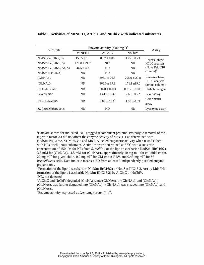
Table 1. Activities of MtNFH1, AtChiC and NtChiV with indicated substrates.
1Data are shown for indicated 6xHis tagged recombinant proteins. Proteolytic removal of the tag with factor Xa did not affect the enzyme activity of MtNFH1 as determined with NodSm-IV(C16:2, S). Mt75352 and MtCRA lacked enzymatic activity when tested either with NFs or chitinous substrates. Activities were determined at 37oC with a substrate concentration of 150 μM for NFs from S. meliloti or the lipo-trisaccharide NodSm-III(C16:2), 3.6 mM for (GlcNAc)6, 4.5 mM for (GlcNAc)5, approximately 10 mg ml-1 for colloidal chitin, 20 mg ml-1 for glycolchitin, 0.9 mg ml-1 for CM-chitin-RBV, and 0.45 mg ml-1 for M. lysodeikticus cells. Data indicate means ± SD from at least 3 independently purified enzyme preparations. 2Formation of the lipo-disaccharides NodSm-II(C16:2) or NodSm-II(C16:2, Ac) by MtNFH1; formation of the lipo-trisaccharide NodSm-III(C16:2) by AtChiC or NtChiV. 3ND, not detected. 4AtChiC and NtChiV degraded (GlcNAc)6 into (GlcNAc)3 or (GlcNAc)2 and (GlcNAc)4; (GlcNAc)4 was further degraded into (GlcNAc)2; (GlcNAc)5 was cleaved into (GlcNAc)3 and (GlcNAc)2. 5Enzyme activity expressed as ΔA550 mg (protein)-1 s-1.
Substrate Enzyme activity (nkat mg-1)1
Assay MtNFH1 AtChiC NtChiV
NodSm-V(C16:2, S) 156.5 ± 8.1 0.37 ± 0.06 1.27 ± 0.23 Reverse-phase HPLC analysis (Nova Pak C18 column)2
NodSm-IV(C16:2, S) 121.8 ± 21.7 ND3 ND
NodSm-IV(C16:2, Ac, S) 46.5 ± 4.2 ND ND
NodSm-III(C16:2) ND ND ND
(GlcNAc)6 ND 393.1 ± 26.8 205.9 ± 29.8 Reverse-phase HPLC analysis (amino column)4 (GlcNAc)5 ND 266.0 ± 19.9 171.1 ±19.0
Colloidal chitin ND 0.020 ± 0.004 0.012 ± 0.001 Ehrlich's reagent
Glycolchitin ND 13.49 ± 3.32 7.66 ± 0.22 Lever assay
CM-chitin-RBV ND 0.83 ± 0.225 1.33 ± 0.03 Colorimetric
assay
M. lysodeikticus cells ND ND ND Lysozyme assay
www.plantphysiol.orgon April 5, 2019 - Published by Downloaded from Copyright © 2013 American Society of Plant Biologists. All rights reserved.
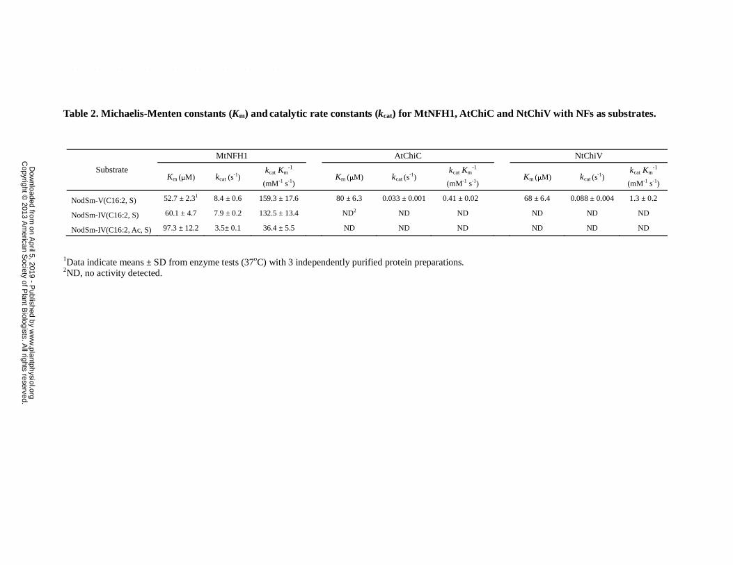
Table 2. Michaelis-Menten constants (Km) and catalytic rate constants (kcat) for MtNFH1, AtChiC and NtChiV with NFs as substrates.
1Data indicate means ± SD from enzyme tests (37oC) with 3 independently purified protein preparations. 2ND, no activity detected.
Substrate
MtNFH1 AtChiC NtChiV
Km (μM) kcat (s-1)
kcat Km-1
(mM-1 s-1) Km (μM) kcat (s
-1)
kcat Km-1
(mM-1 s-1) Km (μM) kcat (s
-1)
kcat Km-1
(mM-1 s-1)
NodSm-V(C16:2, S) 52.7 ± 2.31 8.4 ± 0.6 159.3 ± 17.6 80 ± 6.3 0.033 ± 0.001 0.41 ± 0.02 68 ± 6.4 0.088 ± 0.004 1.3 ± 0.2
NodSm-IV(C16:2, S) 60.1 ± 4.7 7.9 ± 0.2 132.5 ± 13.4 ND2 ND ND ND ND ND
NodSm-IV(C16:2, Ac, S) 97.3 ± 12.2 3.5± 0.1 36.4 ± 5.5 ND ND ND ND ND ND
w
ww
.plantphysiol.orgon A
pril 5, 2019 - Published by
Dow
nloaded from
Copyright ©
2013 Am
erican Society of P
lant Biologists. A
ll rights reserved.
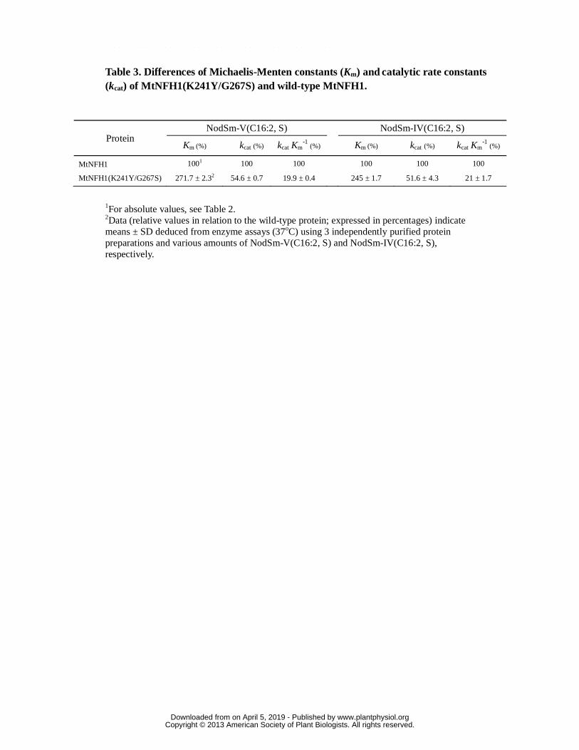
Table 3. Differences of Michaelis-Menten constants (Km) and catalytic rate constants (kcat) of MtNFH1(K241Y/G267S) and wild-type MtNFH1.
1For absolute values, see Table 2. 2Data (relative values in relation to the wild-type protein; expressed in percentages) indicate means ± SD deduced from enzyme assays (37oC) using 3 independently purified protein preparations and various amounts of NodSm-V(C16:2, S) and NodSm-IV(C16:2, S), respectively.
Protein NodSm-V(C16:2, S)
NodSm-IV(C16:2, S)
Km (%) kcat (%) kcat Km-1
(%) Km (%) kcat (%) kcat Km-1
(%)
MtNFH1 1001 100 100 100 100 100
MtNFH1(K241Y/G267S) 271.7 ± 2.32 54.6 ± 0.7 19.9 ± 0.4 245 ± 1.7 51.6 ± 4.3 21 ± 1.7
www.plantphysiol.orgon April 5, 2019 - Published by Downloaded from Copyright © 2013 American Society of Plant Biologists. All rights reserved.
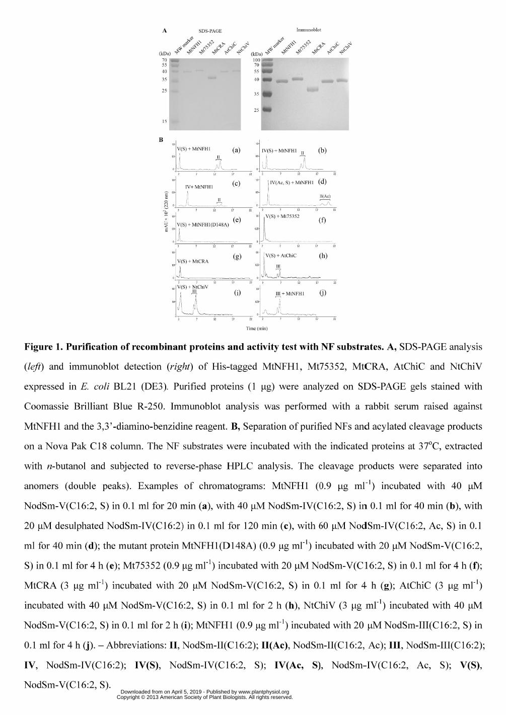
www.plantphysiol.orgon April 5, 2019 - Published by Downloaded from Copyright © 2013 American Society of Plant Biologists. All rights reserved.
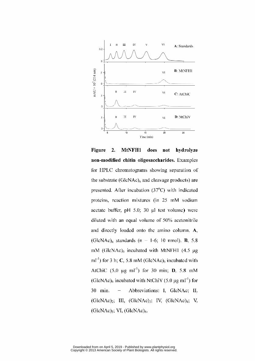
www.plantphysiol.orgon April 5, 2019 - Published by Downloaded from Copyright © 2013 American Society of Plant Biologists. All rights reserved.
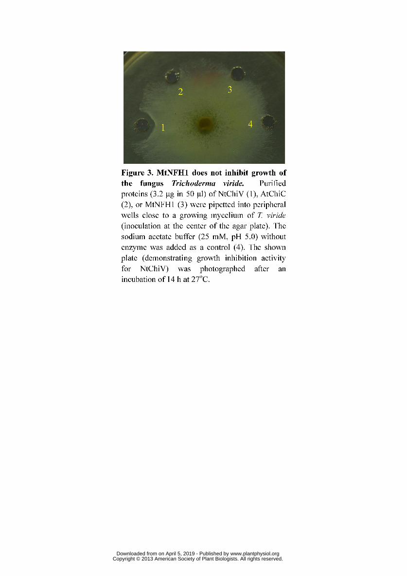
www.plantphysiol.orgon April 5, 2019 - Published by Downloaded from Copyright © 2013 American Society of Plant Biologists. All rights reserved.

www.plantphysiol.orgon April 5, 2019 - Published by Downloaded from Copyright © 2013 American Society of Plant Biologists. All rights reserved.
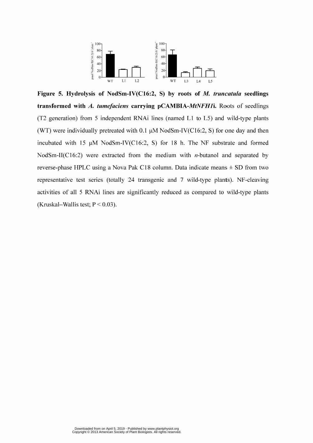
www.plantphysiol.orgon April 5, 2019 - Published by Downloaded from Copyright © 2013 American Society of Plant Biologists. All rights reserved.