Embed Size (px)
Citation preview

RSNA at the Helm of AI Innovation
Register for RSNA's All-Virtual 2020 Meeting— See Page 24
October 2020 Volume 30, Issue 10
3 Year Anniversary
A L S O I N S I D E :
Innovations Abound at RSNA 2020 Virtual Exhibition
RSNA Launches Inaugural Global Learning Center
AI Tool Helps Identify Risk of COVID-19 Complications
COVID-19 Impacts Radiology Research
AI Plays an Important Role in the COVID-19 Pandemic
PET/MRI Method for Prostate Cancer Staging
Diffusion-Weighted MRI in Juvenile Idiopathic Arthritis

Be Part of Radiology that Represents You.
Group Membership Renewals Are Easy
Influence the future of radiology by sharing your unique voice and perspective. Renew your membership, sign up for microvolunteering opportunities, network with radiologists around the world, and share your passion and expertise to advance your career—and the field.
Together, We Can Build a Better Tomorrow for Radiology
Renew your RSNA Membership Today
RSNA.org/Renew We Are Radiology.
MEM832_RenewalAd_2021.indd 1 8/27/20 12:02 PM

OCTOBER 2020 • VOLUME 30, ISSUE 10
UP FRONT 2 First Impression
5 My Turn: Curtis P. Langlotz, MD, PhD
RADIOLOGY’S FUTURE 17 Education and Funding
18 R&E Foundation Donors
19 Your Donations in Action
Radiology's Continued Commitment to COVID-19
Follow us for exclusive news, annual meeting offers and more!
NEWS YOU CAN USE20 Journal Highlights
22 Radiology in Public Focus
24 Annual Meeting Watch
24 Value of Membership
25 Infographic: RSNA Leads in AI
Diffusion-Weighted MRI in Juvenile Idiopathic Arthritis
RSNA Launches Inaugural Global Learning Center
Innovations Abound at RSNA 2020 Virtual Exhibition
RSNA MISSIONRSNA promotes excellence in patient care and health care delivery through education, research and technologic innovation.
FEATURES
PET/MRI Method for Prostate Cancer Staging
EDITOR
Vahid Yaghmai, MD
R&E FOUNDATION CONTRIBUTING EDITOR
Theresa C. McLoud, MD
EXECUTIVE EDITOR
Shelley L. Taylor
MANAGING EDITOR
Beth Burmahl
STAFF WRITER
Jennifer Allyn
GRAPHIC DESIGNER
Eriona Baholli-Karasek
EDITORIAL ADVISORS
Mark G. WatsonExecutive Director
Karena GalvinDeputy Executive Director
Jennifer L. Michalek Assistant Executive Director: Marketing and Communications
Marijo MilletteDirector: Public Information and Communications
EDITORIAL BOARD
Vahid Yaghmai, MDChairman
Tammie S. Benzinger, MD, PhDStephen D. Brown, MDCarlo Catalano, MDAndrew D. Chung, MDAdam E. Flanders, MDDaniel A. Hamstra, MD, PhDLucy Lester, MDAna P. Lourenco, MDTheresa C. McLoud, MDMartin P. Torriani, MDJeffrey S. Klein, MD Board Liaison
2020 RSNA BOARD OF DIRECTORS
Bruce G. Haffty, MD ChairMatthew A. Mauro, MDLiaison for Education
Curtis P. Langlotz, MD, PhDLiaison for Information Technology and Annual Meeting
Umar Mahmood, MD, PhD Liaison for International AffairsJeffrey S. Klein, MD Liaison for Publications and Communications
Carolyn C. Meltzer, MD Liaison for ScienceJames P. Borgstede, MD PresidentMary C. Mahoney, MD President-Elect
3 Year Anniversary
15
8
9,10,12
14
6
Be Part of Radiology that Represents You.
Group Membership Renewals Are Easy
Influence the future of radiology by sharing your unique voice and perspective. Renew your membership, sign up for microvolunteering opportunities, network with radiologists around the world, and share your passion and expertise to advance your career—and the field.
Together, We Can Build a Better Tomorrow for Radiology
Renew your RSNA Membership Today
RSNA.org/Renew We Are Radiology.
MEM832_RenewalAd_2021.indd 1 8/27/20 12:02 PM

2 RSNA News | October 2020
FIRST IMPRESSION
Outstanding Educator and Researcher AnnouncedThe RSNA Board of Directors has announced the Outstanding Educator and Outstanding Researcher who will be recognized during the 106 th Scientific Assembly and Annual Meeting.
Mark E. Mullins, MD, PhDAtlanta, GA
David A. Mankoff, MD, PhDPhiladelphia, PA
OUTSTANDING EDUCATOR OUTSTANDING RESEARCHER
RSNA Focuses on Diversity, Equity and InclusionRSNA believes that a diverse representation of volunteers and leaders can best serve the organization, and the profession. The RSNA Board has long directed its committee leadership to con-sider diversity of gender, age, race/ethnicity, locations/regions, practice type and subspecialty in their recommendations of individuals for committee membership. With this commitment to diverse committee member representation, RSNA’s volunteer and leadership gender composition outpaces the demographics of the U.S. radiologist population.
RSNA’s Committee on Diversity, Equity and Inclusion (CDEI), chaired by Yoshimi Anzai, MD, assesses the DEI environment in RSNA and the profession to identify gaps that RSNA might fill to help radiology leaders understand DEI issues and make positive changes. Recognizing that unconscious bias about gender identity, age, race/ethnicity, sexual orien-tation, religion and ability may influence the evaluation and selection of faculty, committee members and leaders, honorees, awardees and other appointed positions, the RSNA Board enthusiastically approved the committee’s recommendation to conduct unconscious bias training for all RSNA volunteer leaders. In September, RSNA Board members and other Society leaders participated in a new virtual “Everyday Bias” workshop.
Another gap identified by the CDEI was the Society’s limited member demographic data. As a result, RSNA evaluated existing data and implemented improved data point collection to better
reflect the ways that RSNA members identify themselves. We are pleased with the positive response members have shown to our expanded options for gender and race/ethnicity. Watch for many DEI courses and studies being presented in the RSNA 2020 program and the RSNA Online Learning Center.

October 2020 | RSNA News 3
Celebrate the Ninth Annual International Day of Radiology The International Day of Radiology (IDoR), celebrated on Nov. 8, recognizes the anniversary of the discovery of the X-ray. Radiologists and related professionals are encouraged to celebrate IDoR to create greater awareness of the value that radiology research, diagnosis and treatment contribute to safe patient care and to promote better understanding of the vital role radiologists play in health care.
Linver Awarded SBI Gold MedalMichael N. Linver, MD, received the 2020 Gold Medal of the Society of Breast Imaging (SBI).
Dr. Linver is director emeritus of the Breast Imaging Center of X-Ray Associates of New Mexico, PC, and clinical professor of radiology at the University of New Mexico, both in Albu-querque.
He is a former member of the RSNA Radlex Breast Sub-committee and the RSNA Breast Education Exhibits Com-mittee. He also served as a moderator and faculty at numerous RSNA annual meetings.
The award will be presented at SBI's 2021 annual meeting.
RSNA COVID-19 Resources RSNA offers critical resources on the 2019 novel coronavirus (COVID-19), including:
• Online RSNA COVID-19 ResourcesLearn about current research and best practices for managing through COVID-19 and getting your practice back on track at RSNA.org/COVID-19.
• Online Learning CenterAccess available COVID-19 resources including webinars, pre-recorded webinars and videos at RSNA.org/Learning-Center.
• RSNA CommunityJoin RSNA’s online community specifically for COVID-19 discussions. Ask questions, share ideas, get peer-to-peer support and discover lessons learned. All community users are required to have an active RSNA account.
• Industry SolutionsRSNA’s COVID-19 Industry Solutions website is a central hub designed to help radiologists and the radiology community find solutions including equipment sani-tation, teleradiology, AI software, training resources for residents and technologists, equipment guidelines, and 3D printing.
• RSNA Coronavirus Cases View peer-reviewed cases of COVID-19 at Cases.RSNA.org, a free educational and point-of-care tool provided to get more patients the care they need.
SNMMI Names New OfficersAlan B. Packard, PhD, has been named the 2020-21 pres-ident of the Society of Nuclear Medicine and Molecular Imaging (SNMMI). SNMMI introduced a new slate of officers during its annual meeting.
Dr. Packard is an associate professor of radiology at Har-vard Medical School and director of radiopharmaceutical research and senior research associate in nuclear medicine at Boston Children’s Hospital.
President-elect is Richard L. Wahl, MD, the Elizabeth E. Mallinckrodt Professor of Radiology, director of the Mallinckrodt Institute of Radiology and a professor of radiation oncology, all at Washington University School of
Medicine, St. Louis. Vice president-elect is Munir Ghesani, MD, associate professor of radiology
at Mount Sinai Hospital and chief of nuclear medicine and molecular imaging at Mount Sinai Health Enterprise in New York, NY.
Packard
Linver

4 RSNA News | October 2020
FIRST IMPRESSION
October 2020 • Volume 30, Issue10 Published monthly by the Radiological Society of North America, Inc. 820 Jorie Blvd., Suite 200, Oak Brook, IL 60523-2251. Printed in the USA.
Postmaster: Send address corrections or changes to: RSNA News, 820 Jorie Blvd., Suite 200, Oak Brook, IL 60523-2251Non-member subscription rate is $20 per year; $10 of active members’ dues is allocated to a subscription of RSNA News.
RSNA NEWS LETTERS TO THE [email protected] 1-630-571-7837 fax
[email protected] 1-888-600-0064 1-630-590-7770
Contents of RSNA News copyrighted ©2020, RSNA. RSNA is a registered trademark of the Radiological Society of North America, Inc.
REPRINTS AND [email protected] 1-630-571-7829 1-630-590-7724 fax
[email protected] Lisa Lazzaretto Assistant Director: Corporate Relations 1-630-571-7818
RSNA Collaborates on Open-Source COVID-19 Medical Image Database
RSNA and Medical Organizations Encourage Patients to #ReturnToCare
RSNA is collaborating to develop the Medical Imaging and Data Resource Center (MIDRC), an open-source data-base with medical images from tens of thousands of coronavirus (COVID-19) patients. The MIDRC will help physi-cians better understand, diagnose, moni-tor and treat COVID-19.
RSNA will co-lead the effort with the American College of Radiology (ACR) and the American Association of Physi-cists in Medicine (AAPM). The National Institute of Biomedical Imaging and Bioengineering (NIBIB) at the National Institutes of Health (NIH) is funding the
effort through a contract to Maryellen Giger, PhD, of the University of Chicago, which will host the MIDRC.
The initiative includes RSNA represen-tatives Curtis Langlotz, MD, PhD, RSNA Board liaison for information technology and annual meeting, of Stanford Univer-sity, and Adam Flanders, MD, of Thomas Jefferson University Hospital, with co-in-vestigators Etta Pisano, MD, of Beth Israel Deaconess Medical Center, Michael Tilkin, MS, from ACR and Paul Kinahan, PhD, from AAPM.
Funded under the NIH special emer-gency COVID-19 process, the MIDRC
will create an open access platform to col-lect, annotate, store and share COVID- related medical images.
The MIDRC will bring together engi-neers, physicians and scientists to collect and organize the data to answer crucial questions about how imaging could be deployed against COVID-19.
Dr. Langlotz discusses the MIDRC in My Turn on Page 5.
Due to the COVID-19 pandemic, many patients opted to postpone elec-tive, screening and other time-sensitive imaging exams that are important to their health. With imaging centers and health care facilities reopening with strin-gent safety guidelines, RSNA and other radiology organizations are joining their physician colleagues and patient advocacy groups throughout medicine to encourage patients to #ReturnToCare.
Studies have shown dramatic declines in screenings and cancer-related care due to COVID-19. Radiologists are encouraged to contact patients to discuss scheduling imaging exams based on the guidelines in their region, at their hospital and radiology department.
RSNA offers a variety of radiology practice and patient-focused COVID-19 related resources.
The RSNA COVID-19 Resources webpage is updated regularly with links to original research, guidelines, education and multimedia presentations.
RadiologyInfo.org, RSNA's and ACR’s public information website, has a Medical Imaging and Coronavirus Safety page.
RSNA is part of a coalition of medical organizations and patient advocacy groups promoting the #ReturnToCare campaign. The campaign encourages patients to talk to their doctors about scheduling missed or delayed exams, including imaging, where appropriate.
For information about the #ReturnToCare campaign, visit www.returntocarecampaign.org.
WEB EXTRAS Access on-demand RSNA webinars
on the COVID-19 surge and post-surge at RSNA.org/Learning-Center.
WEB EXTRAS Read more about the RSNA International
COVID-19 Open Radiology Database (RICORD) at RSNA.org/Covid-19.

October 2020 | RSNA News 5
My Turn:
Will Artificial Intelligence Play a Role in Imaging of COVID-19?BY CURTIS P. LANGLOTZ, MD, PHD
I am frequently asked how artificial intelligence (AI) can help address the coronavirus pandemic. The questions are everywhere: Could AI improve detection of subtle COVID-19 disease on chest radiography or chest CT? Could AI help distinguish the imaging appearance of COVID-19 from similar diseases? Could AI predict the need for inpatient or intensive care by improving assessment of disease severity?
These COVID-19 conundrums intrigue us all. But so far, the best answer scientists can give is: We don’t know. The earliest attempts to build COVID-19 AI algorithms have lacked reliable testing and training data and have often been based on skewed populations or unrepre-sentative patients, according to a research review published in Radiology: Cardiotho-racic Imaging (see Web Extras).
The Need for DataBecause the answers to crucial questions about COVID-19 require abundant and reliable imaging data, RSNA recently announced the RSNA International COVID-19 Open Radiology Database (RICORD), led by a group chaired by Dr. Matt Lungren. The response was over-whelming — more than 200 institutions expressed interest in contributing imaging data. RSNA has already collected over 600 chest CTs and over 7,400 chest radio-graphs from health care organizations around the globe, including the U.S., Canada, Brazil and Turkey.
Expert thoracic radiologists are seg-menting and classifying the lung disease on these images using a detailed proto-col developed in collaboration with the American College of Radiology (ACR) and a European consortium. The labeled studies are slated for public release in the fall of 2020, with data from additional health care organizations in the pipeline. Thus RSNA is gathering a vast repository of images to answer critical questions about COVID-19.
A Unified National EffortRSNA’s new COVID-19 data repository will be a key component of the newly formed Medical Imaging and Data Resource Center (MIDRC), a collabora-tion with ACR, the American Association of Physicists in Medicine (AAPM), and over 20 research institutions and pro-fessional societies, all sponsored by the National Institute of Biomedical Imaging and Bioengineering (NIBIB). RSNA is delighted to be a founding member of this initiative, which brings together informatics professionals dedi-cated to collecting, organizing and labeling coronavirus data, and making it widely available for research. The MIDRC also will convene teams of scientists from academia, industry and the Food and Drug Administration (FDA) to answer key research questions about COVID-19. Imaging data collected by the MIDRC will be linked to other COVID-19 repositories at the National Institutes of Health (NIH) and elsewhere, enabling the formation of multi-modal data sets to answer complex clinical questions.
RSNA’s primary goal in leading these new initiatives is to make large amounts of COVID-19 imaging data freely avail-able as quickly as possible. We plan to release over 10,000 studies for research-ers around the globe in the next three months. A small fraction of studies will be sequestered to assure reliable scoring of data science challenges (whose tempo will accelerate in the months to come) and to benchmark algorithms from academia and industry.
The MIDRC’s mission may be extended beyond an initial two years to encompass new disease processes, addi-tional organ systems, and the full diversity of imaging modalities. Just as massive publicly available data sets like ImageNet have driven progress in AI outside of medicine, an expanded MIDRC could create a “Medical ImageNet,” a unique resource to supercharge AI research in
clinical imaging.As we establish this massive COVID-
19 imaging repository, we hope you will participate by contributing COVID-19 data, volunteering to annotate images or pursuing key COVID-19 research ques-tions with the freely available data.
With your help, we can address the vital questions posed by this unprece-dented pandemic. When major radiology organizations and their members team up, we are unstoppable!
WEB EXTRAS Access the Radiology: Cardiothoracic Imaging
study “Suboptimal Quality and High Risk of Bias in Diagnostic Test Accuracy Studies on Chest Radiography and Computed Tomography in the Acute Setting of the COVID-19 Pandemic: A Systematic Review,” at RSNA.org/Cardiothoracic.
Read more about RICORD and MIDRC in RSNA News at RSNA.org/News.
Access all RSNA COVID-19 resources at RSNA.org/COVID-19.
Learn about the RSNA AI Challenge at RSNA.org
Access AI educational content at RSNA.org/Learning-Center.
Curtis P. Langlotz, MD, PhD, is the RSNA Board of Directors liaison for information technology and annual meeting. Dr. Langlotz is professor of radiology and biomedical infor-matics, director of the Center for Artificial Intelligence in Medicine and Imaging, and associate chair for information systems in the Department of Radiology at Stanford Uni-versity. As medical informatics director for Stanford Health Care, he is responsible for the information technology that supports the Stan-ford Radiology practice.

6 RSNA News | October 2020
FEATURE
Innovations Abound at RSNA 2020 Virtual ExhibitionVirtual format connects attendees with exhibitors in exciting new waysBY JENNIFER ALLYN
While RSNA's annual meeting Technical Exhibition is always the ideal setting to discover the latest in medical imaging, this year’s virtual format will make it even easier for attendees to explore the latest products, services and supplies and engage with industry experts during RSNA 2020.
Annual meeting attendees will have ample opportunity to connect with exhibitors and discover the latest innovations in equipment and software in the RSNA 2020 Virtual Exhibition.
“Our technical exhibition has always been a huge draw for attendees, and we appreciate the technological advances and innovative solutions our exhibiting com-panies bring to the RSNA meeting each year,” said Curtis P. Langlotz, MD, PhD, RSNA Board liaison for information technology and annual meeting.
This year, RSNA transitioned to a completely virtual meeting to be held from Nov. 29 to Dec. 5. For all seven days of the annual meeting, exhibitors will staff their virtual booths, offering a variety of networking tools — from live chats to video conferencing — to help attendees connect with exhibitors in excit-ing new ways. “This virtual event, just like our meet-ing every year, will enable our industry partners to showcase products that help our attendees enhance their practices and improve patient care," Dr. Langlotz said. Attendees can also virtually access popular RSNA attractions including the AI Showcase, AI Theater and Innovation Theater.
Easy Navigation Offers Complete Access to ExhibitsThe RSNA 2020 Virtual Exhibition will house an expansive lineup of content from the leading vendors who advance
the medical imaging technology that improves patient care. Along with con-necting via social media and live chat, attendees can schedule one-on-one virtual meetings with exhibitors to learn about their products. Those without time to connect with exhibitors at the meeting can leave a virtual “business card” to schedule time afterward.
Virtual attendees of the Innovation Theater can access 15-minute, exhibitor presentations that showcase the latest innovations in medical imaging.
RSNA AI ShowcaseState-of-the-art artificial intelligence (AI) solutions will be featured in the AI Show-case, a virtual collection of AI software and product demonstrations available during RSNA 2020. The AI Showcase will connect attendees with industry lead-ers to explore the possibilities of AI.
The AI Theater will offer on-demand presentations about the AI innovations that are fueling the future of imaging. Each 15-minute presentation will give attendees topline information to learn about the latest AI products and solu-tions. A complete list of AI Theater pre-sentations will be available at RSNA.org/Exhibits.
The winners of the 2020 AI Challenge will be announced during the meeting in the AI Showcase. The 2020 RSNA-STR Pulmonary Embolism Detection Chal-lenge, organized in collaboration with the Society of Thoracic Radiology (STR), is

October 2020 | RSNA News 7
designed to train machine learning algo-rithms to detect and characterize instances of pulmonary embolism. A dataset of over 12,000 CT studies was collected from five research centers based on four different continents.
RSNA 2020 will also include an Imag-ing AI in Practice Demonstration. The multi-vendor interoperability demonstra-tion showcases new technologies and new communications standards needed to inte-grate AI into the diagnostic radiology workflow. Using real-world clinical scenar-ios, the demonstration will show how AI can be used to support improvements in patient care.
Daily Dedicated Exhibitor TimeSelect exhibitors will offer virtual product demonstrations with unique presentations that will mimic in-booth product presen-tations and product launches. Exhibitor demonstrations will be held daily from 9 a.m. to 6 p.m. Central Time (CT) and
may include a live Q&A with company representatives. The demonstrations will also be available on demand. A schedule of the exhibitor demonstrations is available at RSNA.org/Exhibits. Each day, RSNA 2020 will feature a dedicated Industry Hour from noon to 2 p.m. CT, where attendees can interact with the virtual exhibition and participate in industry educational presentations to learn about the latest research and inno-vations. RSNA will not present education sessions during this time. Industry pre-sentations will be held each day in for-mats including panel discussions, virtual demonstrations or lectures with company leadership and medical professionals. The sessions will also be available on demand.
And RSNA events extend beyond offi-cial meeting hours. Watch your email and RSNA social media for announcements of before- and after-hour exhibitor events such as virtual happy hours, round table discussions, breakfast presentations and
live Q&As, all designed to help you learn about and connect with exhibitors.
Virtual Assistant Offers Help 24/7Located on the RSNA 2020 homepage, the RSNA 2020 Virtual Assistant will provide answers 24/7 to help attendees navigate the annual meeting and Virtual Exhibition. The tool helps attendees find sessions, identify exhibits, connect with other attendees and can assist with questions outside of meeting hours.
The Virtual Assistant is sponsored by Change Healthcare.
WEB EXTRAS Access the RSNA 2020 Technical Exhibits
at RSNA.org/Exhibits.
Register and learn more about RSNA 2020 at RSNA.org/Annual-Meeting.

8 RSNA News | October 2020
FEATURE
RSNA Launches Inaugural Global Learning CenterTeams focus on virtual education during pandemic BY MARY HENDERSON
When the COVID-19 pandemic halted international travel last spring, volunteer radiologists with RSNA’s Global Learning Center (GLC) quickly modified their plans to travel to South Africa to visit the inaugural site.Designed to expand radiology education across the world, the RSNA GLC pro-gram was introduced in 2019. RSNA is partnering with established radiology departments based in low- or middle-in-come countries to create the GLCs.
Last year, RSNA chose Stellenbosch University, a public research university located in Stellenbosch, the Western Cape province of South Africa, as the site of the first GLC.
“Given this year’s pandemic and associated travel restrictions, we had to modify our first GLC program to make it even more flexible yet academically robust using all the technological means available,” said Claudio Silva, MD, chair of RSNA’s Committee on International Radiology Education (CIRE). “It’s a tes-tament to the RSNA and GLC teams and the team leaders who have been able to make this work seamlessly.”
The program pairs four RSNA volun-teer radiologists with four members of the GLC’s radiology department to design a customized curriculum tailored to the department’s unique educational needs.
Last March, the GLC team led by pro-gram director, Mark Cresswell, MBBCh, a radiologist at St Paul’s Hospital, Van-couver, BC, Canada, was ready to head to South Africa to meet the Stellenbosch team and tour the facility when the pan-demic took hold. The two groups quickly pivoted and conducted a needs assessment and curriculum planning through a series of phone calls and virtual meetings.
Within a matter of months, the GLC team developed a three-year curriculum focusing on cardiothoracic and musculo-skeletal imaging set to begin with online education and incorporate in-person teaching later in the program.
Online Courses LaunchedIn July, nine radiologists from Stellenbosch
began their first two online courses. Addi-tional courses will be provided every four months for the entirety of the program.
Results of pre- and post-testing will help shape live demonstrations and case-based learning to be conducted virtually following course completion. When travel can safely begin, in-person demos and hands-on training will resume.
“We all have high hopes for the RSNA Global Learning Center program and expect it to make an enormous contribu-tion to global outreach and education,” Dr. Silva said.
A second GLC location in sub-Saharan Africa will be named this fall. Funding for the center will be provided in part by a $750,000 grant from the U.S. Depart-ment of Energy’s National Nuclear Secu-rity Administration (NNSA).
“The grant was a happy surprise that will help us further develop capabilities in nuclear medicine in a sub-Saharan African country,” said Omolola M. Atal-abi, MBBS, chair of the Global Learning Center subcommittee of the CIRE. “Each GLC will have a long-lasting impact on a large group of people, changing attitudes and practices and instilling different per-spectives.”
RSNA members interested in vol-unteering on a GLC team can apply at RSNA.org/GLC. Applications are accepted on a rolling basis.
RSNA Global Learning Center (GLC) Teams in South AfricaRSNA Global Learning Center (GLC) Team• Program Director, Mark Cresswell,
MBBCh, St Paul’s Hospital, Vancouver, BC, Canada
• Brian F. Mullan, MD, FCCP, University of Iowa Hospitals and Clinics, Iowa City
• Prachi P. Agarwal, MD, University of Michigan Health System, Ann Arbor, MI
• Omer A. Awan, MD, University of Maryland Medical Center, Baltimore, MD
Stellenbosch University GLC Team• Program Director, Richard D. Pitcher,
MBChB, PhD
• Christelle Ackerman, MBChB, PhD
• Stephanie Grifith-Richards, MBChB
• Razaan Davis, MBChB
• Sucari Vlok, MBChB
AtalabiSilva
“We all have high hopes for the RSNA Global Learning Center program and expect it to make an enormous contribution to global outreach and education.”
CLAUDIO SILVA, MD, MSc
October 2020 | RSNA News 9
Radiologists Develop AI Tool to Identify Risk of COVID-19 ComplicationsUCI researchers also use RSNA-generated data to develop AI imaging algorithm
BY MIKE BASSETT
Artificial intelligence (AI) tools and technology are playing a key role in many aspects of the COVID-19 pandemic.
At the University of California, Irvine, (UCI), for example, two radiologists are part of a multidisciplinary team of doctors who developed an AI application now being used to help doctors at the UCI Medical Center assess the potential sever-ity of a COVID-19 patient’s condition.
Neuroradiologists Peter D. Chang, MD, and Daniel S. Chow, MD, assistant professors in residence, Department of Radiology at UCI, also serve as co-direc-tors of the Center for Artificial Intelli-gence in Diagnostic Medicine (CAIDM), a multi-specialty initiative to develop and integrate AI technology across the UCI health care system.
Developed at CAIDM, the AI tool, or vulnerability scoring machine, uses machine learning (ML) to calculate the likelihood that a COVID-19 patient will need a ventilator or some form of esca-lated care.
“The idea is that when a patient comes in the hospital and tests positively for COVID, we’d like to know what their hospital stay will look like,” Dr. Chang said. “Will they respond well to treat-ment, like the majority of patients, or will they fall into the smaller cohort of patients who develop severe complications and will need an ICU bed or ventilator?”
The tool is trained on the historic data available at UCI and new data coming from the UCI system, Dr. Chang said. Using that data, the researchers were able to identify 13 lab values and risk factors that have proven to be predictive, and use that data to develop a risk prediction score to help determine the level of care a patient will need.
“Thus far, we have gotten excellent feedback from our clinical colleagues and the tool is now part of the COVID clini-cal pathway,” Dr. Chow said.
The center was able to build, test and deploy a model in about a month. According to Dr. Chow, this tool has been running live at the UCI Medical Center for several months now, and the tool has
been updated monthly.“We have numerous cases of early
detection, where the tool was able to accurately identify vulnerable patients and provide our frontline colleagues a bit of an extra warning,” Dr. Chow added. “This couldn’t come at a more necessary time, as our area of Orange County was becoming very saturated.”
RSNA Data Generates Algorithm The center has also been involved in projects involving imaging and COVID-19. For example, Dr. Chang and his colleagues have developed an algorithm to help with the detection of pneumonia on chest radiographs.
“Sometimes the appearance of pneu-monia can be very subtle, especially if it is not a chest radiologist or an attending radiologist looking at those images,” Dr. Chang said. “The goal was to create a tool that serves as a very accurate algorithm for the detection of pneumonia. So, if a patient comes in with all the signs and symptoms of COVID, and the chest X-ray comes back positive, in all prac-ticality that patient is being treated as a COVID patient before that COVID test comes back.”
Dr. Chang explained that the algorithm actually came out of a course he was teaching in computer science in which the curriculum focused on coronavirus applications. For the final exam, stu-dents were tasked with building the best possible algorithm for coronavirus chest radiographs.
“And the top performing models are the ones we are now using,” he said.
The data used for the project came from the 2018 RSNA Pneumonia Detec-tion Challenge in which teams were tasked with building an algorithm that could detect a visual signal for pneumonia in medical images.
Dr. Chang said both UCI and UC San Diego (UCSD) Medical Center have developed separate chest X-ray algorithms,
although the approach for both is similar. Dr. Chang regularly collaborates with the UCSD team to co-develop and improve on each other’s algorithms.
2020 RSNA AI ChallengeIntroduced in 2017, the RSNA AI Chal-lenge was created to engage radiologists in developing datasets useful for training artificial intelligence (AI) systems and per-forming clinically relevant tasks.
Researchers compete to create applica-tions that perform defined tasks according to specified performance measures. The goal of each challenge is to explore and demonstrate the ways AI can benefit radiology and improve patient care.
The 2020 RSNA-STR Pulmonary Embolism Detection Challenge, organized in collaboration with the Society of Tho-racic Radiology (STR), was designed to train machine learning (ML) algorithms to detect and characterize instances of pulmonary embolism. A dataset of over 12,000 CT studies was collected from five research centers based on four different continents.
WEB EXTRAS Learn more about the RSNA AI Challenge at
RSNA.org/Education.
Access RSNA on-demand AI webinars, a series of four, 60-minute sessions focusing on AI and ML providing insight into what this technology means for radiology, at RSNA.org/Education.
Access a publicly available version of the UCI AI tool at covidrisk.hs.uci.edu.
Access AI educational content at RSNA.org/Learning-Center.
ChowChang

10 RSNA News | October 2020
FEATURE
Vagal
Boettcher
Luker
The Long Road Back to a Pre-Pandemic Normal in Radiology ResearchAs labs begin to ramp up, researchers brace for another potential shutdownBY NICK KLENSKE
It goes without saying that the COVID-19 pandemic has disrupted nearly every aspect of our lives — and radiologic research is no exception. When the pandemic struck in March and much of the country shut down, so too did imaging research.
“For nearly three months, academic medical centers and universities halted all non-es-sential research, potentially resulting in a loss of one to two years of productivity,” said Achala Vagal, MD, radiology professor and vice chair of research at the University of Cincinnati Medical Center.
Dr. Vagal is the corresponding author of the Radiology article, “The Impact of the COVID-19 Pandemic on the Radiology Research Enterprise: Radiology Scientific Expert Panel,” describing how several aca-demic radiology research programs man-aged during COVID-19 shutdowns. But what about post-shutdown?
“As lockdowns are being lifted and researchers cautiously head back into the lab, we’re getting our first glimpse of what the ‘new normal’ for radiologic research might look like,” Dr. Vagal explained. “One thing for sure is that it will likely be a long time before we get back to anything close to a pre-pandemic ‘normal’.”
For example, Dr. Vagal noted that although the University of Cincinnati reopened its research on June 1, their labs are only operating at 50% occupancy, with a significant amount of work still happen-ing remotely. All principal investigators and research vice chairs are required to create detailed reentry plans for protecting per-sonnel. For those projects and clinical trials that plan to enroll patients, an additional
COVID-19 plan must be submitted outlin-ing how participants will be protected.
The Looming Shadow of Another Shutdown Even as some research ramps up, the signif-icant spike in COVID-19 cases happening in many parts of the country is a looming shadow over the research community.
“It’s extremely difficult to start an exper-iment when there is a very real risk that you’ll have to ramp back down,” said Gary Luker, MD, editor of Radiology: Imaging Cancer and co-author of the journal’s edi-torial, “Transitioning to a New Normal after COVID-19: Preparing to Get Back on Track for Cancer Imaging.”
This risk of another shutdown is partic-ularly challenging for animal studies said Adeline Boettcher, PhD, scientific editor of RSNA subspecialty journals and co-author of the editorial.
“Many animal experiments are planned to last from two to eight weeks,” Dr. Boettcher said. “But during a pandemic, a lot can happen in this time span, making it exceptionally difficult to plan these kinds of experiments.”
Getting Patients Back in the ClinicAnother immediate challenge is getting patients to come back into a hospital set-ting for clinical studies. At the University of

October 2020 | RSNA News 11
“For nearly three months, academic medical centers and universities halted all non-essential research, potentially resulting in a loss of up to one to two years of productivity.”
ACHALA VAGAL, MD
WEB EXTRAS Access the Radiology study, “The Impact of the
COVID-19 Pandemic on the Radiology Research Enterprise: Radiology Scientific Expert Panel,” at RSNA.org/Radiology.
Access the Radiology: Imaging Cancer editorial, “Transitioning to a New Normal after COVID-19: Preparing to Get Back on Track for Cancer Imaging, at RSNA.org/Imaging-Cancer.
Access a video with Drs. Luker and Boettcher, discussing their Radiology: Imaging Cancer editorial at YouTube.com
Michigan, as with many other academic centers, studies involving older, high-risk participants cannot resume unless the study has a direct, immediate benefit to the individual, Dr. Luker said.
However, clinical studies of new imag-ing methods typically do not provide a direct benefit to a participant even though the research may be tremendously benefi-cial in the future.
“This means many clinical imaging studies cannot start again or must exclude older participants and others regarded as high risk for COVID-19,” Dr. Luker said. “For imaging studies in cancer and other diseases predominantly affecting older people, this means we have a much smaller population of subjects that we can recruit from.”
To counter this, Dr. Boettcher notes that some facilities are developing targeted messaging that highlights the importance of imaging research and emphasizes the cleanliness of facilities. “You want to make sure patients feel safe going into these facilities, and that’s increasingly difficult to do — especially with cases continuing to spike,” she explained.
Social distancing guidelines present another challenge, as keeping people six feet apart means fewer people in a lab and thus fewer studies being done. It also poses challenges to training new researchers. “How do we train people to do research when they can’t be within six feet of each other and, in some cases, can’t even be in the same room because of space restrictions?” Dr. Luker asked.
Funding and staffing are two more issues of concern. “Many sources of revenue have either gone away or have decreased significantly, which will have a major impact on cancer research, cancer imaging research and research in general,”
Dr. Boettcher said.“With many institutions not hiring,
or even letting junior faculty members go, there’s a concern that there will be a shortage of imaging scientists,” Dr. Luker said. “This will clearly affect the future of clinical practice and patient care as many important discoveries will either not be made or will be significantly delayed.”
Research Community Bands TogetherWith all these challenges, concerns and unknowns, what is the future of imaging research?
“To be honest, I don’t think any of us knows what all the pieces are or what the future will hold,” Dr. Vagal said. “The only thing we can say for certain is that everything is very uncertain.”
But, according to Drs. Vagal and Luker, it is in this uncertainty that radiology can find a silver lining.
According to Dr. Vagal, there’s been so much uncertainty surrounding COVID-19 that it has prompted the research community to collaborate very closely and effectively, often in new ways.
“The result is a remarkably quick, and often global, dissemination of science, with the number of papers, sharing of scientific findings and open access to journals reaching unprecedented levels,” she said. “This is the future of imaging research.”
“Instead of the pre-pandemic prac-tice of everybody keeping their data to themselves, I think we’re seeing more of a sense of community and shared purpose amongst the research community,” Dr. Luker said. “I think everyone realizes that the only way we’re going to overcome this problem and ‘get back to normal’ is by working together.”
DISRUPTION OF NORMAL OPERATIONS
Personal interactions were severely limited for months. Most research projects were shelved and many trainees and staff transitioned to remote work.
• Shutdown of 3-4 months causes 1-2 years lost productivity
o Lost data o Research protocol deviations o Prioritization of COVID-19 research
• Staff redeployed to clinical care
• Delayed ABR Core Exam, tenure, degree completion
• Limited interactions for trainees with faculty/patients

12 RSNA News | October 2020
FEATURE
Kundu
Research Shows AI Plays an Important Role in the COVID-19 Pandemic
Murphy
Mei
AI models aid in detecting, monitoring, interpreting the global virusBY NICK KLENSKE
Due to a combination of cost constraints, concerns about contamination, and availability of other means of testing, chest CT is not recommended as a routine method for diagnosing COVID-19. But CT does serve a purpose in understanding COVID-19.
“Chest imaging has the potential to play a role in the COVID pandemic — one that goes well beyond the diagnostic realm,” said Shinjini Kundu, MD, PhD, a radiol-ogy resident at the Johns Hopkins Uni-versity School of Medicine. “This includes helping exclude other possible causes for COVID-like symptoms, confirming a diagnosis made by another means, and providing critical data for monitoring a patient’s progress.”
In a recent Radiology: Artificial Intel-ligence study, Dr. Kundu argues that the key to leveraging the benefits of chest imaging in caring for COVID-19 patients is artificial intelligence (AI). “AI’s power to generate models from large volumes of information — fusing molecular, clinical, epidemiological and imaging data — may accelerate solutions to detect, contain and treat COVID-19,” Dr. Kundu said.
Chest imaging was used as a primary tool to screen for COVID-19 when the disease first manifested in China. Because there were too many images for individual review, AI was initially used to automate diagnosis and help radiologists understand the findings. Although these early AI sys-tems reported good results, the technology remained limited to screening purposes.
“Unlike other diseases, we still don’t know a lot about COVID-19 and how it affects the rest of the body. We are still learning every day,” Dr. Kundu said. “But with AI, imaging can be used in real time to not only monitor the disease, but to accelerate key insights about its symptoms and treatment.”
Fusing Data, Crunching InformationChest imaging can help monitor a treat-ment’s effectiveness and help researchers develop new treatments. However, lever-aging this potential requires the ability to combine and analyze data from a variety of sources. This is where AI is useful.
“The power of AI lies in both its ability to fuse data from many diverse sources and its capability to crunch through this
information to find patterns that humans could never see on their own,” Dr. Kundu said.
To illustrate, Dr. Kundu points to the correlation between clinical data such as blood tests, which can indicate the level of inflammation in the body and changes in the lungs.
This integration of immunologic mark-ers with chest imaging could help high-light inflammatory markers that change the lungs and other organs or protect against such change. AI can also consider geospatial data and even information crowdsourced from people recording their symptoms, providing data from around the world.
“This is a cooperative, global effort and we all have to do our part,” Dr. Kundu added. “If we can gather all this data at different scales — molecular, clinical, epidemiological — and potentially parse key patterns using AI, we can learn the disease’s dominant trends and use this information to accelerate how we treat COVID-19.”
The Role of Chest Radiography in COVID-19The role of AI and chest imaging in the COVID-19 pandemic is not limited to chest CT scans. According to a recent Radiology study, chest radiography (CXR) can also play an important role in triage for COVID-19, particularly in low- resource settings.
According to the study, CXR is a fast and relatively inexpensive imaging modal-ity that is available in many resource-con-strained health care settings.
“Unfortunately, these settings often lack the radiological expertise needed to inter-pret such images, making AI a potentially invaluable tool,” said Keelin Murphy, PhD, a researcher at Radboud University Medical Center, the Netherlands, and the study’s lead author.
The study evaluated the performance of CAD4COVID-XRay, an AI system
October 2020 | RSNA News 13
Research Shows AI Plays an Important Role in the COVID-19 Pandemic
designed to detect COVID-19 pneumo-nia on CXR.
Based on an AI system originally developed to detect tuberculosis on CXR, CAD4COVID-XRay was retrained to identify COVID-19 using 24,678 CXR images, including 1,540 used for valida-tion while training. The test data con-sisted of a set of continuously acquired CXR images obtained from patients sus-pected of having COVID-19 pneumonia.
During testing, CAD4COVID-XRay performed at a similar level as six radiol-ogists in terms of sensitivity and specific-ity, or even better at high sensitivity oper-ating points. The system developers have made CAD4COVID-XRay tool available free of charge in support of the crisis.
“We expect it to be particularly useful in the developing world for triage where, combined with other markers, it can help identify COVID-19 cases,” Dr. Murphy said. “It will also be beneficial to over-burdened radiology departments during infection peaks.”
AI Algorithm Rapidly Identifies COVID-19 PatientsBecause chest imaging alone has limited negative predictive value to fully exclude infection, there is a need to incorporate clinical information into the diagnosis of COVID-19 patients. AI has the potential to fill this need, according to researchers at Mount Sinai Hospital, New York City.
In a recent study in Nature Medicine, the researchers combined AI with imag-ing and clinical data to analyze patients
with COVID-19, said study co-author Xueyan Mei, MS, a PhD candidate and radiology trainee at the BioMedical Engineering and Imaging Institute, Icahn School of Medicine at Mount Sinai.
“Our goal was to design an AI model that can quickly identify COVID-19 positive patients in the early stage using initial chest CT scans and associated clin-ical information,” Mei said.
The AI algorithm combined chest CT scans and corresponding clinical informa-tion, including travel and exposure his-tory, leukocyte counts, symptomatology, and patient age and sex.
Applied to a test set of 279 cases, the AI algorithm achieved an area under the curve (AUC) of 0.92 and performed equally well in terms of sensitivity (84.3%) compared to a senior thoracic radiologist (74.6%). The algorithm was able to provide a diagnosis within minutes — a notable improvement over the up to 48 hours it can take to get RT-PCR results back — at an impressive 88% accuracy rate.
The AI model could be used as an application that can run on a simple workstation alongside the radiologists, according to Mei.
“The AI system could be imple-mented as a rapid diagnosis tool to flag patients with suspected COVID-19 infection when CT images and/or clinical information are available and radiologists could review these suspected cases iden-tified by AI with a higher priority,” Mei said.
“The power of AI lies in both its ability to fuse data from many diverse sources and its capability to crunch through this information to find patterns that humans could never see on their own.”
SHINJINI KUNDU, MD, PHD
WEB EXTRAS Access the Radiology: Artificial Intelligence
study, “How Might AI and Chest Imaging Help Unravel COVID-19’s Mysteries?” at RSNA.org/AI.
Access the Radiology study, “COVID-19 on the Chest Radiograph: A Multi-Reader Evaluation of an AI System,” at RSNA.org/Radiology.
Access the Nature Medicine study, “Artificial intelligence–enabled rapid diagnosis of patients with COVID-19,” at Nature.com
Learn about RSNA’s RICORD resource for COVID-19 imaging data at RSNA.org/COVID-19.
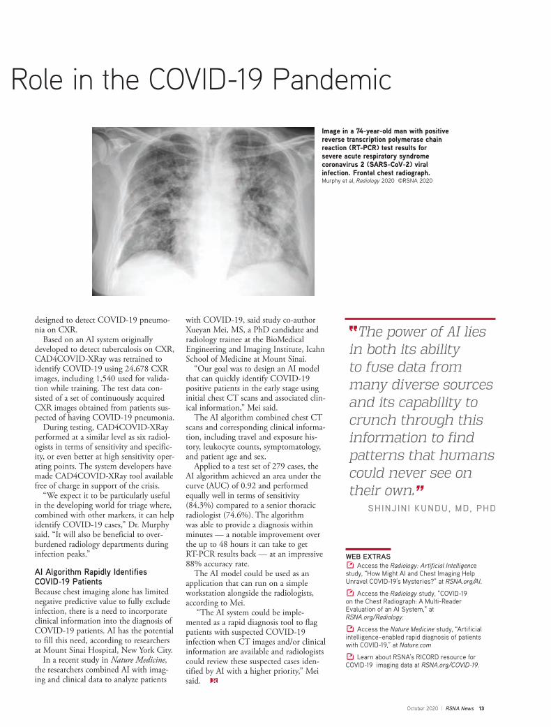
Image in a 74-year-old man with positive reverse transcription polymerase chain reaction (RT-PCR) test results for severe acute respiratory syndrome coronavirus 2 (SARS-CoV-2) viral infection. Frontal chest radiograph. Murphy et al, Radiology 2020 ©RSNA 2020

14 RSNA News | October 2020
FEATURE
Novel PET/MRI Method Promising for Prostate Cancer StagingR&E research shows technology could have clinical value for high-risk prostate cancer patientsBY EVONNE ACEVEDO
A comprehensive nuclear imaging method combining whole-body PET/MRI and regional multiparametric MR of the prostate could offer significant improvements in prostate cancer staging, according to recent research. Pre-treatment staging is currently prob-lematic for many prostate cancer patients. In a significant number of men, con-ventional imaging with CT or MR and nuclear medicine bone scan is falsely neg-ative for regional lymph node metastases, said Samuel J. Galgano, MD, assistant professor, Abdominal Imaging and Molec-ular Imaging and Therapeutics, University of Alabama at Birmingham (UAB).
“Approximately 35% of men with high-risk prostate cancer will have biochemical recurrence even after optimal treatment,” said Dr. Galgano.
In his recent research, “Pretreatment Staging of High-risk Prostate Cancer with F-18 Fluciclovine PET/MRI,” funded by a 2017 RSNA Research Fellow Grant, Dr. Galgano studied the utility of the amino acid PET tracer (18F) fluciclovine for stag-ing high-risk prostate cancer patients prior to therapy and for monitoring response.
“This novel approach using (18F) flu-ciclovine-PET/MRI takes advantage of each strength of the individual imaging modality to improve on the inherent weakness of the other imaging modality,” Dr. Galgano said.
While 18F fluciclovine has been approved by the U.S. Food and Drug Administration (FDA) for use in recurrent prostate cancer, its usefulness in pre-treat-ment staging and for monitoring response to therapy has yet to be established, Dr. Galgano said.
“In addition, the use of PET/MRI for the initial staging of prostate cancer is just beginning to be investigated, but allows for the potential increased sensitivity of detection of metastatic disease on molec-ular imaging combined with high-res-olution images of the prostate for local disease staging,” Dr. Galgano said.
Possible Use in Clinical SettingThe study comprised 14 men with
biopsy-proven prostate cancer that met National Comprehensive Cancer Network high-risk criteria. Dr. Galgano’s team performed comprehensive whole-body 18F fluciclovine-PET/MR imaging plus regional multiparametric MRI of the prostate prior to the patients undergoing treatment. After treatment, the patients underwent repeat 18F fluciclovine-PET/MR imaging.
“Interestingly, 18F fluciclovine-PET/MR demonstrated seminal vesicle invasion in two patients that was not identified on MR imaging alone,” Dr. Galgano said. “In addition, 18F fluciclovine-PET/MR detected suspected metastatic lymph nodes that were not pathologically enlarged by conventional RECIST 1.1 size criteria, which has been demonstrated in prior studies."
Overall findings show that 18F fluci-clovine-PET/MRI has potential value in the initial staging of patients with high-risk prostate cancer, potentially expanding its use in the clinical setting.
“Surgeons may be able to use this technology to develop new treatment algorithms for the optimal management of high-risk prostate cancer patients,” Dr. Galgano said. “Additionally, 18F fluci-clovine PET improves detection of pelvic lymph node metastases when compared to MR, where lymph node metastases are frequently only detected once the lymph node becomes enlarged.
“It is exciting that this comprehensive staging examination can take place in a single imaging exam, which is beneficial to the patient and helps consolidate hos-pital trips and imaging studies.”
RSNA Grant Spurs Biomedical Imaging Research Dr. Galgano said the RSNA Research Fellow grant was not only the catalyst for his research career, but offered the oppor-
tunity to work with scientific advisor, Jonathan McConathy, MD, PhD, direc-tor, Division of Molecular Imaging and Therapeutics at UAB, to gain insight into research methods and techniques.
“Prior to participating in the RSNA Research Fellow Grant program, I had not ever seriously considered a career as a phy-sician-scientist,” said Dr. Galgano, who was a fellow in abdominal imaging at the time of the R&E grant. “After participa-tion, I plan to continue pursuing funded research in biomedical imaging.”
Among other lessons, the experience taught Dr. Galgano the process for run-ning an effective clinical trial.
“From the new drug application, to the FDA approval, to the coordination of patient care with referring providers, the study taught me a lot about how to do — and not to do — good research."
Dr. Galgano, who used the data from his R&E research to formulate an National Institues of Health R01 submis-sion, said the findings from this study are an important first step toward evaluating an approved PET agent for a new appli-cation.
“Improvements in initial staging could affect treatment planning decisions and ultimately lead to improved outcomes in biochemical recurrence and progres-sion-free survival,” Dr. Galgano said.
Galgano
WEB EXTRAS Learn more about RSNA R&E Foundation
grants at RSNA.org/Research.

October 2020 | RSNA News 15
Diffusion-Weighted MRI Accurate in Detecting Juvenile Idiopathic Arthritis of the KneeNon-invasive, non-contrast method shows potential benefits for pediatric patientsBY MELISSA SILVERBERG
Assessing arthritis in the knee can be difficult in adults using traditional MRI methods. In children — who may not be able to stay still or may even need to be sedated during the procedure — the process can be even more difficult.
A recent study published in Radiology, “Juvenile Idiopathic Arthritis: Diffu-sion-weighted MRI in the Assessment of Arthritis in the Knee,” by lead author Anouk Barendregt, MD, in the Depart-ment of Pediatric Immunology and Radiology at the Amsterdam University Medical Center, and colleagues, shows MRI technology may make the process of assessing arthritis easier for pediatric patients without sacrificing accuracy.
“We have been interested in novel methods to assess arthritis without injec-tion of contrast agent for a long time,” Dr. Barendregt said. “This technique is much more patient-friendly, especially for children.”
Diffusion-weighted imaging (DWI) is commonly used in areas such as stroke and prostate but has not been widely tested in the diagnosis and analysis of juvenile idiopathic arthritis. DWI has a number of benefits for pediatric patients, Dr. Barendregt said. It is non-invasive and there is no need to place an IV and administer contrast. The technique is also faster, so children don’t need to stay still as long, reducing the number of young patients that need sedation for a lengthy MRI scan.
Children with juvenile idiopathic arthritis often have autoimmune-mediated inflammation of the synovial membrane. The team studied the diagnostic accuracy of DWI for the detection of arthritis as compared with the clinical reference stan-dard, which included contrast material–enhanced MRI.
The research team conducted a pro-spective study of 45 patients ages 6 to 18 who underwent pre- and post-contrast MRI of the knee with an additional DWI sequence between 2015 and 2018. For the clinical reference standard, a multidisci-plinary team determined the presence or absence of arthritis on the basis of clinical, laboratory and imaging findings, exclud-ing DWI. The two data sets were then scored by two radiologists blinded to all clinical data.
DWI Shows High AccuracyThe study found that DWI was accurate in detecting arthritis in pediatric patients, both in sensitivity and specificity. The results showed sensitivity for detection of arthritis with DWI was 93% (13 of the 14 participants with arthritis were cor-rectly classified). DWI also scored above 80% on specificity for patients without arthritis. The results indicate that DWI could replace contrast-enhanced MRI for imaging of inflammation.
“What we found in our study quite surprised us — it was better than we expected,” said study co-author Robert
Hemke, MD, PhD, musculoskeletal radiologist at Amsterdam University Med-ical Center, who said the team has been researching DWI in arthritis patients for about five years. “During this time, we had regarded contrast-enhanced MRI as the reference standard.”
The Radiology study, which began as a small research project several years ago, was aided significantly by the partici-pation of pediatric rheumatologists and other researchers.
“Working as a team really benefited our research,” Dr. Hemke said. “I’ve learned how important it is to keep your eyes open to other fields of imaging outside of the musculoskeletal domain.” With further and larger validation studies, DWI may be valuable for both adult and pediatric arthritis patients, researchers said.
Hemke
WEB EXTRAS Access the Radiology study, “Juvenile
Idiopathic Arthritis: Diffusion-weighted MRI in the Assessment of Arthritis in the Knee,” at RSNA.org/Radiology.
Barendregt
Image in 12-year-old girl without synovial inflam-mation in whom a functional disorder and/or pain syndrome was eventually diagnosed. The score from the diffusion-weighted imaging (DWI) data set was in agreement with the reference stan-dard: All members of the multidisciplinary team agreed that no arthritis was present in the knee. ADC = apparent diffusion coefficient.Barendregt et al, Radiology 2020 ©RSNA 2020

16 RSNA News | October 2020
FEATUREFEATURE
Industry Focus
SPONSORED CONTENT
ENHANCED COMMUNICATION MAY REDUCE PATIENT ANXIETY DURING MRI EXAMS
Passive magnetic technology can ensure the quality of MRI
Not every challenge the health care community faces requires a massive research investment. In fact, sometimes the simplest solutions are the most elegant.
Consider the patient experience during an MRI exam. Even the patients with the steeliest of nerves can get rattled when placed in the confi ning MRI tube with all its noisy clicking and knocking. The challenge is only inten-sifi ed when the patient is critically ill, unusually young or elderly or claustrophobic.
MRI anxiety can be a serious prob-lem that could stop a complex — and expensive — procedure in its tracks. This makes communication between patients and radiology technologists critical to a successful exam.
Consider all the factors at play here: An MRI machine can cost a hospital as much as $3 million. Running the MRI can cost as much as $300 or $400 an hour. If an exam is interrupted or cut short because of patient anxiety, that is a costly interruption.
Add to that equation the increasingly tight profi t margins many health care institutions deal with today, the need to assure the highest possible care to patients, and the imperative for facilities to make effi cient use of their equip-ment, staff and investments.
How then does the MRI technologist ask the patient to communicate with them amidst all this? Sometimes with a rubber ball placed in their hand at the last minute.
As more and more advanced technolo-gies become available to radiologists, they sometimes run the risk of losing the clearest possible interface with the patient. Sometimes signifi cant innova-tions are required to make sure radiol-ogy obtains and interprets the most accurate information possible from patients. And sometimes it just takes a simple idea well executed.
An example is the iBrain non-magnetic and non-active dual-channel com-munication equipment developed by Shanghai Zhongchangjiang Commu-nication Technology Co., LTD. Under ordinary circumstances, communication
between the MRI technician and patient can be a challenge.
“Conventional MRI communication devices do not have a well set up transmission device on the patient’s earphones,” said iBrain inventor Aidao Zhu. “While some MRI systems have a microphone for two-way communica-tion, most microphones are wired and active, and the electrical signals they transmit near the scanning area can easily interfere with the image quality.”
However, Zhu has developed a non-metallic air tube that can connect the technologist and patient via a micro-phone and speaker near the patient’s face. The audio signal it carries through the air tube is some distance from the scanning device so there is no mag-netic disturbance to the examination, thus allowing the technologist to give the patient instructions and the patient
to explain in real time if they either do not understand or are experiencing discomfort.
“Real-time two-way communication and monitoring can ensure the safety of the examination process,” Zhu said. “At the same time, clear imaging quality can avoid misdiagnosis, the main embodiment of improving the safety of patients.”
The iBrain MRI telephone device con-trols the host machine with full touch technology. The system can also play music at the appropriate time, which helps the patient relax, reducing the patient’s the anxiety during examination and improving the examination result.
As radiology continues to respond to new challenges with new technolo-gies, one of the simplest factors — the interface with the patient — will call for ingenuity and creativity as well.
“Personalized medical services cannot be separated from communication,” Zhu said, “especially in radiology and magnetic resonance imaging.”
Zhu

October 2020 | RSNA News 17
Education and Funding Opportunities
Please review the education announcements carefully as some deadlines have been extended. For up-to-date information, go to RSNA.org/Education.
Writing a Competitive Grant Proposal Workshop Register now for the Writing a Competitive Grant Proposal Workshop designed for researchers in radiology, radiation oncology, nuclear medicine and related sciences who are interested in actively pursuing federal funding. This 1½-day program is guided by a faculty of leading researchers with extensive experience in all aspects of grant applications and funding. Course fee is $225. Register at RSNA.org/WCGP or contact RSNA staff at [email protected] or 1-630-368-3758
April 9–10, 2021 RSNA Headquarters Oak Brook, ILDeadline: January 10
New Education Resources Available Online The RSNA Online Learning Center adds new resources regularly to support your ongoing education needs.
COVID-19 VideosSeveral videos are available on demand. Members can earn available CME for free and non-members pay a nominal fee. Recent videos include:
• COVID-19 and Health Disparities – A Call for Action• Thoracic Imaging Findings of Multisystem Inflammatory Syndrome in Children (MIS-C)• Town Hall Discussion: How COVID-19 is Changing Private Radiology Practice• The Economic Impact of COVID-19 in the Radiology Community • Essentials of Chest Imaging: Pediatric COVID-19 • Crisis Leadership During COVID-19
Access all COVID-19 education at RSNA.org/Learning-Center-COVID-19. Short instructional videos related to COVID-19 have been added to the microlearning playlist on RSNA’s YouTube channel (@RSNAtube):
• Changing Workflows During COVID-19• Workflow Solutions to Keep Patients Safe During COVID-19• Tips to Reduce Stress
Diversity, Equity and Inclusion CoursesCourses from the 2019 RSNA annual meeting focusing on diversity, equity and inclusion (DEI) are available in the Online Learning Cen-ter. These courses are free to members and can be accessed by visiting RSNA.org/Learning-Center-Diversity. Read more about RSNA's com-mitment to DEI on Page 2.
Virtual RSNA/ASNR Comparative Effectiveness Research Training (CERT) Program
Apply now for the Comparative Effectiveness Research Training (CERT) Program, jointly sponsored by RSNA and the American Society of Neuroradiology (ASNR). CERT will be delivered in a combination of online modules, a modified virtual workshop with sessions on Feb. 4–5, 2021, web-based didactic lectures and small group web-based grant proposal review discussions. Beginning in December 2020, the program will continue through 2021. The application deadline has been extended to Oct. 15. There is no fee for this course. For more information, visit RSNA.org/CERT. Contact RSNA staff at [email protected] with questions.
February 4–5, 2021 (virtual) Application Deadline Extended: October 15
While the uncertainty of the COVID-19 situation makes planning difficult, we are hopeful that RSNA will be able to offer this valuable educational opportunity as scheduled. RSNA will continue to monitor the situation and will communicate any change of plans.

18 RSNA News | October 2020
RADIOLOGY’S FUTURERADIOLOGY’S FUTURE
Vanguard ProgramCompanies supporting endowments and term funding for named grants.
Hitachi Healthcare Americas$15,000A Vanguard Member since 1999
The RSNA Research & Education Foundation thanks the following donors for gifts made June 23, 2020 through July 23, 2020.
RESEARCH & EDUCATION FOUNDATION DONORS
Individual DonorsDonors who give $1,500 or more per year qualify for the RSNA Presidents Circle. Their names are shown in bold face.
$5,000 – $9,999Glendon G. & Karen E. Cox
$1,500 – $2,499H. Scott Beasley, MD, FACR In memory of R. Todd BeasleyElizabeth Houser In memory of Robert E. Campbell, MD In memory of David B. Fraser, MD
$500 – $1,499Diana T. Jucas, MDWilliam F. Lytle Jr., MDDeborah Shatzkes, MDIngrid E. & Stephen R. Thomas, PhD In memory of Robert E. Campbell, MD In memory of David B. Fraser, MD
$300 – $499Saleh M. Alreshoodi, MBBSNanette D. DeBruhl, MDKuang-Chun J. Hsieh, MDHenry Irvine, MDPatricia C. & William W. Olmsted, MD In memory of David B. Fraser, MDThomas Pope, MD & Jennifer Cranny, MD
Pierre-Yves J. Sonke, MD
$299 or LessYuki Arita, MDMatthew Barkovich, MDLaura & Adam Berkey In honor of Jerry P. Petasnick, MDLucas K. Buckley, MDAlessandra Caporale, PhDPhilip R. Chapman, MDMarco De Rossi, MScLiten & Eric DeNautWilfod F. Diaz-Martinez, MDCristy Gustas-French, MDKeith A. Hanson, BAMary R. & Donald P. Harrington, MDShams Jubouri, MBChBKlaudia Jumaa, MDHyo-cheol Kim, MDLisa & Marc D. Kohli, MDSena LeachVasantha & Mahadevappa Mahesh, MS, PhD
Rocio Marquez, BAAnika L. McGrath, MDKambiz Motamedi, MDStephen C. O’Connor, MDDulia C. Ortega, MDSheri, Alon, Alex & Oren Panovka In honor of Jerry P. Petasnick, MDE. Russell & Julia R. RitenourRoya Sohaey, MDMarc D. Succi, MD

October 2020 | RSNA News 19
R&E Foundation Accepting Grant Applications Applications for 2021 R&E Foundation grants are being accepted. Deadlines for application are in early 2021.
Education Grants Deadline: Jan. 11
• Derek Harwood-Nash International Education Scholar Grant
• RSNA/AUR/APDR/SCARD Radiology Education Research Development Grant
Research GrantsDeadline: Jan. 15
• Research Scholar Grant• Research Resident/Fellow Grant• Research Seed Grant
Medical Student Research GrantDeadline: Feb. 3
Opportunity for Diverse Medical Student Grant Applicants
The R&E Foundation invites medical students who identify as minorities underrepresented in medicine (UIM) to apply for grant funding through the RSNA Research Medical Student Diversity Grant.
Education GrantsDeadline: Jan. 11
Derek Harwood-Nash International Ed-ucation Scholar Grant. Open to both in-ternational and domestic applicants for projects covering any area of radiologic education with an international scope.
RSNA/AUR/APDR/SCARD Radiol-ogy Education Research Development Grant. Encourages innovation and improvement in health sciences educa-tion by providing research opportunities in radiology education. International applicants welcome.
Visit RSNA.org/Foundation to discover the available grants, learn how to apply and read about current and past funded projects. Questions about the grant submission process should be directed to Keshia Osley, assistant director, grant administration, at 1-630-571-7816 or [email protected].
Clinical Evaluation of Acute Brain Injury with Resting State Functional MRI
Prognostic assessment of acute brain injuries and disorders of consciousness remains a major clinical challenge due to limitations in clinical assessment and conventional neuroimaging. Recently, the use of resting state fMRI to interrogate functional brain network integrity has shown promise in improving neuroprognostication. However, its application in clinical practice remains limited due to a number of factors including cumbersome data analysis typically requiring substantial oversight, and challenges in interpretation at the individual patient level.2017 Siemens Healthineers/RSNA Research Fellow Grant recipient, Jeffrey Ware, MD, University of Pennsylvania, Neuroradiology Division, investigated the “Implementation and Validation
of a Clinical Resting State Functional MRI Protocol for Prognostic Evaluation of Acute Brain Injury.” Dr. Ware and his team developed an automated and clinically feasible resting state fMRI analysis protocol requiring minimal oversight, allowing for a global assessment of functional brain network integrity in individual patients. To validate this protocol, they retrospectively applied it to an existing cohort of comatose patients with acute brain injuries. In a simulated clinical setting, neuroradiologists blinded to clinical status and conventional neuroimaging findings rendered an interpretation of network integrity based on the output of the resting state fMRI pipeline.
The researchers then examined the reliability of radiological resting state fMRI assessment and also compared it with patient clinical status and subsequent neurological outcome to determine the clinical and prognostic significance.
“This research is currently ongoing and if successful, this project will be an important step forward in clinical translation of resting state fMRI and would allow for more widespread clinical use to improve imaging-based prognostication of patients with severe brain injuries due to trauma, cardiac arrest or other causes,” Dr. Ware said. “Regardless of the outcome, this project will inform future efforts to translate resting state fMRI into clinical practice.”
YOUR DONATIONS IN ACTION
Ware

20 RSNA News | October 2020
NEWS YOU CAN USE
Journal Highlights
The following are highlights from the current issues of RSNA’s peer-reviewed journals.
Relationship between Coronary Iodine Concentration Determined Using Spectral CT and the Outcome of Percutaneous Coronary Intervention in Patients with Chronic Total Occlusion
Adhesive Small Bowel Obstruction: Predictive Radiology to Improve Patient Management
Adhesive small bowel obstruction (SBO) remains one of the leading causes of emer-gency room visits and is still associated with high morbidity and mortality rates.
Because the management of adhesive SBO has shifted from immediate surgery to nonoperative treatment in the absence of ischemia, it is crucial to rapidly detect or predict strangulation, which requires emergent surgery. CT is now established as the best imaging technique for the initial assessment of patients suspected of having adhesive SBO. CT helps confirm the diagnosis of mechanical SBO, locate the site of obstruction, establish the cause, and detect complications.
A new article in Radiology reviewed the role of imaging in answering specific questions to help predict the management needs of each patient.
Marc Zins, MD, Saint Joseph Hospital, Paris, France, and col-leagues provided an update on the best CT signs for predicting ischemia and a need for bowel resection.
“When evaluating patients suspected of having adhesive small bowel obstruc-tion, radiologists play a pivotal role and should address the questions that will affect the management strategy. By using CT as the imaging modality of choice and
searching for highly specific CT findings, radiologists can now accurately predict the presence of ischemia, the need for
bowel resection related to bowel infarction, and the outcome of nonoperative management,” the
authors write. Read the full article at RSNA.org/
Radiology. Access, “Adhesive Small Bowel
Obstruction: Predictive Radiology to Improve Patient Management, at RSNA.org/Learning-center.C
Diagrams illustrate the open-loop and closed-loop mechanisms of adhesive small bowel obstruction. (a) In open-loop obstruction, a single transition zone is seen between the dilated afferent loop and the collapsed efferent loop. The risk of vascular compromise is low. (b) In closed-loop obstruction, the bowel lumen is obstructed at two sites located next to each other, at the entry and exit points of the loop. The risk of vascular compromise is high since the adjacent mesenteric vessels (arrow) are affected.Zins et al, Radiology 2020 ©RSNA 2020
Coronary chronic total occlusion (CTO) is an obstructive coronary artery disease for which patients are commonly referred for percutaneous coronary intervention (PCI). But despite recent technological advances and improved interventional strategies, the success rate of PCI for the revascularization of CTO has remained low. Coronary CT angiography is a valu-able imaging method for the character-ization of CTO. However, the visual assessment of contrast enhancement in the occluded segment of the CTO on coronary CT angiograms may be subjec-tive depending on the reader’s experience. Spectral CT based on the dual-energy CT
technique can quantify iodine content, which is a major com-ponent of the contrast media used at coronary CT angiogra-phy. A new study in Radiology: Cardio-thoracic Imaging measured the coronary iodine concentration (CIC) of CTO lesions with coronary CT angiography by using spectral CT and evaluated the feasibility of CTO-CIC in the assessment of the outcome of PCI for CTO. Jeong Yoon Lee, MD, Korea University Anam Hospital, Seoul, Republic of Korea, and colleagues looked at 50 consecutive patients who underwent preprocedural
coronary CT angiography with spectral CT prior to their staged PCI for CTO. Iodine-no-water maps with spectral CT provided the CIC at proximal CTO.
“Our results showed that patients with a failed PCI had significantly lower mean CTO-CIC than those who underwent a successful PCI. By using spectral CT for coronary angiography before PCI for CTO, a low CIC at the entry of the CTO lesion is associated with failure of success-ful antegrade PCI for the management of CTO,” the authors conclude. To read the full article, go to RSNA.org/Cardiothoracic.
a b

October 2020 | RSNA News 21
Anatomy, Imaging, and Pathologic Conditions of the Brachial Plexus
The brachial plexus is an intricate anatomic structure with an important function: pro-viding innervation to the upper extremity, shoulder and upper chest. Owing to its complex form and longitudinal course, the brachial plexus can be challenging to conceptualize in three dimensions, which complicates standard orthogonal imaging planes.
A new article in RadioGraphics provides radiologists with a firm understanding of the anatomy of the various components of the brachial plexus to facilitate accurate detection and localization of pathologic entities.
Brian M. Gilcrease-Garcia, MD, Northwestern University, Chicago, and colleagues, reviewed the brachial plexus anatomy and the spectrum and categories of brachial plexopathies and provided an image-rich review of the bra-chial plexus, with emphasis on multimodality comparison of ultrasound CT and MRI findings and tech-niques.
“Brachial plexopathy can be challenging to diagnose and manage. The medical his-tory and physical examination findings are the mainstays for diagnosis, but imaging can be used to confirm physical examina-tion and electrodiagnostic findings, localize
sites of involvement to assist in preoperative planning, and assess for underlying struc-tural or neoplastic anomalies. Familiarity
with the anatomy and nerve distributions of the brachial plexus and the spectrum of
associated imaging appearances is import-ant for radiologic evaluation of this struc-ture,” the authors conclude.
Read the full article at RSNA.org/RadioGraphics.
To access the corresponding RadReport, go to Radreport.org/home.
Complete rupture of the entire left brachial plexus at the level of the trunks after a fall, representing neurotmesis with fifth-degree injury. (a) Axial nonenhanced CT image of the cervical spine shows a hematoma (long arrow) obliterating the fat within the left interscalene triangle, compared with a normal-appearing right interscalene triangle (short arrow); this finding is highly suggestive of neurovascular injury. (b) Coronal CT angiogram (MIP render-ing) of the left upper extremity shows cutoff of the left axillary artery (arrow), consistent with arterial transection, which was confirmed surgically.Gilcrease-Garcia et al, RadioGraphics 2020:40;6 ©RSNA 2020
Publish Your Research in the Radiology Suite of Journals The Radiology suite of journals are among the most highly respected and widely read radiology publications. Each peer-reviewed journal upholds the highest editorial stan-dards to keep readers up-to-date on the latest research and advances in radiology.
Submissions of your original research and editorial commentary are welcome to Radiology, RSNA’s premier journal featuring the most current, clinically relevant and highest quality research, and RadioGraphics, RSNA’s primary education journal focused on a variety of radiologic subspecialties to promote lifelong learning.
You can also submit to RSNA’s three online journals, Radiology: Artificial Intelligence, Radiology: Cardiothoracic Imaging and Radiology: Imaging Cancer. Each journal focuses on specific areas that impact radiology and patient care.
RSNA Case Collection, designed to serve as an educational resource for the global radiology community, is also open for submissions.
Impact factors for Radiology and RadioGraphics continued to improve in 2019. The latest impact factor for Radiology, is 7.931, up from 7.608 in 2018, while the impact factor for RadioGraphics, is 4.967, up from 3.923 in 2018.
For more information, visit RSNA.org/Journals.
Don’t Miss Radiology, RadioGraphics Podcasts The editors of Radiology, Radiology: Artificial Intelligence and RadioGraphics highlight original research and interview authors on their podcasts. These eight- to 10-minute episodes keep you updated on the latest research and news in radiology.
Find the Radiology and Radiology: Artificial Intelligence podcasts at: RSNARadiology.libsyn.com. RadioGraphics podcasts are available at RSNARadiographics.libsyn.com.
Visit RSNA.org/Journals to access the peer-reviewed original research, authoritative reviews and editorials featured on the podcasts.

22 RSNA News | October 2020
NEWS YOU CAN USE
Radiology in Public Focus
Press releases were sent to the medical news media for the following articles appearing in recent issues of Radiology.
Brain Iron Accumulation Linked to Cognitive Decline in Alzheimer’s PatientsRadiology researchers using MRI have found that iron accu-mulation in the outer layer of the brain is associated with cognitive deterioration in people with Alzheimer’s disease (AD).
Reinhold Schmidt, MD, professor of neurology and chairman of the Department of Neurology at the Medical University of Graz in Graz, Austria, and colleagues devel-oped an approach using a 3T MRI scanner that allowed the best tradeoff between resolution and scan time, along with postprocessing to correct the influence of the distortions.
The technique enabled the researchers to create a map of brain iron, determining iron levels in parts of the brain like the temporal lobes and the occipital lobes in the back of the head.
The results point to a potential role in AD treatment for drugs that reduce the iron burden in the brain. These drugs, known as chelators, can remove excess iron from the body.
“Our study provides support for the hypothesis of impaired iron homeostasis in Alzheimer’s disease and indicates that the use of iron chela-tors in clinical trials might be a promising treatment target,” Dr. Schmidt said. “MRI-based iron mapping could be used as a biomarker for Alzhei-mer’s disease prediction and as a tool to monitor treatment response in therapeutic studies.”
WEB EXTRASAccess the Radiology study, “Cross-sectional and Longitudinal Assessment of Brain Iron Level in
Alzheimer Disease using 3-T MRI,” at RSNA.org/Radiology.
Cardiac CT Can Double as Osteoporosis Test
The Mindways Solid phantom with volume of interest in the quality assurance phantom (red circles, left side). A partici-pant’s noncontrast-enhanced axial CT (right side) with volume of interest (yellow circles) in the trabecular bone compartment of three vertebrae for bone mineral density measurements.Therkildsen et al, Radiology 2020 ©RSNA 2020
Cardiac CT exams performed to assess heart health also provide an effec-tive way to screen for osteoporosis, potentially speeding treatment to the previously undiagnosed.
Bone mineral density (BMD) tests can diagnose osteoporosis, but the number of people who get these tests is suboptimal. Josephine Ther-kildsen, MD, from Herning Hospital, Hospital Unit West, in Herning, Denmark, and colleagues studied cardiac CT as a method for screening for osteoporosis. Because the cardiac CT scan also visualizes the thoracic vertebrae, it is relatively easy to add a BMD test to the procedure.
Of the 1,487 people in the study, 179, or 12%, had very low BMD. During follow-up of just over three years on average, 80 of the partici-pants, or 5.3%, were diagnosed with a fracture. The fracture was osteo-porosis-related in 31 of the 80 people.
The association between a very low BMD and a higher rate of frac-ture strongly suggests that thoracic spine BMD may be used to guide osteoporosis preventive measures and treatment decisions, the study authors said.
“We believe that opportunistic BMD testing using routine CT scans can be done with little change to normal clinical practice and with the benefit of identifying individuals with a greater fracture rate,” Dr. Therkildsen said.
WEB EXTRASAccess the Radiology study, “Thoracic Bone Mineral Density Derived from Cardiac CT
Is Associated with Greater Fracture Rate,” at RSNA.org/Radiology.
Schematic representation of MRI process-ing. Left: FreeSurfer-derived cortical and subcortical segmentations overlaid on a T1-weighted scan were affinely registered into R2* space. Right: R2* map correct-ed for macroscopic field inhomogeneities, where median R2* values were calculated for each region and then used for further statistical analyses. Schmidt et al, Radiology 2020 ©RSNA 2020

October 2020 | RSNA News 23
Chest X-Rays Show More Severe COVID-19 in Non-White PatientsRacial/ethnic minority patients admitted to the hospital with COVID-19 infection are more likely to have more severe disease on chest X-rays than white/non-Hispanic patients, increasing the likelihood of adverse outcomes, such as intubation or death.
Emerging data show that racial/ethnic minorities have been dis-proportionately affected by COVID-19. Socioeconomic factors and pre-existing medical conditions like hypertension are likely contribut-ing factors to this disparity. Furthermore, limited English proficiency may introduce additional linguistic and health literacy barriers to care, potentially resulting in delays seeking medical attention and greater severity of disease at the time of admission to the hospital with COVID-19 infection.
Efren J. Flores, MD, a radiologist at Massachusetts General Hospi-tal, Boston, and colleagues studied these disparities with an eye toward developing a better understanding of some of the factors. They looked at data from 326 patients hospitalized with confirmed COVID-19 infection between March 27 and April 10, 2020. Analysis of chest X-ray results revealed that non-white patients had significantly more severe lung disease on admission than white/non-Hispanic patients. Increased disease severity on chest X-rays increased the likelihood of adverse clinical outcomes, including admission to the intensive care unit, intubation and death.
As expected, the increased severity of lung disease on chest X-rays among non-white patients correlated with a combination of factors, including delay in seeking care at the hospital, higher prevalence of pre-existing comorbidities and limited English proficiency.
“Limited English proficiency is an additional socioeconomic factor that really influences many aspects of access to care,” Dr. Flores said.
Dangerous Blood Clots Form in Leg Arteries of COVID-19 Patients
Breast Cancer Awareness Month: Share RadiologyInfo.org With Your PatientsVisit RadiologyInfo.org, the public information website pro-duced by RSNA and ACR, for easy-to-read patient informa-tion about the risk factors, available screening methods and treatment options for breast cancer.
Number of admitted patients with confirmed COVID-19 infection by zip code, B, Hispanic or Latino Fraction by Population by Zip Code (2018 American Community Survey 5yr Survey) C, Persons-per-room by zip code expressed in percent of individuals living in overcrowded conditions (PPR > 1.01; 2018 American Community Survey 5yr Survey), D, Median income by zip code expressed in U.S. dollars. Majority of patients admitted for COVID-19 infection included in the study cohort were living in home addresses with zip codes with a relatively higher proportion of Hispanic or Latino individuals, higher persons per room and lower median household income.Flores et al, Radiology 2020 ©RSNA 2020
COVID-19 is associated with life-threat-ening blood clots in the arteries of the legs. COVID-19’s association with blood clots in the pulmonary arteries is well-es-tablished. Less is known about the virus’ connection to lower extremity arterial thrombosis.
In a recent Radiology article, Inessa A. Goldman, MD, a radiologist at Mon-tefiore and assistant professor at Albert Einstein College of Medicine in New York City, and colleagues identified 16 COVID-19-positive patients, average age 70, who underwent CT angiography of the lower extremities for symptoms of leg
ischemia. These patients were compared with 32 COVID-19-negative patients, average age 71, who underwent CT angiography with similar symptoms in previous years and who were well matched with COVID-19 cohort for demographic and clinical characteristics.
All patients with COVID-19 infection undergoing lower extremity CT angi-ography had at least one clot in the leg, compared with only 69% of controls. The clots in the COVID-19 patients were significantly larger and affected arteries higher up in the leg with greater frequency than those in controls. Death
or limb amputation was more common in the COVID-19 patients. “We found that arterial thrombosis associated with COVID-19 infection was characterized by dire outcomes, namely strikingly increased rates of amputation and death, which in our series were 25% and 38%, respec-tively,” Dr. Goldman said.
WEB EXTRASAccess the Radiology study, “Lower Extremity
Arterial Thrombosis Associated with COVID-19 is Characterized by Greater Thrombus Burden and Increased Rate of Amputation and Death,” at RSNA.org/Radiology.
WEB EXTRASAccess the Radiology study, “Racial/Ethnic Disparities in Disease
Severity on Admission Chest Radiographs among Patients Admitted with Confirmed COVID-19: A Retrospective Cohort Study,” at RSNA.org/Radiology.
Access, “Chest X-Rays Show More Severe COVID-19 in Non-White Patients,” at RSNA.org/Learning-Center.
Media Coverage of RSNA In June, 487 RSNA-related news stories were tracked in the media. These stories reached an audience of 249 million.
Coverage included U.S. News & World Report, San Francisco Chronicle, The Arizona Republic, Houston Chronicle, HealthDay, Drugs.com, Pittsburgh Post-Gazette, ScienceDaily, Auntminnie.com, Healthcare Business News, Diagnostic Imaging, Radiology Business, Applied Radiology and Health Imaging News.

24 RSNA News | October 2020
NEWS YOU CAN USE
Attend RSNA 2020 VirtuallyRSNA 2020 will be delivered virtually, which means connecting with your peers and industry representatives at the world’s largest medical imaging conference is easier and more affordable than ever.
This year’s meeting provides a seamless experience while delivering high-quality educational opportunities. Interactive sci-ence and education offerings including Q&A with presenters during many sessions and chat features for networking will bring RSNA 2020 to life.
The sample schedule at left demonstrates how sessions will be organized during a typical day of the virtual meeting. Watch for the Annual Meeting Preview issue of RSNA News mailing in November for more infor-mation about how to navigate the meeting.
The Virtual Exhibition offers an expan-sive lineup of the leading vendors in medical
imaging with exclusive exhibitor and cor-porate programming throughout the week. Discovery and networking opportunities include Lunch & Learns, product demon-strations, live chat and meeting scheduling. Attendees will have ample opportunities to network with exhibitors one-on-one and discover the latest innovations in equipment and software.
IMPORTANT DATES FOR RSNA 2020
Oct. 1Early-bird registration deadline for non-members
Nov. 29-Dec. 5RSNA 2020
RSNA 2020 Registration Open There is no better time to be a member of RSNA, because members who reg-ister receive free premium access to RSNA 2020. This includes a seven-day, all-access virtual conference pass with CME opportunities, and access to on-demand Virtual Meeting sessions through April 30, 2021. Invite a colleague to enjoy the quality education and connections you have come to expect from RSNA. Non-members who register by Oct. 1 will have access to early-bird rates. All conference packages will increase by $100 on Oct. 2. To view packages and prices and to register for RSNA 2020, go to RSNA.org/Register.
5k Fun RunWe may not be in Chicago this year, but we can still come together for a good cause by participating in the virtual 5k Fun Run.
Your $40 entry fee sup-ports the R&E Foundation grants and awards that advance radiologic research, education and practice.
Whether you run or walk, you can com-plete the Fun Run at any point during RSNA 2020 on the course — or treadmill — of your choice. Share your photos using #RSNA20. Sign up at RSNA 2020 registration at RSNA.org/Register
Use Career Connect to Find Job and Fellowship PositionsWith RSNA’s online resource, Career Connect, residents and practicing radiologists can search for positions by specialty, location and institution. There are over 170 job or fellowship listings currently available.
Users can read institutional profiles, find out if positions are available and access contact information. RSNA members can log in and personalize searches by entering key words such as institution name, state or specialty.
Career Connect provides a print feature and save option that allows members to store search results for later viewing.To access RSNA’s Career Connect, visit Jobs.RSNA.org.
TIME (CST) Sample Meeting Day7–8 am Corporate Programming
8–8:15 am Daily Opening
8:30–11 am Science & Education Sessions Industry Presentations
11 am–Noon Plenary Session
Noon–2 pm Industry HourDedicated time to visit Virtual Exhibition
2–6 pm Science & Education Sessions Industry Presentations
6–9 pm Corporate Programming
Annual Meeting Watch
The Value of Membership

RSNA Driving AI Innovation
RSNA leads the way, convening radiologists, data scientists and industry toward the shared goal of gathering, organizing and labeling volumes of data to help solve some of today’s most pressing health problems. Data is the foundation for building artificial intelligence (AI) algorithms and tools that will impact every area of health care. RSNA is leading collaborations to gather COVID-19 imaging data to fight the ongoing pandemic. Read more on pages 4-5.
RSNA AI Challenges
Structured Reporting Resources
SHARING KNOWLEDGE
RSNA keeps members on the leading edge
of AI innovation.
Education
Industry Partnerships
RSNA Annual Meeting
RSNA AI in Practice
Demo
RSNA AI Showcase
Industry webinars
QIBA Quantitative
ImagingBiomarkers
Alliance
Medical ImageNet
Future vision of MIDRC expansion
RICORDRSNA
InternationalCOVID-19 Open
Radiology Database
GATHERING DATA
RSNA data initiatives tackle COVID-19 and lay groundwork for future
solutions.
FORGING TOOLS
RSNA provides the data and spurs the creation of
tools that will improve the efficiency and
quality of care.
Research
Radiology: Artificial
IntelligenceOn-Demand AI Webinars
Online Learning Center
R&E Foundation
Grants
MIDRC Medical Imaging
and Data ResourceCenter
collaboration

MTG1313 KE
Learn about new products and technology that will help you deliver the best patient care.
Attend product demos, the Innovation Theater and industry presentations
Network, schedule one-on-one meetings and chat live with exhibitors
Visit virtual booths to see the latest advancements in technology and equipment
Register now at RSNA.org/Register
EXPLORE THE LATEST SOLUTIONS IN MEDICAL IMAGING AT RSNA 2020