Embed Size (px)
Citation preview

Rotational stability and visual outcomeafter implantation of a new toric intraocularlens for the correction of corneal astigmatism
during cataract surgeryAlexander Bachernegg, MD, Theresa R€uckl, MD, Wolfgang Riha, MD,
G€unther Grabner, MD, Alois K. Dexl, MD, MSc
PURPOSE: To evaluate rotational stability and the refractive and visual outcomes of a new aspherictoric intraocular lens (IOL) for correction of preexisting corneal astigmatism during routine cataractsurgery.
SETTING: Department of Ophthalmology, ParacelsusMedical University Salzburg, Salzburg, Austria.
DESIGN: Case series.
METHODS: Aspheric Bi-Flex T toric IOLs were monolaterally or bilaterally implanted afterphacoemulsification in patients with topographic corneal astigmatism between 1.5 diopters (D)and 4.0 D. Preoperative IOL calculations were performed by laser interference biometry (Haigisformula). Appropriate IOL–torus alignment was facilitated by combined imaging and eye-trackingtechnology. Refraction and uncorrected distance (UDVA) and corrected distance visual acuitieswere measured 1 day, 1 week, and 1 and 3 months postoperatively. At each visit,photodocumentation in retroillumination was performed to evaluate torus position and potentialtoric IOL rotation.
RESULTS: The mean refractive astigmatism decreased from 1.93 D G 0.90 (SD) (range 0.5 to4.0 D) to 0.30 G 0.54 D (range 0.0 to 1.5 D) at 3 months. Patients achieved a mean UDVA of0.05 G 0.12 logMAR (range �0.18 to 0.30 logMAR [w20/20]). Intraoperative to 3-monthpostoperative comparison of IOL axis alignment showed low levels of rotation (mean 2.12 G3.45 degrees; range �2 to C5 degrees).
CONCLUSIONS: Implantation of the new aspheric toric IOL was effective, safe, and stable in correct-ing preexisting regular corneal astigmatism during cataract surgery. Combined imaging and eyetracking seems to be a promising technology to evaluate the correct axis for IOL torus alignment.
Financial Disclosure: No author has a financial or proprietary interest in any material or methodmentioned.
J Cataract Refract Surg 2013; 39:1390–1398 Q 2013 ASCRS and ESCRS
According to numerous estimations, 15% to 29% ofcataract patients have more than 1.5 diopters (D) of re-fractive astigmatism.1 Several techniques exist to cor-rect corneal astigmatism. These include limbalrelaxing incisions,2 opposite clear corneal incisions,3
excimer laser refractive procedures,4,5 femtosecondlaser–assisted astigmatic keratotomy,6,7 and toric in-traocular lens (IOL) implantation.8
The use of toric IOLs to reduce visually significantcorneal astigmatism offers a rational, predictable,and stable method of refractive correction.9
Implanting a toric IOL allows the correction of notonly the refractive spherical equivalent (SE) but alsoits astigmatic component. The success of a toric IOLcan be judged by its ability to reduce refractive astig-matism immediately after surgery as well as by itsability to maintain a stable position in the capsularbag over the longer term. The most frequent cause ofIOL rotation after uneventful cataract surgery is cap-sular bag shrinkage due to fibrosis.10 The majority offibrosis occurs within the first 3 months after implan-tation.11 Even a small rotational deviation of the toric
Q 2013 ASCRS and ESCRS 0886-3350/$ - see front matter
Published by Elsevier Inc. http://dx.doi.org/10.1016/j.jcrs.2013.03.033
1390
ARTICLE
PIIS0886335013005245_002.indd 1PIIS0886335013005245_002.indd 1 2016.03.31. 9:56:472016.03.31. 9:56:47

IOL from its intended axis can result in large reductionin the astigmatic correction and potential axis shift.Hence, rotation stability of toric IOLs and accuracyin marking procedures are essential.A
The purpose of this study was to evaluate the rota-tional stability and the refractive and visual outcomesof the new Bi-Flex T aspheric toric IOL (MediconturMedical Engineering, Ltd., Inc.) for correction of pre-existing corneal astigmatism during routine cataractsurgery.
PATIENTS AND METHODS
Patient Population
In this prospective interventional case series, consecutivepatients had monocular or binocular implantation of thenew aspheric toric IOL after phacoemulsification performedby the same surgeon (G.G.). All patients gave written in-formed consent before enrollment. The studywas performedin accordancewith the Declaration of Helsinki and approvedby an institutional review board (Ethics Committee, Countyof Salzburg).
Inclusion criteria were senile cataract and preexisting reg-ular topographic corneal astigmatism between 1.5 diopters(D) and 4.0 D. Exclusion criteria were irregular cornealastigmatism, diabetic retinopathy, iris neovascularization,serious intraoperative complications, congenital eye abnor-mality, glaucoma, pseudoexfoliation syndrome, amblyopia,uveitis, long-term antiinflammatory treatment, advancedage-relatedmacular degeneration, retinal detachment, previ-ous ocular surgery, severe corneal and retinal disease, anda history of eye trauma.
Preoperative Assessment
Before surgery, a complete medical history was takenand all patients had a full ophthalmologic examination in-cluding refraction and uncorrected (UDVA) and corrected(CDVA) distance visual acuity measurements. This studyused the Early Treatment of Diabetic Retinopathy Study
charts. The number of optotypes identified correctlywas counted and converted into logMAR values. In addi-tion, intraocular pressure (IOP) (contact Goldman tonome-try), corneal topography (Keratron, Optikon OphthalmicEquipment), and laser interference biometry (IOLMaster,Carl Zeiss Meditec AG) using the Haigis formulaB wereperformed. Preoperative images were taken with anSMI-Surgery Guidance unit SG3000 (Sensomotoric Instru-ments GmbH) for intraoperative imaging and eye trackingto facilitate the appropriate positioning of the toric IOL.Intraocular lens cylinder power and axis placement werecalculated using a program available from the IOL manu-facturer,C taking into account the IOLMaster keratometryreadings. One eye was calculated with a target refractionof �3.0 D because the patient requested residual postoper-ative myopia. All other eyes were calculated for emmetro-pia. No surgically induced astigmatism was assumedpreoperatively because of the corneoscleral tunnel incisionused.
Intraocular Lens
The Bi-Flex T toric IOL has an overall diameter of13.0 mm, double-loop haptics, and an optic diameter of6.0 mm without haptic angulation. The refractive index ofthe optic material at 23�C is 1.46. The single-piece IOL isof a hydrophilic acrylic copolymer with integrated cova-lently bound benzophenone as an ultraviolet filter. The to-ric component of the IOL is located on the posterior surfaceof the optic. The torus is marked with 2 marks at the edgeof the optic. The toric IOL is available in cylinder powers of1.5 to 9.0 D. (Toric IOLs with cylinder powers O9.0 D areproduced on request.) The IOL is aspheric with neutralasphericity and has a sharp 360-degree edge to preventmigration of lens epithelial cells and therefore preventposterior capsule opacification.D
Surgical Technique
A 5.0 mm continuous curvilinear capsulorhexis was cre-ated in all cases. The cataract was removed by phacoemul-sification. The toric IOLs were folded and implanted in thebag through a 2.2 mm incision. Sutureless wound closurewas performed. After IOL implantation and ophthalmicviscoelastic device (OVD) removal, the toric IOL wasrotated to its final position by exactly aligning the toricreference according to the intraoperative imaging andeye-tracking unit.
The SMI Surgery Guidance unit was used to orient thetoric IOL. The reference unit took reference measures, in-cluding keratometry readings and high-definition imagesof the eye. The unit’s surgery pilot was used to place digitalmarks on the patient’s eye during surgery. The surgerypilot directly displayed the data in the oculars of the micro-scope. At the end of surgery, an intraoperative picturein retroillumination was taken to document the torusposition.
Postoperative Assessment
Postoperative examinations were performed at 1 day,1 week, and 1 and 3 months. The examinations includedUDVA, CDVA, and IOP measurements. A slitlamp exam-ination, subjective refraction, and photography of the IOLin retroillumination were also performed.
Submitted: February 13, 2013.Final revision submitted: March 14, 2013.Accepted: March 14, 2013.
From the Department of Ophthalmology, Paracelsus MedicalUniversity Salzburg, Salzburg, Austria.
Supported by the Fuchs-Foundation for the Promotion ofResearch in Ophthalmology, Salzburg, Austria. MediconturMedical Engineering Ltd., Inc., Zs�amb�ek, Hungary, financiallysupports the Fuchs-Foundation as the clinical research center ofthe Department of Ophthalmology of Paracelsus Medical Univer-sity Salzburg, Austria.
Presented at the XXX Congress of the European Society of Cataract& Refractive Surgeons, Milan, Italy, September 2012.
Corresponding author: Alois K. Dexl, MD, MSc, Department ofOphthalmology, Paracelsus Medical University Salzburg, M€ullnerHauptstraße 48, A-5020 Salzburg, Austria. E-mail: [email protected].
1391ROTATIONAL STABILITY AND OUTCOMES OF A NEW TORIC IOL
J CATARACT REFRACT SURG - VOL 39, SEPTEMBER 2013
PIIS0886335013005245_002.indd 2PIIS0886335013005245_002.indd 2 2016.03.31. 9:56:482016.03.31. 9:56:48

Rotation of the toric IOL was evaluated as follows. First,postoperative photographs were compared with thepicture indicating the torus position directly at the end ofsurgery. Clockwise rotation was counted as positiverotation and counterclockwise as negative rotation. Second,every rotation (clockwise or counterclockwise) was re-garded as positive rotation, and the rotation from 1 time-point to another (end of operation to 1 day; 1 day to1 week; 1 week to 1 month; 1 month to 3 months) wasplotted to check whether there was significant rotationwithin a particular period. Rotation between the torusposition at the end of surgery and the first postoperativevisit at 1 day was regarded as misalignment. Rotationbetween 1 day and the following visits was regarded astoric IOL rotation.
Statistical Analysis
Statistical analysis was performed using SPSS for Win-dows software (version 15.0, SPSS, Inc.). The mean valuesand standard deviations were calculated for every parame-ter. Normal distribution of all data samples was firstchecked using the Kolmogorov-Smirnov test. Parametricanalysis was possible. To analyze the data from preopera-tive examinations and postoperative examinations and be-tween consecutive postoperative visits in each IOL group,1-way analysis of variance (ANOVA) for repeated mea-sures was used. If sphericity could not be assumed,Greenhouse-Geisser estimates were used as a correctionfactor. Post hoc comparisons were performed using theBonferroni procedure. In all instances, the level of statisticalsignificance was a P value less than .05. For comparisonsbetween the IOL groups, the 1-way ANOVA with Bonfer-roni post hoc comparison procedure was used whenparametric analysis was possible. If variances were not ho-mogeneous (checked by the Levene test), Tamhane posthoc analysis was used. When parametric analysis was notpossible, the Kruskal-Wallis test was used to compare theIOL groups, with a P value less than .05 indicating statisti-cal significance. For post hoc analysis, the Mann-Whitneytest with Bonferroni adjustment was used to avoid an ex-perimental error rate.
RESULTS
This study enrolled 30 eyes of 20 consecutive patients.Table 1 shows the demographic data and implantedtoric IOL powers.
Visual Acuity
The UDVA data were calculated from the 29 eyestargeted for postoperative emmetropia. All 30 eyeswere available for CDVA calculation.
Themean UDVA increased statistically significantlyto 0.05 G 0.12 logMAR (range �0.18 to 0.30 logMAR[w20/22.5]) after 3 months (P!.01) (Figure 1, A).Postoperatively, the UDVA was 20/25 (0.1 logMAR)or better in 22 eyes (73%) and 20/20 (0.0 logMAR)or better in 17 eyes (56%) (Figure 2).
The mean CDVA increased significantly to �0.01G 0.10 logMAR (range �0.18 to 0.22 logMAR[w20/20]) after 3 months P!.01) (Figure 1, B).
The CDVA was 20/32 (0.2 logMAR) or better in30 eyes (100%), 20/25 (0.1 logMAR) or better in 26eyes (87%), and 20/20 or better (0.0 logMAR) in23 eyes (77%) (Figure 2).
There was a 0.5-line difference between the UDVAand the CDVA postoperatively, indicating a goodrefractive result. The UDVA and CDVA values stayedstableduring the 3-month follow-up (Figure 1,AandB).
Refraction
The mean SE did not significantly change frompreoperative values and was �0.16 G 0.33 D (range
Table 1. Preoperative demographics (20 patients, 30 eyes).
Demographic Value
Age (y)Mean G SD 63.9 G 11.1Range 37, 81
Sex (n)Female 12Male 8
Operated eye (n)Right 5Left 5Both 10
Refractive astigmatism (D)Mean G SD 1.93 G 0.90Range 0.50, 4.00
Topographic astigmatism (D)Mean G SD 2.36 G 0.50Range 1.53, 3.28
Keratometric astigmatism (D)Mean G SD 3.29 G 0.84Range 1.94, 4.74
MRSE (D)Mean G SD �1.21 G 2.83Range �6.38, C4.50
UDVA (logMAR)Mean G SD 0.97 G 0.56Range 0.10, 2.00
CDVA (logMAR)Mean G SD 0.44 G 0.31Range 0.00, 1.30
IOL power (sphere)Mean G SD 18.50 G 4.58Range 12.50, 29.00
IOL power (cylinder)Mean G SD 2.98 G 0.87Range 1.50, 4.50
Axial length (mm)Mean G SD 23.75 G 1.67Range 20.57, 26.51
CDVA Z corrected distance visual acuity; IOLZ intraocular lens; MRSEZ mean refraction spherical equivalent; UDVA Z uncorrected distancevisual acuity
1392 ROTATIONAL STABILITY AND OUTCOMES OF A NEW TORIC IOL
J CATARACT REFRACT SURG - VOL 39, SEPTEMBER 2013
PIIS0886335013005245_002.indd 3PIIS0886335013005245_002.indd 3 2016.03.31. 9:56:482016.03.31. 9:56:48

�0.75 to C0.50 D) 3 months postoperatively (PZ.07)(Figure 3, A). Twenty-seven eyes (90%) were withinG0.50 D of emmetropia, and 30 eyes (100%) werewithin G1.00 D (Figure 4).
There was no statistically significant difference be-tween the refractive astigmatism at the 3-month visit(mean 0.30G 0.54 D) and the calculated residual astig-matism (mean 0.38 G 0.52 D) (PZ.31) (Figure 3, B).Postoperatively, the refractive astigmatism was0.50 D or less in 24 eyes (80%), 1.00 D or less in 29eyes (97%), and 1.50 D or less in 30 eyes (100%)(Figure 5).
Misalignment and Intraocular Lens Rotation
Analysis of the toric IOL axis position showeda mean rotation of 2.12 G 3.45 degrees (range �2 toC5 degrees) from the end of surgery to the lastfollow-up; the difference was not statistically signifi-cant (Figure 6,A). Themagnitude of rotation (regardedas misalignment) could be seen within the first24 hours, whereas minimal rotation was seen betweenthe 1-day visit and the 3-month visit (Figure 6, B).
The median misalignment (ie, absolute difference intoric IOL axis between intended placement at time ofsurgery and measured IOL axis 1 day after surgery)was 0 degree (range 0 to C5 degrees) (Figure 6, B).The alignment was withinG2 degrees of the intendedaxis in 27 eyes (90%). The median IOL rotation be-tween 1 day and 3 months was also 0 degrees. NoIOL rotated more than 2 degrees within this time pe-riod (Figure 6, B). There were no statistically signifi-cant differences in the intended, 1-day, and 3-monthIOL axes (repeated-measure ANOVAwith Bonferronipost hoc test).
Complications
Due to the learning curve, 2 trial IOLs had a partiallybroken double-loop haptic after placement in the pos-terior chamber with the injector (Medicontur Medjet Binjector; Medicontur Medical Engineering. Ltd., Inc.);no secondary procedure was required within thefollow-up. No eye had other intraoperative or postop-erative complications, and no patient required
Figure 1. Distance visual acuity (A: uncorrected; B: corrected) over time after phacoemulsification and toric IOL implantation.
Figure 2. The UDVA and CDVA 3 months postoperatively(CDVA Z corrected visual acuity; UDVA Z uncorrected visualacuity).
1393ROTATIONAL STABILITY AND OUTCOMES OF A NEW TORIC IOL
J CATARACT REFRACT SURG - VOL 39, SEPTEMBER 2013
PIIS0886335013005245_002.indd 4PIIS0886335013005245_002.indd 4 2016.03.31. 9:56:482016.03.31. 9:56:48

a secondary procedure because of rotational instabilityor torus misalignment. In addition, no eye requireda neodymium:YAG (Nd:YAG) capsulotomy up tothe last postoperative visit.
DISCUSSION
Corneal astigmatism is frequent in cataract patients1
and contributes significantly to the refractive out-comes of surgery. The use of toric IOLs is one ofmany surgical options to correct corneal astigmatismand provide improved visual outcomes. Because ofthe unpredictability of surgical methods that flattenthe cornea,12 stable and effective toric IOLs
implanted in the capsular bag during routine cataractsurgery (requiring no modification of the cornea) arean important advancement in modern cataract sur-gery. Precise measurement, IOL calculation,IOL placement, and IOL rotational stability are man-datory for success.
The use of toric IOLs to correct corneal astigmatismis frequently complicated by rotation of the IOL in thecapsular bag.9 Rotation occurs in the early postopera-tive period, before the anterior and posterior leaves ofthe capsule fuse. Several mechanismsmay result in un-desirable postoperative IOL rotation. These include in-complete OVD clearance, which causes reducedfriction between the haptics and the capsular bag,13
Figure 3. Stability of SE (A) and refractive astigmatism (B) over time after phacoemulsification and implantation of the trial toric IOL. Thehorizontal line in A indicates the calculated mean postoperative target refraction (SE). The horizontal line in B indicates the calculated meanpostoperative residual refractive astigmatism (cylinder).
Figure 4. Spherical equivalent 3 months postoperatively. Figure 5. Preoperative and postoperative refractive astigmatism.
1394 ROTATIONAL STABILITY AND OUTCOMES OF A NEW TORIC IOL
J CATARACT REFRACT SURG - VOL 39, SEPTEMBER 2013
PIIS0886335013005245_002.indd 5PIIS0886335013005245_002.indd 5 2016.03.31. 9:56:482016.03.31. 9:56:48

and early postoperative IOP fluctuations14; both arelinked to rotational instability. Capsulorhexis sizeand IOL design and material are other influencing fac-tors.15 With time, postoperative capsule shrinkage
compresses the IOL haptics and may cause rotationof IOLs of certain designs and materials.
Different methods can be used to accurately deter-mine the position of a toric IOL. Weinand et al.16
Figure 6. A: Intraocular lens rotation in relation to the torus position at the end of surgery (rotation clockwise was regarded as positive andcounterclockwise as negative). B: Rotation between different follow-up visits (every rotation was regarded as positive whether rotation wasclockwise or counterclockwise).
Table 2. Publications reporting rotational stability after implantation of a toric IOL.
Study* YearIOL Model
(Manufacturer)
Mean G SD
FU(Mo)
Eyes/Patients (n)
Mean G SD(Range)
PostopUDVA
(LogMAR)
Preop Cylinder (D) PostopRefractive
Cylinder (D)IOL
Rotation (�)Refractive Keratometric Topographic
Current 2013 Bi-Flex T(Medicontur)
0.05 G 0.12 1.93 G 0.90 3.29 G 0.84 2.36 G 0.50 0.30 G 0.54 3 30/20 2.12 G 3.45(C4, �2)
Alberdi21 2012 T-flex 573T&623T(Rayner)
0.10 G 0.12 2.81 G 0.87 NR NR 0.52 G 0.63 3 27/22 3.11 G 3.57(0, 12)
De Silva20 2006 MicroSil 611TU(Humanoptics)
0.32 G 0.24 3.52 G 1.11 3.08 G 0.76 NR 1.23 G 0.90 6 21/14 NR
Entabi23 2011 T-flex 623T(Rayner)
0.28 G 0.23 3.35 G 1.20 3.98 G 1.89 NR 0.95 G 0.66 4 33/25 3.4 G NR(0, 12)
Hoffmann24 2011 Acrysof toric(Alcon)
0.20 G 0.11 3.49 G 1.31 3.55 G 0.73 3.50 G 0.78 0.67 G 0.32 3 40/30 0.23 G 1.9(0, 5)
Mendicute8 2009 Acrysof toric(Alcon)
0.11 G 0.15 1.75 G 0.71 NR NR 0.62 G 0.46 3 20/20 3.53 G 1.97(0, 8)
Sheppard22 2013 Tecnis toric1-piece (Abbott)
0.15 G 0.17 1.91 G 1.07 2.21 G 0.91 NR 0.67 G 0.54 1–2 67/60 3.4 G NR(0, 12)
Chua25 2012 AA4203-TF/TL(Staar)
NR NR NR 1.68 G 0.52 0.54 G 0.54 3 26/NR 9.42 G 7.80(NR)
Chua25 2012 AcrySof toric(Alcon)
NR NR NR 1.60 G 0.27 0.52 G 0.36 3 24/NR 4.23 G 4.28(NR)
FU Z follow-up; IOL Z intraocular lens; NR Z not reported; UDVA Z uncorrected distance visual acuity*First author
1395ROTATIONAL STABILITY AND OUTCOMES OF A NEW TORIC IOL
J CATARACT REFRACT SURG - VOL 39, SEPTEMBER 2013
PIIS0886335013005245_002.indd 6PIIS0886335013005245_002.indd 6 2016.03.31. 9:56:482016.03.31. 9:56:48
and Becker et al.17 analyzed digital and conventionalphotographs taken preoperatively and postopera-tively through the slitlamp and operating microscope.Analyzing photographs in retrograde illuminationhas become widely used in evaluating the centrationand axial positioning of a toric IOL. It is reasonableto consider that misalignment of fewer than 5 degreesmight be attributed to observational errors whena slitlamp photographic technique is used for axismeasurement; these errors might be related to reasonsother than IOL rotation (reference marking andreading, preoperative keratometry readings), as men-tioned in the literature.18,19
In this study, the SMI Surgery Guidance unitSG3000 was used to align the toric IOL to the correctposition. The equipment was developed to increaseprecision and objective evaluation outcomes of cata-ract surgery, mainly the refractive part. The equipmentprovides automated visual guidance for the surgeonwithout manual markers. Based on a preoperative ref-erence image, the unit allows automatic registrationand control of the eye position under the surgicalmicroscope.
The low level of rotation in the present study(mean 2.12 G 3.45 degrees; range �2 to C5 degrees)is in agreement with results in relatively recentreports of postoperative toric IOL stability. Table 2is a summary of published data on toric IOL rota-tion.8,20–25 Mendicute et al.8 found a mean IOL rota-tion of 3.6 degrees in 30 eyes with the Acrysof SN60TIOL (Alcon Laboratories, Inc.) at 12 weeks; 97% ofthe IOLs rotated less than 10 degrees. Entabi et al.23
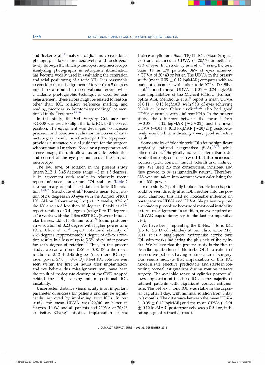
report rotation of 3.4 degrees (range 0 to 12 degrees)at 16 weeks with the T-flex 623T IOL (Rayner Intraoc-ular Lenses, Ltd.). Hoffmann et al.24 found postoper-ative rotation of 0.23 degree with higher power toricIOLs. Chua et al.25 report rotational stability of4.23 degrees. Approximately 1 degree of off-axis rota-tion results in a loss of up to 3.3% of cylinder powerfor each degree of rotation.15 Thus, in the presentstudy, we can attribute 0.06 G 0.02 D to the meanrotation of 2.12 G 3.45 degrees (mean toric IOL cyl-inder power 2.98 G 0.87 D). Most IOL rotation wasseen within the first 24 hours after implantation,and we believe this misalignment may have beenthe result of inadequate clearing of the OVD trappedbehind the IOL, causing minor positional IOLinstability.
Uncorrected distance visual acuity is an importantparameter of success for patients and can be signifi-cantly improved by implanting toric IOLs. In ourstudy, the mean UDVA was 20/40 or better in30 eyes (100%) and all patients had CDVA of 20/25or better. Chang26 studied implantation of the
1-piece acrylic toric Staar TF/TL IOL (Staar SurgicalCo.) and obtained a CDVA of 20/40 or better in92% of eyes. In a study by Sun et al.27 using the toricStaar TF in 130 patients, 84% of eyes achieveda CDVA of 20/40 or better. The UDVA in the presentstudy (mean 0.05 G 0.12 logMAR) compares with re-ports of outcomes with other toric IOLs. De Silvaet al.20 found a mean UDVA of 0.32 G 0.24 logMARafter implantation of the Microsil 6116TU (Human-optics AG). Mendicute et al.8 report a mean UDVAof 0.11 G 0.15 logMAR, with 93% of eyes achieving20/40 or better. Other studies21,22 also had goodUDVA outcomes with different IOLs. In the presentstudy, the difference between the mean UDVA(C0.05 G 0.12 logMAR [w20/25]) and the meanCDVA (�0.01 G 0.10 logMAR [w20/20]) postopera-tively was 0.5 line, indicating a very good refractiveresult.
Some studies of foldable toric IOLs found significantsurgically induced astigmatism (SIA),28,29 whileothers did not.30 Surgically induced astigmatism is de-pendent not only on incisionwidth but also on incisionlocation (clear corneal, limbal, scleral) and architec-ture. We used 2.3 mm corneoscleral incisions, andthey proved to be astigmatically neutral. Therefore,SIA was not taken into account when calculating thetoric IOL power.
In our study, 2 partially broken double-loop hapticscould be seen directly after IOL injection into the pos-terior chamber; this had no noticeable influence onpostoperative UDVA and CDVA. No patient requireda secondary procedure because of rotational instabilityor torus misalignment. In addition, no eye required anNd:YAG capsulotomy up to the last postoperativevisit.
We have been implanting the Bi-Flex T toric IOL(1.5 to 4.5 D of cylinder) at our clinic since May2011. It is a single-piece hydrophilic acrylic toricIOL with marks indicating the plus axis of the cylin-der. We believe that the present study is the first todescribe application of this toric IOL in a cohort ofconsecutive patients having routine cataract surgery.Our results indicate that implantation of this IOLmodel is safe, effective, predictable, and stable in cor-recting corneal astigmatism during routine cataractsurgery. The available range of cylinder powers al-lows application of this toric IOL in the majority ofcataract patients with significant corneal astigma-tism. The Bi-Flex T toric IOL was stable in the capsu-lar bag after 1 day, with minimal rotation from 1 dayto 3 months. The difference between the mean UDVA(C0.05 G 0.12 logMAR) and the mean CDVA (�0.01G 0.10 logMAR) postoperatively was a 0.5 line, indi-cating a good refractive result.
1396 ROTATIONAL STABILITY AND OUTCOMES OF A NEW TORIC IOL
J CATARACT REFRACT SURG - VOL 39, SEPTEMBER 2013
PIIS0886335013005245_002.indd 7PIIS0886335013005245_002.indd 7 2016.03.31. 9:56:482016.03.31. 9:56:48

WHAT WAS KNOWN
� Approximately 60% of cataractous eyes have more than0.75 D of corneal astigmatism.
� Stable and effective toric IOLs implanted in the capsularbag during routine cataract surgery are therefore an im-portant requirement for modern cataract surgery, allowingcorrection of corneal astigmatism during cataract surgery.
WHAT THIS PAPER ADDS
� The new Bi-Flex T hydrophilic acrylic toric IOL effectivelyand predictably managed corneal astigmatism during rou-tine cataract surgery.
� The difference between the mean UDVA (C0.05 G 0.12logMAR) and the mean CDVA (�0.01 G 0.10 logMAR)postoperatively was a 0.5 line, indicating a good refractiveresult. The mean IOL rotation was 2.12 G 3.45 degreesover the 3-month follow-up.
REFERENCES1. Hoffer KJ. Biometry of 7,500 cataractous eyes. AmJOphthalmol
1980; 90:360–368; correction, 890
2. OuchiM,KinoshitaS.AcrySof IQ toric IOL implantationcombined
with limbal relaxing incision during cataract surgery for eyes with
astigmatismO2.50 D. J Refract Surg 2011; 27:643–647
3. Qammar A, Mullaney P. Paired opposite clear corneal incisions
to correct preexisting astigmatism in cataract patients.
J Cataract Refract Surg 2005; 31:1167–1170
4. Norouzi H, Rahmati-Kamel M. Laser in situ keratomileusis for
correction of induced astigmatism from cataract surgery.
J Refract Surg 2003; 19:416–424
5. GunvantP,AblamowiczA,GollamudiS.Predicting thenecessity
of LASIK enhancement after cataract surgery in patients with
multifocal IOL implantation. Clin Ophthalmol 2011; 5:1281–
1285. Available at: http://www.ncbi.nlm.nih.gov/pmc/articles/
PMC3180499/pdf/opth-5-1281.pdf. Accessed April 9, 2013
6. Kim P, Sutton GL, Rootman DS. Applications of the femtosec-
ond laser in corneal refractive surgery. Curr Opin Ophthalmol
2011; 22:238–244
7. R€uckl T, Dexl AK, Bachernegg A, Reischl V, Riha W,
Ruckhofer J, Binder PS, Grabner G. Femtosecond laser–assis-
ted intrastromal arcuate keratotomy to reduce corneal astigma-
tism. J Cataract Refract Surg 2013; 39:528–538
8. Mendicute J, Irigoyen C, Ruiz M, Illarramendi I, Ferrer-Blasco T,
Mont�es-Mic�o R. Toric intraocular lens versus opposite clear cor-
neal incisions to correct astigmatism in eyes having cataract sur-
gery. J Cataract Refract Surg 2009; 35:451–458
9. Horn JD. Status of toric intraocular lenses. Curr Opin Ophthal-
mol 2007; 18:58–61
10. Ohmi S. Decentration associated with asymmetric capsular
shrinkage and intraocular lens size. J Cataract Refract Surg
1993; 19:640–643
11. Strenn K, Menapace R, Vass C. Capsular bag shrinkage after
implantation of an open-loop silicone lens and a poly(methyl
methacrylate) capsule tension ring. J Cataract Refract Surg
1997; 23:1543–1547
12. Carvalho MJ, Suzuki SH, Freitas LL, Branco BC, Schor P,
H€offling-Lima AL. Limbal relaxing incisions to correct corneal
astigmatism during phacoemulsification. J Refract Surg 2007;
23:499–504
13. Myers TD, Olson RJ. Comparison of the effects of viscoelastic
agents on clinical properties of the Unfolder lens injection sys-
tem. J Cataract Refract Surg 1999; 25:953–958
14. Pereira FAS, Milverton EJ, Coroneo MT. Miyake-Apple study of
the rotational stability of the Acrysof toric intraocular lens after
experimental eye trauma. Eye 2010; 24:376–378. Available at:
http://www.nature.com/eye/journal/v24/n2/pdf/eye2009150a.
pdf. Accessed April 9, 2013
15. Shimizu K, Misawa A, Suzuki Y. Toric intraocular lenses: cor-
recting astigmatism while controlling axis shift. J Cataract
Refract Surg 1994; 20:523–526
16. Weinand F, Jung A, Stein A, Pf€utzner A, Becker R, Pavlovic S.
Rotational stability of a single-piece hydrophobic acrylic intraoc-
ular lens: new method for high-precision rotation control.
J Cataract Refract Surg 2007; 33:800–803
17. Becker KA, Auffarth GU, V€olcker HE. Messmethode zur Bestim-
mung der Rotation und der Dezentrierung von Intraokularlinsen
[Measurement method for the determination of rotation and de-
centration of intraocular lenses]. Ophthalmologe 2004;
101:600–603
18. Auffarth GU, Rabsilber TM. Torische Hinterkammerlinsen nach
Kataraktoperation und refraktivem Linsenaustausch [Toric IOLs
after cataract surgery and refractive lens exchange]. Ophthal-
mologe 2007; 104:1024–1031
19. Tehrani M, Dick HB, Krummenauer F, Pfirrmann G, Boyle T,
Stoffelns BM. Capsule measuring ring to predict capsular bag
diameter and follow its course after foldable intraocular lens
implantation. J Cataract Refract Surg 2003; 29:2127–2134
20. De Silva DJ, Ramkissoon YD, Bloom PA. Evaluation of a toric
intraocular lens with a Z-haptic. J Cataract Refract Surg 2006;
32:1492–1498
21. Alberdi T, Mac�ıas-Murelaga B, Bascar�an L, Go~ni N, S�aez de
Arregui S, Mendicute J. Rotational stability and visual quality
in eyes with Rayner toric intraocular lens implantation.
J Refract Surg 2012; 28:696–701
22. Sheppard AL,Wolffsohn JS, Bhatt U, Hoffmann PC, Scheider A,
H€utzWW, Shah S. Clinical outcomes after implantation of a new
hydrophobic acrylic toric IOL during routine cataract surgery.
J Cataract Refract Surg 2013; 39:41–47
23. Entabi M, Harman F, Lee N, Bloom PA. Injectable 1-piece
hydrophilic acrylic toric intraocular lens for cataract surgery:
efficacy and stability. J Cataract Refract Surg 2011;
37:235–240
24. Hoffmann PC, Auel S, H€utz WW. Results of higher power toric
intraocular lens implantation. J Cataract Refract Surg 2011;
37:1411–1418
25. ChuaW-H, Yuen LH, Chua J, Teh G, Hill WE. Matched compar-
ison of rotational stability of 1-piece acrylic and plate-haptic sili-
cone toric intraocular lenses in Asian eyes. J Cataract Refract
Surg 2012; 38:620–624
26. ChangDF. Comparative rotational stability of single-piece open-
loop acrylic and plate-haptic silicone toric intraocular lenses.
J Cataract Refract Surg 2008; 34:1842–1847
27. Sun X-Y, Vicary D, Montgomery P, Griffiths M. Toric intraoc-
ular lenses for correcting astigmatism in 130 eyes. Ophthal-
mology 2000; 107:1776–1781; discussion by RM Kershner,
1781–1782
28. Ruhswurm I, Scholz U, ZehetmayerM, HanselmayerG, Vass C,
Skorpik C. Astigmatism correction with a foldable toric intraocu-
lar lens in cataract patients. J Cataract Refract Surg 2000;
26:1022–1027
1397ROTATIONAL STABILITY AND OUTCOMES OF A NEW TORIC IOL
J CATARACT REFRACT SURG - VOL 39, SEPTEMBER 2013
PIIS0886335013005245_002.indd 8PIIS0886335013005245_002.indd 8 2016.03.31. 9:56:482016.03.31. 9:56:48

29. Bauer NJC, de Vries NE, Webers CAB, Hendrikse F,
Nuijts RMMA. Astigmatism management in cataract surgery
with the AcrySof toric intraocular lens. J Cataract Refract Surg
2008; 34:1483–1488
30. Ali�o JL, AgdeppaMCC, Pongo VC, El Kady B. Microincision cat-
aract surgery with toric intraocular lens implantation for correct-
ing moderate and high astigmatism: pilot study. J Cataract
Refract Surg 2010; 36:44–52
OTHER CITED MATERIALA. Spalton D, Chan E. Latest developments in intraocular lenses.
Continuing Education & Training Module 12 Part 10: Vision in the
aged. October 2009; 28–34. Available at: http://www.optometry.
co.uk/uploads/articles/CET091009.pdf. Accessed April 9, 2013
B. Hill W. IOLMaster lens constants; the Haigis formula. Available
at: http://doctor-hill.com/zeiss_iolmaster/haigis_formula.htm.
Accessed April 9, 2013
C. Medicontur Medical Engineering Ltd., Inc. Medicontur toric IOL
calculator. Available at: http://medicontur.hu/toric-iol-calculator/.
Accessed April 9, 2013
D. Package insert. Medicontur Medical Engineering Ltd., Inc.,
Zs�amb�ek, Hungary
First author:Alexander Bachernegg, MD
FromtheDepartment ofOphthalmology,Paracelsus Medical UniversitySalzburg, Salzburg, Austria
1398 ROTATIONAL STABILITY AND OUTCOMES OF A NEW TORIC IOL
J CATARACT REFRACT SURG - VOL 39, SEPTEMBER 2013
PIIS0886335013005245_002.indd 9PIIS0886335013005245_002.indd 9 2016.03.31. 9:56:482016.03.31. 9:56:48