Embed Size (px)
Citation preview

J. exp. Biol. 163, 15-31 (1992) 15Printed in Great Britain © The Company of Biologists Limited 1992
ROTATIONAL MOVEMENT OF A SPERMATOZOON AROUNDITS LONG AXIS
BY SUMIO ISHIJIMA, MIYAKO S. HAMAGUCHI, MASAKAZUNARUSE, SANAE A. ISHIJIMA* AND YUKIHISA HAMAGUCHI
Biological Laboratory, Tokyo Institute of Technology, O-okayama, Meguro-ku,Tokyo 152, Japan
Accepted 16 September 1991
Summary
The rotational movement of a spermatozoon around its longitudinal axis wasinvestigated by two methods: by observing a spermatozoon attached vertically to acoverslip by the tip of its head, and by observing a spermatozoon freely swimmingin a medium by means of 'double-focal microscopy', which yielded simultaneousimages at two different focal planes.
Similar results were obtained by these two methods. Sea urchin, starfish,medaka, human, golden hamster and bull spermatozoa rolled in both clockwiseand counterclockwise directions, although there was a large difference in theproportion of spermatozoa rolling in each direction in the different species. Themajority of sea urchin and starfish spermatozoa rolled in a clockwise directionwhen an observer viewed the cell from its anterior end, whereas the majority ofmedaka, golden hamster, human and bull spermatozoa rolled in a counterclock-wise direction relative to the same observer. Moreover, some spermatozoaoccasionally changed their rotational direction.
These results suggest that the mechanism regulating the direction of rotation ofthe spermatozoa is lax. As rotational movement of a spermatozoon around itslongitudinal axis is due to the three-dimensional component of the beat of theflagellum, the direction of the three-dimensional movement presumably changesas the spermatozoa swim.
Introduction
Most spermatozoa roll around their longitudinal axis as they swim freely (Gray,1955, 1958; Bishop, 1958; Rikmenspoel, 1965; Phillips, 1972). This is very strongpresumptive evidence that sperm flagellar movement is not confined to a singleplane (Taylor, 1952; Gray, 1962; Chwang and Wu, 1971), although the unlikely
* Present address: Department of Biology, College of Arts and Sciences, the University ofTokyo, Komaba, Meguro-ku, Tokyo 153, Japan.
Key words: flagellar waves, rolling motion, rotation frequency, spermatozoon, three-dimen-sional geometry.
16 S. ISHIJIMA AND OTHERS
possibility that the asymmetrical shape of the sperm head causes the rotation of thewhole spermatozoon cannot be ruled out.
Several investigators have tried to determine the sense of rotation of aspermatozoon, using a variety of approaches, because information on thedirection and the rate of rotation are fundamental to the determination of thethree-dimensional geometry of flagellar movement (Bishop, 1958; Drake, 1974;Woolley, 1977; Yeung and Woolley, 1984). However, there is no agreement as tothe direction of rotation reported in these studies (Woolley, 1979).
The geometry of the flagellar waveform is technically difficult to determineprecisely, although several methods have been used to overcome this problem,including high-speed cinemicrography, video micrography, stroboscopic illumi-nation, micromanipulative techniques, and so on (Woolley, 1979; Ishijima andMohri, 1990).
In order to elicit detailed information on the shape of the flagellar waveform toaid our understanding of the basic control mechanisms of the flagellar movement,the following micromanipulative technique has been developed (Ishijima andHiramoto, 1982; Ishijima and Mohri, 1985; Ishijima etal 1986; Ishijima andWitman, 1987). A spermatozoon is held by its head with a sucking micropipetteand its flagellar movement observed from various directions under the microscopewhile the spermatozoon is alive and beating. This method was very useful forstudying whether flagellar movement occurred within a single plane or, if not, howfar the waveform of the beating flagellum deviated from the major beating planewithin the limit of resolution of the light microscope. However, it could not beused to ascertain the handedness or three-dimensional shape of the beatingflagellum.
In the present study, rotational movement of a spermatozoon around itslongitudinal axis was investigated by two methods in order to reconstruct the exactthree-dimensional geometry of flagellar movement of various spermatozoa. Thepresent studies will not only help to clarify both the existence and nature of therotational movement of spermatozoa, but will also suggest ways for analyzingaspects of their three-dimensional movement.
Materials and methods
Sperm preparations
Bull sperm were kindly provided as a frozen straw of semen in liquid nitrogen bythe Snow Brand Embryotransfer Laboratory (Tomakomai, Japan). For eachexperiment, a straw was thawed in warm (37°C) water before use. To select motilespermatozoa from the semen by 'swimming-up' procedures (Lopata et al. 1976;Ishijima and Witman, 1991), the thawed semen was placed at the bottom of a shortglass tube (15 mm x 50 mm) and carefully covered with 3 ml of warm (37 °C)Tyrode's solution. After leaving the tube to stand for almost lOmin at roomtemperature, the sperm suspension in the upper 1.0-1.5 ml in each tube wascollected and transferred into another tube.
Sperm rotation around its long axis 17
Golden hamster spermatozoa were obtained from a cauda epididymis removedsurgically from a mature male under ether anesthesia (Yanagimachi, 1982;Ishijima and Witman, 1991). A dense mass of the 'dry' sperm, which had oozed outwhen the cauda epididymis was punctured with a sharp needle, was placed at thebottom of a short glass tube (15 mmx50mm). The dry sperm were covered with3 ml of warm (37°C) Tyrode's solution, and then actively motile golden hamsterspermatozoa were selected by the swimming-up method outlined above.
Human semen was collected by masturbation from four healthy men aftersexual abstinence for at least 4 days. The semen was washed three times inTyrode's solution by centrifugation at 250g for lOmin. Approximately 30^1samples of the loosely packed sperm pellet were suspended in 3 ml of Tyrode'ssolution.
The spermatozoa of the medaka, Oryzias latipes, were obtained as follows(Yamamoto, 1961). The testis was isolated by dissecting the abdomen of an adultmale from the opening of the rectum, after pithing the brain with the fine point ofstraight iris scissors, and then put into Ringer's solution for the medaka (7.5 gNaCl, 0.2g KC1, 0.2g CaCl2 and 0.02g NaHCO3 per liter of deionized water,pH7.3) in a plastic culture dish (35 mmx 10 mm). The spermatozoa were liberatedby teasing apart the testis in the Ringer's solution.
Concentrated spermatozoa of the sea urchin, Hemicentrotus pulcherrimus, andthose of the starfish, Asterina pectinifera, were obtained by dissecting out thegonad and placing it in a plastic culture dish (35 mmx 10mm).
Observations and recording
The rotational movement of various spermatozoa was investigated both byobserving a spermatozoon attached vertically to the coverslip at the tip of its headand by observing a spermatozoon swimming freely in a medium by means ofdouble-focal microscopy (see below). Using a suitably pretreated coverslip (seebelow), sperm could be found that were stuck by the anterior surface of their headwith the wave axes of the beating flagellum oriented nearly vertical to the coverslipsurface. The anterior regions of the beating flagella of these spermatozoa wereobserved using a Nikon Optiphot microscope equipped with a phase-contrastcondenser, objectives (plan 40x BM and plan 20x BM) and lOx eyepieces. In thecase of the experiments on rotational movement of the head of bull spermatozoa,differential interference contrast (DIC) microscopy was used with objectives (plan40x DIC and plan 20x DIC) and lOx eyepieces. For mammalian spermatozoa,the condenser was modified for a microscope stage warmer; the top lens wasremoved and home-made phase annuli corresponding to the phase plates ofobjectives were inserted. Images were recorded on video tape with a Nationalvideo camera (WV-1300A, Matsushita Communication Industrial Co., Ltd,Yokohama, Japan), a video timer (VTG-33, For-A Corp., Tokyo) and a Nationaltime-lapse video tape recorder (NV-8030, Matsushita Electric Industrial Co., Ltd,Oosaka) which yielded 60 images per second. Images were displayed on a NationalTN-96 monitor (Matsushita Communication Industrial Co., Ltd). Illumination

18 S. ISHIJIMA AND OTHERS
was provided by stroboscopic flashes from a Chadwick-Helmuth model 100(Chadwick-Helmuth Corp., El Monte, CA), the intensity of the flash beingmodulated by a number of neutral density filters placed between the condenserand the lamp. A heat-absorption filter affording some thermal protection was alsoplaced in this position. Turning a rotating mechanical stage confirmed that thedirection of rotation of a specimen was identical to that of the images formed bythe video system and shown on the monitor.
Double-focal microscopy was used for medaka, bull and human spermatozoa.This method yielded images at two different focal planes at the same time(Hamaguchi etal. 1991). One video camera (National WV-1300A) equipped witha Nikon zoom lens was mounted on one of the binocular eyepiece tubes whileanother video camera (AVC-1550, Sony Corp., Tokyo) with a Nikon zoom lenswas placed on the other binocular eyepiece tube; the second lens had a mechanicaltube length different from that of the first. The difference in the mechanical tubelength between the two lenses was adjusted so that the distance between two focalplanes was approximately 60 % of the maximum displacement of beating flagellain medaka, bull and human spermatozoa. The output of the two video cameraswas montaged using a National special effects generator (WJ-545, MatsushitaCommunication Industrial Co., Ltd) and then recorded on the video tape with aNational time-lapse video tape recorder (NV-8030) after first being fed through avideo timer. Images were displayed on a National WV-5360 monitor. The samesystem of illumination and filters as described for the other method was used forobservation and recording. Rotating a wire model of a spermatozoon around itslong axis confirmed that the rolling direction of a specimen was identical to that ofimages formed by the video system and appearing on the monitor.
The observation chamber was constructed by gluing three strips of glass onto aglass slide. The spermatozoa, prepared as described above, were diluted to aconcentration of approximately 106 sperm ml"1 and the sperm suspension wastransferred to the chamber. In the experiments in which we observed aspermatozoon attached to a coverslip by the tip of its head, the coverslips wereprecoated in different ways for the different species. The coverslips used for bulland human spermatozoa were precoated with 0.5 % agar dissolved in 0.9 % NaCl.For golden hamster spermatozoa, the coverslips were coated with 0.5 % agardissolved in 0.9 % NaCl and then dried. 0.5 % agar dissolved in artificial sea water(Jamarin U, Jamarin Laboratory, Oosaka) was used for precoating coverslips inthe case of sea urchin and starfish spermatozoa, and 0.5 % agar dissolved in theRinger's solution was used in the case of medaka spermatozoa. The presence ofagar on the coverslip did not change the rolling direction of spermatozoa.
Observations and recording were made at 37°C for mammalian spermatozoaand at room temperature (23°C) for medaka, sea urchin and starfish spermatozoa.
Data analysis
The rotational movement of each spermatozoon was recorded for intervals ofapproximately 15 s. For each spermatozoon, three parts of the video tape record

Sperm rotation around its long axis 19
were randomly chosen for analysis. For detailed field-by-field analysis, images ofthe flagellum and head were traced from the video monitor onto transparentplastic sheets using a fine-point marker. The direction of sperm rotation around itslongitudinal axis was determined from the direction of rotation of flagellar images.For analysis by double-focal microscopy, the beating plane was first determined byreconstructing a bending wave fitting the trace of images focused on the twodifferent planes. The direction of sperm rotation was determined from therotational direction of this beating plane. Rotation frequency of the beating planeor the head was calculated from the mean period required for one completerevolution of the beating plane or the head, respectively.
Results
Rotational movement of medaka, golden hamster, bull and human spermatozoaattached vertically to the cover slip
Most spermatozoa (58-100%) of medaka, golden hamster, bull and humanrolled in a counterclockwise direction around their longitudinal axes, when the cellwas viewed from the anterior end, but the remainder of them rolled in a clockwisedirection when they were attached vertically to a coverslip at the tip of the head(Table 1).
Bull spermatozoa
Bull spermatozoa fairly easily attached vertically to the surface of the coverslipby the anterior surface of their head and rolled around their longitudinal axis. Theaxis of rotation did not oscillate to any great extent, suggesting that flagellar wavesof bull spermatozoa were fairly symmetrical on the two sides of the tail. When theanterior region of the beating flagellum of these spermatozoa was brought intofocus, cross sections of beating flagella were recorded on the video tape as brightspots (Fig. 1). Judging the rolling direction of the spermatozoon from the directionin which the bright spots moved revealed that bull spermatozoa rolled in bothdirections; clockwise and counterclockwise relative to the observer, althoughcounterclockwise rotation was predominant over clockwise rotation (Table 1).The average rotation frequency determined from the rolling flagellum was9.78±4.35 Hz (Table 1).
Bull spermatozoa have broad, flat heads and it was possible to determine thedirection and frequency of rotation of the sperm head by taking advantage of thismorphological characteristic (Fig. 2). The sperm head rotated in the samedirection as the tail. The rotation frequency of the head (9.25±6.09Hz, iV=12)was, moreover, little different from that of the tail. These results suggest that thehead of a bull spermatozoon does not rotate freely but rolls together with the tailaround its longitudinal axis.
Golden hamster spermatozoa
Few golden hamster spermatozoa attached vertically to the surface of the

20 S. ISHIJIMA AND OTHERS
Table 1. Characteristics of rotational movement of various spermatozoa
Species
Bull
Golden hamster
Human
MedakaOryzias latipes
Sea urchin
Direction of sperm
Counterclockwise(%)
73.3(57.1)
76.2
57.1(58.8)
88.2(100)
5.0Hemicentrotus pulcherrimus
StarfishAsterina pectinifera
30.4
rotation
Clockwise(%)
26.7(42.9)
23.8
42.9(41.2)
11.8(0)
95.0
69.6
N
30(84)
21
21(51)
17(20)
20
23
Rotation frequency(Hz)
9.78±4.35(9.08±1.75)
1.48±0.69
9.33±4.85(10.18±1.91)
3.77±2.82(5.30±1.21)
2.55±0.98
0.50±0.57
N
19(49)
8
15(51)
17(20)
16
15
Measurements were carried out at 37 °C for mammalian spermatozoa and at roomtemperature (23°C) for medaka, sea urchin and starfish spermatozoa.
N, number of spermatozoa measured. The figures in parentheses are data measured by meansof double-focal microscopy.
Direction of a spermatozoon rotation around its long axis was noted by an observer viewingthe cell from its anterior end. Spermatozoa that rotated in both directions were included in boththe clockwise and counterclockwise categories.
Values of rotation frequency are mean±s.D.
coverslip because of their hook-shaped heads. Thus, the axis of rotation tended tooscillate back and forth when a spermatozoon rotated around its longitudinal axis.In those spermatozoa whose axis of rotation did not oscillate too much, thedirection of roll of most (76.2%) was counterclockwise, whereas the remainder(23.8%) rolled in a clockwise direction (Table 1). In this experiment, onespermatozoon was found to change its direction of roll from counterclockwise toclockwise, suggesting that each spermatozoon can occasionally change its direc-tion of roll. The average rotation frequency of golden hamster spermatozoa was1.48±0.69Hz (Table 1). Observations of the rotational movement of the spermhead and tail reveal that the sperm head and tail rolled together (data not shown).
Human spermatozoa
A human spermatozoon attached vertically to the coverslip by its head rolledaround its longitudinal axis with small and rapid oscillations. This oscillation of therotational axis was probably because flagellar movement in human spermatozoadid not occur within the plane containing the head and proximal region of theflagellum (see Ishijima et al. 1986). 57.1% of human spermatozoa rotated in a

Sperm rotation around its long axis
D
,4m
3Fig. 1. Phase-contrast video micrographs of rotational movement of bull spermflagellum observed from the sperm long axis. (A) Clockwise movement. (B) Counter-clockwise movement. The time interval between successive images is 1/60s. In C andD, tracings made from A and B, respectively, have been superimposed. Numbersindicate the positions of successive images. The scale bar represents 10|<m.
counterclockwise direction, the remainder in a clockwise direction. The averagerotation frequency of human spermatozoa was 9.33±4.85Hz, similar to that ofbull spermatozoa.
Medaka spermatozoa
Medaka spermatozoa have nearly spherical heads with an average diameter of

S. ISHIJIMA AND OTHERS
D
2 3 *4
Fig. 2. Differential interference contrast video micrographs of rotational movement ofbull sperm head. (A) Clockwise movement. (B) Counterclockwise movement. Thetime interval between successive images is 1/60 s. In C and D, tracings made from Aand B, respectively, have been superimposed. Numbers indicate the positions ofsuccessive images. The scale bar represents 10/mi.
3.71±0.60ium and tails of 30.9±1.60,um (7V=17) long. These spermatozoa movedforward at a rate of 86.6±20.1,ums~1 with a maximum displacement of beatingflagella of 5.14±1.73^m (yv=16) near the coverslip at 23°C. These values weremuch smaller than those of sea urchin spermatozoa, which are approximately thesame size as medaka spermatozoa. When medaka spermatozoa attached verticallyto the coverslip, most rolled counterclockwise (88.2%), the remainder (11.8%)undergoing clockwise rotation. Average rotation frequency of medaka spermato-zoa was 3.77±2.82Hz at 23°C.

Sperm rotation around its long axis 23
Rotational movement of sea urchin and starfish spermatozoa attached verticallyto the coverslip
In contrast to medaka and mammalian spermatozoa, the majority of sea urchinand starfish spermatozoa rolled in a clockwise direction when the spermatozoaattached vertically to the surface of a coverslip by the tip of their head.
Sea urchin spermatozoa
Most sea urchin spermatozoa attached vertically to the surface of a coverslip bythe tip of their head rolled around their longitudinal axis with little or nooscillation of the rolling axis. The beating plane of flagellar movement wasobserved as a rolling short segment because of the high beat frequency of flagellarwaves compared to the rotation frequency. The rolling motion of this segment wassometimes interrupted by intermittent pauses and, moreover, changed direction ofrotation from clockwise to counterclockwise. Judging the sense of rotation ofspermatozoa from the direction of rotation of the short segment, 95 % of seaurchin spermatozoa rolled clockwise and the spermatozoa of only 5 % rolledcounterclockwise. Average rotation frequency was 2.55±0.98 Hz.
Starfish spermatozoa
Most starfish spermatozoa did not attach vertically to a coverslip because of thealmost spherical shape of the sperm head, so the rolling axis underwent a'precession' movement, i.e. a slow, rotary motion of the rolling axis of a spinningspermatozoa, so that the axis of rotation described a cone and the poles of rotationdescribed circles in the medium. The rolling movement of starfish spermatozoa(Fig. 3) was rather slow (average rotation frequency=0.50±0.57Hz) compared tothat of the other species studied (Table 1). The rolling direction of the majority ofstarfish spermatozoa (69.6%) was clockwise, with 30.4% rolling counterclock-wise. One spermatozoon of 22 examined in this experiment rolled in a clockwisedirection after first rolling counterclockwise.
Rotational movement of medaka, bull and human spermatozoa investigated bymeans of double-focal microscopy
Spermatozoa swimming freely in a medium were recorded by double-focalmicroscopy, which yielded different images of beating flagella on two differentfocal planes (Fig. 4). A set of these images enabled us to determine the directionof the beating plane of the sperm flagellum (see Fig. 4). Several successive sets ofthese images yielded the sense of rotation of the sperm flagellum.
The direction of sperm rotation around its long axis determined by this methodwas counterclockwise for most medaka, human and bull spermatozoa (Table 1).These results were similar to those obtained from observing the spermatozoaattached vertically to the coverslip, although there were some differences in thepercentages rotating clockwise and counterclockwise (Table 1).
The rotation frequency determined by this method was almost identical to thatobtained by the other method (see Table 1).

S. ISHIJIMA AND OTHERS
5 '. 5
4 *3
Fig. 3. Phase-contrast video micrographs of rotational movement of starfish spermflagellum observed from the sperm long axis. (A) Clockwise movement. (B) Counter-clockwise movement. The time interval between successive images is 0.4s. In C and D,tracings made from A and B, respectively, have been superimposed. Numbers indicatethe positions of successive images. The scale bar represents 10 ,um.
DiscussionThere are three important findings in the present study. (1) Spermatozoa of all
species examined rolled both clockwise and counterclockwise. (2) The proportionof spermatozoa rotating counterclockwise to that rotating clockwise was differentin the different species. (3) The sense of rotation of most sea urchin and starfishspermatozoa was opposite to that of most medaka and mammalian (bull, goldenhamster and human) spermatozoa.

Sperm rotation around its long axis
Ai Bi Aii
25
Cii
Dii
Bp
.Uf
.Lf
Fig. 4. Determination of the direction of roll of a bull spermatozoon from the imagesobtained by double-focal microscopy. A, Ai and Aii are micrographs of a bullspermatozoon focused on the upper focal plane and B, Bi and Bii are images focusedon the lower focal plane. The spermatozoon is swimming in the direction of the arrowin A. The distance between the two focal planes is 4/j,m. The time interval betweensuccessive images (A, Ai and Aii or B, Bi and Bii) is 1/60s. The scale bar representslO^m. The superimposed tracings made from these micrographs are shown in C, Ciand Cii, where the filled areas represent the flagellar parts focused on the upper focalplane and the open traces are the counterparts on the lower focal plane. The majorbeating plane of a sperm flagellum is determined from these superimposed tracings asshown in D, Di and Dii, which are the side views; the image on the upper focal plane(Uf) and that on the lower focal plane (Lf) lie in the major beating plane (Bp). Thedirection of sperm rotation is determined from the direction of rotation of this majorbeating plane. The specimen in this figure rotates clockwise when viewed from theanterior end because the clockwise angle (arrow) from the focal plane increased withtnfflfc
Bidirectional rotation of spermatozoa
The observation that spermatozoa of all species examined could roll in bothdirections suggests that the mechanism controlling the direction of rotation ofbeating flagella is not fixed but, on the contrary, is easily changeable. This wasconfirmed by the observations that several golden hamster, sea urchin and starfish
26 S. ISHIJIMA AND OTHERS
spermatozoa changed their direction of roll from counterclockwise to clockwise orvice versa. Taking into account that the rotational movement of spermatozoa wasdue to the three-dimensional movement of their beating flagella (Taylor, 1952;Chwang and Wu, 1971; Woolley, 1979; and see below), the observation that spermcould rotate in both directions suggests that the three-dimensional geometry offlagellar movement of these spermatozoa changes while the spermatozoa arebeating.
The results obtained in the present experiments explain earlier confusion overthe rolling direction of spermatozoa. The observation by Drake (1974) that bullspermatozoa rolled in either a clockwise or a counterclockwise direction whenviewed from in front is the same as that obtained in the present study, althoughthat report did not contain any quantitative data. The suggestion that humanspermatozoa roll counterclockwise (Linnet, 1979) agrees with a part of our results.
The significance of changing the direction of sperm rotation is unclear.However, when the direction of sperm rotation changes, sperm can quickly andeffectively change their direction of movement. This kind of sperm behaviorimplies a tactical response, although no such response has yet been established inthe species examined in the present experiments (see Miller, 1985).
Some cilia show an almost half-funnel-shaped movement (Sleigh, 1974), theeffective stroke of the ciliary movement taking place in the vertical plane whilst inthe recovery stroke the cilium swings out to the side relative to an observer lookingdown onto the cell surface. The direction of the recovery stroke in such three-dimensional ciliary movement is opposite in different species. Thus, a Parameciumcilium rotates counterclockwise during the recovery stroke, as viewed from theciliary tip (Parducz, 1967; Machemer, 1972), while the lateral cilia oiMytilus edulisgill rotate clockwise (Aiello and Sleigh, 1972). The observation that cilia can beatin both directions during the recovery stroke may correspond to the finding that asperm flagellum can change the direction of its bending waves.
Factors regulating the direction of sperm rotation
Even though spermatozoa rolled in both directions, the proportion of spermato-zoa rolling counterclockwise to those rolling clockwise was not equal. Clockwiserotation was predominant to counterclockwise rotation in sea urchin and starfishspermatozoa, and vice versa in medaka, human, golden hamster and bullspermatozoa. Much more work is required to explain the difference in theproportion of counterclockwise to clockwise rotation. Some biochemical factorsmay be involved in regulating the ratio because the proportion of counterclock-wise to clockwise rotation in sea urchin and starfish spermatozoa was opposite tothat in the others. Calcium ion concentration is a possible candidate for such aregulating factor because intracellular calcium ion concentration in sea urchinspermatozoa is greater than 2xlO~~6molF1 (Schackmann and Chock, 1986),whereas that in mammalian spermatozoa is less than 4xlO~7moll~1: human,1.46xlO-7moir1 (Irvine and Aitken, 1986); rabbit, 1.44xlO~7moll"1 (Mahanesetal. 1986); ram, 1.93xlO~7moir1 (Simpson and White, 1988); ram,

Sperm rotation around its long axis 27
4xlO~7mol I"1 (Babcock and Pfeiffer, 1987). Therefore, a relatively high concen-tration of intracellular calcium, more than 10~6 mol I"1 for example, could induceclockwise rotation whereas a relatively low concentration, less than 10~6moll~1
for example, could induce counterclockwise rotation. Our preliminary exper-iments (Ishijima and Hamaguchi, 1990) revealed that Ca2+ in the reactivationsolution changed the direction of rotational movement of demembranated seaurchin spermatozoa. Cilia of Paramecium change the direction of the effectivestroke according to the intracellular calcium ion concentration (Naitoh andKaneko, 1972), and this type of regulating mechanism may also exist in flagella.
Three-dimensional geometry of sperm flagella and cilia
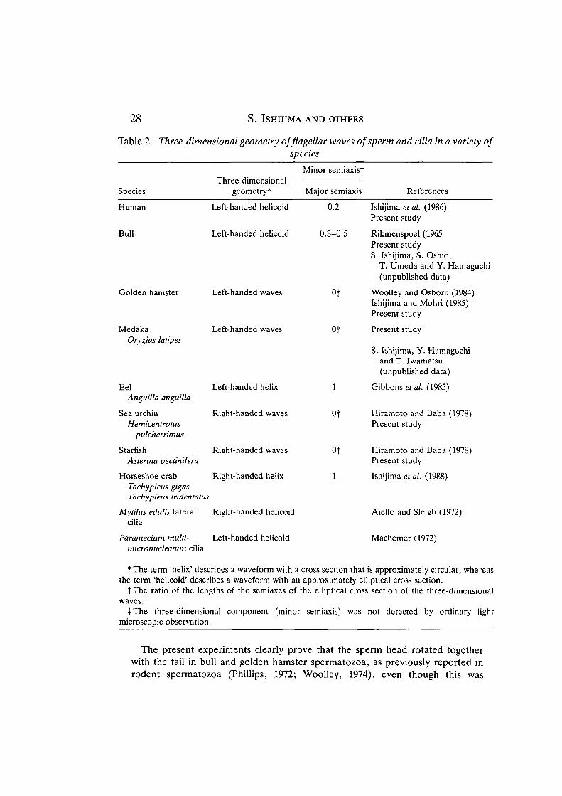
We cannot completely rule out the possibility that sperm rotation is caused bythe asymmetrical shape of the sperm head, especially in a free-swimmingspermatozoon (Phillips and Olson, 1975), but the more likely explanation is that itis caused by the three-dimensional movement of the flagellum (Gray, 1958;Chwang and Wu, 1971; Phillips and Olson, 1975, and see below). A knowledge ofthe direction of sperm rotation obtained from the present study therefore gives usthe sense of the three-dimensional geometry of flagella beating; that is, aspermatozoon rotates in the direction opposite to that of the revolving motion ofsegments of the sperm tail (see Fig. 1 of Gray, 1962). Most medaka, bull, goldenhamster and human spermatozoa beat with left-handed waves (i.e. with the sametwist as a left-handed screw), whereas most sea urchin and starfish spermatozoabeat with right-handed waves (i.e. with the same twist as a right-handed screw).This agrees with previous observations of bending flagella of golden hamsterspermatozoa (Woolley and Osborn, 1984). The three-dimensional geometry of theflagellar waves of some spermatozoa and the ciliary bends in each species aresummarized in Table 2. The results imply that most cilia and flagella of freshwateranimals beat with left-handed waves while those of marine animals beat with right-handed waves.
Influence of the sperm head on sperm rotation
In principle, the shape of the sperm head could be involved in promotingrotation of spermatozoa that swim freely (Phillips and Olson, 1975), just as apinwheel revolves in the wind. Assuming that the rotation frequency of spermato-zoa will increase as a result of the acceleration of rotation caused by the interactionbetween the asymmetrical sperm head and the stream of medium, it is possible tofind some differences in beat frequency from the two methods used in this study.Double-focal microscopy measured the rotation frequency of spermatozoa swim-ming freely in a stream of medium, whereas the other method measured that ofspermatozoa attached vertically to the coverslip, in which situation the spermheads were rotating in little or no stream. But the observation that the rotationfrequencies of spermatozoa obtained by the two methods were almost the samedoes not support this conjecture, at least for the spermatozoa examined in thisstudy.
28 S. ISHIJIMA AND OTHERS
Table 2. Three-dimensional geometry offlagellar waves of sperm and cilia in a variety ofspecies
Species
Human
Bull
Golden hamster
MedakaOryzias latipes
EelAnguilla anguilla
Sea urchinHemicentrotus
pulcherrimus
StarfishAsterina pectinifera
Horseshoe crabTachypleus gigasTachyplens tridentatus
Mytilus edulis lateralcilia
Three-dimensionalgeometry*
Left-handed helicoid
Left-handed helicoid
Left-handed waves
Left-handed waves
Left-handed helix
Right-handed waves
Right-handed waves
Right-handed helix
Right-handed helicoid
Minor semiaxisj
Major semiaxis
0.2
0.3-0.5
ot
0$
1
ot
ot
1
References
Ishijima et al. (1986)Present study
Rikmenspoel (1965Present studyS. Ishijima, S. Oshio,
T. Umeda and Y. Hamaguchi(unpublished data)
Woolley and Osborn (1984)Ishijima and Mohri (1985)Present study
Present study
S. Ishijima, Y. Hamaguchiand T. Iwamatsu(unpublished data)
Gibbons et al. (1985)
Hiramoto and Baba (1978)Present study
Hiramoto and Baba (1978)Present study
Ishijima et al. (1988)
Aiello and Sleigh (1972)
Paramecium multi-micronudeatum cilia
Left-handed helicoid Machemer (1972)
* The term 'helix' describes a waveform with a cross section that is approximately circular, whereasthe term 'helicoid' describes a waveform with an approximately elliptical cross section.
t The ratio of the lengths of the semiaxes of the elliptical cross section of the three-dimensionalwaves.
tThe three-dimensional component (minor semiaxis) was not detected by ordinary lightmicroscopic observation.
The present experiments clearly prove that the sperm head rotated togetherwith the tail in bull and golden hamster spermatozoa, as previously reported inrodent spermatozoa (Phillips, 1972; Woolley, 1974), even though this was

Sperm rotation around its long axis 29
presumed from the fact that the major beating plane, which was parallel to themajor axis of the sperm head cross section in bull, golden hamster, human andram, did not rotate when the sperm head was captured by a sucking micropipette(Ishijima and Mohri, 1985; Ishijima et al. 1986; Ishijima and Witman, 1987;S. Ishijima, S. Oshio, T. Umeda and Y. Hamaguchi, unpublished data).
Comparison of the two methods
Each method used in the present study has some advantages and disadvantages.The observation technique that examined the spermatozoon attached vertically toa coverslip is very useful for studying rotational movement of spermatozoabecause it is easy to do and clearly establishes the rotation of the spermatozoa. Butthis method can be applied only to those spermatozoa with a more or less roundedtip to the sperm head. Double-focal microscopy can be employed for almost allspermatozoa, although it involves somewhat troublesome procedures to deter-mine the direction of rotation of spermatozoa.
Although there is no agreement, as mentioned above, about the direction ofrotation of spermatozoa, there are several reports presenting the values ofrotational frequency of spermatozoa (see Table 3 of Ishijima et al. 1986, or Table Iof Ishijima and Mohri, 1990, for details). There are no significant differencesbetween these data and those obtained in the present study.
As shown in Table 2, we can now determine the three-dimensional geometry offlagellar movement of spermatozoa by combining the handedness of bendingwaves of sperm flagella with the information obtained by means of a micromanipu-lative technique developed by one of us. Moreover, the handedness of bendingwaves of sperm flagella and the rotation frequency obtained in this study arenecessary for describing sperm behavior in a medium.
We thank Dr S. Oshio for supplying bull spermatozoa and Dr T. Iwamatsu forhis advice on the preparation of medaka spermatozoa. We are also grateful to DrJ. S. Hyams for his critical reading of the manuscript.
ReferencesAIELLO, E. AND SLEIGH, M. A. (1972). The metachronal wave of lateral cilia of Mytilus edulis.
J. Cell Biol. 54, 493-506.BABCOCK, D. F. AND PFEIFFER, D. R. (1987). Independent elevation of cytosolic [Ca2+] and pH
of mammalian sperm by voltage-dependent and pH-sensitive mechanisms. J. biol. Chem. 262,15041-15047.
BISHOP, D. W. (1958). Motility of the sperm flagellum. Nature 182, 1638-1640.CHWANG, A. T. AND W U , T. Y. (1971). A note on the helical movement of micro-organisms.
Proc. R. Soc. Lond. B 178, 327-346.DRAKE, A. D. (1974). Observations on bull sperm rotation. Biol. Reprod. 10, 78-84.GIBBONS, B. H., BACCETTI, B. AND GIBBONS, I. R. (1985). Live and reactivated motility in the
9+0 flagellum oiAnguilla sperm. Cell Modi. 5, 333-350.GRAY, J. (1955). The movement of sea-urchin spermatozoa. J. exp. Biol. 32, 775-801.GRAY, J. (1958). The movement of the spermatozoa of the bull. J. exp. Biol. 35, 96-108.GRAY, J. (1962). Flagellar propulsion. In Spermatozoon Motility (ed. D. W. Bishop), pp. 1-12.
Washington, DC: American Association for the Advancement of Science.

30 S. ISHIJIMA AND OTHERS
HAMAGUCHI, M. S., ISHIJIMA, S., HAMAGUCHI, Y. AND HIRAMOTO, Y. (1991). Double-focalvideomicroscopy: a simple video system for analyzing the dynamics of cell motility. J. exp.Biol. 161, 537-541.
HIRAMOTO, Y. AND BABA, S. A. (1978). A quantitative analysis of flagellar movement inechinoderm spermatozoa. 7. exp. Biol. 76, 85-104.
IRVINE, D. S. AND AITKEN, R. J. (1986). Measurement of intracellular calcium in humanspermatozoa. Gamete Res. 15, 57-71.
ISHIJIMA, S. AND HAMAGUCHI, Y. (1990). Effects of Ca2+ on the direction of rotationalmovement of demembranated sea urchin spermatozoa. Zool. Sci. 7, 1043.
ISHIJIMA, S. AND HIRAMOTO, Y. (1982). Mechanical properties of sperm flagella. Cell Motil.(Suppl.) 1, 149-152.
ISHIJIMA, S. AND MOHRI, H. (1985). A quantitative description of flagellar movement in goldenhamster spermatozoa. J. exp. Biol. 114, 463-475.
ISHIJIMA, S. AND MOHRI, H. (1990). Beating patterns of mammalian spermatozoa. In Controls ofSperm Motility (ed. C. Gagnon), pp. 25-38. Boca Raton: CRC Press.
ISHIJIMA, S., OSHIO, S. AND MOHRI, H. (1986). Flagellar movement of human spermatozoa.Gamete Res. 13, 185-197.
ISHIJIMA, S., SEKIGUCHI, K. AND HIRAMOTO, Y. (1988). Comparative study of the beat patterns ofAmerican and Asian horseshoe crab sperm: Evidence for a role of the central pair complex informing planar waveforms in flagella. Cell Motil. Cytoskel. 9, 264-270.
ISHIJIMA, S. AND WITMAN, G. B. (1987). Flagellar movement of intact and demembranated,reactivated ram spermatozoa. Cell Motil. Cytoskel. 8, 375-391.
ISHIJIMA, S. AND WITMAN, G. B. (1991). Demembranation and reactivation of mammalianspermatozoa from golden hamster and ram. Methods Enzymol. 196, 417-428.
LINNET, L. (1979). Human spermatozoa: Unidirectional rotation of the tail as indicated by head-to-head agglutinates. Arch. Androl. 2, 157-161.
LOPATA, A., PATULLO, M. J., CHANG, A. AND JAMES, B. (1976). A method for collecting motilespermatozoa from human semen. Fertil. Steril. 27, 677-684.
MACHEMER, H. (1972). Ciliary activity and the origin of metachrony in Paramecium: Effects ofincreased viscosity. J. exp. Biol. 57, 239-259.
MAHANES, M. S., OCHS, D. L. AND ENG, L. A. (1986). Cell calcium of ejaculated rabbitspermatozoa before and following in vitro capacitation. Biochem. biophys. Res. Comimm.134, 664-670.
MILLER, R. L. (1985). Sperm chemo-orientation in the metazoa. In Biology of Fertilization, vol.2 (ed. C. B. Metz and A. Monroy), pp. 275-337. Orland: Academic Press.
NAITOH, Y. AND KANEKO, H. (1972). Reactivated Triton-extracted models of Paramecium:Modification of ciliary movement by calcium ions. Science 176, 523-524.
PARDUCZ, B. (1967). Ciliary movement and coordination in ciliates. Int. Rev. Cytol. 21, 91-128.PHILLIPS, D. M. (1972). Comparative analysis of mammalian sperm motility. J. Cell Biol. 53,
561-573.PHILLIPS, D. M. AND OLSON, G. (1975). Mammalian sperm motility - structure in relation to
function. In The Functional Anatomy of The Spermatozoon (ed. B. A. Afzelius), pp. 117-126.Oxford: Pergamon Press.
RIKMENSPOEL, R. (1965). The tail movement of bull spermatozoa. Observations and modelcalculations. Biophys. J. 5, 365-392.
SCHACKMANN, R. W. AND CHOCK, P. B. (1986). Alteration of intracellular [Ca2+] in sea urchinsperm by the egg peptide speract. Evidence that increased intracellular Ca2+ is coupled toNa+ entry and increased intracellular pH. J. biol. Chem. 261, 8719-8728.
SIMPSON, A. M. AND WHITE, I. G. (1988). Measurement and manipulation of cytoplasmic freecalcium of ram and boar spermatozoa using quin 2. Cell Calcium 9, 45-56.
SLEIGH, M. A. (1974). Patterns of movement of cilia and flagella. In Cilia and Flagella (ed. M. A.Sleigh), pp. 79-92. London: Academic Press.
TAYLOR, G. (1952). The action of waving cylindrical tails in propelling microscopic organisms.Proc. R. Soc. Lond. A 211, 225-239.
WOOLLEY, D. M. (1974). Freeze-substitution: a method for the rapid arrest and chemicalfixation of spermatozoa. J. Microsc. 101, 245-260.

Sperm rotation around its long axis 31
WOOLLEY, D. M. (1977). Evidence for 'twisted plane' undulations in golden hamster sperm tails.J. Cell Biol. 75, 851-865.
WOOLLEY, D. M. (1979). Interpretations of the pattern of sperm tail movements. In TheSpermatozoon (ed. D. W. Fawcett and J. M. Bedford), pp. 69-79. Baltimore-Munich: Urban& Schwarzenberg.
WOOLLEY, D. M. AND OSBORN, I. W. (1984). Three-dimensional geometry of motile hamsterspermatozoa. J. Cell Sci. 67, 159-170.
YAMAMOTO, T. (1961). Physiology of fertilization in fish eggs. Int. Rev. Cytol. 12, 361-405.YANAGIMACHI, R. (1982). In vitro sperm capacitation and fertilization of golden-hamster eggs in
a chemically defined medium. In In Vitro Fertilization and Embryo Transfer (ed. E. S. E.Hafez and K. Semm), pp. 65-76. Lancaster: MTP Press.
YEUNG, C.-H. AND WOOLLEY, D. M. (1984). Three-dimensional bend propagation in hamstersperm models and the direction of roll in free-swimming cells. Cell Modi. 4, 215-226.