Embed Size (px)
Citation preview

Contents lists available at ScienceDirect
Surface & Coatings Technology
journal homepage: www.elsevier.com/locate/surfcoat
Room-temperature deposition of hydroxyapatite/antibiotic compositecoatings by vacuum cold spraying for antibacterial applications
Deyan Lia,b, Yongfeng Gonga, Xiuyong Chena,⁎, Botao Zhangc, Haijun Zhangb, Peipeng Jinb,Hua Lia,⁎
a Key Laboratory of Marine Materials and Related Technologies, Key Laboratory of Marine Materials and Protective Technologies of Zhejiang Province, Ningbo Institute ofMaterials Technology and Engineering, Chinese Academy of Sciences, Ningbo 315201, Chinab School of Mechanical Engineering, Qinghai University, Xining 810016, Chinac Cixi Institute of Biomedical Engineering, Institute of Materials Technology and Engineering, Chinese Academy of Sciences, Ningbo 315201, China
A R T I C L E I N F O
Keywords:HydroxyapatiteAntibioticNanocomposite coatingsVacuum cold sprayAntibacterial property
A B S T R A C T
To develop implants with long-term antibacterial property, hydroxyapatite (HA)-gentamicin sulfate (GS) com-posite powder were synthesized and deposited onto titanium substrates by vacuum cold spraying at roomtemperature. The successful fabrication of the HA-GS composite powder and coatings was confirmed by trans-mission electron microscopy (TEM), field-emission scanning electron microscopy (FE-SEM), Fourier transforminfrared spectroscopy (FTIR) and X-ray photoelectron spectroscopy (XPS) analysis, respectively. Gentamicinrelease kinetics, antibacterial property and biocompatibility were systematically investigated in vitro. Resultsindicate that we can effectively load the coating with antibiotic and the coating shows long-term antibacterialcapacity. Moreover, the HA-GS composite coatings displayed excellent biocompatibility. The approach in thisstudy provides an alternative to fabricate an efficient bioactive drug delivery system for biomedical applications.
1. Introduction
Bacteria-induced inflammatory reaction is one of the main factorsthat result in the failure of implantation [1–3]. To prevent bacterialinfection, conventional methods including debridement, irrigation andsystemic antibiotics therapy treatment are generally used [4]. Never-theless, bacterial infection develops in as many as 5–33% of implantsurgeries [5]. Local antibiotic therapy has become an effective way toprevent infection owing to high local antibiotic concentration andnontoxicity, providing sustained delivery of antibiotics right at the siteof implantation [6–8].
It is well known that gentamicin sulfate (GS) has strong anti-bacterial effect towards both gram-positive and negative bacteria and anumber of strains of mycoplasma [9,10]. Thus, it is commonly used toinhibit bacterial infection around the implant. To date, many platformsincluding both organic and inorganic materials have been reported toload drugs for local delivery of antibiotics, such as poly-methylmethacrylate (PMMA) beads [11–13], polyethylene ter-ephthalate [14], polycaprolactone fiber [4], graphene nanosheets [15],titania nanotubes [5,16,17], tricalcium phosphate [8,18] and hydro-xyapatite [19]. Among them, hydroxyapatite (HA) has been widelyused in orthopedic applications, due to its similarity in chemistry to
human skeletal bones and teeth [20,21]. Traditionally, plasma spraytechnology has been successfully utilized for HA coating construction inclinical applications [22]. However, the processes of plasma spraymethod which carried out at a very high temperature are not suitablefor fabrication of composite coatings loaded with temperature sensitivedrugs. In recent years, vacuum cold spraying (VCS) has been employedto fabricate temperature sensitive nanostructured coatings owing to itsunique advantages of retaining the chemistry of the temperature sen-sitive materials [23–26]. Nevertheless, to the best of our knowledge,this is the first study that employs VCS to construct antibiotic loadedcomposite coatings for antibacterial applications.
Herein, we report a new technical route for preparing hydro-xyapatite-antibiotic composite coatings by VCS at room temperature forlocal delivery of gentamicin. Microstructural and chemical compositioncharacterization of the powder and coatings were performed. The ki-netics release of gentamicin from the coatings and its effect onEscherichia coli (E. coli) adhesion were investigated. Furthermore, thebiocompatibility of the coatings was also evaluated in vitro.
http://dx.doi.org/10.1016/j.surfcoat.2017.09.085Received 21 August 2017; Received in revised form 28 September 2017; Accepted 30 September 2017
⁎ Corresponding authors.E-mail addresses: [email protected] (X. Chen), [email protected] (H. Li).
Surface & Coatings Technology 330 (2017) 87–91
Available online 02 October 20170257-8972/ © 2017 Elsevier B.V. All rights reserved.
MARK

2. Experimental part
2.1. Sample preparation
HA powder in nanosizes was synthesized using a wet chemical ap-proach as previously described [20]. Briefly, (NH4)2HPO4 (purchasedfrom Sinopharm Chemical Reagent Co., Ltd., China) was slowly drippedinto the Ca(NO3)2 4H2O solution, followed by adjustment of the pH to11 using NH3·H2O. The solution was then mechanically stirred for 2 h tocomplete reaction and settled overnight. The resulting slurry wasthoroughly washed with distilled water to remove ammonium hydro-xide and freeze dried for 24 h. HA-gentamicin sulfate (GS, SinopharmChemical Reagent Co., Ltd., China) composite powder were producedby adding HA powder to the GS solution, followed by freeze drying.
Titanium alloy (Ti-6Al-4V) with the dimension of 10 × 10 × 1 mmwas used as the substrates. The VCS 2000 system (developed by Xi'anJiaotong University, China) was used to deposit the HA and the HA-GSpowder onto titanium substrates at room temperature according toprevious studies [21,25,26]. Helium was used as the carrier gas with aflow rate of 5 L/min. The scanning speed was 10 mm/s and the spraydistance was 10 mm.
2.2. Sample characterization
Microstructure of the powder and the coatings were characterizedby transmission electron microscopy (TEM, FEI Tecnai F20, theNetherlands) and field emission scanning electron microscopy (FESEM,FEI Quanta FEG250, the Netherlands), respectively. The infraredspectra of the powder and the coatings were obtained by Fouriertransform infrared spectroscopy (FTIR, Nicolet 6700, Thermo FisherScientific, USA) with a resolution of 8 cm−1 and a scan number of 4 at aspectral region ranging from 400 to 4000 cm−1. Chemical compositionof the powder and the coatings was detected using X-ray photoelectronspectroscopy (XPS, AXIS ULTRA DLD, Japan).
2.3. Cumulative release of GS
In order to investigate the release of GS from the HA-GS coatings,the samples were put in wells of a 6-well plate with 5 mL phosphate-buffered saline (PBS, Sinopharm Chemical Reagent Co., Ltd., China)and gently shaken at 37 °C. At predetermined time intervals of 1, 3, 7,14, 21, 28 and 31 days, the GS contained PBS solution was collectedand fresh PBS was added to the wells. The GS contained PBS solutionwas analyzed using a UV–vis spectrophotometer (Lambda 950, PerkinElmer, USA) at the wavelength of 248 nm.
2.4. Antibacterial assay
Gram-negative E. coli bacteria (ATCC25922), which have beenwidely used for antibacterial assay [2,15,27–30], were used as a sim-plified model to investigate the antibacterial effects of the samples.Native titanium substrates were used as control. Bacteria were initiallycultured in LB media which was prepared by dissolving 5 g yeast ex-tract, 10 g NaCl and 10 g peptone in 1000 mL deionized water. The
culture medium of nutrient agar was prepared by dissolving 33 g nu-trient agar (Sinopharm Chemical Reagent Co., Ltd., China) in 1000 mLdeionized water. And all samples were firstly sterilized by UV irradiatedboth sides for 120 min, respectively. For antibacterial rate assay [30],the HA coatings, the HA-GS coatings and the HA-GS coatings afterimmersed in PBS for 31 days (HA-GS(R) coating) were exposed to 40 μL(106 cells/mL) bacteria suspension and incubated at 37 °C. E. coli ad-hered on the samples were removed with 4 mL PBS by vigorous shakingfor 10 min. 100 μL of the E. coli suspension of each sample was thenspread onto nutrient agar plates and incubated at 37 °C for 24 h. Thenumber of colonies formed units (CFUs) was counted. The antibacterialrate was calculated according to the equation: A = ×
−( ) 100%N NN
00
,where A is the antibacterial rate; N0 is the CFUs of the uncoated tita-nium substrate; N is the CFUs of the coated titanium substrates.
2.5. Cell culture test
Attachment of the human osteoblast cells (HFOB 1.19 SV40 trans-fected osteoblasts) on the coating samples was examined. All sampleswere firstly sterilized by UV irradiated both sides overnight, respec-tively. Osteoblasts were cultured in a α-minimum medium (α-MEM)(SH30265.01B, HyClone, USA) supplemented with 10% heat-in-activated fetal bovine serum, 100 U/mL penicillin and 100 μg/mLstreptomycin in an atmosphere of 100% humidity and 5% CO2 at 37 °C.Osteoblasts were seeded onto the sterilized samples at an initial densityof 1 × 10⁴ cells/mL in 12-well culture plates with 1.5 mL media in eachwell. For SEM observation of osteoblasts adhesion on the surfaces of thesamples, the cells after incubated for 1 day and 3 days were fixed with2.5% glutaraldehyde for 24 h, and dehydrated through the critical pointdrying using 25%, 50%, 75%, 90%, and 100% ethanol solution. Thedehydrated samples were finally sputter-coated with gold for SEM ob-servation. Native titanium substrates were used as control.
Fig. 1. TEM images of (a) the HA powder, (b) the GS powder and (c)the HA-GS composite powder.
Fig. 2. FTIR spectra of the HA powder, the HA coating, the HA-GS powder and the HA-GScoating.
D. Li et al. Surface & Coatings Technology 330 (2017) 87–91
88

3. Results and discussion
3.1. Characterization of the prepared powder and coatings
To prepare the HA-GS composite powder, the synthesized HApowder were dispersed in GS PBS solution and then dried. Themorphologies of untreated and GS treated HA nanoparticles are shownin Fig. 1a and c, respectively. The TEM image of the synthesized HAnanoparticles showed a rod-like shape with the size of ~20–100 nm inlength and ~10 nm in diameter (Fig. 1 a). Clear surface modification isseen for the HA nanoparticles after the treatment with GS (Fig. 1 c).FTIR analyses of the HA powder and coating, and the HA-GS powderand coating are shown in Fig. 2. The peaks at 1093 cm−1, 1032 cm−1,961 cm−1, 602 cm−1, 564 cm−1 and 472 cm−1 are attributed to thestretching vibration of PO4
3−. The characteristic adsorption at3572 cm−1 and 633 cm−1 are corresponding to OH− group of HApowder [20]. The presence of GS were not observed in the FTIR spectraof the HA-GS composite powder and coatings, which might be due to
the small quantity of GS in the composite powder and coatings or themasking of low-intensity resonances of GS by the HA resonances. Si-milar result was reported [31]. Nevertheless, it is worthwhile to notethat almost identical FTIR peaks are shown for the powder and corre-sponding coatings (Fig. 2), suggesting unique advantages of excellentcontrol over the chemistry of the antibiotic-loaded nanocoatings offeredby the VCS.
To further confirm the successful introduction of GS into the HA-GS
Fig. 3. XPS spectra of the powder and the coatings (a), and high resolution XPS spectra of N1 s (b).
Table 1XPS chemical compositions of the samples.
Samples O (at. %) N (at. %) Ca (at. %) C (at. %)
Ha powder 59.85 0 17.86 22.3Ha coating 60.69 0 17.61 20.7Ha-GS powder 57.46 1.03 17.11 24.4Ha-GS coating 54.24 1.93 13.74 30.1
Fig. 4. FE-SEM images of (a) the HA coating and(b) the HA-GS coating. (−1 is enlarged view ofselected area in −1, −3 is cross-sectional viewof the coating).
Fig. 5. Cumulative releases of GS from the HA-GS coating in vitro.
D. Li et al. Surface & Coatings Technology 330 (2017) 87–91
89
composite powder and coatings, XPS analysis was performed. Fig. 3shows the XPS spectra of the HA powder, the HA-GS powder, HAcoating and the HA-GS coating. The quantitative data acquired from theXPS detection are listed in Table 1. XPS spectra of the HA powder andthe HA coating exhibit the peaks for elements O, Ca and C. For the HA-GS powder and the HA-GS coating, an additional peak around 399 eVfor the N element was observed from high resolution XPS spectra (Fig. 3b), which is derived from amino groups of GS molecules [32]. Althoughthe presence of the peak of N element is not clear in the high resolutionXPS spectra of the HA-GS composite powder and coating, the content ofN at 1.93% suggests the successful loading of GS into the coating(Table 1). Since the experiments were carried out under air condition,the high C content in the composite coating might result from atmo-sphere CO2. Similar result was reported [31,33]. The result once againconfirms that the HA-GS coatings were successfully fabricated.
FESEM views from top surfaces of the coatings (Fig. 4) show similarmicrostructural features with relatively rough topographical morphol-ogies for the coatings with/without addition of GS. In addition, na-nostructured HA particles which are similar to the starting rod-like HAfeedstock can be clearly seen on the surfaces of the coatings (Fig. 4 a-2and b-2). The special hybrid structure comprising both microstructureand nanostructure might play crucial roles in promoting protein inter-action and cell responses [34]. It is noted that the morphologies could
be generally observed during the cold spray processing, owing to thefact that cold sprayed coatings are composed of individual particlesaccomplished by tamping effect [25]. The cross-sectional morphologiesof the coatings show a thickness of ~40 μm and suggest a densestructure (Fig. 4 a-3 and b-3). This phenomenon could be explained bythe high speed impingement and plastic deformation of particles duringthe VCS process, resulting in strong adhesion and cohesion [25].
3.2. Cumulative release of GS from the coatings and their antibacterialproperties
Long-term release of antibiotic from the coatings is essential for theapplication of antibacterial biomaterials. The release of GS from theHA-GS coatings in PBS solution was measured using UV–vis spectro-photometer, as shown in Fig. 5. It is noted that the HA-GS coatingsfacilitated long-term release of GS, even after the samples immersed inPBS for 31 days.
In this study, the model gram-negative bacterium E. coli were usedto evaluate the antibacterial properties of the samples (Fig. 6). Titaniumsubstrates were used as controls. The antibacterial rate of the HAcoatings was around 19.6%. The antibacterial rate of the as-sprayedHA-GS coatings was significantly improved to around 99%, mainly dueto the good antibacterial property of GS [9]. Although the antibacterialrates of the HA-GS coatings after immersed in PBS for 31 days (the HA-GS(R) coating) showed a slight decrease, favorable antibacterial prop-erty was still observed, since the HA-GS coatings showed long-termrelease of GS. Therefore, the results of both gentamicin measurementand antibacterial test confirm that the gentamicin released was stillbioactive. This result implies that coating fabrication by VCS with an-tibacterial agent-incorporated HA composite might open a new windowfor producing antibacterial biomedical materials.
3.3. Biocompatibility of the coatings
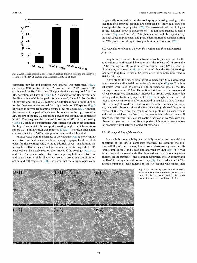
Favorable biocompatibility is essentially required for potential ap-plications of the HA-GS composite coatings. To examine the bio-compatibility of the coatings, human osteoblasts were grown on dif-ferent samples for 1 and 3 days and analyzed by SEM (Fig. 7). It wasfound that cells showed a similar flattened and well spreading mor-phology on the surfaces of the titanium substrates, the HA coating andthe HA-GS coating after culture for 1 day (Fig. 7 a-1, b-1 and c-1). Theaverage number of cells adhered to the HA coating was higher than
Fig. 6. Antibacterial rates of E. coli for the HA coating, the HA-GS coating and the HA-GScoating (R) (the HA-GS coating after incubated in PBS for 31 days).
Fig. 7. FE-SEM micrographs of human osteo-blasts cultured on the surfaces of (a) the Ti sub-strate, (b) the HA coating, and (c) the HA-GScoating for 1 day (−1) and 3 days (−2).
D. Li et al. Surface & Coatings Technology 330 (2017) 87–91
90

those of the titanium substrates, suggesting better biocompatibility ofthe HA coatings. Furthermore, the number and morphology of cellscultured on the HA-GS coating was similar to those on the HA coating.In addition, overall samples were fully covered by cells after culturedfor 3 days, indicating outstanding biocompatibility of the HA-GScoating. Taken together, we have successfully deposited the HA-GScomposite coating onto titanium substrate that displayed a long-termantibacterial property and excellent biocompatibility.
4. Conclusions
In this study, HA-GS composite coatings were constructed by VCS ontitanium substrates with the successful incorporation of GS.Comprehensive characterization showed that the physical and chemicalcharacteristics of the as-synthesized composite powder were completelyretained in the as-deposited coatings. The GS-containing HA coatingwas further confirmed to show a long-term antibacterial property andfavorable biocompatibility. The construction of the novel HA-GS com-posite antibacterial coatings by the VCS approach might facilitate thedevelopment of long-term drug-releasing coatings for medical implantswith exceptional properties in the future.
Acknowledgements
This work was supported by National Natural Science Foundation ofChina (grant # 51401232 and 41706076), CAS-Iranian Vice Presidencyfor Science and Technology Joint Research Project (grant #174433KYSB20160085), and Ningbo Municipal Major Projects onIndustrial Technology Innovation (grant # 2015B11054).
References
[1] W. Liu, J. Chang, In vitro evaluation of gentamicin release from a bioactive tri-calcium silicate bone cement, Mat. Sci. Eng. C-Mater. 29 (2009) 2486–2492 col.
[2] X. Chen, K. Cai, J. Fang, M. Lai, Y. Hou, J. Li, Z. Luo, Y. Hu, L. Tang, Fabrication ofselenium-deposited and chitosan-coated titania nanotubes with anticancer andantibacterial properties, Colloid. Surf. B 103 (2013) 149–157.
[3] J.H. Chu, J. Lee, C.C. Chang, Y.C. Chan, M.L. Liou, J.W. Lee, J.S.C. Jang, J.G. Duh,Antimicrobial characteristics in Cu-containing Zr-based thin film metallic glass,Surf. Coat. Technol. 259 (2014) 87–93.
[4] H.I. Chang, Y.C. Lau, C. Yan, A.G.A. Coombes, Controlled release of an antibiotic,gentamicin sulphate, from gravity spun polycaprolactone fibers, J. Biomed. Mater.Res. A 84A (2008) 230–237.
[5] K.C. Popat, M. Eltgroth, T.J. Latempa, C.A. Grimes, T.A. Desai, Decreased staphy-lococcus epidermis adhesion and increased osteoblast functionality on antibiotic-loaded titania nanotubes, Biomaterials 28 (2007) 4880–4888.
[6] R. Cristescu, C. Popescu, G. Socol, A. Visan, I.N. Mihailescu, S.D. Gittard,P.R. Miller, T.N. Martin, R.J. Narayan, A. Andronie, I. Stamatin, D.B. Chrisey,Deposition of antibacterial of poly(1,3-bis-(p-carboxyphenoxy propane)-co-(sebacicanhydride)) 20:80/gentamicin sulfate composite coatings by maple, Appl. Surf. Sci.257 (2011) 5287–5292.
[7] Z.S. Nurkeeva, V.V. Khutoryanskiy, G.A. Mun, M.V. Sherbakova, A.T. Ivaschenko,N.A. Aitkhozhina, Polycomplexes of poly(acrylic acid) with streptomycin sulfateand their antibacterial activity, Eur. J. Pharm. Biopharm. 57 (2004) 245–249.
[8] L.D. Silverman, L. Lukashova, O.T. Herman, J.M. Lane, A.L. Boskey, Release ofgentamicin from a tricalcium phosphate bone implant, J. Orthop. Res. 25 (2007)23–29.
[9] E. Wers, H. Oudadesse, B. Lefeuvre, O. Merdrignac-Conanec, A. Barroug, Evaluationof the kinetic and relaxation time of gentamicin sulfate released from hybrid bio-material bioglass-chitosan scaffolds, Appl. Surf. Sci. 353 (2015) 200–208.
[10] A. Rudin, C.A. Phillips, D.W. Gump, B.R. Forsyth, Antibacterial activity of genta-micin sulfate in tissue culture, Appl. Microbiol. 20 (1970) 989–990.
[11] P. Frutos Cabanillas, E. Diez Pena, J.M. Barrales-Rienda, G. Frutos, Validation and
in vitro characterization of antibiotic-loaded bone cement release, Int. J. Pharm. 209(2000) 15–26.
[12] W.C. Liu, C.T. Wong, M.K. Fong, W.S. Cheung, R.Y.T. Kao, K.D.K. Luk, W.W. Lu,Gentamicin-loaded strontium-containing hydroxyapatite bioactive bone cement-anefficient bioactive antibiotic drug delivery system, J. Biomed. Mater. Res. B 95B(2010) 397–406.
[13] E. Diez Pena, G. Frutos, P. Frutos, J.M. Barrales Rienda, Gentamicin sulphate releasefrom a modified commercial acrylic surgical radiopaque bone cement. I. influenceof the gentamicin concentration on the release process mechanism, Chem. Pharm.Bull. 50 (2002) 1201–1208.
[14] G. Ginalska, M. Osinska, A. Uryniak, T. Urbanik Sypniewska, A. Belcarz, W. Rzeski,A. Wolski, Antibacterial activity of gentamicin-bonded gelatin-sealed polyethyleneterephthalate vascular prostheses, Eur. J. Vasc. Endovasc. 29 (2005) 419–424.
[15] H. Pandey, V. Parashar, R. Parashar, R. Prakash, P.W. Ramteke, A.C. Pandey,Controlled drug release characteristics and enhanced antibacterial effect of gra-phene nanosheets containing gentamicin sulfate, Nano 3 (2011) 4104–4108.
[16] M. Yoshinari, Y. Oda, T. Kato, K. Okuda, Influence of surface modifications to ti-tanium on antibacterial activity in vitro, Biomaterials 22 (2001) 2043–2048.
[17] C. Moseke, F. Hage, E. Vorndran, U. Gbureck, TiO2 nanotube arrays deposited on Tisubstrate by anodic oxidation and their potential as a long-term drug deliverysystem for antimicrobial agents, Appl. Surf. Sci. 258 (2012) 5399–5404.
[18] V. Uskokovic, Nanostructured platforms for the sustained and local delivery ofantibiotics in the treatment of osteomyelitis, Crit. Rev. Ther. Drug 32 (2015) 1–59.
[19] H. Duan, Y. Ma, X. Liu, L. Hao, N. Zhao, Hierarchically nanostructured hydro-xyapatite microspheres as drug delivery carriers and their effects on cell viability,RSC Adv. 5 (2015) 83522–83529.
[20] Y. Liu, J. Huang, H. Li, Synthesis of hydroxyapatite-reduced graphite oxide nano-composites for biomedical applications: oriented nucleation and epitaxial growth ofhydroxyapatite, J. Mater. Chem. B 1 (2013) 1826–1834.
[21] Y. Liu, Z. Dang, Y. Wang, J. Huang, H. Li, Hydroxyapatite/graphene-nanosheetcomposite coatings deposited by vacuum cold spraying for biomedical applications:Inherited nanostructures and enhanced properties, Carbon 67 (2014) 250–259.
[22] L. Sun, C.C. Berndt, K.A. Gross, A. Kucuk, Material fundamentals and clinical per-formance of plasma-sprayed hydroxyapatite coatings: a review, J. Biomed. Mater.Res. 58 (2001) 570–592.
[23] J. Akedo, Room temperature impact consolidation (RTIC) of fine ceramic powderby aerosol deposition method and applications to microdevices, J. Therm. SprayTechn. 17 (2008) 181–198.
[24] S.D. Cook, K.A. Thomas, J.F. Kay, M. Jarcho, Hydroxyapatite-coated titanium fororthopedic implant applications clinical orthopaedics and related research, Clin.Orthop. Relat. R. (1988) 225–243.
[25] Y. Liu, Y.Y. Wang, G.J. Yang, J.J. Feng, K. Kusumoto, Effect of nano-sized TiNadditions on the electrical properties of vacuum cold sprayed SiC coatings, J.Therm. Spray Techn. 19 (2010) 1238–1243.
[26] S.Q. Fan, G.J. Yang, C.J. Li, G.J. Liu, C.X. Li, L.Z. Zhang, Characterization of mi-crostructure of nano-TiO2 coating deposited by vacuum cold spraying, J. Therm.Spray Techn. 15 (2006) 513–517.
[27] G.A. Buckholtz, N.A. Reger, W.D. Anderton, P.J. Schimoler, S.L. Roudebush,W.S. Meng, M.C. Miller, E.S. Gawalt, Reducing Escherichia coli growth on a com-posite biomaterial by a surface immobilized antimicrobial peptide, Mater. Sci. Eng.C 65 (2016) 126–134.
[28] Z. Li, D. Lee, X. Sheng, R.E. Cohen, M.F. Rubner, Two-level antibacterial coatingwith both release-killing and contact-killing capabilities, Langmuir 22 (2006)9820–9823.
[29] K.D. Park, Y.S. Kim, D.K. Han, Y.H. Kim, E.H.B. Lee, H. Suh, K.S. Choi, Bacterialadhesion on PEG modiÞed polyurethane surfaces, Biomaterials 19 (1998) 851–859.
[30] X. Chen, K. Cai, J. Fang, M. Lai, J. Li, Y. Hou, Z. Luo, Y. Hu, L. Tang, Dual actionantibacterial TiO2 nanotubes incorporated with silver nanoparticles and coatedwith a quaternary ammonium salt (QAS), Surf. Coat. Technol. 216 (2013) 158–165.
[31] X. Cui, Y. Gu, L. Li, H. Wang, Z. Xie, S. Luo, N. Zhou, W. Huang, M.N. Rahaman, Invitro bioactivity, cytocompatibility, and antibiotic release profile of gentamicinsulfate-loaded borate bioactive glass/chitosan composites, J. Mater. Sci.-Mater.Med. 24 (2013) 2391–2403.
[32] D.W. Lee, Y.P. Yun, K. Park, S.E. Kim, Gentamicin and bone morphogenic protein-2(BMP-2)-delivering heparinized-titanium implant with enhanced antibacterial ac-tivity and osteointegration, Bone 50 (2012) 974–982.
[33] D. Chen, Z. Jiang, J. Geng, Q. Wang, D. Yang, Carbon and bitrogen Co-doped TiO2
with enhanced visible-light photocatalytic activity, Ind. Eng. Chem. Res. 46 (2007)2741–2746.
[34] T.J. Webster, C. Ergun, R.H. Doremus, R.W. Siegel, R. Bizios, Specific proteinsmediate enhanced osteoblast adhesion on nanophase ceramics, J. Biomed. Mater.Res. 51 (2000) 475–483.
D. Li et al. Surface & Coatings Technology 330 (2017) 87–91
91