Embed Size (px)
Citation preview

Flexor Tendon Rehabilitation
Romina Astifidis MS, PT,CHT
Curtis National Hand CenterBaltimore, MD
October 6-8, 2017
Keys to successful treatment
• Doing the wrong thing can lead to injury
• Not doing enough of the right thing can cause poor outcomes
• Use the following resources
• Mentors
• Surgeons
• Protocols
• Evidence
Tendon Healing
Tendon healing
• Extrinsic healing
• Adhesion formation between tendon and surrounding tissue
• Potenza and Peacock (1960‐70s)• Tendons healed by fibroblastic response (adhesions)
• Tendon cells were incapable of proliferating
• “One wound” concept = tendon healing though adhesion formation
Tendon healing
• Tendons ability to heal without adhesions
• Intrinsic vascularity and synovial diffusion
• Fibroblasts needed for healing
• Supplied by the endotenon and epitenon
• Tenocytes appearing at 2‐3 weeks
Gelberman et al., Manske et al., Lundborg et al. (1980s)
Factors that affect tendon healing
• Age
• Individual biochemical response
• Nutrition
• Mechanism/type of injury• Crush or untidy laceration
• Associated fractures or blood vessel injury
• Controlled stress
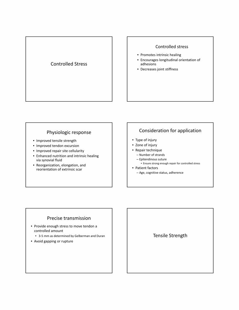
Controlled Stress
Controlled stress
• Promotes intrinsic healing
• Encourages longitudinal orientation of adhesions
• Decreases joint stiffness
Physiologic response
• Improved tensile strength
• Improved tendon excursion
• Improved repair site cellularity
• Enhanced nutrition and intrinsic healing via synovial fluid
• Reorganization, elongation, and reorientation of extrinsic scar
Consideration for application
• Type of injury
• Zone of injury
• Repair technique– Number of strands
– Epitendinous suture • Ensure strong enough repair for controlled stress
• Patient factors– Age, cognitive status, adherence
Precise transmission
• Provide enough stress to move tendon a controlled amount
• 3‐5 mm as determined by Gelberman and Duran
• Avoid gapping or rupture
Tensile Strength
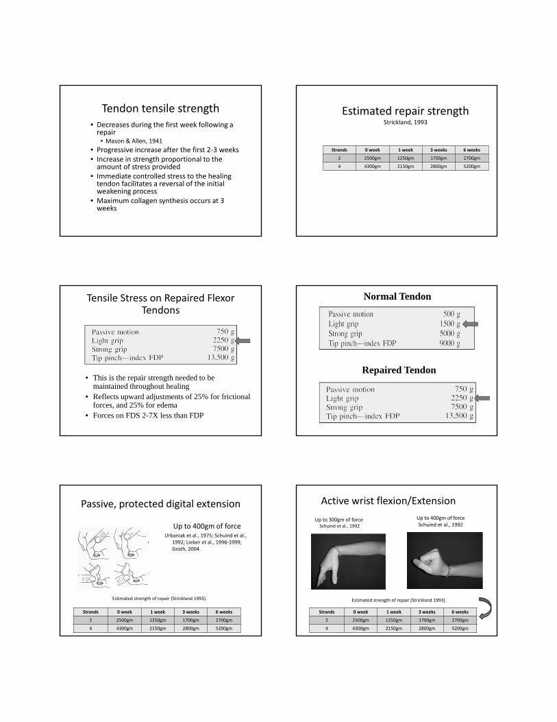
Tendon tensile strength• Decreases during the first week following a repair
• Mason & Allen, 1941
• Progressive increase after the first 2‐3 weeks• Increase in strength proportional to the amount of stress provided
• Immediate controlled stress to the healing tendon facilitates a reversal of the initial weakening process
• Maximum collagen synthesis occurs at 3 weeks
Estimated repair strengthStrickland, 1993
Strands 0 week 1 week 3 weeks 6 weeks
2 2500gm 1250gm 1700gm 2700gm
4 4300gm 2150gm 2800gm 5200gm
Tensile Stress on Repaired Flexor Tendons
• This is the repair strength needed to be maintained throughout healing
• Reflects upward adjustments of 25% for frictional forces, and 25% for edema
• Forces on FDS 2-7X less than FDP
Normal Tendon
Repaired Tendon
Passive, protected digital extension
Up to 400gm of forceUrbaniak et al., 1975; Schuind et al.,
1992; Lieber et al., 1996‐1999; Groth, 2004
Strands 0 week 1 week 3 weeks 6 weeks
2 2500gm 1250gm 1700gm 2700gm
4 4300gm 2150gm 2800gm 5200gm
Estimated strength of repair (Strickland 1993).
Active wrist flexion/Extension
Up to 300gm of forceSchuind et al., 1992
Up to 400gm of forceSchuind et al., 1992
Strands 0 week 1 week 3 weeks 6 weeks
2 2500gm 1250gm 1700gm 2700gm
4 4300gm 2150gm 2800gm 5200gm
Estimated strength of repair (Strickland 1993).
Active straight fist
Up to 1100gm of force
Greenwald et al.,1994
Groth, 2004
Strands 0 week 1 week 3 weeks 6 weeks
2 2500gm 1250gm 1700gm 2700gm
4 4300gm 2150gm 2800gm 5200gm
Active hook fist
Up to 1300gm of force
Greenwald et al.,1994
Groth, 2004
Strands 0 week 1 week 3 weeks 6 weeks
2 2500gm 1250gm 1700gm 2700gm
4 4300gm 2150gm 2800gm 5200gm
Active composite fist
400‐4000gm of force
Urbaniak et al., 1975
Schuind et al., 1992;
Greenwald et al., 1994
Evans, 1997
Silva et al., 1998
Gelberman et al., 1999
Boyer et al., 2001
Groth, 2004
Strands 0 week 1 week 3 weeks 6 weeks
2 2500gm 1250gm 1700gm 2700gm
4 4300gm 2150gm 2800gm 5200gm
Active, isolated joint motion
Up to 1900gm of force
Schuind et al.,1992Groth, 2004
Strands 0 week 1 week 3 weeks 6 weeks
2 2500gm 1250gm 1700gm 2700gm
4 4300gm 2150gm 2800gm 5200gm
Factors That Affect Flexor Tendon Repair Outcomes
• Mechanism/Type of Injury• Multiple digits/ concomitant injuries
• Age• Patient motivation/socioeconomic factors
• Nutrition… smoking may cause vasoconstriction
• Timing of repair, timing of therapy
• Controlled Stress (mobilization)• Without tendon gapping and rupture
****Amount of scar formation****
Factors that Cause Resistance to Flexor Tendon
Gliding
• Surgical repairs
• Tendon bulkiness
• Smoothness of tendon gliding surface
• Healing responses of the tendons
• Presence of intact annular pulleys
• Edema formation
• Adhesion formation
• Joint stiffness
• Extensor tendon tethering
• Splints and bandages and speed, frequency and methods of postoperative care
Wu YF and Tang JB. Hand Clinics. 2013
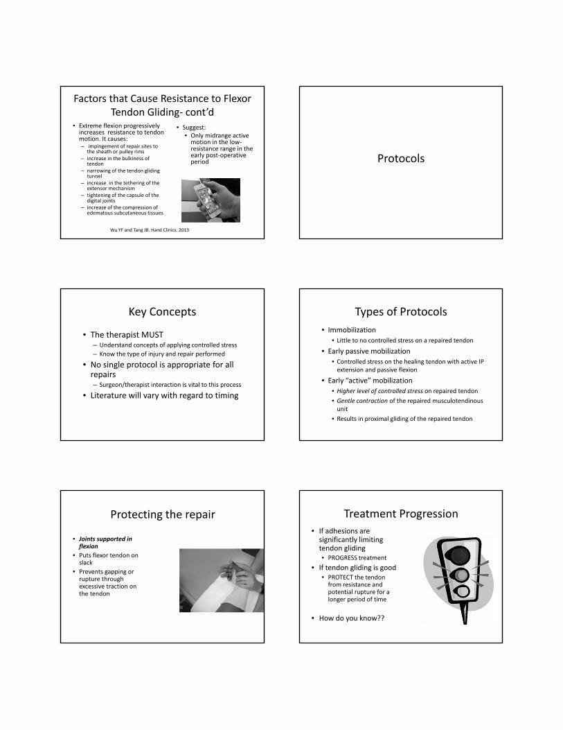
Factors that Cause Resistance to Flexor Tendon Gliding‐ cont’d
• Extreme flexion progressively increases resistance to tendon motion. It causes:– impingement of repair sites to
the sheath or pulley rims– increase in the bulkiness of
tendon– narrowing of the tendon gliding
tunnel– increase in the tethering of the
extensor mechanism– tightening of the capsule of the
digital joints – increase of the compression of
edematous subcutaneous tissues
• Suggest:• Only midrange active motion in the low‐resistance range in the early post‐operative period
Wu YF and Tang JB. Hand Clinics. 2013
Protocols
Key Concepts
• The therapist MUST– Understand concepts of applying controlled stress
– Know the type of injury and repair performed
• No single protocol is appropriate for all repairs– Surgeon/therapist interaction is vital to this process
• Literature will vary with regard to timing
Types of Protocols
• Immobilization
• Little to no controlled stress on a repaired tendon
• Early passive mobilization
• Controlled stress on the healing tendon with active IP extension and passive flexion
• Early “active” mobilization
• Higher level of controlled stress on repaired tendon
• Gentle contraction of the repaired musculotendinousunit
• Results in proximal gliding of the repaired tendon
Protecting the repair
• Joints supported in flexion
• Puts flexor tendon on slack
• Prevents gapping or rupture through excessive traction on the tendon
Treatment Progression
• If adhesions are significantly limiting tendon gliding• PROGRESS treatment
• If tendon gliding is good• PROTECT the tendon from resistance and potential rupture for a longer period of time
• How do you know??
Measurement
Strickland & Glogovac, 1980
Active PIP + DIP flexion – extension lag x 100175°
= % of normal active PIP and DIP motion
Excellent: 85‐100%Good: 70‐84%Fair: 50‐69%Poor: <50%
Immobilization
Immobilization
• Rationale/Used for:• Children (those under age 10‐12)
• Cognitively impaired
• Non‐adherent patients (????)
• EXTRINSIC HEALING
Immobilization
• Early stage (up to 4 weeks)• Dorsal blocking orthotic or cast
• Wrist 10‐30° flexion• MPs 40‐60° flexion• IPs in extension
• If therapy is provided:• Passive flexion of the digits • Mobilization of uninvolved joints• Wound/scar management
Cifaldi‐Collins & Shwarze, 1991
Immobilization
• Intermediate Stage (3 to 6 weeks)
• Orthosis modified to wrist neutral
• Removed for hourly exercises to include:• Passive flexion and extension of fingers with wrist in 10 ° extension
• Active flexion: hook, straight, and full fist.
• Synergistic motion
• BE GENTLE…immobilized tendon is generally weaker
Immobilization
• After 3‐4 days, assess tendon gliding
• Measure full MP/PIP/DIP flexion passively and actively
• If >50 ° difference is present, move to the late stage
• If <50 ° difference noted, continue with intermediate phase of the program until 6 weeks post‐op

Immobilization
• Late Stage (5 to 6 weeks)• D/C dorsal blocking orthosis
• Add serial extension splinting
• Begin gentle blocking exercises
• After 1 week of gentle blocking, may initiate light resistance
• If tendon gliding is good, delay any resistance
Early passive mobilization
Early Passive Mobilization
• Rationale: • Promoting synovial diffusion for healing
• Inhibit dense adhesion formation
• Facilitate a stronger repair at an earlier stage
• Two main protocols• Duran & Houser
• Kleinert
“Original” Duran & Houser
• 0‐ 4 ½ Weeks• Orthosis
• Dorsal block with wrist in 20° flexion, and MPs in a relaxed state of flexion:
• Orthosis ends at PIP joints to allow full IP extension
• Rubber band traction to the injured finger (loosely) during the day
• Between exercises stockinette is applied over the fingers and pinned to forearm
• All fingers resting in flexion within stockinette to prevent impulsive grasping
“Original” Duran & Houser
• Exercises: 6‐8 repetitions, 2x/day within orthosis that blocks MP in flexion– Passively extend DIP while PIP is held passively in flexion
– Passively extend PIP while DIP rests in flexion
“Original” Duran & Houser
• 4 ½ Weeks• Replace dorsal block with a wrist band with rubber band traction
• Exercises: 10 repetitions every 2 hours as previous
• Add gentle active extension against the rubberbandtraction.

“Original” Duran & Houser
• 5 ½ Weeks:• Hourly exercises: 10‐12 repetitions• Remove wrist band and nail suture for rubber band attachment
• Active flexion is initiated: gentle blocking, FDS gliding, and composite fist
• Passive flexion of all joints• IP passive extension with MP flexed
“Original” Duran & Houser
• 6 Weeks• Begin gentle PIP extension
• Dynamic splinting if needed
• 7 ½ Weeks• Initiate gentle resistance
• No strong resistance to the tendon for another 2‐4 weeks
“Modified” Duran
• Eliminate the rubber‐band traction
• Extend the DBS hood to the fingertips
• Strap the fingers in IP extension at night
• Exercises:
– Passive flexion: isolated and composite
– Active IP extension
– Passive protected extension
– Protected tenodesis in therapy if appropriate
Protected Tenodesis
Passive composite flex with wrist extension 20‐30 degrees followed bypassive wrist flexion, fingers extended passively by tenodesis effect
Modified Kleinert Protocol
• Dorsal blocking orthosis
• Wrist in 45° flexion
• MPs 40° flexion
• IPs allowed full extension
• Volarly applied “PFT” (postoperative flexor tendon)

Modified Kleinert Protocol• The PFT is a prefabricated orthosis• Rubber band traction runs from the fingernail, under a rolling bar at the palm, to a coiled lever at the forearm.
• Coiled lever and rolling bar on the PFT • Designed to minimize resistance within the rubber band during IP extension
Modified Kleinert Protocol• Exercises: 20 repetitions per hour• 0‐4 to 6 weeks
• Active IP extension against rubber bands
• 3‐6 weeks• Remove orthosis for wrist motion at 4 weeks• Begin gentle active flexion
• 6 weeks• Discontinue orthosis• Add differential tendon gliding exercises
• 6‐8 weeks• Begin gentle resistance
Washington Regimen
• Dorsal blocking orthosis– Wrist at 20‐45° flexion
– MPs at 40‐60 ° flexion
– IPs allowed full extension
• A safety pin is applied to the palmar strap at the distal palmar crease, and on the forearm strap– A nylon line is run from the fingernail of the injured finger(s) only, under the safety pin at the DPC, attaching to 2 rubber bands
– One rubber band is cut, so that it is only a single strand
– One rubber band with exercise; 2 at rest
Washington Regimen
• Full finger flexion to the distal palmar crease strap is attempted with singular rubber band traction
Washington Regimen
• 0‐3 weeks– Therapist performs protected passive flexion and extension
– Active extension against traction x10 reps, hourly– Rubber band traction on 24 hours/day
• 4 weeks– Discontinue rubber band traction – Begin active flexion with an active hold in flexion for 10 seconds, passive flexion, and active extension
Washington Regimen• 5 weeks
–May be allowed out of orthosis for hygiene and light activity
• 6 weeks– Discontinue orthosis
• 8 weeks– Add blocking if needed– Gradual increase in use and resistance– Heavy lifting above 5lbs not allowed until after week 12 post‐op

Zone I Protocol: LEAF
• Limited extension active flexion (LEAF)– Evans, 1990
• Rationale: – Place the repaired FDP tendon in a shortened position
– 4.5mm proximal to normal resting length
– Decrease gap formation
• Therapy initiated at 24 – 48 hours post op
Zone I Protocol: LEAF
• Early Stage (0‐3 Weeks)– Dorsal blocking othosis– Wrist at 30‐40° flexion– MPs at 30° flexion– Full IP extension allowed
• A separate finger based dorsal gutter is taped on with the DIP joint at 40‐45° flexion
Zone I Protocol: LEAF
Weeks 0‐4• Exercises‐ 10‐20 reps/hour:
• Passive DIP flexion to 75° in orthosis• Passive composite flexion• Passive IP flexion with MPs resting at 30° in orthosis (modified hook position)
• Full active PIP extension while other hand holds MP’s at 90° flexion
• With distal strap holding adjacent fingers in extension, place and hold PIP joint flexion of injured finger
Zone I Protocol: LEAF
Weeks 0‐4
• In therapy, orthosis removed for:– Passive wrist tenodesis
– Slow repetitive motions to loosen finger
– Short arc motion (SAM) place and hold against 15‐20g of force in the following position:
• Wrist extension = 20
• MP flexion = 75‐80
• PIP flexion = 70‐75
• DIP flexion = 40
Zone I Protocol: LEAF
• Weeks 3‐4 • Discontinue DIP dorsal blocking gutter
• Add gentle place/hold flexion
• Week 4• Add synergistics, hook fist, and gentle DIP blocking for FDP glide
• Orthosis remolded to wrist neutral
• Week 4 ½ • May begin DIP extension orthotic PRN
• Week 6‐7• Discharge orthosis
Zones III through V
• Repairs are most commonly placed in the preferred Zone II protocols
• Less complications and better results
– Do not have the tight pulley/sheath system
– Adhesions are often less dense
• Watch intrinsic scarring and/or paradoxical extension in zone III

Early “active” mobilization
“What” is Early Active Motion?
• Place and hold– ½ fist, whole fist
– Optional tenodesis splint
• Active fist– 1/3 first 1‐2 weeks, increasing to 2/3 the third week. Full ROM at week 4‐5.
– Full IPJ flex and ext with MCP extension blocked at 60‐80° flex
– Finger method?
• Active initiation of fist to 50% with PROM to full fist
• Active fist with 45° MPJ, 45° PIPJ and 45° DIPJ
Indiana Protocol
• Repair technique
– Tajima core suture plus horizontal mattress
– Equal to 4 strand repair plus epitendinous suture
• Criteria
• Motivated, understanding patients
• Minimal to moderate edema which does not restrict passive flexion
• Minimal wound complications
Indiana Protocol
• Week 0‐4–Dorsal blocking orthosis–Wrist 20° flexion, MPs 50° flexion, IPs allowed full extension
–Worn continuously • Once hourly: remove and apply hinged wrist splint
• Immediately reapply dorsal blocking splint after exercises
Indiana Protocol
• Synergistic orthosis with hinge
– Allows full wrist flexion and 30° extension
– MPs blocked at 60° flexion
– IPs allowed 0° extension
Indiana Protocol
Week 0‐4• Passive:
– 15 reps of passive flexion/extension to the PIP joint, then the DIP joint, then entire digit
• Apply synergistic orthosis for 25 reps of place/hold– Passively flex digits & simultaneously extend wrist
– Gentle place/hold contraction for 5 seconds
– Simultaneous wrist flexion with digit extension to orthosis

Indiana Protocol
• Week 4:
– Discharge synergistic orthosis
– Continue dorsal blocking orthosis between exercise
• Exercises
– Synergistic motion: 25 reps every 2 hours
– Add light active finger flexion and extension
– Avoid combined finger and wrist extension
Indiana Protocol
• Week 5• Exercises
• Continue week 4 exercises• Add tendon gliding and hook fisting
• Week 6• Discontinue dorsal blocking orthosis• Exercises
• Continue previous exercises• Add blocking exercises • Do not perform blocking exercises to the small finger FDP
Indiana Protocol
• Week 7
• Add passive extension exercises
• Week 8
• Add light resistance
• Week 14
• Return to normal activity
Pyramid of Progressive Force
• Pyramidal series of eight exercise levels in ascending order of increasing force
• The patient progresses up a level in the pyramid if the tendon is unresponsive • Unresponsive = < 10% resolution of active lag between therapy sessions
• Continue progression until the tendon is responsive • > 10% resolution of active lag between therapy sessions
Pyramid of Progressive Force(Groth, 2004) Pyramid of Progressive Force
• The active lag is measured:
Current DIP flexion – previous DIP flexionprevious DIP flexion
= % resolution of active lag between therapy sessions

Other protocols
• Tang JB, Journal of the Brittish Society for Surgery of the Hand 32 (2) 2007, 118‐129
• Critical aspects• Partial release of A2 pulley or
complete release of A4 pulley• 4‐6 strand repair for FDP• Active motion under least
tension, synergistic with wrist position
• DBS with wrist 20‐30° flex, MPJ slight flex, IPJ ext‐therapy starts at 3‐5 days post‐op
• For 2.5 weeks RX includes PROM 10 reps f/u by 10 A fists in comfortable range 4 times a day
• From 2.5‐5 weeks, wrist splinted in 30° ext and focus on PROM, AROM for 2/3rds of fist‐ assistance as needed to achieve full fist to prevent rupture
• D/C splint and full AROM at 5‐6 weeks
Nantong Tang, 2007
Other protocols
• Tang JB. Wide Awake Primary Flexor Tendon Repair, Tenolysis, and Tendon Transfer. Clinics in Orthopedic Surgery 2015.
• Short forearm splint with wrist 20‐30 flexion, MPJ at slight flex and IPJ in extension. Optional wrist in slight ext.
• Day 3‐5: Start with full PROM (10‐30 reps) followed by active finger flexion (20‐30 reps) for 1/3 total range 5‐6x/day. Encourage full IPJ extension.
• Week 3‐4: Increase active range to 2/3 total range and progress to full by week 4.
Delay full active flexion to week 5 if multiple fingers are repaired or if a late tendon repair or a primary tendon rupture.
Other protocols
• Lalonde, D. Wide Awake Flexor Tendon Repair and Early Tendon Mobilization in Zones 1 and 2. Hand Clinics. (2013)29(2): 207‐213• Patients perform full range of motion during surgery to ensure no gapping or lack of glide
• Post op days 2‐4: hand is elevated and splinted with wrist in comfortable ext, MPJ flexed to 80‐90° and PIP/DIPJ extended
• Post op days 3‐21: passive warm up of fingers followed by active mid‐range movement: 45° flex of each of the MP, PIP and DIP with full IPJ extension. No place and hold.
• Finger wrap
Mass & Saint JohnCoats et al., 2005; Clancy & Mass, 2013; LaLonde, 2013
Manchester Short SplintPeck, 2014
• 62 forearm‐based
• 40 Manchester short
• Significantly less flexion contracture at PIP at 6 and 12 weeks
• Significantly greater arc of flexion at DIP
• Greater proportion of excellent/good results
Studies to note
• Wu YF and Tang JB. Tendon Healing, Edema and Resistance to Flexor Tendon Gliding. Hand Clinics. (2013) 29(2): 167‐178• Severe edema in subcutaneous tissue and tendon as measured
by circumference adds to resistance of tendon gliding by 2x‐3x• Resistance to motion increased progressively for the first 4 days
and remained consistent from days 4‐7 therefore recommend best time to start motion is 4th to7th day
• Repetitive PROM of the digit as “warm‐up” greatly reduced the force and energy of the digital flexion
• Tendon swelling is worse in a delayed repair so sheath release or venting may be necessary
• Self adhesive tapes increase digital flexion energy 4x baseline and should be removed for exercise.
• Adhesions do not form before day 9 so digital motion can be started as late as day 7‐9
FPL Repair
• Goal is minimum of 30‐40 degrees of IPJ motion• Direct repair at all levels of injury may be possible as late as 3 to 6 weeks after injury but may need tendon lengthening because of proximal retraction is greater in FPL
• Rate of rupture of repair in Zone 2 of thumb is twice as common as Zone 1 likely secondary to zone of avascularity.
• Splint positioning usually includes wrist in 10‐20 ° flexion and CMC and MCP in neutral. Typically the IPJ is dorsally blocked at 20‐25°
• Treatment options include early passive, place and hold or early active based on the type of suture and should be guided by physician.
Take Home Message
• Measure…measure…measure
• Never follow a protocol blindly‐ use clinical judgment
• Start therapy close to post op day 5 and before post op day 10
• Make sure to do a passive warm‐up of finger prior to exercises which can include passive modified tenodesis and passive modified hook.
• Always position the wrist in neutral or slight extension for active exercises
• When doing place and hold or AAROM start with a looser fist
• Monitor interossei tightness and retrain hook fist for differential gliding
Thank you!