Embed Size (px)
Citation preview

Review ArticleRoles of the Phosphorylation of Transcriptional Factors inEpithelial-Mesenchymal Transition
Rong Xu, Jae-YeonWon, Chang-Hyeon Kim, Da-Eun Kim, and Hyungshin Yim
Department of Pharmacy, College of Pharmacy, Institute of Pharmaceutical Science and Technology, Hanyang University,Ansan, Gyeonggi-do, Republic of Korea
Correspondence should be addressed to Hyungshin Yim; [email protected]
Received 29 March 2019; Revised 3 May 2019; Accepted 9 May 2019; Published 2 June 2019
Guest Editor: Daniele Vergara
Copyright © 2019 Rong Xu et al.This is an open access article distributed under the Creative Commons Attribution License, whichpermits unrestricted use, distribution, and reproduction in any medium, provided the original work is properly cited.
Epithelial-to-mesenchymal transition (EMT) is the first step in the development of the invasive and migratory properties of cancermetastasis. Since the transcriptional reprogramming of a number of genes occurs in EMT, the regulation of EMT transcriptionfactors has been intensively investigated. EMT transcriptional factors are commonly classified by the direct or indirect repressionof E-cadherin because one of hallmarks of EMT is the loss of E-cadherin. This facilitates the expression of genes for EMT, tumorinvasion, andmetastasis.Theposttranslationalmodification of EMT transcriptional factors, such as Snail and Slug, directly regulatestheir functions, including their stability, nuclear localization, protein-protein interaction, and ubiquitination for the promotionor termination of EMT at the specific points. Here, we discuss how posttranslational modifications regulate gene expression ina dynamic and reversible manner by modifying upstream signaling pathways, focusing in particular on the posttranslationalmodifications of Snail, Slug, ZEB1, ZEB2, and TWIST1.This review demonstrates that EMT transcription factors regulatemetastasisthrough their posttranslational modifications and that the flexibility and reversibility of EMT can be modified by phosphorylation.
1. Introduction
Cancer metastasis begins with the migration and invasion ofcancer cells to the surrounding tissues and involves the loss ofcell-cell adhesion. For this, cancer cells acquire mesenchymalcharacteristics by altering the levels of genetic expressionduring the epithelial-to-mesenchymal transition (EMT).Thisalteration of gene expression increases cell migration andinvasion during EMT, such that EMT is considered as thefirst step of cancer metastasis. EMT is driven by variouscytokines and growth factors, including transforming growthfactor-𝛽 (TGF-𝛽), Wnt, Notch, EGF, FGF, and HGF [1]. Wntsignals generate the translocation of 𝛽-catenin to the nucleus,which triggers the transcriptional activation of TCF/LCF1transcriptional complex. This activation is also generated byTGF-𝛽 signals in cancer cells through the activation of Smadprotein by phosphorylation [2]. The TGF-𝛽 signals crosstalkwith Wnt, Notch, and receptor tyrosine kinase signals toinduce the specific expression of EMT transcription factorand its functions in cancer metastasis, depending on the
cellular context [1]. The treatment of TGF-𝛽 in cancer cellsresults in the genetic alteration of important genes throughtranscriptional regulation, primarily targeting TGFB1 andTGFB3, which are repressed by c-Myc andOct4/Klf4, respec-tively [3]. EMT-inducing transcription factors interact withepigenetic regulators to control the expression of genesassociated with cell polarity, cell adhesion, the cytoskeleton,and extracellular matrix degradation via the repression ofepithelial genes [4].
EMT transcription factors are commonly classifiedaccording to the direct or indirect repression of E-cadherin,since a hallmark of EMT is the loss of E-cadherin, which isassociated with the acquisition of metastatic activity. Severalstudies suggest that the posttranslational modification of theE-cadherin gene (CDH1), such as CDH1 silencing, is a criticalmechanism for EMT. The CDH1 promoter contains E-boxelements that are responsible for transcriptional repression[5]. The binding of the zinc finger transcription factor Snailto the E-box elements of the CDH1 promoter has been foundto repress the transcription of CDH1 [5]. Slug, a member
HindawiJournal of OncologyVolume 2019, Article ID 5810465, 11 pageshttps://doi.org/10.1155/2019/5810465

2 Journal of Oncology
of the Snail family [6], as well as ZEB1 [7], and ZEB2 [8]can repress the transcription of CDH1, thereby promotingthe dissociation of cell adhesion, consequently inducing cellinvasion andmigration. E47 [9] andKLF8 [10] are also knownas direct repressors of the CDH1 promoter. Although thebHLH factor TWIST1 can induce EMT, it indirectly repressesthe transcription of CDH1 [11], which is an indirect repressorof the CDH1 promoter. Similar to TWIST1, goosecoid [12],FOXC2 [13], SIX1 [14], and bHLH factor E2.2 [15] cansuppress the transcription of CDH1 in a seemingly indirectmanner. The activation of these transcription factors drivestumor invasion and metastasis, inducing the transition tomesenchymal characteristics by repressing the transcriptionof CDH1 and activation of mesenchymal CDH2.
Although these several transcription factors can inducethe EMT, their specific functions display in a differentway [16,17]. In melanoma, the difference between ZEB1 and ZEB2 hasbeen studied well [18]. ZEB1 and ZEB2 have different bindingcorepressors or coactivators based on their structural differ-ence and cell context. Basically ZEB2 is expressed in normalmelanocyte, while ZEB1 is highly expressed in melanoma[16, 18]. Like their expression patterns, their functions showan opposing way. ZEB1 cooperates with TWIST1 for theironcogenic properties, while ZEB2 and Slug work togetherfor tumor-suppression [16, 18]. The different expression andfunctions of transcription factors are connected with thediverse EMT signaling pathways, depending on tissues andcontext. In addition, the signaling pathways triggered byWnt,Notch, TGF-𝛽, EGF, and several stresses are not linear, butcomplex and crosstalk. Their differences of expression andfunctions make the regulation of EMT more precise andtighter.
The activity, specificity, and accuracy of transcriptionfactors are commonly regulated by posttranslational modifi-cations, especially phosphorylation. In response to changes inextracellular and/or intracellular signaling, activated proteinkinases such as extracellular signal-regulated kinases andcasein kinases in EMT signaling pathway phosphorylatetranscription factors and related coregulators, facilitating aprogram of gene expression. These posttranslational modi-fications regulate the physiology of transcription factors toinduce mesenchymal characteristics while repressing epithe-lial characteristics. The phosphorylation and dephosphory-lation of EMT transcription factors, such as Snail and Slug,can directly regulate their function, including their stability,nuclear localization, protein-protein interaction, and ubiqui-tination for the termination of their function at a specific timepoint. In this review, we discuss the mechanisms by whichposttranslational modifications dynamically and reversiblyregulate gene expression activity by modifying upstreamsignaling pathways. In particular, this review focuses onthe posttranslational modifications of Snail, Slug, ZEB1,ZEB2, and TWIST1, because the factors trigger the classicEMT process through activation of mesenchymal factorsand suppression of epithelial factors by regulation of geneexpression, as the core EMT transcriptional factors. As theresults, invasiveness and migrating activity are acquired incancer cells [16, 17]. In addition, they are connected withthe several characters related with the stemness of cancer
stem cell and changing cell metabolism for cell survival [16].Moreover, their functions are converged on the suppres-sion of E-cadherin, but their regulatory functions for EMTare diverse in the different context with specific manner.This review provides an overview of how these core EMTtranscription factors regulate metastasis via posttranslationalmodifications.
2. Snail (Snail1; SNAI1)
2.1. Function and Structure of Snail. Numerous studies haveshown that Snail (product of SNAI1), a member of the Snailsuperfamily of zinc finger transcription factors, functions as astrong inducer of EMT,which converts epithelial cells tomes-enchymal cells by the acquisition of migratory and invasiveproperties through the repression of CDH1, switching fromCDH2 expression [5, 47, 48]. Snail has several transcriptionaltargets for EMT with epithelial factors such as desmoplakin[47] and Muc-1 [49] and mesenchymal factors, includingvimentin and fibronectin [47]. The importance of Snail isbased on its ability to induce EMT and its positive correlationwith malignancy. Its expression is sufficient to induce EMT[47] and is positively correlated with tumors and metastasis[50–52]. Snail is highly expressed in high grade tumors,metastatic cancer, and recurring cancer [50, 51].
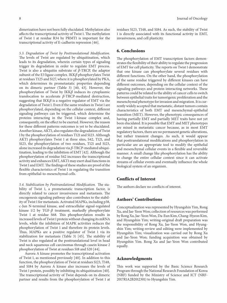
Snail was first identified in Drosophila melanogaster [53]and Snail homologues have been detected in several species,from Drosophila melanogaster to Homo sapience. The Snailsuperfamily includes Snail (Snail 1), Slug (Snail 2), and Smuc(Snail 3), which share several common structures, with a C-terminal DNA-binding domain and an N-terminal SNAGdomain (Figure 1(a)). The SNAG domain, originally definedas a repressor motif in zinc finger proteins, such as Snail andGfi-1 [54], is critical for the binding of transcriptional core-pressor complexes, including histone deacetylase 1/2 [55], 14-3-3 [56], and Ajuba [57]. The C-terminal domain has fourzinc fingers and an E-box motif-binding region [58]. Nuclearexport sequences (NES) and a serine-rich domain (SRD)are located at the central region of Snail [59] (Figure 1(a)).Detailed studies show that the activity and stability of Snailare regulated by posttranslational modification in severalresidues of Snail, depending on the extracellular signaling andtumor microenvironment, as discussed below.
2.2. Transcriptional Repressor Activity by PosttranslationalModification. The most important function of Snail is thetranscriptional repression of the CDH1 gene, an epithelialmarker, by binding to the E-element of the CDH1 promoterregion via its SNAG domain [55]. When the SNAG domainof Snail binds to the CDH1 promoter region, the recruitmentof the mSin3A/HDAC1/2 corepressor complex is required[5, 47, 55]. The phosphorylation of Snail at the S11 residuenear the SNAGdomain by PKA and at the S92 residue byCK2increases the efficiency for the recruitment of the mSin3Acorepressor, which is required for the repressive activity ofCDH1 (Table 1) [19].The role of CK2 in EMT is demonstratedby the unbalanced expression of the CK2 subunit, whichdrives EMT in breast epithelial cells [60] and is related to

Journal of Oncology 3
SNAI1
SNAI2
.1
.1
#264
#268
SNAG
SNAG
11
8 54 87 92 96100104 116 158 247 251 254
82 90 92 11296100104 107 203 206 234 235 246
SLUG
SRD NES ZF
ZF ZF ZF ZF ZF
ZF ZF ZF
phosphorylationglycosylation
acetylationubiquitin
(a)
ZEB1
ZEB2
.1
.1
#1125
#1214
38
191
585 867
CBD
CBD
N-ZF
N-ZF
SID
SID
HD
HD
CID
CID
C-ZF
C-ZF
phosphorylation
(b)
TWIST1 .1
18 20 34 42 68 121 123 125 127 148 184
109 121 138 153 163
GRR bHLH Twist WR
#202
NLS NLS
Basic Helix 1 Loop Helix II
phosphorylationmethylation
(c)
Figure 1: Structures of the core EMT-TFs. (a) Snail has N-terminal SNAG domain and C-terminal DNA binding domain of four C2H2 zincfinger (ZF) motifs. Nuclear export sequences (NES) and a serine-rich domain (SRD) are located at the central region of Snail. Slug hasN-terminal SNAG domain as a transcriptional repressor, proline-rich SLUG domain in the central region, and C-terminal five zinc fingermotifs. (b) ZEB has two zinc finger clusters that are N-terminal zinc finger (NZF) and C-terminal zinc finger (CZF) and a centrally locatedhomeodomain (HD).The corepressors bind to the protein binding domain of ZEB1, which are CtBP interaction domain (CID) at C-terminus,Smad interaction domain (SID) and homeodomain (HD) at the center, and CAF/p300 binding domain (CBD) at N-terminus of ZEB1. (c)Twist1 has a DNA-binding basic region (amino acids 109–121) and a bHLH domain (amino acids 122-163) and TwistWR domain (amino acids182-202) for the transcriptional activity. SRD, serine-rich domain; ZF, zinc finger domain; NES, nuclear export sequence; CBD, coactivatorbinding domain; SID, Smad interaction domain; BD, homeodomain; OD, CtBP interaction domain; NLS, nuclear localization signals.
the modification of Snail. In addition, PAK1, an interactingpartner of the motility regulators, GTPase, Rac1, or Cdc42,also phosphorylates Snail at the S246 residue in the zinc fingerdomain, promoting the transcriptional repression of Snailtargeting the E-cadherin and occludin promoters [26]. Basedon these studies, the phosphorylation sites of Snail relatedwith repression are not limited to the SANG domain, whichis critical for the binding of the transcriptional corepressor,but are also distributed on the SRD and the C-terminalDNA-binding domain. Although the phosphorylation ofSnail mostly promotes EMT through E-cadherin suppressiveactivity, PKD1-mediated phosphorylation of Snail at S11suppressed EMT and reduced its transcriptional repressiveactivity [20, 21], which is associated with the epigeneticsuppression of PKD1 in several cancers.
2.3. Nuclear Accumulation by Posttranslational Modification.Nucleocytoplasmic shuttling is themainmechanism that reg-ulates the spatial-dependent function of transcription factors.
The nuclear location is mandatory for the transcriptionalregulation of transcription factors. Snail localizes in thenucleus primarily via nuclear localization signals (NLS). Snailhas three NLS motifs in the N-terminal region (amino acids8-16), which overlap with the SNAG domain, and a middleregion (amino acids 151-152) that is proximal to the DNA-binding domain [61]. NES is located in the middle regionof Snail for export to the cytoplasm [59]. In addition toNLS and NES, nucleocytoplasmic shuttling is also regulatedby the phosphorylation of Snail by several kinases. PAK1phosphorylates Snail at the S246 residue, whichmodulates itstranscriptional activity by increasing its accumulation in thenucleus, which in turn increases the E-cadherin repressionactivity of Snail [26].The S246 residue is also phosphorylatedby PI3K, induced by growth-regulated protein 𝛼, which hasbeen found to increase EMT and bladder cancer recurrence[28]. The phosphorylation of Snail at T203 in the nucleusby Lats2, a serine/threonine kinase in mitosis, was found toincrease its retention in the nucleus in response to multiple

4 Journal of Oncology
Table 1: The posttranslational modification sites of Snail for EMT.
Residues-modification Kinase/Enzyme Function Cell Type ReferencePhosphorylation
S11-pPKA TF activity HEK 293T, MDCK [19]
Stabilization
PKD1 Nuclear export of snail (Destabilization) Breast epithelial cell [20, 21]Suppression of EMT
S82-p ERK2 Nuclear accumulation Breast cancer [22]
S92-p CK2 TF activity HEK 293T, MDCK [19]Stabilization
S96/S100/ GSK3𝛽 Degradation prostate cancer [23, 24]S104/S107-p Breast cancerS104/S107-p CK1 Degradation prostate cancer [24]S104-p ERK2 Nuclear accumulation Breast cancer [22]
T203-p Lats2 Nuclear accumulation [25]Stabilization
S246-pPAK1
Nuclear accumulation Breast cancer[26, 27]TF activity Non-small lung cancer
StabilizationPI3K Nuclear accumulation Bladder cancer [28]
Other posttranslational modificationsS112-gl O-GlcNAc Stabilization [29]K206/K234/ A20 Monoubiquitylation Breast cancer [30]K235-ub Stabilization
signals, including TGF-𝛽-induced EMT [25]. In responseto the collagen receptor DDR2, ERK2 is activated andphosphorylates Snail at residues S82 and S104, leading tothe nuclear accumulation of Snail and the suppression ofE-cadherin expression [22]. Thus the nuclear accumulationinduced by phosphorylation is directly connected with thetranscriptional activity of Snail and its function.
2.4. Ubiquitination and Degradation by PosttranslationalModification. Snail is a highly unstable protein with a shorthalf-life approximately 25 min [62]. Snail has two consensusmotifs binding with GSK3𝛽, which phosphorylate Snail atS92, S96, S100, and S104, located in the SRD, which regulatesits stability [23, 24, 62].The phosphorylation of the first motifat S96 and S100 regulates its 𝛽-transducin repeat-containingprotein- (𝛽-TRCP-) mediated ubiquitination [23, 24]. ForGSK3𝛽-mediated phosphorylation, Snail is phosphorylatedat S104 and S107 by CK1 as a priming site for the subsequentphosphorylation by GSK3𝛽 [24]. CK1-mediated primingphosphorylation allows for GSK3𝛽-mediated phosphoryla-tion of residues S100 and S96, as well as residue S92 as a sub-sequent phosphorylation reaction. The phosphorylation ofresidues S96 and S100 demonstrates that𝛽-TRCP recognitionsites play a role in protein polyubiquitination anddegradation[24].
2.5. Stabilization of Snail by Posttranslational Modification.The phosphorylation of Snail at residues S82 and S104 by
ERK2, residue S246 by PAK1, and residue T203 by Lats2induced the nuclear retention of Snail and increased itsactivity. The nuclear localization of Snail by phosphorylationincreases its stability as it allows it to escape from 𝛽-TRCP-mediated polyubiquitination and degradation. For stability,Snail is also monoubiquitinated at K206, K234, and K235by ubiquitin-editing enzyme A20 [30]. MonoubiquitinatedSnail1 has a reduced affinity for GSK3𝛽, and thus, Snail isstabilized in the nucleus due to the decreased phosphory-lation mediated by GSK3𝛽 [30]. In addition, the O-GlcNAcmodification of Snail increases its stability by suppressingprotein degradation [29]. The O-GlcNAc modification atS112 disrupts the CK1-mediated priming phosphorylation atresidues S104 and S107, inhibiting GSK3𝛽-mediated degra-dation [29]. Consequently,O-GlcNAcmodification stabilizesSnail to avoid the protein degradation and thereby increasesits transcriptional repressor activity for CDH1 expression.
The phosphorylation of Snail can be influenced by envi-ronmental conditions. In terms of the functional regulationof Snail by phosphorylation, PKA, CK2, PAK1, PI3K, Lats2,and ERK2 are positive regulators of Snail that support itstranscriptional activity, nuclear localization, and stabiliza-tion.However, the phosphorylation of Snail by PKD1,GSK3𝛽,and CK1 suppresses its transcriptional activity and inducesits degradation. Consequently, they function as negativeregulators of Snail and EMT. The balance between a positiveand negative regulator is thus dependent on the cellularcontext.

Journal of Oncology 5
Table 2: The posttranslational modification sites of Slug for EMT.
Residues-modification Kinase/Enzyme Function Cancer Type ReferencePhosphorylation
S158/S254-p PAK4 Stabilization Prostate [31]TF activity
S87/S104-p ERK2 TF activity Breast [32]
S54/S104-p Cyclin Degradation Lung [33]E/CDK2
S92/S96/S100/S104-p GSK-3𝛽Degradation Non-small cell lung [34]Degradation Breast [35]Degradation Liver, breast [36]
S247/S251/S254-p PAK1 Stabilization Breast, bladder [37]Acetylation
K8/K116-Ac Deacetylase Stabilization Basal-like [38]SIRT2 breast
3. Slug (Snail2; SNAI2)
3.1. Function and Structure of Slug. Slug (product of SNAI2)is a member of the Snail family and has a zinc finger domainwith transcriptional repressor activity. Slug shares commoncharacters with Snail based on its structure. Slug has C-terminal five zinc finger DNA-binding domains with an E-box motif (CAGGTG)-binding region which is required fortranscriptional activity as a transcription factor [63, 64].In the N-terminus, Slug also has a SNAG domain, whichacts as a transcriptional repressor (amino acids 1-32) andwhich is separated from the C-terminal zinc finger domain[64]. Slug has similar NLS as Snail [65]. In spite of thesimilarities between Snail and Slug, Slug has specific proline-rich domains in the central region, i.e., the SLUG domain,although the function of SLUG domain is uncovered [66]. AsSnail binds to mSin3A, a corepressor for CDH1 repression,Slug also interacts with the corepressors NCoR and CtBP1to transcriptionally repress CDH1 [66]. NCoR and CtBP1are recruited as transcriptional regulators at different Slugbinding regions. NCoR binds to Slug through the SLUGdomain, whereas CtBP1 is recruited to the SNAG domain[66]. Depending on the specific cellular conditions andenvironmental context, the expression of corepressors, suchas NCoR, CtBP1, and mSin3A, may differ, which determinesthe dominant role of either Snail or Slug for the repressionof CDH1 and EMT. Although the amounts of detailed Slugstudies are fewer than those for Snail, the phosphorylation-mediated transcriptional regulation, stability, and degrada-tion of Slug are described in the following section.
3.2. Regulation of Transcriptional Activity by PosttranslationalModification. The transcriptional repressor activity of Slug issupported by PAK4- and ERK2-mediated phosphorylation.A recent study revealed that PAK4 phosphorylates Slug atresidues S158 and S254, which increases its transcriptionalactivity as a repressor of the CDH1 promoter (Table 2) [31]. Aphosphomimetic mutant of Slug at two residues suppressedthe expression of CDH1, indicating that the phosphoryla-tion of Slug at residues S158 and S254 is important for
the regulation of its transcriptional activity. In addition toPAK4, ERK2 also activated Slug as a transcriptional regulatorthrough the phosphorylation of residue S87; however, thisphosphorylation does not regulate its stability or nuclearlocalization [32]. The phosphorylation of Slug at residue S87is essential for its ability to induce vimentin or Axl expressionfor EMT, although this is a separate mechanism from the roleof Slug as a transcriptional repressor of CDH1. Additionalphosphorylation sites of Slug were detected at residues S4 andS88 based on an in vivo analysis, and the phosphorylation ofSlug at residue S4 was found to increase the transcriptionalrepression of CDH1 expression [66].
3.3. Degradation of Slug by Posttranslational Modification.Like Snail, the stability of Slug is regulated by GSK3𝛽-mediated phosphorylation throughCHIP (carboxyl terminusof Hsc70-interacting protein) at residues S92, S96, S100, andS104 (Table 2) [34, 36]. The phosphorylation of Slug byGSK3𝛽 provides the recognition sites of 𝛽-TRCP-mediatedubiquitination and proteasomal degradation. The activity ofGSK3𝛽 limits the intracellular concentration of Slug, thusmodulating its turnover by direct phosphorylation. Non-degradable Slug promotes cell migration, invasion, and can-cer metastasis of lung adenocarcinoma [34]. Non-phospho-mimetic Slug at residues S92 and S96 was found to inhibitthe degradation of Slug, while non-phospho-mimetic Slugat residues S100 and S104 was found to accumulate inthe nucleus. Thus, the phosphorylation of Slug at residuesS92 and S96 negatively affects its stability, while the phos-phorylation of residues S100 and S104 affects its cytosoliclocalization and stability [36]. This indicates that GSK3𝛽-mediated phosphorylation had a negative impact on CDH1repression. During the cell cycle of cancer cells during cancerprogression, Slug is also phosphorylated by cyclin E/CDK2,which promotes its proteasomal degradation at the G1/Sphase transition [33].
3.4. Stabilization of Slug by Posttranslational Modification.In terms of posttranslational modifications, the stability of

6 Journal of Oncology
proteins is regulated by the balance between prodegrada-tive modifications and defensive modifications against thedegradation. In the case of Slug, PAK-mediated phosphory-lation stabilizes Slug protein. PAK4 phosphorylates Slug atresidues S158 and S254 for its stabilization by blocking itsubiquitination after GSK3𝛽-mediated phosphorylation [31].The expression of non-phospho-mimetics upregulates thetranscriptional expression of CDH1 and reduces EMT, dueto the reduced stability of Slug. Thus, PAK4-mediated phos-phorylation of Slug at residues S158 and S254 is importantto maintain the stability and its transcriptional repressionactivity (Table 2) [31]. Based on the phosphorylation of Snailby PAK1, Thaper and his colleagues observed whether PAK1phosphorylates Slug around residue S246 of Snail.The kinaseassay showed that PAK1 also phosphorylates Slug at residuesS247, S251, and S254 [37].The activation of PAK1 is dependenton the activation of Lyn tyrosine kinase, which triggers thephosphorylation of Slug and, therefore, its stabilization. Sincethe nuclear localization and stability of Slug is dependenton the activity of Lyn, PAK1 and Lyn kinases are positiveregulators of Slug in EMT. In addition to phosphorylation,deacetylation modifications induce the stabilization of Slug.Deacetylation of Slug at residues K8 and K116 by SIRT2prevents the degradation of Slug, which in turn increases itsstability. SIRT2-mediated deacetylation of Slug is sufficientto increase the protein half-life and activity of Slug viastabilization [38].
Although Slug has beenwidely studied, these studies havebeen limited to breast cancer due to its constitutive overex-pression in aggressive breast cancer. However, this proteinneeds to be studied in a range of different cancer systemsto determine its unique function compared to Snail. Furtherstudies could be used to demonstrate the importance of Slugin EMT. Furthermore, the posttranslational modification ofSlug may account for its functional flexibility in EMT.
4. ZEB1/2
4.1. Function and Structure of ZEB1/2. The zinc finger E-boxbinding homeobox (ZEB) family is composed of ZEB1 (alsoknown as TCF8, or deltaEF1) and ZEB2 (also named SIP1).They are transcription factors characterized by the presenceof a zinc finger DNA-binding domain, which is required fortranscriptional activity [67, 68]. ZEB1 was first identified asa repressor of the delta 1-crystallin enhancer and expressedin mesodermal tissues, suggesting that ZEB1 is involvedin embryogenesis and development [69]. Loss of functionexperiments with ZEB1 demonstrated that the developmen-tal defects related with mesenchymal-epithelial transitionshowed E-cadherin expression and vimentin depletion inembryonic tissues [70], suggesting that ZEB1 could have animportant function in EMT. In several human cancer celllines, the expression of ZEB1 induces EMT, as well as cancercell invasion and metastasis [7, 71, 72]. ZEB expression istriggered by diverse growth factors and signaling pathways,including TGF-𝛽, Wnt, and Notch signaling [73]. ZEB1 hastwo zinc finger domains in the N- and C-terminals, both ofwhich bind to the E-box motif of the CDH1 promoter region.During EMT, ZEB1 and ZEB2 suppressed the expression of
CDH1 directly through the recruitment of the corepressorsCtBP and HDAC1 [74]. These corepressors bind to theprotein binding domains of ZEB1, which are a CtBP inter-action domain (CID) at the C-terminus, a Smad interactiondomain (SID), and a homeodomain (HD) in the middleand a CAF/p300 binding domain (CBD) at N-terminus ofZEB1 [75] (Figure 1(b)). The recruitment of corepressors tospecific binding sites allows ZEB1 to function specifically asa transcription factor of EMT. ZEB2 has similar structuredomains with two zinc finger domains, an N-terminal CBD,Smad-binding domain (SBD), and HD and a C-terminalCID [76]. The function and stability of ZEB1 are regulatedby the posttranscriptional modification of miRNA-200 andmiRNA-203 [77]. ZEB1 needs to be studied further in order tofully elucidate the correlation between the posttranslationalmodification of this protein and its functions.
4.2. Regulation of Activity, Stability, and Location of ZEBby Posttranslational Modification in EMT. The ZEB post-translational modification studies are relatively fewer thanthose of other transcription factors, including Snail, Slug, andTwist. Interestingly, the majority of the results related withthe posttranslational modification of ZEB1/2 are found thatZEB1/2 function is negatively regulated. The transcriptionalrepression of ZEB1 is disrupted by phosphorylation andsumoylation, which suppresses its transition from epithelial-to-mesenchymal characteristics.
First, the phosphorylation of ZEB1 at residues T851,S852, and S853 by PKC inhibits the transcriptional activityinduced by IGF-1 treatment [76]. Under IGF-1 treatment,the ERK1/2 pathway is activated and phosphorylates ZEB1 atT867. ERK1/2-mediated phosphorylation of ZEB1 disrupts itsnuclear localization by IGF-1 treatment, which consequentlydisrupts its transcriptional activity [76]. In Llorens’s study,two NLS were detected at two different regions, the firstat amino acids 111–241 prior to the zinc finger domain andthe second at amino acids 869–875 after the phosphory-lation site at residue T867 [76]. Thus, it is plausible thatthe phosphorylation of residue T867 directly disrupts theinteraction between its NLS regions and importin, althoughmore evidence is needed.
Second, the sumoylation of ZEB1/2 is triggered by thepolycomb protein Pc2, which reduces the transcriptionalactivity [78]. ZEB1 is sumoylated at residues K347 andK774, while ZEB2 is sumoylated at residues K391 and K866,which relieve E-cadherin repression [78]. As a mechanism ofrelieving E-cadherin repression, the sumoylation of ZEB2 atresidue K866 disrupts the recruitment of corepressor CtBP,since this site is near a CtBP-binding motif. Thereby, Pc2-mediated sumoylation reduces the recruitment of CtBP1 fortranscriptional repression [78].
Although ERK1/2 and PKC are negative regulators ofZEB1 in EMT, DNA damage sensing kinase ATM positivelyregulates ZEB1 function in response to DNA damage. ZEB1function has been investigated in relation to radioresistancesince ZEB1 is highly expressed in radioresistant-cancer cells[75]. ATM phosphorylates ZEB1 at residue S585 in responseto DNA damage, which accelerates its interaction with USP7,

Journal of Oncology 7
Table 3: The posttranslational modification sites of Twist 1 for EMT.
Sites Kinase/Enzyme Function ReferencePhosphorylation
S123/T148-p AURKATF activity (Partner choice)
[39]StabilizationHomodimerization
S184-p AURKA TF activity (Direct) [39]Stabilization
T125/S127-p PKA TF activity (Partner choice) [40]Heterodimerization
S42-p AKT1 TF activity [41]T121/S123-p AKT1 Degradation [42]
T125/S127-p IKK𝛽 Degradation [43]Cytoplasm translocalization
S18/S20-p CK2 Stabilization [44]S68-p P38, JNK, ERK1/2 Stabilization [45]
MethylationR34-Me PRMT1 E-cadherin repression [46]
a deubiquitinase, thus increasing the stability of ZEB1 [75],which may contribute to radioresistance in cancer cells.
5. Twist1/2
5.1. Function and Structure of Twist1/2. As an indirect repres-sor of theCDH1 promoter, the basic helix-loop-helix (bHLH)transcription factor Twist1/2 is well-characterized comparedto other indirect repressors, such as FOXC2 [13], SIX1 [14],and bHLH factor E2.2 [15]. Twist is evolutionarily conservedin species ranging from the fruit fly to humans. In mammals,two types of Twist, Twist 1 and Twist 2, exist. Twist 1 was firstdetected in Drosophila as an essential gene for early embryodevelopment. Twist 1 has a DNA-binding basic region (aminoacids 109–121), a bHLH domain (amino acids 122-163), and aTwist WR domain (amino acids 182-202) for transcriptionalactivity [79] (Figure 1(c)). The two Twists share a similarityof 100% in the C-terminal Twist box, 95% in the bHLHdomain, and 54% in the N-terminal region [80]. The majorstructural differences of Twist 1 and Twist 2 are in the proteinsize and the N-terminal domains. Twist 1 has two glycine-rich regions (GRR) with 202 amino acids, while Twist 2does not have a glycine-rich region and has 160 amino acids[80]. The transcriptional activity of Twist is activated by thedimerization of the Twist WR domain, which recognizes aunique tandem E-box motif in the proximal region of thepromoters of target genes [81, 82]. Its binding efficiency tothe E-box motif of the target gene’s promoter is much betterwhen Twist forms a heterodimer with another helix-loop-helix domain containing transcription factors [80–82]. OnceTwist binds to these E-boxes, it can transcriptionally repressthe expression of E-cadherin and consequently disrupts celladhesion for the cell dissemination from the primary tumorsite and subsequent metastasis [83]. Clinically, Twist func-tions as a prometastatic factor whose expression is associated
with a poor clinical prognosis in several types of cancer[84, 85].
5.2. Regulation of Dimerization and Transcriptional Activityby Posttranslational Modification. Since Twist 1 forms aheterodimer or homodimer for transcriptional activation, theeffects of Twist 1 on phosphorylation have been investigatedin terms of its preference between heterodimer and homod-imer formation. According to Wang and colleagues, AuroraA kinase directly phosphorylates Twist 1 at residues S123,T148, and S184 (Table 3) [39]. These modifications result inan increased transcriptional activity and inhibited ubiquity-lation and favor homodimerization over heterodimerizationwith E12 and Hand2 [39]. Notably, p-S123 and p-T148 mayregulate its partner binding, while p-S184 may affect itstranscriptional activity directly. In addition, the choice ofpartner and the DNA-binding capacity of Twist 1 may beaffected by the phosphorylation of residues T125 and S127of the Thr-Gln-Ser (TQS) motif in the bHLH domain [40].During development, PKA regulates the partner preferenceof Twist 1 and its DNA-binding capacity by phosphorylatingresidues T125 and S127 at a highly conserved TQS motif.The phosphorylation of Twist 1 at the TQS motif inducesTwist 1–E12 heterodimerization [40], suggesting that Twist1 phosphorylation at residues T125 and S127 determines itsdimeric partner choice, which affects the induced metastaticactivity of Twist 1.
As a positive regulatory phosphorylation of transcrip-tional activity, Akt-mediated phosphorylation of Twist 1 atresidue S42 modulates its transcriptional target TGF-𝛽2,resulting in prometastasis [41, 42]. Furthermore, hyaluronicacid binding to CD44-induced c-Src activation promotesTwist 1 phosphorylation, which increases its transcriptionalactivity and nuclear localization in breast cancer [86]. Despitethis, the exact phosphorylation sites and preferences of
8 Journal of Oncology
dimerization have not been fully elucidated.Methylation alsoaffects the transcriptional activity of Twist 1. Themethylationof Twist 1 at residue R34 by PRMT1 is important for thetranscriptional activity of E-cadherin repression [46].
5.3. Degradation of Twist by Posttranslational Modification.The levels of Twist are regulated by ubiquitination, whichleads to its degradation, wherein certain types of signalingtrigger its degradation in order to regulate EMT process.Twist is also a ubiquitin substrate of 𝛽-TRCP, the adaptorsubunit of the E3 ligase complex. IKK𝛽 phosphorylates Twistat residues T125 and S127, where it is phosphorylated by PKA,which determines its prometastatic properties dependingon its dimeric partner (Table 3) [40, 43]. However, thephosphorylation of Twist by IKK𝛽 induces its cytoplasmictranslocation to accelerate 𝛽-TRCP-mediated destruction,suggesting that IKK𝛽 is a negative regulator of EMT via thedegradation of Twist 1. Even if the same residues in Twist 1 arephosphorylated, depending on the cellular context, differentsignaling pathways can be triggered, which determine theproteins interacting in the Twist 1-kinase complex and,consequently, on the effect to be exerted. However, the reasonfor these different patterns outcomes is yet to be elucidated.Another kinase, AKT1, also regulates the degradation of Twist1 by the phosphorylation of residues T121 and S123. AlthoughAKT1 phosphorylates Twist 1 at three sites, S42, T121, andS123, the phosphorylation of two residues, T121 and S123,alone increased its degradation via𝛽-TRCP-mediated ubiqui-tination, leading to the inhibition of EMT [42]. Although thephosphorylation of residue S42 increases the transcriptionalactivity and enhances EMT,AKT1may exert dual functions inTwist 1 and EMT.The findings of these studies are proof of theflexible characteristics of Twist 1 in regulating the transitionfrom epithelial-to-mesenchymal cells.
5.4. Stabilization by Posttranslational Modification. The sta-bility of Twist 1, a prometastatic transcription factor, isdirectly related to cancer invasiveness and metastasis. Theoncogenesis signaling pathway also contributes to the stabil-ity of Twist 1 formetastasis. ActivatedMAPKs, including p38,c-Jun N-terminal kinase, and extracellular signal-regulatedkinase 1/2 by TGF-𝛽 treatment, markedly phosphorylateTwist 1 at residue S68. This phosphorylation results inincreased levels of Twist 1 proteinwithout changing itsmRNAlevels, while the inhibition of MAPK activities reduces thephosphorylation of Twist 1 and therefore its protein levels.Thus, MAPKs are a positive regulator of Twist 1 via itsstabilization for metastasis (Table 3) [45]. The stability ofTwist is also regulated at the posttranslational level in headand neck squamous cell carcinomas through casein kinase 2phosphorylation of Twist at residues S18 and S20 [44].
Aurora A kinase promotes the transcriptional activationof Twist 1, as mentioned previously [40]. In addition to thisfunction, the phosphorylation of Twist at residues S123, T148,and S184 by Aurora A kinase also increases the levels ofTwist 1 protein, possibly by inhibiting its ubiquitination [40].The transcriptional activity of Twist depends on its dimericpartner and results from the phosphorylation of Twist 1 at
residues S123, T148, and S184. As such, the stability of Twist1 is directly associated with its functional activity in EMT,invasiveness, and cell plasticity.
6. Conclusions
The phosphorylation of EMT transcription factors demon-strates the flexibility of their ability to regulate the progressionof EMT for cell plasticity.The reports on Twist 1 demonstratethat one kinase can phosphorylate several residues withdifferent functions. On the other hand, the phosphorylationof the same residue triggered by different kinases can havedifferent outcomes, depending on the cellular context of thesignaling pathways and protein interacting networks. Thesepatterns could be related to the ability of cancer cells to switchbetween epithelial traits for tumorigenic proliferation and themesenchymal phenotype for invasion andmigration. It is cur-rently widely accepted thatmetastatic, distant tumors containcharacteristics of both EMT and mesenchymal-epithelialtransition (MET). However, the phenotypic consequences ofhaving partially EMT and partially MET traits have not yetbeen elucidated. It is possible that EMT andMET phenotypesare mixed in metastatic cancer because, as in most EMTregulatory factors, there are no permanent genetic alterations,but rather transient changes. As such, it would appearthat posttranslational modifications and phosphorylation inparticular are an appropriate tool to modify the epithelialand mesenchymal cellular events in a flexible and reversiblemanner. A small change like phosphorylation has the abilityto change the entire cellular context since it can activatestreams of cellular events and eventually influence the wholebiological system of an organism.
Conflicts of Interest
The authors declare no conflicts of interest.
Authors’ Contributions
Conceptualization was represented by Hyungshin Yim, RongXu, and Jae-YeonWon; collection of resources was performedbyRongXu, Jae-YeonWon,Da-EunKim,Chang-HyeonKim,and Hyungshin Yim; writing-original draft preparation wasthe responsibility of Rong Xu, Jae-Yeon Won, and Hyung-shin Yim; writing-review and editing were implemented byHyungshin Yim; visualization was carried out by Rong Xuand Jae-Yeon Won; funding acquisition was obtained byHyungshin Yim. Rong Xu and Jae-Yeon Won contributedequally.
Acknowledgments
This work was supported by the Basic Science ResearchProgram through theNational Research Foundation of Korea(NRF) funded by the Ministry of Science and ICT (NRF-2017R1A2B2012301) to Hyungshin Yim.

Journal of Oncology 9
References
[1] J. Yang and R. A. Weinberg, “Epithelial- mesenchymal transi-tion: at the crossroads of development and tumor metastasis,”Developmental Cell, vol. 14, no. 6, pp. 818–829, 2008.
[2] J. M. Lee, S. Dedhar, R. Kalluri, and E. W. Thompson, “Theepithelial-mesenchymal transition: new insights in signaling,development, and disease,”The Journal of Cell Biology, vol. 172,no. 7, pp. 973–981, 2006.
[3] R. Li, J. Liang, S. Ni et al., “A mesenchymal-to-epithelial transi-tion initiates and is required for the nuclear reprogramming ofmouse fibroblasts,” Cell Stem Cell, vol. 7, no. 1, pp. 51–63, 2010.
[4] W. L. Tam and R. A. Weinberg, “The epigenetics of epithelial-mesenchymal plasticity in cancer,” Nature Medicine, vol. 19, no.11, pp. 1438–1449, 2013.
[5] L. A. Giroldi, P. Bringuier, M. de Weijert, C. Jansen, A. vanBokhoven, and J. A. Schalken, “Role of E boxes in the repressionof E-cadherin expression,”Biochemical and Biophysical ResearchCommunications, vol. 241, no. 2, pp. 453–458, 1997.
[6] K. M. Hajra, D. Y. Chen, and E. R. Fearon, “The SLUG zinc-finger protein represses E-cadherin in breast cancer,” CancerResearch, vol. 62, no. 6, pp. 1613–1618, 2002.
[7] A. Eger, K. Aigner, S. Sonderegger et al., “DeltaEF1 is atranscriptional repressor of E-cadherin and regulates epithelialplasticity in breast cancer cells,” Oncogene, vol. 24, no. 14, pp.2375–2385, 2005.
[8] J. Comijn, G. Berx, P. Vermassen et al., “The two-handed E boxbinding zinc finger protein SIP1 downregulates E-cadherin andinduces invasion,” Molecular Cell, vol. 7, no. 6, pp. 1267–1278,2001.
[9] M. A. Perez-Moreno, A. Locascio, I. Rodrigo et al., “A newrole for E12/E47 in the repression of E-cadherin expression andepithelial-mesenchymal transitions,” The Journal of BiologicalChemistry, vol. 276, no. 29, pp. 27424–27431, 2001.
[10] S. K. Lahiri and J. Zhao, “Kruppel-like factor 8 emerges as animportant regulator of cancer,” American Journal of Transla-tional Research, vol. 4, pp. 357–363, 2012.
[11] J. Yang, S. A.Mani, J. L.Donaher et al., “Twist, amaster regulatorof morphogenesis, plays an essential role in tumor metastasis,”Cell, vol. 117, no. 7, pp. 927–939, 2004.
[12] K. A. Hartwell, B. Muir, F. Reinhardt, A. E. Carpenter, D. C.Sgroi, and R. A. Weinberg, “The Spemann organizer gene,Goosecoid, promotes tumor metastasis,” Proceedings of theNational Acadamy of Sciences of the United States of America,vol. 103, no. 50, pp. 18969–18974, 2006.
[13] S. A. Mani, J. Yang, M. Brooks et al., “Mesenchyme Forkhead1 (FOXC2) plays a key role in metastasis and is associated withaggressive basal-like breast cancers,” Proceedings of the NationalAcadamy of Sciences of the United States of America, vol. 104, no.24, pp. 10069–10074, 2007.
[14] D. S. Micalizzi, K. L. Christensen, P. Jedlicka et al., “TheSix1 homeoprotein induces human mammary carcinoma cellsto undergo epithelial-mesenchymal transition and metastasisin mice through increasing TGF-𝛽 signaling,” The Journal ofClinical Investigation, vol. 119, no. 9, pp. 2678–2690, 2009.
[15] V. R. Sobrado, G. Moreno-Bueno, E. Cubillo et al., “The class IbHLH factors E2-2A and E2-2B regulate EMT,” Journal of CellScience, vol. 122, no. 7, pp. 1014–1024, 2009.
[16] M. P. Stemmler, R. L. Eccles, S. Brabletz, and T. Brabletz, “Non-redundant functions of EMT transcription factors,” Nature CellBiology, vol. 21, no. 1, pp. 102–112, 2019.
[17] P. Simeone,M. Trerotola, J. Franck et al., “Themultiverse natureof epithelial to mesenchymal transition,” Seminars in CancerBiology, 2018.
[18] J. Caramel, E. Papadogeorgakis, L. Hill et al., “A switch in theexpression of embryonic EMT-Inducers drives the developmentof malignant melanoma,” Cancer Cell, vol. 24, no. 4, pp. 466–480, 2013.
[19] M. R. MacPherson, P. Molina, S. Souchelnytskyi et al., “Phos-phorylation of serine 11 and serine 92 as new positive regulatorsof human Snail1 function: Potential involvement of caseinkinase-2 and the cAMP-activated kinase protein kinase A,”Molecular Biology of the Cell (MBoC), vol. 21, no. 2, pp. 244–253,2010.
[20] C. Du, C. Zhang, S. Hassan, M. H. U. Biswas, and K. C. Balaji,“Protein kinase D1 suppresses epithelial-to-mesenchymal tran-sition through phosphorylation of snail,” Cancer Research, vol.70, no. 20, pp. 7810–7819, 2010.
[21] L. I. Bastea,H.Doppler, B. Balogun, andP. Storz, “Protein kinaseD1 maintains the epithelial phenotype by inducing a DNA-bound, inactive SNAI1 transcriptional repressor complex,” PLoSONE, vol. 7, no. 1, article no. e30459, 2012.
[22] K. Zhang, C. A. Corsa, S. M. Ponik et al., “The collagen receptordiscoidin domain receptor 2 stabilizes SNAIL1 to facilitatebreast cancer metastasis,” Nature Cell Biology, vol. 15, no. 6, pp.677–687, 2013.
[23] I. Y. Jong, X.-Y. Li, I. Ota, E. R. Fearon, and S. J. Weiss, “Wnt-dependent regulation of the E-cadherin repressor snail,” TheJournal of Biological Chemistry, vol. 280, no. 12, pp. 11740–11748,2005.
[24] Y. Xu, S. Lee, H. S. Kim et al., “Role of CK1 in GSK3𝛽-mediatedphosphorylation and degradation of Snail,” Oncogene, vol. 29,no. 21, pp. 3124–3133, 2010.
[25] K. Zhang, E. Rodriguez-Aznar, N. Yabuta et al., “Lats2 kinasepotentiates Snail1 activity by promoting nuclear retention uponphosphorylation,” EMBO Journal, vol. 31, no. 1, pp. 29–43, 2012.
[26] Z. Yang, S. Rayala, D. Nguyen, R. K. Vadlamudi, S. Chen, andR. Kumar, “Pak1 phosphorylation of Snail, a master regulator ofepithelial-to- mesenchyme transition, modulates Snail’s subcel-lular localization and functions,” Cancer Research, vol. 65, no. 8,pp. 3179–3184, 2005.
[27] E.Kim,H.Youn, T.Kwon et al., “PAK1 tyrosine phosphorylationis required to induce epithelial-mesenchymal transition andradioresistance in lung cancer cells,” Cancer Research, vol. 74,no. 19, pp. 5520–5531, 2014.
[28] L. Chen, X.-W. Pan, H. Huang et al., “Epithelial-mesenchymaltransition induced by GRO-𝛼-CXCR2 promotes bladder cancerrecurrence after intravesical chemotherapy,” Oncotarget , vol. 8,no. 28, pp. 45274–45285, 2017.
[29] S. Y. Park, H. S. Kim, N. H. Kim et al., “Snail1 is stabilized byO-GlcNAc modification in hyperglycaemic condition,” EMBOJournal, vol. 29, no. 22, pp. 3787–3796, 2010.
[30] J.-H. Lee, S. M. Jung, K.-M. Yang et al., “A20 promotesmetastasis of aggressive basal-like breast cancers throughmulti-monoubiquitylation of Snail1,” Nature Cell Biology, vol. 19, no.10, pp. 1260–1273, 2017.
[31] J.-J. Park, M.-H. Park, E. H. Oh et al., “The p21-activated kinase 4-Slug transcription factor axis promotesepithelial−mesenchymal transition and worsens prognosis inprostate cancer,” Oncogene, vol. 37, no. 38, pp. 5147–5159, 2018.
[32] R. Virtakoivu, A. Mai, E. Mattila et al., “Vimentin-ERK signal-ing uncouples slug gene regulatory function,” Cancer Research,vol. 75, no. 11, pp. 2349–2362, 2015.
10 Journal of Oncology
[33] W.-L. Wang, H.-C. Huang, S.-H. Kao et al., “Slug is temporallyregulated by cyclin e in cell cycle and controls genome stability,”Oncogene, vol. 34, no. 9, pp. 1116–1125, 2015.
[34] S.-H. Kao, W.-L. Wang, C.-Y. Chen et al., “GSK3𝛽 controlsepithelial-mesenchymal transition and tumor metastasis byCHIP-mediated degradation of slug,” Oncogene, vol. 33, no. 24,pp. 3172–3182, 2014.
[35] Z.-Q. Wu, X.-Y. Li, C. Y. Hu, M. Ford, C. G. Kleer, and S.J. Weiss, “Canonical Wnt signaling regulates Slug activity andlinks epithelial-mesenchymal transition with epigenetic BreastCancer 1, Early Onset (BRCA1) repression,” Proceedings of theNational Acadamy of Sciences of the United States of America,vol. 109, no. 41, pp. 16654–16659, 2012.
[36] J. Y.Kim,Y.M.Kim,C.H.Yang, S. K.Cho, J.W. Lee, andM.Cho,“Functional regulation of Slug/Snail2 is dependent on GSK-3𝛽-mediated phosphorylation,” FEBS Journal, vol. 279, no. 16, pp.2929–2939, 2012.
[37] D. Thaper, S. Vahid, K. M. Nip et al., “Targeting Lyn regulatesSnail family shuttling and inhibits metastasis,” Oncogene, vol.36, no. 28, pp. 3964–3975, 2017.
[38] W. Zhou, T. K. Ni, A. Wronski et al., “The SIRT2 deacetylasestabilizes slug to control malignancy of basal-like breast cancer,”Cell Reports, vol. 17, no. 5, pp. 1302–1317, 2016.
[39] J. Wang, K. Nikhil, K. Viccaro et al., “The Aurora-A-Twist1axis promotes highly aggressive phenotypes in pancreatic car-cinoma,” Journal of Cell Science, vol. 130, no. 6, pp. 1078–1093,2017.
[40] R. P. Gajula, S. T. Chettiar, R. D. Williams et al., “Structure-Function Studies of the bHLH Phosphorylation Domain ofTWIST1 in Prostate Cancer Cells,” Neoplasia, vol. 17, no. 1, pp.16–31, 2015.
[41] G. Xue, D. F. Restuccia, Q. Lan et al., “Akt/PKB-mediated phos-phorylation of Twist1 promotes tumor metastasis via mediatingcross-talk betweenPI3K/Akt andTGF-𝛽 signaling axes,”CancerDiscovery, vol. 2, no. 3, pp. 248–259, 2012.
[42] C. W. Li, W. Xia, S. O. Lim et al., “AKT1 inhibits epithelial-to-mesenchymal transition in breast cancer throughphosphorylation-dependent twist1 degradation,” CancerResearch, vol. 76, no. 6, pp. 1451–1462, 2016.
[43] J. Zhong, K. Ogura, Z. Wang, and H. Inuzuka, “Degradationof the transcription factor twist, an oncoprotein that promotescancer metastasis,” Discovery Medicine, vol. 15, no. 80, pp. 7–15,2013.
[44] Y.-W. Su, T.-X. Xie, D. Sano, and J. N. Myers, “IL-6 stabilizestwist and enhances tumor cell motility in head and neck cancercells through activation of casein kinase 2,” PLoS ONE, vol. 6,no. 4, article no. e19412, 2011.
[45] J. Hong, J. Zhou, J. Fu et al., “Phosphorylation of serine 68 oftwist1 by MAPKs stabilizes twist1 protein and promotes breastcancer cell invasiveness,” Cancer Research, vol. 71, no. 11, pp.3980–3990, 2011.
[46] S. Avasarala,M. Van Scoyk,M. K. K. Rathinam et al., “PRMT1 isa novel regulator of epithelial-mesenchymal-transition in non-small cell lung cancer,”The Journal of Biological Chemistry, vol.290, no. 21, pp. 13479–13489, 2015.
[47] A.Cano,M.A. Perez-Moreno,A. Locascio et al., “The transcrip-tion factor Snail controls epithelial-mesenchymal transitions byrepressing E-cadherin expression,” Nature Cell Biology, vol. 2,no. 2, pp. 76–83, 2000.
[48] A. Barrallo-Gimeno andM.A.Nieto, “The Snail genes as induc-ers of cell movement and survival: implications in developmentand cancer,” Development, vol. 132, no. 14, pp. 3151–3161, 2005.
[49] S. Guaita, I. Puig, C. Francı et al., “Snail induction of epithelialto mesenchymal transition in tumor cells is accompaniedby MUC1 repression and ZEB1 expression,” The Journal ofBiological Chemistry, vol. 277, no. 42, pp. 39209–39216, 2002.
[50] S. E. Moody, D. Perez, T.-C. Pan et al., “The transcriptionalrepressor Snail promotes mammary tumor recurrence,” CancerCell, vol. 8, no. 3, pp. 197–209, 2005.
[51] S. Jiralerspong, S. Liu, S. L. Palla et al., “Correlation of Snailexpression and survival in patients with early-stage triple-negative breast cancer (TNBC),” Journal of Clinical Oncology,vol. 28, no. 15 suppl, pp. 10525-10525, 2010.
[52] E. Batlle, E. Sancho, C. Franci et al., “The transcription factorSnail is a repressor of E-cadherin gene expression in epithelialtumour cells,”Nature Cell Biology, vol. 2, no. 2, pp. 84–89, 2000.
[53] Y. Grau, C. Carteret, and P. Simpson, “Mutations and chromo-somal rearrangements affecting the expression of snail, a geneinvolved in embryonic patterning in Drosophila melanogaster,”Genetics, vol. 108, no. 2, pp. 347–360, 1984.
[54] H. Nakayama, I. C. Scott, and J. C. Cross, “The transition toendoreduplication in trophoblast giant cells is regulated by themSNA zinc finger transcription factor,” Developmental Biology,vol. 199, no. 1, pp. 150–163, 1998.
[55] H. Peinado, E. Ballestar, M. Esteller, and A. Cano, “Snailmediates E-cadherin repression by the recruitment of theSin3A/histone deacetylase 1 (HDAC1)/HDAC2 complex,”Molecular and Cellular Biology, vol. 24, no. 1, pp. 306–319, 2004.
[56] Z. Hou, H. Peng, D. E. White et al., “14-3-3 binding sites inthe snail protein are essential for snail-mediated transcriptionalrepression and epithelial-mesenchymal differentiation,” CancerResearch, vol. 70, no. 11, pp. 4385–4393, 2010.
[57] Z.Hou,H. Peng, K. Ayyanathan et al., “TheLIMproteinAJUBArecruits protein argininemethyltransferase 5 tomediate SNAIL-dependent transcriptional repression,” Molecular and CellularBiology, vol. 28, no. 10, pp. 3198–3207, 2008.
[58] M. A. Nieto, “The snail superfamily of zinc-finger transcriptionfactors,”Nature Reviews Molecular Cell Biology, vol. 3, no. 3, pp.155–166, 2002.
[59] D. Domınguez, B. Montserrat-Sentıs, A. Virgos-Soler et al.,“Phosphorylation regulates the subcellular location and activityof the snail transcriptional repressor,” Molecular and CellularBiology, vol. 23, no. 14, pp. 5078–5089, 2003.
[60] A. Deshiere, E. Duchemin-Pelletier, E. Spreux et al., “Unbal-anced expression of CK2 kinase subunits is sufficient todrive epithelial-to-mesenchymal transition by Snail1 induc-tion,” Oncogene, vol. 32, no. 11, pp. 1373–1383, 2013.
[61] H. Ko, H. S. Kim, N. H. Kim et al., “Nuclear localization signalsof the E-cadherin transcriptional repressor snail,” Cells TissuesOrgans, vol. 185, no. 1-3, pp. 66–72, 2007.
[62] B. P. Zhou, J. Deng, W. Xia et al., “Dual regulation of Snailby GSK-3𝛽-mediated phosphorylation in control of epithelial-mesenchymal transition,” Nature Cell Biology, vol. 6, no. 10, pp.931–940, 2004.
[63] K. Hemavathy, S. I. Ashraf, and Y. T. Ip, “Snail/Slug family ofrepressors: slowly going into the fast lane of development andcancer,” Gene, vol. 257, no. 1, pp. 1–12, 2000.
[64] K. Hemavathy, S. C. Guru, J. Harris, J. D. Chen, and Y. T.Ip, “Human slug is a repressor that localizes to sites of activetranscription,” Molecular and Cellular Biology, vol. 20, no. 14,pp. 5087–5095, 2000.
[65] J.-M. Mingot, S. Vega, B. Maestro, J. M. Sanz, and M. A.Nieto, “Characterization of snail nuclear import pathways

Journal of Oncology 11
as representatives of C2H2 zinc finger transcription factors,”Journal of Cell Science, vol. 122, no. 9, pp. 1452–1460, 2009.
[66] P. Molina-Ortiz, A. Villarejo, M. MacPherson et al., “Charac-terization of the SNAG and SLUG domains of Snail2 in therepression of E-cadherin and EMT induction: Modulation byserine 4 phosphorylation,” PLoS ONE, vol. 7, no. 5, article no.e36132, 2012.
[67] K. Ikeda and K. Kawakami, “DNA binding through distinctdomains of zinc-finger-homeodomain protein AREB6 hasdifferent effects on gene transcription,” European Journal ofBiochemistry, vol. 233, no. 1, pp. 73–82, 1995.
[68] P. Zhang, Y. Wei, L. I. Wang et al., “ATM-mediated stabilizationof ZEB1 promotes DNA damage response and radioresistancethrough CHK1,” Nature Cell Biology, vol. 16, no. 9, pp. 864–875,2014.
[69] J.-I. Funahashi, R. Sekido, K. Murai, Y. Kamachi, and H.Kondoh, “𝛿-crystallin enhancer binding protein 𝛿EF1 is a zincfinger-homeodomain protein implicated in postgastrulationembryogenesis,”Development, vol. 119, no. 2, pp. 433–446, 1993.
[70] Y. Liu, S. El-Naggar, D. S. Darling, Y. Higashi, and D. C. Dean,“Zeb1 links epithelial-mesenchymal transition and cellularsenescence,” Development, vol. 135, no. 3, pp. 579–588, 2008.
[71] S. Spaderna, O. Schmalhofer, M. Wahlbuhl et al., “The tran-scriptional repressor ZEB1 promotes metastasis and loss of cellpolarity in cancer,” Cancer Research, vol. 68, no. 2, pp. 537–544,2008.
[72] Y. Liu, N. Zhang, Y. Wang et al., “Zinc finger E-box bindinghomeobox 1 promotes invasion and bone metastasis of smallcell lung cancer in vitro and in vivo,” Cancer Science, vol. 103,no. 8, pp. 1420–1428, 2012.
[73] K. Polyak and R. A. Weinberg, “Transitions between epithelialand mesenchymal states: acquisition of malignant and stem celltraits,” Nature Reviews Cancer, vol. 9, no. 4, pp. 265–273, 2009.
[74] J. Wang, S. Lee, C. E.-Y. Teh, K. Bunting, L. Ma, and M. F.Shannon, “The transcription repressor, ZEB1, cooperates withCtBP2 and HDAC1 to suppress IL-2 gene activation in T cells,”International Immunology, vol. 21, no. 3, pp. 227–235, 2009.
[75] P. Zhang, Y. Sun, and L. Ma, “ZEB1: at the crossroadsof epithelial-mesenchymal transition, metastasis and therapyresistance,” Cell Cycle, vol. 14, no. 4, pp. 481–487, 2015.
[76] M. C. Llorens, G. Lorenzatti, N. L. Cavallo et al., “Phosphory-lation regulates functions of ZEB1 transcription factor,” Journalof Cellular Physiology, vol. 231, no. 10, pp. 2205–2217, 2016.
[77] U. Wellner, J. Schubert, U. C. Burk et al., “The EMT-activatorZEB1 promotes tumorigenicity by repressing stemness-inhibiting microRNAs,” Nature Cell Biology, vol. 11, no. 12, pp.1487–1495, 2009.
[78] J. Long, D. Zuo, and M. Park, “Pc2-mediated sumoylation ofSmad-interacting protein 1 attenuates transcriptional repressionof E-cadherin,”The Journal of Biological Chemistry, vol. 280, no.42, pp. 35477–35489, 2005.
[79] Q. Qin, Y. Xu, T. He, C. Qin, and J. Xu, “Normal and disease-related biological functions of Twist1 and underlying molecularmechanisms,” Cell Research, vol. 22, no. 1, pp. 90–106, 2012.
[80] H. L. Franco, J. Casasnovas, J. R. Rodrıguez-Medina, and C. L.Cadilla, “Redundant or separate entities?—roles of Twist1 andTwist2 asmolecular switches during gene transcription,”NucleicAcids Research, vol. 39, no. 4, pp. 1177–1186, 2011.
[81] I. Castanon, S. Von Stetina, J. Kass, and M. K. Baylies,“Dimerization partners determine the activity of the TwistbHLH protein during Drosophila mesoderm development,”Development, vol. 128, no. 16, pp. 3145–3159, 2001.
[82] A. T. Chang, Y. Liu, K. Ayyanathan et al., “An evolutionarilyconservedDNA architecture determines target specificity of theTWIST family bHLH transcription factors,” Genes & Develop-ment, vol. 29, no. 6, pp. 603–616, 2015.
[83] T. T. Onder, P. B. Gupta, S. A. Mani, J. Yang, E. S. Lander,and R. A. Weinberg, “Loss of E-cadherin promotes metastasisvia multiple downstream transcriptional pathways,” CancerResearch, vol. 68, no. 10, pp. 3645–3654, 2008.
[84] K. Shibata, H. Kajiyama, K. Ino et al., “Twist expression inpatients with cervical cancer is associated with poor diseaseoutcome,” Annals of Oncology, vol. 19, no. 1, pp. 81–85, 2008.
[85] K. Nordfors, J. Haapasalo, K. Makela et al., “Twist predicts pooroutcome of patients with astrocytic glioma,” Journal of ClinicalPathology, vol. 68, no. 11, pp. 905–912, 2015.
[86] L. Y. W. Bourguignon, G. Wong, C. Earle, K. Krueger, andC. C. Spevak, “Hyaluronan-CD44 interaction promotes c-Src-mediated twist signaling, microRNA-10b expression, andRhoA/RhoC up-regulation, leading to Rho-kinase-associatedcytoskeleton activation and breast tumor cell invasion,” TheJournal of Biological Chemistry, vol. 285, no. 47, pp. 36721–36735,2010.

Stem Cells International
Hindawiwww.hindawi.com Volume 2018
Hindawiwww.hindawi.com Volume 2018
MEDIATORSINFLAMMATION
of
EndocrinologyInternational Journal of
Hindawiwww.hindawi.com Volume 2018
Hindawiwww.hindawi.com Volume 2018
Disease Markers
Hindawiwww.hindawi.com Volume 2018
BioMed Research International
OncologyJournal of
Hindawiwww.hindawi.com Volume 2013
Hindawiwww.hindawi.com Volume 2018
Oxidative Medicine and Cellular Longevity
Hindawiwww.hindawi.com Volume 2018
PPAR Research
Hindawi Publishing Corporation http://www.hindawi.com Volume 2013Hindawiwww.hindawi.com
The Scientific World Journal
Volume 2018
Immunology ResearchHindawiwww.hindawi.com Volume 2018
Journal of
ObesityJournal of
Hindawiwww.hindawi.com Volume 2018
Hindawiwww.hindawi.com Volume 2018
Computational and Mathematical Methods in Medicine
Hindawiwww.hindawi.com Volume 2018
Behavioural Neurology
OphthalmologyJournal of
Hindawiwww.hindawi.com Volume 2018
Diabetes ResearchJournal of
Hindawiwww.hindawi.com Volume 2018
Hindawiwww.hindawi.com Volume 2018
Research and TreatmentAIDS
Hindawiwww.hindawi.com Volume 2018
Gastroenterology Research and Practice
Hindawiwww.hindawi.com Volume 2018
Parkinson’s Disease
Evidence-Based Complementary andAlternative Medicine
Volume 2018Hindawiwww.hindawi.com
Submit your manuscripts atwww.hindawi.com


![The therapeutic potential of targeting brain tumour ... · mutated in meningioma [23]. Like many enzymes, LDHA post-transcriptional activity is regulated by phosphorylation and acetylation](https://img.dokumen.tips/doc/110x75/5e5f5a01b72cf77a2b21d432/the-therapeutic-potential-of-targeting-brain-tumour-mutated-in-meningioma-23.jpg)