Embed Size (px)
Citation preview

APPLIED AND ENVIRONMENTAL MICROBIOLOGY, Jan. 2004, p. 542–549 Vol. 70, No. 10099-2240/04/$08.00�0 DOI: 10.1128/AEM.70.1.542–549.2004Copyright © 2004, American Society for Microbiology. All Rights Reserved.
Role of Two G-Protein Alpha Subunits, TgaA and TgaB, in theAntagonism of Plant Pathogens by Trichoderma virens
Prasun K. Mukherjee,1 Jagannathan Latha,1 Ruthi Hadar,2 and Benjamin A. Horwitz2*Nuclear Agriculture and Biotechnology Division, Bhabha Atomic Research Centre, Mumbai 400 085, India,1 and
Department of Biology, Technion—Israel Institute of Technology, Haifa 32000, Israel2
Received 22 May 2003/Accepted 3 October 2003
G-protein � subunits are involved in transmission of signals for development, pathogenicity, and secondarymetabolism in plant pathogenic and saprophytic fungi. We cloned two G-protein � subunit genes, tgaA andtgaB, from the biocontrol fungus Trichoderma virens. tgaA belongs to the fungal G�i class, while tgaB belongsto the class defined by gna-2 of Neurospora crassa. We compared loss-of-function mutants of tgaA and tgaB withthe wild type for radial growth, conidiation, germination of conidia, the ability to overgrow colonies ofRhizoctonia solani and Sclerotium rolfsii in confrontation assays, and the ability to colonize the sclerotia of thesepathogens in soil. Both mutants grew as well as the wild type, sporulated normally, did not sporulate in thedark, and responded to blue light by forming a conidial ring. The tgaA mutants germinated by straightunbranched germ tubes, while tgaB mutants, like the wild type, germinated by wavy and highly branched germtubes. In confrontation assays, both tgaA and tgaB mutants and the wild type overgrew, coiled, and lysed themycelia of R. solani, but tgaA mutants had reduced ability to colonize S. rolfsii colonies. In the soil plate assay,both mutants parasitized the sclerotia of R. solani, but tgaA mutants were unable to parasitize the sclerotia ofS. rolfsii. Thus, tgaA is involved in antagonism against S. rolfsii, but neither G protein subunit is involved inantagonism against R. solani. T. virens, which has a wide host range, thus employs a G-protein pathway in ahost-specific manner.
Trichoderma spp. are well established as plant disease bio-control agents that can parasitize many plant-pathogenic fungi(7). Trichoderma virens, an important member of this group,produces several antifungal antibiotics and is a hyperparasite(or mycoparasite) of plant pathogens like Rhizoctonia solani,Sclerotium rolfsii, Sclerotinia sclerotiorum, and Pythium spp. (2,9, 17). At least one commercial product (SOILGARD) hasbeen formulated with T. virens (13). Mycoparasitism is, essen-tially, a host-parasite interaction. The interaction begins withrecognition of the host or of molecules released by the host byenzymatic action of the mycoparasite. Such signals could beprovided by fungal cell wall degradation products releasedupon contact with or approach to the host (4, 29).
Regardless of the chemical nature of the signals, they arelikely to be perceived by conserved eukaryotic signal transduc-tion pathways. Fungal G-protein subunits, mitogen-activatedprotein kinases (MAPKs), and the components of cyclic AMP(cAMP) signaling are required for virulence of plant pathogens(3, 14, 27). Signaling roles have been clearly defined for a classof fungal G-protein � subunit genes with homology to themammalian Gi class that includes visual transducin. gna-1 ofNeurospora crassa is involved in development and fertility (1).Activation of fadA of Aspergillus down-regulates mycotoxinproduction and conidiation but stimulates transcription of agene required for penicillin production (26). The homolog inthe rice blast fungus, Magnaporthe grisea, magB, is required forappressorium formation, mating, and virulence on rice (16).Additional genes, with no obvious homology to any particular
mammalian G� class, have been found for several species. InN. crassa, the function of gna-2 overlaps with that of gna-1, butloss of gna-2 alone has no discernible phenotype (1). Loss ofthe third G� subunit gene of Neurospora, gna-3, leads to pre-mature, dense conidiation and other developmental pheno-types, some of which are rescued by exogenous cAMP (12).Deletion of the M. grisea homolog of this gene, magA, preventsproduction of mature asci but has no apparent effect on viru-lence or development (16). A similar pattern is seen for Botrytiscinerea, for which the G�i homolog bcg1, but not bcg2, thehomolog of Neurospora gna-2 and Magnaporthe magC, is re-quired for virulence (6). A recurring theme that has emergedfrom the study of fungal signal transduction is that a highlyconserved signaling protein can perform a variety of tasks indifferent species (15).
Antagonistic fungal-fungal interactions, including those thatcould be applied for biocontrol, may provide novel modes ofregulation by these conserved signaling elements. When accu-mulation of tga1, the Trichoderma atroviride G�i homolog, wasblocked by antisense expression, hyphal extension growth wasinhibited and the mutant colonies underwent conidiation pro-fusely (23). This phenotype is similar to that of Neurosporagna-3 (12). Signaling through fadA of Aspergillus also repressesconidiation (28). Immunoblot analysis indicated that theamount of G�i homolog tga1 was decreased in the T. atrovirideantisense lines (23), while the level of a second G� subunit wasnormal. Nevertheless, antisense technology has some limita-tions for obtaining loss-of-function mutants. More than oneGTP-binding protein gene could be silenced completely orpartially by the antisense RNA. We therefore obtained loss-of-function mutants by homologous integration.
We have recently shown (19) that a MAPK homolog of T.
* Corresponding author. Mailing address: Department of Biology,Technion—Israel Institute of Technology, Haifa 32000, Israel. Phone:972 4 8293976. Fax: 972 4 8225153. E-mail: [email protected].
542
on April 4, 2018 by guest
http://aem.asm
.org/D
ownloaded from

virens represses conidiation in the dark, is not involved inhyphal parasitism, and plays a role in the parasitism of sclerotia ofR. solani and S. rolfsii. Thus, other members of the G-protein �subunit family might be required for mycoparasitism of differenthosts or different stages in the life cycle. The objectives of thisstudy were to isolate G-protein � subunit genes and to investigatetheir role in development and mycoparasitism. We have ad-dressed, for the first time, the question of host-specific roles of aG-protein pathway in mycoparasitism.
MATERIALS AND METHODS
Fungal strains and culture conditions. T. virens IMI 304061 was isolated fromsoil of Pantnagar, India, with S. rolfsii as bait (18), and is a mycoparasite on thesclerotia of S. rolfsii and R. solani and the hyphae of R. solani (17). S. rolfsii wasisolated from a ginger rhizome and has been deposited at the Microbial TypeCulture Collection of India (20), and R. solani (ITCC 4110) was obtained fromthe Indian Type Culture Collection, New Delhi, India. The fungi were routinelymaintained on potato dextrose agar (PDA; Difco) at room temperature andstored as glycerol (20%) stocks at �80°C for long-term storage.
Genomic and cDNA clones, nucleic acid manipulations, constructs, and trans-formation. Degenerate primers were designed based on conserved motifs (oMP19and oMP20) in the GTP-binding site (25) and were used to amplify a DNA fragmentencoding part of the G-protein � subunit to be used as a probe. The template wasgenomic DNA of the wild-type strain. These partial sequences were used to screena cDNA library (19). Using the cDNA clones as probes, we screened a cosmid library(19), and cosmid clones with inserts of �45 kb were obtained. tgaA was subcloned intwo pieces as an approximately 3.5-kb EcoRV fragment and a 5-kb SacI fragment.tgaB was subcloned as an approximately 5-kb SacI fragment (the 5� SacI site camefrom the cosmid vector). Southern and Northern blot analyses were performedaccording to standard methods (24); hybridization was done in 7% sodium dodecylsulfate–0.25 M phosphate buffer (pH 7) on a Hybond N� membrane (Amersham),according to the manufacturer’s instructions. PCR products were cloned in pCR-ScriptAmp SK(�) (Stratagene). Genomic DNA and total RNA were isolated fromT. virens as described previously (19).
The deletion construct for tgaA was made by replacing the XbaI fragment from thecoding region with the selection marker gene for hygromycin resistance (hph underthe control of the Aspergillus nidulans trpC promoter and trpC transcription termi-nation signals). The disruption construct for tgaB was made by insertion of the hphgene with the trpC promoter (this cassette lacked the trpC termination signals) at theBglII site inside the coding region. Protoplast transformation was performed aspreviously described (19); DNA for the transformation was prepared by PCR am-plification from the constructs with standard primers T3 and T7.
Growth rate, confrontation assay, coiling, and sclerotial parasitism. Thegrowth rate of the mutants relative to the wild type was determined by placing a5-mm-diameter mycelial disk of the fungus in the center of a PDA plate andmeasuring the colony diameter every 24 h. Confrontation assays to assess theability of T. virens to overgrow S. rolfsii and R. solani were done as describedpreviously (17). The ability of T. virens strains to parasitize R. solani hyphae wasstudied after staining of the interacting fungi with cotton blue in lactophenol.The ability of T. virens to parasitize the sclerotia of R. solani and S. rolfsii in soilwas assessed by using the soil plate assay, as described previously (17), exceptthat autoclaved soil and 109 Trichoderma conidia were used. Sclerotia of thepathogens were put in the middle of the soil plate after mixing of the conidia withthe soil. For monitoring of germination and early development, conidia weresuspended in sterile distilled water and incubated on a glass slide in a moistchamber at room temperature. The germinated conidia were stained with cottonblue in lactophenol and photographed after 18 h of incubation.
Photoinduction. Mycelial colonies were grown and then photoinduced (23).Cultures were inoculated in 9-cm-diameter petri dishes containing 3 ml of PDbroth (Difco), at the center of an 8-cm-diameter disk of Whatman no. 3 filterpaper. After 36 h of growth, the cultures were exposed to blue light at 480 �molm�2 and fixed in ethanol 24 h later.
Phylogenetic analysis. Protein sequences were aligned by use of CLUSTALW1.7 (http://npsa-pbil.ibcp.fr). A distance matrix was computed from this alignmentby PROTDIST from the PHYLIP package (5) (http://bioweb.pasteur.fr). The modelused by PROTDIST is empirical and is based on the probabilities of change fromone amino acid to another (11). Bootstrapping was used to create 100 data sets. Theresulting distance was scaled in units of the expected fraction of amino acidschanged. The FITCH program (PHYLIP; Fitch-Margoliash method, with bootstrap-ping; 100 data sets) was used to generate a phylogenetic tree; a human G�i was
chosen as the outgroup species. One of the possible trees is plotted in Fig. 1, and thepercentages indicated on the major branches are derived from a consensus treegenerated by the program CONSENSE (PHYLIP package). The tree was plottedwith TREEVIEW (22).
Nucleotide sequence accession numbers. The sequences for tgaA and tgaBhave been deposited under GenBank accession numbers AY186729 (tgaA) andAY168002 (tgaB).
RESULTS
Isolation of tgaA and tgaB. Using degenerate primers, weobtained two products, of 189 and 235 bp, which showed high
FIG. 1. Phylogenetic relationships of tgaA and tgaB to other G-protein � subunits based on protein sequences. Binomial names areindicated by a two-letter code, followed by the gene name, as follows(the corresponding protein database accession numbers are also indi-cated): Mg, M. grisea (magB, O13315; magA, AAB65425; magC,AAB65427); Cp, Cryphonectria parasitica (cpg1, Q00580; cpg3,AAM14395); Nc, N. crassa (gna1, QO5425; gna2, Q05424; gna3,Q9HFW7); Ta, T. atroviride (tga1, AAK74191; tga3, AAM69919); Tv,T. virens (tgaA, AAO18659; tgaB, AAN65182); Bc, B. cinerea (G1,CAC19871; G2, CAC19872); Ch, C. heterostrophus (cga1, O74227);An, A. nidulans (fadA, Q00743; ganA, AAD34893; ganB, AAF12813).Gi, a human gene encoding G�i subunit 2 (accession no. NP�006487).The scale bar indicates 0.1 nucleotide substitutions per site, and thenumbers at the forks indicate the number of times the group consistingof the species which are to the right of that fork occurred among thetrees, of 100 trees generated from the distance matrix. The groups havenot yet been assigned standard names, so they are labeled here, arbi-trarily, according to the nomenclature of M. grisea, a plant pathogenfor which all three genes have been characterized.
VOL. 70, 2004 ROLE OF G PROTEINS IN TRICHODERMA VIRENS BIOCONTROL 543
on April 4, 2018 by guest
http://aem.asm
.org/D
ownloaded from

levels of sequence similarity to N. crassa gna1 and gna2, re-spectively. Full-length cDNA clones named pG1204 (1,836 bp,for tgaA) and pG1602 (2,246 bp, for tgaB) were identified afterscreening of a T. virens cDNA library (19) with the probes. tgaAcDNA had a 273-bp 5� untranslated region (UTR) and a501-bp 3� UTR, while the tgaB cDNA had a 449-bp 5� UTRand a 732-bp 3� UTR. tgaA and tgaB code for 353 and 354amino acids, respectively. tgaA is almost identical to tga1 fromT. atroviride (differs by two amino acids), while it differs by nineamino acids from its N. crassa homolog. tgaB, on the otherhand, is quite different from its N. crassa homolog (77% aminoacid identity). tgaA and tgaB of T. virens have about 52%similarity to each other (183 of 354 amino acids are identical).tgaA belongs to a very highly conserved group of fungal G�subunits (G�i group) (Fig. 1). A mammalian G�i (human Gi2)included in the calculation as an outgroup falls closer to theascomycete Gi class than to the other ascomycete sequences.tgaB belongs to the second group of fungal G� subunits de-fined in Fig. 1, which has been tentatively related to the Gsclass based on sequence similarity to Drosophila Gs (8). Com-parison of cDNA and genomic sequences showed that tgaA hasa long (292 bp) intron in the 5� UTR and that the coding regionis interrupted by three introns, of 100, 78, and 63 bp. tgaB,however, does not have an intron in the 5� UTR, and thecoding region is interrupted by three introns, of 207, 56, and 63bp. The intron positions are conserved, in part, between eachT. virens gene and its close fungal homologs. The locations withrespect to the amino acid sequence of all three introns of tgaAare identical to those of N. crassa gna-1, and their lengths arequite similar. All three tgaA introns are conserved in Coch-liobolus heterostrophus cgal, but the latter has an additionalintron preceding the last, conserved one. Likewise, all threeintrons of tgaB are found at the same positions in magC of M.grisea, but M. grisea has an additional intron near the 5� end.
Isolation of TgaA and TgaB loss-of-function mutants. Loss-of-function mutants were obtained by homologous integrationthrough double-crossover recombination (Fig. 2 and 3). Afterthe transformation of protoplasts, we obtained three stablemutants each for tgaA and tgaB. The homologous recombina-tion events were confirmed by both PCR and Southern hybrid-ization (Fig. 2). Primer pairs designed to amplify a fragmentinternal to the tgaA coding region gave no products when DNAfrom the homologous recombinants was used as template,while a fragment of the hph gene could be amplified from thesame samples (Fig. 2B). A probe corresponding to part of thetgaA coding region gave no signal on a Southern blot of the
FIG. 2. Deletion of tgaA by gene replacement. (A) Strategy fordouble-crossover integration. (B) PCR amplification showing absenceof the wild-type band for the tgaA mutant strains GAT6, GAT38, andGAT39 (lanes 1 to 3). The amplification was performed with the
primer pair g12for (GGA AAG TCA ACC ATT CTC AAG) andg12intas (TCAGGATGTAGTCACACGCG) or hphfor (GAGGGCGAAGAATCTCGTGC) and hphrev (CACTGACGGTGTCGTCCATC). (C) Southern blot analysis of transformants using the tgaA probeindicated in panel A or hph as probe. Map positions are labeled,starting with the first EcoRV site. Genomic DNA was digested withEcoRV, and the blot was hybridized with the labeled fragments am-plified from the genomic clone (for tgaA) or the transformation vector(for hph), using the primer pairs indicated above. Size markers arefrom a � HindIII/EcoRI digest. (D) Northern blot of total RNA,hybridized to a probe generated by PCR with primers g12for andg12intas and the tgaA cDNA clone as template. Lanes 1 to 3, tgaAtransformant strains GAT6, GAT38, and GAT39, respectively.
544 MUKHERJEE ET AL. APPL. ENVIRON. MICROBIOL.
on April 4, 2018 by guest
http://aem.asm
.org/D
ownloaded from
tgaA deletion mutants (Fig. 2C), while the hygromycin resis-tance cassette was detected for the mutants but not the wildtype (Fig. 2C). As shown in Fig. 2C, two differently sized bandswere obtained for the transformants. The reason for this is notclear and would require additional mapping of several morekilobases of sequence downstream from the gene, but it may berelated to a rearrangement following integration of the con-struct. Northern blot analysis detected two transcripts, perhapsdiffering in size as a result of alternative splicing of the largeintron in the 5� UTR. Both signals were completely absentfrom all three tgaA mutants (Fig. 2D).
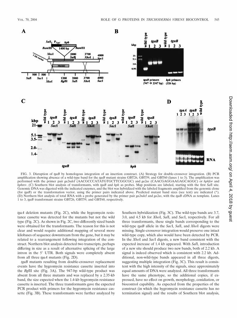
tgaB mutants resulting from double-crossover replacementevents have the hygromycin resistance cassette inserted intothe BglII site (Fig. 3A). The 947-bp wild-type product wasabsent from all three mutants and was replaced by a 2.35-kbband, the size expected when the 1.4-kb hygromycin resistancecassette is inserted. The three transformants gave the expectedPCR product with primers for the hygromycin resistance cas-sette (Fig. 3B). These transformants were further analyzed by
Southern hybridization (Fig. 3C). The wild-type bands are 3.7,3.0, and 4.5 kb for XhoI, SalI, and SacI, respectively. For allthree transformants, these single bands corresponding to thewild-type tgaB allele in the SacI, SalI, and XhoI digests weremissing. Single-crossover integration would preserve one intactwild-type copy, which also would have been detected by PCR.In the XhoI and SacI digests, a new band consistent with theexpected increase of 1.4 kb appeared. With SalI, introductionof a new site should produce two new bands, both of 2.2 kb. Asignal is indeed observed which is consistent with 2.2 kb. Ad-ditional, non-wild-type bands appeared in all three digests,suggesting multiple integration (Fig. 3C). This result is consis-tent with the high intensity of the signals, since approximatelyequal amounts of DNA were analyzed. All three transformantshave the same phenotype, so the additional copies, if ex-pressed, have no effect on growth, morphology, conidiation, orbiocontrol capability. As expected from the properties of theconstruct (in which the hygromycin resistance cassette has notermination signal) and the results of Southern blot analysis,
FIG. 3. Disruption of tgaB by homologous integration of an insertion construct. (A) Strategy for double-crossover integration. (B) PCRamplification showing absence of a wild-type band for the tgaB mutant strains GBT26, GBT59, and GBT60 (lanes 1 to 3). The amplification wasperformed with the primer pair ga2ndeI (AACGCCCATATGTGCTTCGGCGC) and ga2as (CAACGAGGAAGAGCAGGC) or hphfor andhphrev. (C) Southern blot analysis of transformants, with tgaB and hph as probes. Map positions are labeled, starting with the first SalI site.Genomic DNA was digested with the indicated enzymes, and the blot was hybridized with the labeled fragments amplified from the genomic clone(for tgaB) or the transformation vector, using the primer pairs indicated above. Predicted mutant band sizes (see text) are indicated (*).(D) Northern blot analysis of total RNA with a probe generated by the primer pair ga2ndeI and ga2as, with the tgaB cDNA as template. Lanes1 to 3, tgaB transformant strains GBT26, GBT59, and GBT60, respectively.
VOL. 70, 2004 ROLE OF G PROTEINS IN TRICHODERMA VIRENS BIOCONTROL 545
on April 4, 2018 by guest
http://aem.asm
.org/D
ownloaded from
Northern blot analysis detected four transcripts of differentsizes hybridizing to the tgaB probe; none appeared to be iden-tical to the wild-type transcript size. The largest of these was ofseveral kilobases and could span the entire region shown inFig. 3A, including the tgaB gene and the inserted selectionmarker. tgaB transcripts were not altered in tgaA mutants (Fig.3D). The insertion in the tgaB mutants is located in the codingsequence, in a well-conserved domain of the G� subunit in-volved in nucleotide binding. Should any stable protein beproduced from the transcripts in the tgaB mutants, it is veryunlikely that it could function in the finely tuned G-proteinheterotrimer. We note, furthermore, that insertions and dele-tions at cga1 of C. heterostrophus (the homolog of tgaA) re-sulted in identical phenotypes (8; B.A.H., unpublished data).
Growth, colony morphology, conidiation, and conidial ger-mination. The wild type and the tgaA and tgaB mutants weresimilar in growth rate and appearance, except that the coloniesof tgaA mutants appeared greener than those of the wild typeor the tgaB mutants. Neither tgaA nor tgaB mutants underwentconidiation in the dark, and both responded to blue light by
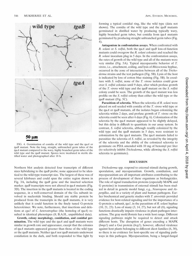
forming a typical conidial ring, like the wild type (data notshown). The conidia of the wild type and the tgaB mutantsgerminated in distilled water by producing typically wavy,highly branched germ tubes, but conidia from tgaA mutantsgerminated by producing straight unbranched germ tubes (Fig.4).
Antagonism in confrontation assays. When confronted withR. solani or S. rolfsii, both the tgaA and tgaB loss-of-functionmutants could overgrow the R. solani colonies and reached theR. solani inoculum plug in 5 days. In the confrontation assays,the rates of growth of the wild type and all of the mutants werevery similar (Fig. 5A). Typical mycoparasitic behavior of T.virens, i.e., attachment, coiling, and lysis of Rhizoctonia hyphae,occurred in the zone of interaction between all of the Tricho-derma strains and the test pathogen (Fig. 5B). Lysis of the hostis indicated by loss of cotton blue staining (Fig. 5B). In cocul-ture with S. rolfsii, none of the T. virens isolates could growover S. rolfsii colonies until 9 days, after which profuse growthof the T. virens wild type and the tgaB mutant on the S. rolfsiicolony could be seen. The growth of the tgaA mutant was lessprolific on the S. rolfsii colony than either the wild type or thetgaB mutant (Fig. 5C).
Parasitism of sclerotia. When the sclerotia of R. solani wereplaced on soil seeded with conidia of the T. virens wild type orthe tgaA or tgaB mutants, all the isolates began colonizing thesclerotia within 2 days, and profuse growth of T. virens on thesclerotia could be seen after 6 days (Fig. 6). Colonization of thesclerotia by the tgaA mutant appeared to be slightly delayed,but this delay is difficult to quantitate in our assay system. Incontrast, S. rolfsii sclerotia, although readily colonized by thewild type and the tgaB mutants in 5 days, were resistant tocolonization by the tgaA mutants. The tgaA mutants failed toparasitize the sclerotia of S. rolfsii, as revealed by the integrityof the sclerotia and the ability of the colonized sclerotia togerminate on PDA amended with 10 mg of benomyl per liter(to selectively inhibit Trichoderma, while allowing the S. rolfsiisclerotia to germinate).
DISCUSSION
Trichoderma spp. respond to external stimuli during growth,sporulation, and mycoparasitism. Growth, conidiation, andmycoparasitism are all important attributes contributing to theprocess of development of these organisms as biofungicides.The role of signal transduction proteins (especially MAPK andG proteins) in transmission of external stimuli has been stud-ied in detail in genetic model fungi, e.g., Neurospora and As-pergillus, and in a variety of plant and human pathogens. Ear-lier biochemical and genetic studies with T. atroviride providedevidence for host-related signaling and for the importance of aG-protein � subunit, tga1, in the parasitism of R. solani hyphae(10, 21, 23). Loss of many (3, 14, 27), but not all (8), signalingfunctions drastically impairs virulence in plant-pathogen inter-actions. The gray mold Botrytis has a wide host range. Differentsignaling pathways might be required to detect and attackdifferent hosts. The disruption of genes encoding a MAPK(bmp1) and a G�i protein (bcg1), however, affected virulenceagainst host plants belonging to different dicot families (6, 30),so there is no evidence for host-specific use of signaling path-ways in this pathogen. Mycoparasitism, being a fungal-fungal
FIG. 4. Germination of conidia of the wild type and the tgaA ortgaB mutant. Note the long, straight, unbranched germ tubes of thetgaA mutant compared to the wavy, highly branched germ tubes of thewild type and the tgaB mutant. Spores were incubated in sterile dis-tilled water and photographed after 18 h.
546 MUKHERJEE ET AL. APPL. ENVIRON. MICROBIOL.
on April 4, 2018 by guest
http://aem.asm
.org/D
ownloaded from

interaction, provides a different type of host-pathogen interac-tion. Trichoderma species are effective against a wide range ofsoilborne plant pathogens, from oomycetes to ascomycetes (7).These hosts probably differ in their surface properties and mayprovide chemically distinct signals to the advancing mycopara-site. A MAPK of T. virens, tmkA, was not involved in hyphal
FIG. 5. Confrontation assays with T. virens and host plant patho-genic fungi. The host was inoculated at the right-hand side of the plate.
FIG. 6. Parasitism of sclerotia by T. virens. The sclerotia were put inthe middle of a soil plate after mixing of conidia into the soil. Controlplates, sclerotia only, with no Trichoderma conidia. Plates were pho-tographed after 7 days of incubation. Mycelial growth is visible as alight-colored colony contrasting with the dark background of the soil.(A) Growth on R. solani sclerotia. (B) Growth on S. rolfsii sclerotia.
(A) Colony interaction between R. solani and T. virens 5 days afterinoculation. Control, host only. (B) Wild type and mutants on R. solanihyphae (after 5 days of inoculation). Note the lysis of host hyphae fromconfrontation plates, indicated by the absence of stain. Control, R.solani hyphae only. (C) Growth of T. virens in confrontation plates withS. rolfsii.
VOL. 70, 2004 ROLE OF G PROTEINS IN TRICHODERMA VIRENS BIOCONTROL 547
on April 4, 2018 by guest
http://aem.asm
.org/D
ownloaded from

parasitism of R. solani, but was essential for the parasitism ofsclerotia of this fungus and of S. rolfsii (19).
We cloned two G-protein � subunits from T. virens. tgaA ortgaB loss-of-function mutants had normal growth and conidia-tion, did not undergo conidiation in the dark, and were myco-parasitic on R. solani hyphae. In contrast, T. atroviride mutantsin which the level of tga1 (the homolog of tgaA) (Fig. 1) wasgreatly reduced through antisense technology grew very slowly,sporulated in the dark, and were deficient in hyphal parasitism(23). Loss of the homologs of tgaA from two plant pathogens,C. heterostrophus and M. grisea, resulted in abnormally straightgrowth of germ tubes that appear to branch less (8, 16), whichis similar to the �tgaA phenotype (Fig. 4). Cochliobolus andMagnaporthe have large multicellular conidia that attach to thehost and other surfaces. The observation of a similar pheno-type for Trichoderma, which has small unicellular conidia thatdo not attach tightly to the substrate, suggests that the G�iclass has a general role in controlling hyphal growth pattern.No phenotypes related to tgaB were detected in this study.Disruption or deletion of the homologs of tgaB in most otherfungi, likewise, conferred no major phenotypic defects (forexamples, see references 6 and 16).
The MAPK (tmkA) loss-of-function mutants of T. virens, incontrast to mutants of tgaA, had near normal growth but un-derwent conidiation in the dark. They also had normal myco-parasitic behavior on R. solani hyphae. The MAPK loss-of-function mutants, however, were less effective in parasitizationof the sclerotia of R. solani and could not parasitize the scle-rotia of S. rolfsii (19). The role of tga1 of T. atroviride in theparasitism of sclerotia is not yet known. The present studyclearly establishes the involvement of tgaA-mediated pathwaysin the parasitism of sclerotia of S. rolfsii, but not of R. solani.tgaA and its homolog in T. atroviride, which belongs to the samegenus, apparently have different functions.
T. virens thus employs both a MAPK pathway (19) and aG-protein pathway (this study) in a host-specific manner. Thebasic structural differences between the sclerotia of S. rolfsiiand R. solani might explain the differential mycoparasitic be-havior against S. rolfsii and R. solani sclerotia. The sclerotia ofS. rolfsii are highly compact and well-differentiated structures(comprised of cortex, medulla, and rind), while those of R.solani are aggregates of a rather loose mass of mycelia (17).Since R. solani hyphae are readily parasitized by tmkA, tgaA,and tgaB mutants, these signal transduction mutants are alsocapable of colonizing (though at a reduced level in the case oftmkA mutants) the sclerotia. On the other hand, since thesclerotia of S. rolfsii are highly melanized specialized structuresthat are difficult to penetrate, some specific enzyme(s) is prob-ably required for the penetration and degradation of thesestructures. A gene(s) for this enzyme could be the downstreamtarget of the tmkA and tgaA pathways. The sclerotia are im-portant survival structures of these pathogens, and their de-struction in soil is of utmost importance for obtaining effectivebiocontrol of these highly damaging plant pathogens with avery broad host range. Therefore, in addition to tmkA, tgaAcould also be a target for gene manipulation (overexpressionand/or expression of the constitutively active allele) of T. virensfor improved biocontrol potential. Furthermore, identificationof the genes that are regulated downstream of the signalingpathways should identify the enzymes that degrade the host
fungi and the transcriptional regulators that act between thesignal transducers and their ultimate downstream targets.
ACKNOWLEDGMENTS
P.K.M. thanks the Department of Science and Technology, Govern-ment of India, for a BOYSCAST Visiting Scientist Fellowship (1999-2000), during which this work was initiated. This study was supportedin part by grant 233/00-2 from the ISF (Israel Academy of Sciences)and grants from the Department of Biotechnology, Government ofIndia, and the Israel Ministry of Sciences, in the form of a jointIndo-Israel research project.
We thank S. F. D’Souza, Head, Nuclear Agriculture and Biotech-nology Division, Bhabha Atomic Research Centre, Mumbai, India, forencouragement and support.
REFERENCES
1. Baasiri, R. A., X. Lu, P. S. Rowley, G. E. Turner, and K. A. Borkovich. 1997.Overlapping functions for two G protein � subunits in Neurospora crassa.Genetics 147:137–145.
2. Baek, J. M., C. R. Howell, and C. M. Kenerley. 1999. The role of anextracellular chitinase from Trichoderma virens Gv29–8 in the biocontrol ofRhizoctonia solani. Curr. Genet. 35:41–50.
3. Bolker, M. 1998. Sex and crime: heterotrimeric G proteins in fungal matingand pathogenesis. Fung. Genet. Biol. 25:143–156.
4. Cortes, C., A. Gutierrez, V. Olmedo, J. Inbar, I. Chet, and A. Herrera-Estrella. 1998. The expression of genes involved in parasitism by Tricho-derma is triggered by a diffusible factor. Mol. Gen. Genet. 260:218–225.
5. Felsenstein, J. 1993. PHYLIP (phylogeny inference package) version 3.5c.Department of Genetics, University of Washington, Seattle.
6. Gronover, C. S., D. Kasulke, P. Tudzynski, and B. Tudzynski. 2001. The roleof G protein � subunits in the infection process of the gray mold fungusBotrytis cinerea. Mol. Plant-Microbe Interact. 14:1293–1302.
7. Herrera-Estrella, A., and I. Chet. 1998. Biocontrol of bacteria and phyto-pathogenic fungi, p. 263–282. In A. Altman (ed.), Agricultural bio/technol-ogy. Marcel Dekker, Inc., New York, N.Y.
8. Horwitz, B. A., A. Sharon., S.-W. Lu, V. Ritter, T. Sandrock, B. G. Turgeon,and O. C. Yoder. 1999. A G protein � subunit gene of Cochliobolus het-erostrophus involved in mating and appressorium formation. Fung. Genet.Biol. 26:19–32.
9. Howell, C., R. Stipanovic, and R. Lumsden. 1993. Antibiotic production bystrains of Gliocladium virens and its relation to biocontrol of cotton seedlingdiseases. Biocontrol Sci. Technol. 3:435–441.
10. Inbar, J., and I. Chet. 1992. Biomimics of fungal cell-cell recognition by useof lectin-coated nylon fibers. J. Bacteriol. 174:1055–1059.
11. Jones, D. T., W. R. Taylor, and J. M. Thornton. 1992. The rapid generationof mutation data matrices from protein sequences. Comput. Appl. Biosci.8:275–282.
12. Kays, A. M., P. S. Rowley, R. A. Baasiri, and K. A. Borkovich. 2000. Regu-lation of conidiation and adenylyl cyclase levels by the G� protein GNA-3 inNeurospora crassa. Mol. Cell. Biol. 20:7693–7705.
13. Koch, E. 1999. Evaluation of commercial products for microbial control ofsoil-borne plant pathogens. Crop Prot. 18:119–125.
14. Lengeler, K. B., R. C. Davidson, C. D’Souza, T. Harashima, W. C. Shen, P.Wang, X. Pan, M. Waugh, and J. Heitman. 2000. Signal transduction cas-cades regulating fungal development and virulence. Microbiol. Mol. Biol.Rev. 64:746–785.
15. Lev, S., A. Sharon, R. Hadar, H. Ma, and B. A. Horwitz. 1999. A mitogen-activated protein kinase of the corn leaf pathogen Cochliobolus heterostro-phus is involved in conidiation, appressorium formation, and pathogenicity:diverse roles for mitogen-activated protein kinase homologs in foliar patho-gens. Proc. Natl. Acad. Sci. USA 96:13542–13547.
16. Liu, S., and R. Dean. 1997. G protein � subunit genes control growth,development, and pathogenicity of Magnaporthe grisea. Mol. Plant-MicrobeInteract. 10:1075–1086.
17. Mukherjee, P. K., A. N. Mukhopadhyay, D. K. Sarmah, and S. M. Shresh-tha. 1995. Comparative antagonistic properties of Gliocladium virens andTrichoderma harzianum on Sclerotium rolfsii and Rhizoctonia solani—its rel-evance to understanding the mechanisms of biocontrol. J. Phytopathol. 143:275–279.
18. Mukherjee, P. K., S. M. Shreshtha, and A. N. Mukhopadhyay. 1993. Baitingwith Sclerotium rolfsii for selective isolation of Gliocladium virens from nat-ural soil. Biocontrol Sci. Technol. 3:101–104.
19. Mukherjee, P. K., J. Latha, R. Hadar, and B. A. Horwitz. 2003. tmkA, a MAPkinase of Trichoderma virens, is involved in biocontrol properties and repres-sion of conidiation in the dark. Eukaryot. Cell 2:446–455.
20. Mukherjee, P. K., P. Thomas, and K. Raghu. 1995. Shelf-life enhancementof fresh ginger rhizomes at ambient temperatures by combination of gamma-irradiation, biocontrol and closed polyethylene bag storage. Ann. Appl. Biol.127:375–384.
548 MUKHERJEE ET AL. APPL. ENVIRON. MICROBIOL.
on April 4, 2018 by guest
http://aem.asm
.org/D
ownloaded from
21. Omero, C., J. Inbar, V. Rocha-Ramírez, A. Herrera-Estrella, I. Chet, andB. A. Horwitz. 1999. G protein activators and cAMP promote mycoparasiticbehaviour in Trichoderma harzianum. Mycol. Res. 103:1637–1642.
22. Page, R. D. M. 1996. TREEVIEW: an application to display phylogenetictrees on personal computers. Comput. Appl. Biosci. 12:357–358.
23. Rocha-Ramírez, V., C. Omero, I. Chet, B. A. Horwitz, and A. Herrera-Estrella. 2002. Trichoderma atroviride G-protein �-subunit gene tga1 is in-volved in mycoparasitic coiling and conidiation. Eukaryot. Cell 1:594–605.
24. Sambrook, J., E. F. Fritsch, and T. Maniatis. 1989. Molecular cloning: alaboratory manual, 2nd ed. Cold Spring Harbor Laboratory, Cold SpringHarbor, N.Y.
25. Strathmann, M., and M. I. Simon. 1990. G protein diversity: a distinct classof � subunits is present in vertebrates and invertebrates. Proc. Natl. Acad.Sci. USA 87:9113–9117.
26. Tag, A., J. Hicks, G. Garifullina, C. Ake, Jr., T. D. Phillips, M. Beremand,
and N. Keller. 2000. G-protein signaling mediates differential production oftoxic secondary metabolites. Mol. Microbiol. 38:658–665.
27. Xu, J.-R. 2000. MAP kinases in fungal pathogens. Fung. Genet. Biol. 31:137–152.
28. Yu, J. H., S. Rosen, and T. H. Adams. 1999. Extragenic suppressors ofloss-of-function mutations in the Aspergillus flbA regulator of G-proteinsignaling domain protein. Genetics 151:97–105.
29. Zeilinger, S., C. Galhaup, K. Payer, S. L. Woo, R. L. Mach, C. Fekete, M.Lorito, and C. P. Kubicek. 1999. Chitinase gene expression during myco-parasitic interaction of Trichoderma harzianum with its host. Fung. Genet.Biol. 26:131–140.
30. Zheng, L., M. Campbell, J. Murphy, S. Lam, and J.-R. Xu. 2000. The BMP1gene is essential for pathogenicity in the gray mold fungus Botrytis cinerea.Mol. Plant-Microbe Interact. 13:724–732.
VOL. 70, 2004 ROLE OF G PROTEINS IN TRICHODERMA VIRENS BIOCONTROL 549
on April 4, 2018 by guest
http://aem.asm
.org/D
ownloaded from