Embed Size (px)
Citation preview

University of Nebraska - Lincoln University of Nebraska - Lincoln
DigitalCommons@University of Nebraska - Lincoln DigitalCommons@University of Nebraska - Lincoln
Publications from USDA-ARS / UNL Faculty U.S. Department of Agriculture: Agricultural Research Service, Lincoln, Nebraska
2010
Role of the Oviduct in Maintaining Sustained Fertility in Hens Role of the Oviduct in Maintaining Sustained Fertility in Hens
M. R. Bakst USDA-ARS, [email protected]
Follow this and additional works at: https://digitalcommons.unl.edu/usdaarsfacpub
Part of the Agricultural Science Commons
Bakst, M. R., "Role of the Oviduct in Maintaining Sustained Fertility in Hens" (2010). Publications from USDA-ARS / UNL Faculty. 670. https://digitalcommons.unl.edu/usdaarsfacpub/670
This Article is brought to you for free and open access by the U.S. Department of Agriculture: Agricultural Research Service, Lincoln, Nebraska at DigitalCommons@University of Nebraska - Lincoln. It has been accepted for inclusion in Publications from USDA-ARS / UNL Faculty by an authorized administrator of DigitalCommons@University of Nebraska - Lincoln.

Murray R. Bakst
Role of the Oviduct in Maintaining Sustained Fertility in Hens
published online Dec 17, 2010; J Anim Sci
http://jas.fass.orgthe World Wide Web at:
The online version of this article, along with updated information and services, is located on
www.asas.org

1
Running head: Role of the oviduct in hen fertility
Role of the Oviduct in Maintaining Sustained Fertility in Hens1
Murray R. Bakst2
Animal Biosciences and Biotechnology Laboratory, ARS-USDA
Beltsville, MD 20705
1 Based on a presentation at the Physiology and Endocrinology Symposium, “Sperm-Oviduct
Interactions in Livestock and Poultry,” at the joint annual meeting of the American Dairy
Science Association, Poultry Science Association, Asociación Mexicana de Producción
Animal, Canadian Society of Animal Science, and American Society of Animal Science, July
11-15, 2010, Denver, CO.
2 Corresponding author: [email protected]
Published Online First on December 17, 2010 as doi:10.2527/jas.2010-3663

2
ABSTRACT: In poultry, sperm transferred by natural mating or AI into the distal end of
the vagina immediately begin their ascent to the utero-vaginal junction (UVJ) at the anterior end
of the vagina. However, due to an intense selection process in the vagina, less than 1% of the
sperm transferred actually reach the UVJ. Those sperm that do reach the UVJ enter numerous
tubular invaginations of the vagina’s surface epithelium located in the UVJ mucosa, collectively
referred to as the sperm-storage tubules (SST). Sperm residing in the SST lumen are capable of
surviving up to several weeks while retaining their fertilizing capacity. Resident sperm are
released gradually from the SST while the hen is in egg production, ascend to the site of
fertilization, and interact with the next ovulated ovum. In this manner, given the absence of an
estrus to synchronize ovulation with copulation, poultry are assured a population of sperm at the
site of fertilization around ovulation. Over the past decade, several new and diverse observations
have been published addressing the microanatomy of the UVJ and SST, and the cellular and
molecular mechanisms orchestrating oviductal sperm selection and storage. These include: role
of sperm mobility in selection and transport; SST numbers in different poultry species and lines
of high and low fertility; roles of the immune system and possibly neuro-endocrine-like cells in
the vagina in sperm selection and storage, and the roles of aquaporins and a fluid exchange
mechanisms contributing to sperm release from the SST. The objective of this paper is to review
and integrate these observations into a comprehensive understanding of the cellular and
molecular events influencing the fate of sperm in the hen’s oviduct, particularly with regard to
oviductal sperm selection and storage.
Key words: avian, oviductal poultry, sperm storage, sperm storage tubules

3
INTRODUCTION
The biology of reproduction is vastly different between birds and mammals. In the
absence of a mammalian-type estrous cycle for the synchronization of copulation with ovulation,
birds rely on oviductal sperm storage. In the domestic and the non-domestic birds examined, the
surface epithelium lining the anterior 2 cm of the vagina, referred to as the uterovaginal junction
(UVJ), is modified to form numerous tubular invaginations referred to collectively as the sperm-
storage tubules (SST). Shortly before and during egg production, sperm residing in the SST will,
upon release from the SST, ascend the oviduct to the site of fertilization in the infundibulum.
Here sperm interact with a nearly daily succession of ovulated ova over days to several weeks,
depending on the species. For more detailed reviews of the events leading up to fertilization, see
Bakst et al. (1994), Wishart and Horrocks (2000), Stepinska and Bakst (2007) for domestic birds,
as well as Birkhead and Moller (1992) and Birkhead and Brillard (2007) for non-domestic birds.
A better understanding of the fundamental cellular and molecular mechanisms regulating
oviductal sperm selection, transport, and storage would have a profound effect on the breeding
sector of the commercial poultry industry. This information could lead to: 1) more innovative
approaches to the development of semen extenders that maintain sperm viability at ambient
temperature for greater than 24 h; 2) the use of gene markers for the selection of the most fecund
breeders; 3) providing sound scientific information rather than empirical observations to poultry
flock managers when confronting fertility problems, and 4) implementation of novel approaches
to poultry management, ultimately increasing the ratio of breeder females per male.
Since the mid-1990s, several intriguing observations regarding the fate of sperm in the
domestic bird oviduct have been published that resulted in both the re-evaluation of former and
the introduction of new concepts in the understanding of reproduction in birds. The objectives of

4
this paper are to review these new observations and then integrate them into a more unified
understanding of oviductal-sperm interactions and sustained fertility in birds in general and
poultry in particular.
ANATOMY AND HISTOLOGY OF THE UTERUS AND VAGINA
The structure and function of the avian oviduct has been extensively described (see
Introduction). Surprisingly, the gross anatomy of the avian vagina, which is now understood to
be quite complex, was not described in detail until recently. In describing the duck vagina,
Brennan et al. (2007) observed a spiral-shaped tube that was further characterized by blind
pouches stemming from its distal half. When considering the waterfowl penis is cork-screwed
with an opposite orientation to the spirals in the vagina (Brennan et al., 2010), the authors
concluded that the anatomical incongruity between the phallus and oviduct would be used by the
female to impede or block penetration of the penis during attempts at forced copulation.
Although Bakst (1998) alluded to the turkey vagina as being tightly coiled and enveloped
by connective tissue, it was the work of Brennan et al. (2007) that motivated a more detailed
anatomical examination of the turkey vagina and uterus. Bakst and Akuffo (2009) fixed the
turkey vagina and uterus in toto, with and without an egg mass in the uterus. The connective
tissue binding the vagina and uterus was then removed and revealed a spiral configuration
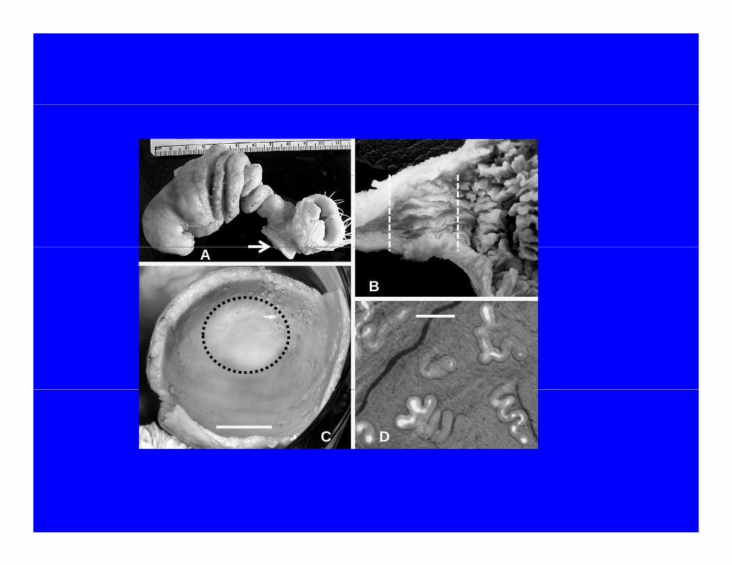
regardless of whether or not an egg mass was present in the uterus (Figure 1A). Without an egg
mass in the uterus, the UVJ mucosal folds containing SST clearly did not extend into the uterus
and were contiguous with the vaginal mucosa (Figure 1B). Alternatively, when an egg mass was
present in the uterus, the UVJ folds containing SST were contiguous with the uterine mucosa and
clearly within the uterus pouch (Figure 1C). This anatomical configuration would indicate that
sperm exiting the SST are subjected to uterine fluids known to stimulate chicken sperm motility

5
in vitro (Brillard et al., 1987).
The cellular and molecular mechanisms responsible for the morphogenesis of the SST are
not known. The anatomical differentiation of the SST has briefly been described by Bakst
(1992) in 30 wk old turkey hens prior to the onset of photostimulation to bring hens into egg
production. Both elongated SST and bud-like surface invaginations, presumptive SST, were
observed (Figure 1D). A more detailed description of SST morphogenesis in Japanese quail
prior to and during the period corresponding to ovarian maturation was described by Holm and
Ridderstrale (2002). At 28 d of age, the time coinciding with the onset of tubular gland
formation in the magnum, low columnar cells were observed at the base of the folds at the UVJ.
Within 10 d, these cells had differentiated into bud-like projections and then tubular structures
consisting of non-ciliated columnar cells, the presumptive SST. Females housed with males
possessed sperm in their SST before the first ovulation (approximately 42 d of age). This
confirmed earlier observations of sperm in the SST of turkey hens inseminated artificially before
the onset of photostimulation and possessing a juvenile oviduct (Bakst 1988; 1992).
The cell signaling pathways controlling SST differentiation and proliferation during the
oviduct’s maturation prior to the onset of egg production remain unknown. Given the similarity
between the cellular organization of luminal mucosae of the intestine and oviduct, one could
assume that stromal trophic factors regulate the differentiation and proliferation of the SST in a
manner similar to that suggested for the intestine by Simmons et al. (1999). These authors
suggested that intestinal crypt cell proliferation and renewal of the cells forming the luminal
epithelium were influenced by insulin-like growth factors originating from the sub-epithelial
stroma cells. If such interactions are found to contribute to SST morphogenesis, we then may be
able to explain why there exists intra- and inter-line variations in SST numbers (Bakst et al.,
6
2010).
The total number of SST in the UVJ varies between species (Birkhead and Moller, 1992).
More recently, Bakst et al. (2010) calculated the total numbers of SST in 4 strains of broilers of
differing fertility and one commercial strain of Large White turkeys. Unlike the numbers of SST
for the chicken (13,533) and turkey (20,000) reported by Birkhead and Moller (1992), Bakst et
al. (2010) observed that the broiler lines averaged 4,900 SST per hen and turkeys averaged
30,600 SST per hen. Furthermore, Bakst et al. (2010) authors observed no statistical differences
in the SST numbers among the 4 strains of broilers. From these data, Bakst et al. (2010)
proposed the following: 1) the longer duration of fertility in turkeys compared with broilers is
due, in part, to a greater number SST and a slower daily release of sperm; 2) in a commercial hen
flock, variation in fertility is not associated with SST numbers; 3) in contrast, when selected
solely for high and low fertility, the number of SST in the high fertility line of hens is
significantly greater than the low fertility line of hens (Brillard et al., 1998), and 4) factors other
than SST numbers play a role in sustained fertility in commercial strains of broilers and turkeys.
VAGINA: SPERM SELECTION, TRANSPORT, AND STORAGE
Within the 30 min following the transfer of semen into the vagina, 84% of sperm flow
back out of the vagina (Howarth, 1971), most often embedded in a plug of mucous (Figure 2A)
(J. P. Brillard, INRA, Nouzilly, France; personal communication). The remaining sperm are
transported in an adovarian direction by a combination of their intrinsic mobility (i.e., capacity to
move through a viscous medium; Froman et al., 2006) and the sperm transport mechanisms of
the vagina that include smooth muscle activity and the activity of the ciliated cells lining the
vagina’s luminal mucosal surfaces.
The abovarian transport of the egg mass through the infundibulum, magnum, and

7
is due to peristaltic activity initiated by local distention of the oviductal smooth muscle layer
(Arjamaa and Talo, 1983). Bakst et al. (1994) speculated that intrinsic sperm motility coupled
with a fluid transport mechanism in the troughs between tightly apposed mucosal folds were
responsible for rapid sperm transport to the UVJ at the distal end of the vagina. This was based
on observations that sperm transferred to the distal end of the vagina of an excised turkey oviduct
were observed in the infundibulum (i.e., a distance of about 80 cm) in less than 10 min (M. R.
Bakst, unpublished results).
The cellular and molecular basis of sperm mobility and its role in oviductal sperm storage
and transport was recently reviewed by Froman et al. (2010) and will not be addressed in detail
in this review. They developed a compelling argument for sperm mobility as being the dominant
factor in sperm selection within the vagina. Interestingly, Denk et al. (2005) suggested that the
swimming speed and motility of mallard sperm figured more prominently in paternity than post-
copulatory sperm selection by the female. Notwithstanding the role of sperm mobility and
motility, other factors do influence the numbers of sperm that reach the SST. If vaginal
insemination is 2 h before or 2 h after oviposition, oviductal sperm transport is altered and sperm
filling of the SST is reduced (Birkhead et al., 1996). We also know that the efficiency of sperm
transport in the turkey vagina, as measured by the percentage of SST that are filled, partially
filled, or empty following artificial insemination, is most efficient before the onset of egg
production. Obviously there are mechanisms (i.e., neuro-muscular, cellular, endocrine, and/or
the presence of an egg-mass in the oviduct) that impact sperm selection and transport after the
onset of lay.
Differences in luminal pH may influence sperm mobility. Bakst (1980) observed
significant differences in the pH of the broiler’s mid-vaginal mucosa that ranged from pH 7.15

8
within 20 min post-oviposition (PO) to pH 7.51 at 8 to 12 hr PO. In the manually-everted turkey
vaginal mucosa (i.e., the distal 1 to 2 cm of the vagina), there are significant pH differences that
range from pH 6.95 at (8 to 12 h and 18 to 22 h PO) to pH 7.30 within 10 min following
oviposition. These variations in pH may modulate the mobility of sperm following semen
transfer. The localization of carbonic anhydrase in UVJ, SST, vaginal epithelia also indicates a
role for pH in the modulation of sperm motility possibly with a higher pH augmenting sperm
motility in the vagina and a lower pH depressing sperm motility in the SST lumen (Holm et al.,
1996).
There is evidence throughout the animal kingdom that the female is able to exert some
influence on which male will fertilize her ova (see Eberhard, 1996). This is not only possible
through female mate choice, but also in the post-copulation selection of sperm in the female’s
reproductive tract. The latter is referred to as cryptic female choice (Eberhard, 1996). Based on
what is known about birds, one would assume that cryptic female choice is most likely to be
observed in the vagina, although there may be further sperm selection after release from the SST
(Birkhead and Brillard, 2007). A local signal, possibly initiated by a component in seminal
plasma or sperm, coupled to a response by the vaginal epithelial cells may trigger a cascade of
events that favor the adovarian transport of these sperm. Sperm signaling may be associated
with the sperm plasmalemma glycoproteins, or lack of them (Bakst et al., 1994; Wishart and
Horrocks, 2000; Pelaez and Long, 2008).
Serotonin-positive non-neuronal endocrine-like cells have been localized in turkey
vaginal and UVJ epithelia but not the SST epithelia (Bakst and Akuffo, 2008). Similar cells,
known as enterochromaffin cells, are observed in the gut epithelium of other species and appear
to regulate a local peristaltic reflex (Olsson and Holmgren, 2001). We are currently examining

9
the possibility that the serotonin-positive cells in the hen’s vaginal epithelium may exert a local
impact on sperm motility and transport to the SST. In addition to augmenting localized peristaltic
activity, serotonin has been shown to stimulate both cilia and sperm beat frequency in a variety
of species (Stephens and Prior, 1992). Interestingly, using a computer-assisted sperm motility
analysis system, serotonin (at 10–4M but not at 10–6M) statistically increased turkey sperm curvi-
linear velocity and tail beat frequency (M. R. Bakst, unpublished results). Thus, in the context of
cryptic female choice, serotonin-containing cells in the vagina and UVJ but not in SST epithelia
may augment local sperm motility, vaginal cilia beat frequency, and smooth muscle activity
facilitating sperm transport to SST (Bakst and Akuffo, 2008).
While the question of how sperm survive within the SST for prolonged periods of time
has yet to be definitively explained, it is assumed that resident sperm metabolize endogenous
fatty acids (Froman, 2010) or other lipids derived from the apical microvilli of the SST
epithelium (Bakst et al., 1994). Liposome-like vesicles appear to pinch-off the microvillar tips
of the SST epithelial cells and appear to interact with the luminal sperm. This region of the SST
epithelium is alkaline phosphatase (AP)-positive (Bakst and Akuffo, 2007) and corresponds to
the localization of AP in the rat intestinal luminal epithelium (Narisawa et al., 2003). These
authors suggested that AP may function in the transfer of lipid across the enterocytes brush
border and it is speculated that AP may have a similar role in the SST epithelium (Bakst and
Akuffo, 2007).
The mechanism(s) of sperm release from the SST has been the subject of speculation for
years (see Bakst et al., 1994 for review). More recently, it has been suggested that sperm release
from the SST may be a neural mediated mechanism that initiates contraction of the actin-rich
band in the apical cytoplasm of the SST epithelium (Freedman et al., 2001), thus expelling sperm

10
from the SST lumen. Alternatively, Froman et al. (2010) suggested that the sperm residing in the
SST lumen are subjected to a fluid current moving toward the SST orifice (Figure 2B). Sperm
will remain in the SST lumen as long as their swimming velocity is greater than that of the
luminal fluid’s flow rate. Sperm release from the SST would take place when, possibly as a
result of waning levels of ATP, sperm motility deceases and they are carried out of the SST with
the luminal fluids. The localization of aquaporin-3 in the apical region of the SST epithelium
(Zaniboni and Bakst, 2004) would support the suggestion that there is a transfer of fluids from
the SST epithelium to the SST lumen.
IMMUNOLOGICAL ASPECTS OF SPERM SELECTION AND STORAGE
Bakst et al. (1994) provided a comprehensive review of possible roles of the oviduct’s
immune system on sperm selection and storage. They noted that there were conflicting
observations regarding the role, if any, on sperm antibodies and the decline in hen fertility.
Since then, Robertson et al. (2000) demonstrated that chicken and turkey sperm must possess the
proper array of plasmalemma-associated proteins and glycoproteins, each with their respective
saccharide groups in order to reach the SST and also to interact with the ovum at the time of
fertilization. Furthermore, Steele and Wishart (1992) observed that vaginally inseminated sperm
bound immunoglobulin (IgA or IgG) and that 84% of those sperm were dead. Of the remaining
viable sperm recovered, 7% bound immunoglobulins but only sperm devoid of immunoglobulins
were observed in the SST. It is unlikely that the antibodies binding to sperm were sperm-
specific antibodies because immunoglobulins were also observed associated with sperm
recovered from virgin hens
The vaginal insemination of heterologous semen into chickens resulted in few sperm
reaching the SST, presumably due to the absence of the specific sperm surface glycoprotein

11
array compatible with the hen’s oviductal sperm selection mechanism. However, when added to
explants of UVJ folds, heterologous sperm entered the SST indicating that sperm selection
process is orchestrated by the vagina and not the SST (Wishart and Horrocks, 2000).
In the past decade, there has been a resurgence of studies addressing the role of the
immune system in the hen’s oviduct with respect to reproductive function (see review by Das et
al., 2008). Classes of immuno-competent cells (i.e., macrophages, antigen-presenting cells
expressing MHC class II, CD4+ and CD8+ T cells, premature B cells, and plasma cells) and cell
products associated with both acquired (Zheng et al., 1998; 1999) and innate immunity (i.e.,
avian β-defensins; Abdel-Mageed et al., 2008) are expressed within the oviductal mucosa,
particularly in the vagina (Figure 2C). The vaginal orifice, as well as the coprodeum (i.e., the
anterior compartment of the cloaca and extension of the large intestine), communicates directly
with the urodeum, the central compartment of the cloaca. Therefore, it is not surprising that the
vaginal mucosa’s immune system is highly differentiated (Bakst and Akuffo, 2009) and
histologically reminiscent of the gut-associated lymphoid tissue described for birds (Befus et al.,
1980).
The role of estrogens in the cyto-differentiation of the oviduct’s luminal and subluminal
epithelia has been established for many years (Berg et al., 2001). Of interest is that the numbers
of immuno-competent cells associated with acquired immunity are greater in laying than non-
laying hens and the observation that this has also been associated with elevated levels of estrogen
(Zheng et al., 1998). In their review, Das et al. (2008) indicated that the storage of sperm in the
SST necessitate an immuno-suppression of sperm antigenicity. Using a low fertility line of hens
subjected to repeated inseminations, Das et al. (2005a) observed swollen SST lacking resident
sperm and lymphocyte infiltration of the SST. In addition, these authors also noted that the

12
mucosa surrounding the SST possessed increased numbers of antigen-presenting cells expressing
MHC class II, CD4+, and CD8+ T phenotypes. Concurrent with the increased numbers of these
immuno-competent cells and the abnormal appearance of the SST was a decrease in the
abundance of mRNA for estrogen receptor (ER)-α (Das et al., 2006a). Their observations
prompted the suggestion that because SST structure and function is estrogen dependent, the
decreased mRNA expression of ER-α, coupled with the increased numbers of immuno-
competent cells in and around the SST, may be directly related to the absence of significant
sperm storage within the SST in low fertility hens. These authors also observed that
transforming growth factor-β (TGF-β and their receptors (TβR) increased in the UVJ when
sperm reside in the SST (Das, 2006b). Given the immunosuppressive properties of TGF-β, and
that lymphocytes in the UVJ mucosa possess TβR, Das et al. (2006b) suggested that TGF-β may
suppress immune-responses to resident sperm in the SST by UVJ lymphocytes. Das et al. (2008)
concluded that this suppression of UVJ lymphocytes by TGF-β may contribute to successful
sperm storage in the SST.
SUMMARY AND CONCLUSIONS
Collating the observations discussed previously into a more comprehensive model of the
fate of sperm following transfer to the vagina, the following is suggested: 1) following semen
transfer, the majority of sperm are rejected by the vagina and the remaining, high-mobility sperm
begin transport to the UVJ; 2) these more ‘fit’ sperm may be subjected to other sperm selection
mechanisms, but the single dominant phenotypic trait affecting transit to the UVJ is sperm
mobility; 3) successful sperm storage in the SST is dependent on the establishment of an
immuno-privileged status for sperm residing in the SST and this may be estrogen- and TGF--

13
dependent; 4) sperm residing in the SST subsist on lipid derived from the SST epithelium; 5)
sperm residence within the SST is dependent on sperm motility exceeding the velocity of the
luminal fluid flow exiting the SST orifice; 6) sperm are transported out of the SST when
mitochondrial ATP begins to deplete, motility wanes and the SST luminal fluid velocity exceeds
that of the sperm; 7) sperm released from the SST are exposed to calcium-rich uterine fluids,
activated and ascend to the infundibulum; 8) differences in sustained fertility in different lines of
commercial broilers are not a function of SST numbers, and 9) the longer fertile period of
turkeys compared to broilers is due to turkeys possessing 5 times as many SST as broilers.
To conclude, sustained fertility in the hen is a complex series of temporal and spatial
events that ultimately result in a relatively low number of highly-selected sperm at the site of
fertilization at the time of ovulation. As sperm numbers in the infundibulum decrease, either due
to low sperm numbers in the SST, impaired sperm transport and selection by the vagina, or the
inability of the SST to store sperm, fertilization rates will fall. While AI technology has not
progressed significantly over the past two decades, we now have the capability to select males
producing sperm with high mobility, the most significant phenotypic trait associated with sperm
fecundity. This one advance may eventually contribute to greater fertility levels and the
possibility of longer intervals between successive inseminations.
LITERATURE CITED
Abdel-Mageed, A. M., N. Isobe, and Y. Yoshimura. 2008. Expression of avian B-defensins in
the oviduct and effects of lipopolysaccharide on their expression in the vagina of hens.
Poult. Sci. 87:979-984.
Arjamaa, O., and A. Talo. 1983. Smooth muscle of the quail oviduct functions as a stretch
receptor during ovum transport. Biol. Reprod. 29:472-478.

14
Bakst, M. R. 1980. Chicken and turkey oviductal pH at known times postoviposition. Poult. Sci.
59:2793-2796.
Bakst, M. R. and H. Cecil. 1985. A microscopic examination of the male turkey proctodeal
gland. J. Morphol. 186:361-368.
Bakst, M. R. 1988. Duration of fertility of turkeys inseminated at different times after the onset
of photostimulation. J. Reprod. Fertil. 84:531-537.
Bakst, M. R. 1992. Observations on the turkey oviductal sperm-storage tubule using differential
interference contrast microscopy. J. Reprod. Fertil. 95:877-883.
Bakst, M. R., G. Wishart, and J. P. Brillard. 1994. Oviducal sperm selection, transport, and
storage in Poultry. Poult. Sci. Rev. 5:117-143.
Bakst, M. R. 1998. Structure of the avian oviduct with emphasis on sperm storage in poultry. J.
Exp. Zool. 282:618-626.
Bakst, M. R., and V. Akuffo. 2007. Alkaline phosphatase reactivity in the vagina and
uterovaginal junction sperm-storage tubules of turkeys in egg production: implications
for sperm storage. Brit. Poult. Sci. 48:515-518.
Bakst, M. R., and V. Akuffo. 2008. Serotonin localization in the turkey vaginal but not sperm
storage tubule epithelia. Poult. Sci. 87:356-561.
Bakst, M. R. and V. Akuffo. 2009. Morphology of the turkey vagina with and without an egg
mass in the uterus. Poult. Sci. 2009. 88:631-635.
Bakst, M. R., A. M. Donoghue, D. E. Yoho, J. R. Moyle, S. M. Whipple, M. J. Camp, G. Q. Lin,
and R. K. Bramwell. 2010. Comparisons of sperm storage tubule distribution and
number in 4 strains of mature broiler breeders and in turkey hens before and after the
onset of photostimulation. Poult. Sci. 89:986-992.

15
Befus, A. D., N. Johnston, G. A. Leslie, and J. Bienenstock. 1980. Gut-associated lymphoid
tissue in the chicken. I. Morphology, ontogeny, and some functional characteristics of
Peyer’s patches. J. Immun. 125:2626-2632.
Berg, C., L. Holm, I. Brandt, and B. Brunstrom. 2001. Anatomical and histological changes of
the oviducts of Japanese quail, Coturnix japonica, after embryonic exposure to
ethynyloestradiol. Reproduction 121:155-165.
Birkhead, T. R. and A. P. Moller. 1992. Sperm Competition in Birds: Evolutionary Causes and
Consequences. Academic Press, New York.
Birkhead, T. R., E. J. A. Cunningham and K. M. Cheng. 1996. The insemination window
provides a distorted view of sperm competition in birds. Proc. Biol. Sci. 263:1187-1192.
Birkhead, T. R. and J. P. Brillard. 2007. Reproductive isolation in birds: postcopulatory
prezygotic barriers. Trends Ecol Evol. 22:266-272.
Brennan, P. L. R., R. O. Prum, K. G. McCracken, M. D. Sorenson, R. E. Wilson., and T. R.
Birkhead. 2007. Coevolution of male and female genital morphology in waterfowl. PLoS
ONE 2, e418. doi:10.1371/journal.pone.0000418).
Brennan, P. L. R, C. J. Clark, and R.O. Prum. 2010. Explosive eversion and functional
morphology of the duck penis supports sexual conflict in waterfowl genitalia. Proc. R.
Soc. B277:1309-1314.
Brillard, J. P., O. Galut, and Y. Nys. 1987. Possible cause of subfertility in hens following
insemination near the time of oviposition. Br. Poult. Sci: 28:307-318.
Brillard, J. P., C. Beaumont, and M. F. Scheller. 1998. Physiological responses of hens
divergently selected On the number of chicks obtained from a single insemination.
J.Reprod. Fert. 114:111-117.

16
Das, S. C., N. Nagasaka, Y. Yoshimura. 2005a. Effects of repeated artificial insemination on the
structure and function of oviductal sperm storage tubules in hens. J. Poult. Sci. 42:39-47.
Das, S. C., N. Nagasaka, Y. Yoshimura. 2005b. Changes in the localization of antigen presenting
cells and T cells in the utero-vaginal junction after repeated artificial insemination in
laying hens. J. Reproduct. Dev. 51:683-687.
Das, S. C., N. Nagasaka, Y. Yoshimura. 2006a. Changes in the expression of estrogen receptor
mRNA in the utero-vaginal junction containing sperm storage tubules in laying hens after
repeated artificial insemination. Theriogenology 65:893-900.
Das, S. C., N. Isobe, M. Nishibori, Y. Yoshimura. 2006b. Expression of TGFisoforms and their
receptors in the utero-vaginal junction of the hen oviduct in the presence or absence of
resident sperm with reference to sperm storage. Reproduction 132:781-790.
Das, S. C., N. Isobe, and Y. Yoshimura. 2008. Mechanism of prolonged sperm storage and
sperm survivability in the hen’s oviduct: A review. Am. J. Reprod. Immunol. 60:477-481.
Denk, A. G., A. Holzmann, A. Peters, E. L. M. Vermeirssen, and B. Kempenaers. 2005.
Paternity in Mallards: effects of sperm quality and female sperm selection for inbreeding
avoidance. Behav. Ecol. 16:825-833.
Eberhard, W. G. 1996. Selection on cryptic female choice. Pages 44–79 in Female Control:
Sexual Selection by Cryptic Female Choice. Princeton University Press, Princeton, NJ.
Freedman, S., V. Akuffo, and M. R. Bakst. 2001. Evidence for the innervation of sperm-storage
tubules in the oviduct of the turkey (Meleagris gallopavo). Reproduction 121:809-814.
Froman, D. P., J. C. Wardell, and A. J. Feltmann. 2006. Sperm mobility: Deduction of a model
explaining phenotypic variation in roosters (Gallus domesticus). Biol. Reprod. 74:487-
491.

17
Froman, D. P., A. J. Feltmann, K. Pendarvis, S. M. Cooksey, S. C. Burgess, and D. D. Rhoads.
2010. A proteome-based model for sperm mobility phenotype. J. Anim. Sci. [Epub
ahead of print] doi: 10.2527/jas. 2010-3367.
Howarth, B., Jr. 1971. Transport of spermatozoa in the reproductive tract of turkey hens. Poult.
Sci.50:84-89.
Holm, L., and Y. Ridderstråle. 2002. Development of sperm storage tubules in the quail during
sexual maturation. J. Exp. Zool. 292:200-205, 2002.
Holm, L., Y. Ridderstråle, P-G. Knutsson. 1996. Localisation of carbonic anhydrase in the
sperm storing segions of the domestic hen oviduct. Acta Anat. 156:253-260.
Kitazawa, T., H. Ukai, S. Komori, and T. Taneike. 2006. Pharmacological characterization of 5-
hydroxytryptamine-induced contraction in the chicken gastrointestinal tract. Auton. Auto-
coid. Pharmacol. 26:157-168.
Narisawa, S., L. Huang, A. Iwasaki, H. Hasegawa, D. H. Alpers, and J. L. Millán. 2003.
Accelerated fat absorption in intestinal alkaline phosphatase knockout mice. Mol Cell
Biol. 23:7525-7530.
Olsson, C. and S. Holmgren. 2001. The control of gut motility. Comp. Biochem. Physiol. 128:
479-501.
Pelaez, J. and J. A. Long. 2008. Characterizing the glycocalyx of poultry spermatozoa: II. In
vitro storage of turkey semen and mobility phenotype affects the carbohydrate
component of sperm membrane glycoconjugates. J. Androl. 29: 431-439.
Robertson, L., G. J. Wishart, and A. J. Horrocks. 2000. Identification of perivitelline N-linked
glycans as mediators of sperm-egg interactions in chickens. Reproduction 120:397-403.
Rzas, J., H. Paczoska-Ekiasiewicz, and K. Kieres. 1991. Serotonin concentration in the chicken

18
oviduct (Gallus domesticus). Acta Agraria Silvestria-Zootech. 29:113-120.
Simmons, J. G., J. B. Pucilowska, and P. K. Lund. 1999. Autocrine and paracrine actions of
intestinal fibroblast-derived insulin-like growth factors. Am. J. Physiol Gastrointest.
Liver Physiol. 278:G817-G827.
Steele, M. G. and G.J. Wishart. 1992. Evidence for a species-specific barrier to sperm transport
within the vagina of the chicken hen. Theriogenology 38: 1107-1114.
Stepinska, U. and M. R. Bakst. 2007. Fertilization. Pages 149-179 in Reproductive Biology and
Phylogeny of Aves. Volume A. B. G. M. Jamieson, Editor. Science Publishers, Enfield,
NH.
Stephens, R. E. and G. Prior. 1992. Dynein from serotonin-activated cilia and flagella: Extraction
characteristics and distinct sites for cAMP-dependent protein phosphorylation. J. Cell
Sci. 103:999-1012.
Wishart, G. J. and A. J. Horrocks. 2000. Fertilization in birds. Pages 193-222 in Fertilization in
Protozoa and Metazoan Animals. J. J. Tarin and A. Caro, Editors. Springer, New York,
NY.
Zaniboni, L. and M. R. Bakst. 2004. Localization of aquaporins in the sperm storage tubules in
the turkey oviduct. Poult. Sci. 83:1209-1212.
Zheng, W.M., Y. Yoshimura, and T. Tamura. 1998. Effects of age and gondal steroids on the
localization of antigen-presenting cells, T and B cells in the chicken oviduct. J. Reprod.
Fertil. 114:45-54.
Zheng, W.M. and Y. Yoshimura. 1999. Localization of macrophages in the chicken oviduct:
effects of age and gonadal steroids. Poult. Sci.78:1014-1018.

19
FIGURE CAPTIONS
Figure 1. Panel A: the turkey uterus and vagina without an egg mass (was excised as one
segment, fixed, and partially dissected free of the connective tissue capsule enveloping their
respective folds. Using the ruler as a guide, the uterus extends from 0 to 5 cm, and the vagina
from 6 to 8 cm. The densely coiled uterovaginal junction is located between 5 and 6 cm. The
arrow highlights the distal end of the coprodeum which joins, along with the vagina, with the
urodeum, the central compartment of the cloaca. The dorsal lip (identified by the pin-feathers)
and the ventral lip of the cloaca are also observed. Removal of the enveloping connective tissue
reveals the cork-screw shaped vagina and the larger diameter uterus characterized by deep
circumferential folds. Panel B: a fixed specimen identical to that in Panel A, with no egg mass,
was cut along its longitudinal axis and pinned back to reveal the luminal mucosal folds of the
uterovaginal junction UVJ and uterus. The UVJ folds (between the two lines) are narrow and
contiguous with the vagina folds. In contrast to the vagina, the uterine folds are more
voluminous and the longitudinal orientation is absent due to the deep transverse folding (see
Panel B) of the uterine wall. The distance between the two vertical lines is 14 mm. Panel C: a
turkey uterus with egg mass present and vagina was excised as one segment, fixed, and the
uterus was cut transversely to visualize the anatomical position of the uterovaginal junction UVJ.
The UVJ folds (circled) are now clearly contiguous with the uterine mucosa. The presence of
sperm-storage tubules were confirmed microscopically in the UVJ folds. Bar = 15 mm. Panel
D: observed is an unfixed squash preparation of a single uterovaginal junction fold (turkey;
ciliated surface of mucosa is against the slide) containing SST with varying numbers of sperm.
The luminal sperm, which are fluorescing intensely, were stained with a nuclear fluorescent dye
prior to insemination. Elongated, pleomorphic SST are observed surrounding a shorter, bud-like

20
SST. Bar = 120 m.
Figure 2. Panel A: observed is a mucous plug containing chicken sperm (heads are slightly
curved and filiform in shape) recovered from the cloacal within 1-hr of semen transfer, thus
accounting, in part, for the significant decline in sperm numbers ascending to the uterovaginal
junction. The micrograph was provided by J. P. Brillard (INRA, Nouzilly, France). Bar = 30
m. Panel B: a similar preparation as described in Panel A except the distal portion of a single
sperm storage tubule (SST) with luminal sperm is observed. The SST epithelium is non-
secretory, non-ciliated and columnar. The arrows indicate the direction of the fluid flow through
the SST. In such a preparation, the tails of the closely aligned sperm move slowly in synchrony.
Bar = 40 m. Panel C: this histological section of a single SST with luminal sperm (arrow in
SST) also reveals the surrounding loose connective tissue containing several different immune
cells and is reminiscent of the intestinal mucosa. Immune cell types observed include
macrophages (mac), plasma cells (pc), and lymphocytes (lc). An intra-epithelial lymphocyte (ie)
is also observed at the base of the SST epithelium. Bar = 20 m.