Embed Size (px)
Citation preview

Role of Serologic Testing in Rheumatic Diseases
Debendra Pattanaik MD FACP Associate professor of Medicine
UTHSC, Memphis TN

Disclosure
None

Objectives
Discuss commonly available serologic testing useful in daily clinical practice
Recognize the serologic associations of rheumatic diseases
Recognize their diagnostic utilities and limitations

Arch Intern Med. 2004;164:2435-2441
476 patients were evaluated at Autoimmunity Center of University ofFlorida, Gainesville for 13 months which were by from primary carephysicians
SLE was over diagnosed on many patients on the basis of + ANA
39 patients are taking prednisone 60 mg/day who have noautoimmune disease but only have + ANA
Inappropriate diagnosis leads to inappropriate therapy, emotional andfinancial consequences
The authors suggested continuing education in screening forautoimmune disease and identify patients who may benefit from earlyreferral.
Diagnostic Accuracy for Lupus and other autoimmune diseases in the community setting

Connective Tissue Disease Disease incidence per million population Estimated new BC cases/year *
Systemic lupus erythematosus 56 259Scleroderma 19 88
Dermatomyositis & polymyositis < 10 < 46
Incidence and Estimated New Cases in B.C. for Selected CTDs
British Columbia Population: 4.631 million. More than 94,000 ANA tests were performed in B.C. in fiscal year 2011/12 at a cost of $2.24 million annually.
Eighteen percent of first-time tested outpatients underwent unnecessaryrepeat testing in 2010/2011. In 57.2% of the repeat testing, both the initialand the repeat ANA tests were ordered by a GP. In 24.8% the initial test wasordered by a GP and the repeat test was ordered by a specialist, and in10.2% both the initial and the repeat test were ordered by the samespecialist.
Antinuclear Antibody (ANA) Testing for Connective Tissue Disease

The sensitivity and specificity of ANA has beenreported as 40% and 66% (PPV = 29%, NPV = 77%)in a study looking at the diagnosis of any CTD asrequested by primary care.
More selective ordering of ANA tests would not onlyimprove the predictive value of the test, but alsoreduce the volume of tests performed, unnecessaryreferrals, misdiagnosis, and inappropriate therapy.
ANA Testing for Connective Tissue Diseases

Prevalence and Sociodemographic Correlates of Antinuclear Antibodies in the United States
Serum samples from the US National Health and Nutrition Examination Survey(NHANES) from 1999 to 2004
~32 Million Americans (13.8%) have +ANA
The most common specific autoantibodies were anti-Ro (3.9%) and anti-Su (2.4%).
Prevalence is higher among females, older individuals, African Americans, and thosewith a normal body weight.
No significant associations of ANA with education, family income, alcohol use, smokinghistory, serum levels of cotinine, or C-reactive protein were observed.
Arthritis & rheumatism; 2012; 64 ( 7), 2319–2327

Shmerling RH. N Engl J Med 2003;349:1499-1500.
Antinuclear Antibody (ANA) Test Results in a Hypothetical Population.

ANA+, No Rheumatic DX-Possibilities
Autoimmune Rheumatic Disease, not yet diagnosesOther autoimmune disease with + ANA Autoimmune thyroid, liver
Transient, e.g., post-viral infection Positive end of bell-shaped normal distribution (low titer) Lab factors Pre-or post-analytical: wrong specimen or report Analytical: lab method
ANA autoAb not associated with rheumatic disease

Many years ago there was a television commercial for apopular light beer in which two groups of football playersshouted out what they liked about the product. “Tastesgreat!” one team yelled. “Less filling!” the other said. Whatwas so wondrous, the advertisement implied, was that onebeer could satisfy both groups.
Does it apply to ANA testing ?

Representative ANA IFA quantification: US Lab proficiency test result 2015

How Often is a ‘Positive ANA’ Reported as Negative?
Recent U.S. survey: usual, expected result 1:160 -Range: negative to 1:5,120
Negative result reported by 2.6% of labs EIA as ANA screen: 6.5% called negative IFA ANA screen: 1.5% called negative Multiplex bead assays: 2.0% called negative
2015 data

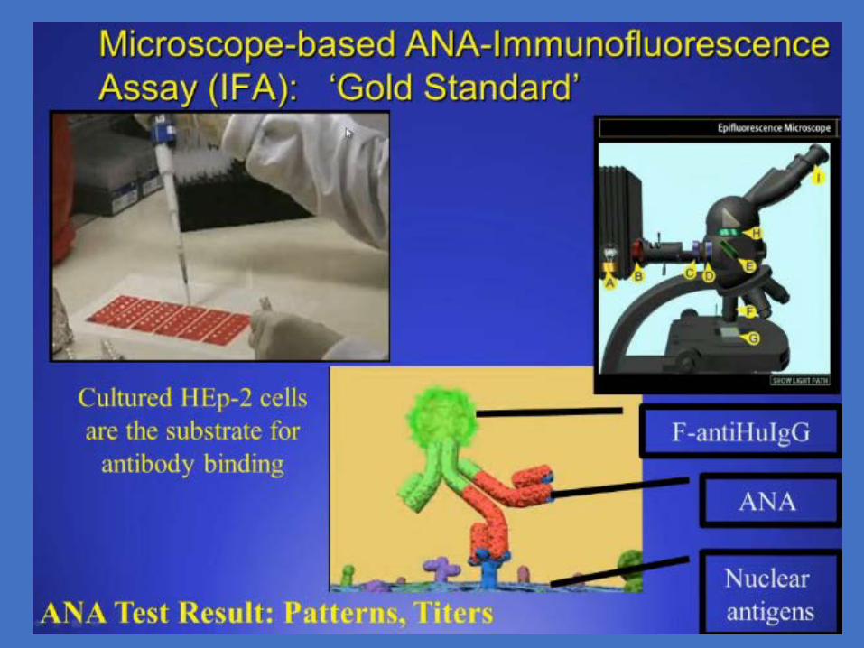
Immunofluorescence microscopy
ELISA
Multiplex assaysBead assays / addressable laser bead assays (‘Flow bead-ometer’)
What Are the Current Methods for ANA Screening?
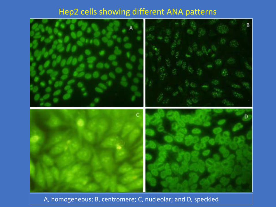
A, homogeneous; B, centromere; C, nucleolar; and D, speckled
Hep2 cells showing different ANA patterns


AutomatedMultiple results from one sampleDifferent manufacturers have similar design,but not identicalUsed to report individual autoAb results, andalso by some labs to replace the IFA-ANA
Multiplex Testing Dominant in Large Labs


How to Handle Variability of Lab Tests for ANAs
Labs differ. “Know your lab”
Worth repeating tests that do not support your clinical impression
Feedback to lab director if questions or problems
Professional interaction between ACR and the College of American Pathologists (CAP) to improve and coordinate


Antinuclear Antibody Testing: A Study of Clinical Utility
The sensitivity of the ANA test for SLE was high, but overall the positive predictive valuewas low for SLE or other rheumatic diseases. Sensitivity was low for ANA testing amongpatients with non—SLE rheumatic disease. More selective test ordering might improve theclinical utility of this test. Clinicians ordering the ANA test should be aware of the test'slow-positive predictive value in settings with a low prevalence of rheumatic disease,particularly among older patients.
Arch Intern Med. 1996;156:1421-1425


Presence of a high titer (>1:640) increases suspicion of anautoimmune disease, but is not diagnostic
Titers can fluctuate– This is not reflective of disease activity, and is not indicated to
follow serially
– Titers that disappear are less clinically significant
For diagnosis of SLE, sensitivity of ~95% and specificity of57%
+ Primary utility diagnostically is the NPV for SLE if ANA isnegative
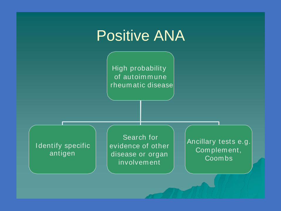
Positive ANA
High probabilityof autoimmune
rheumatic disease
Search forevidence of other disease or organ
involvement
Ancillary tests e.g.Complement,
Coombs
Identify specificantigen
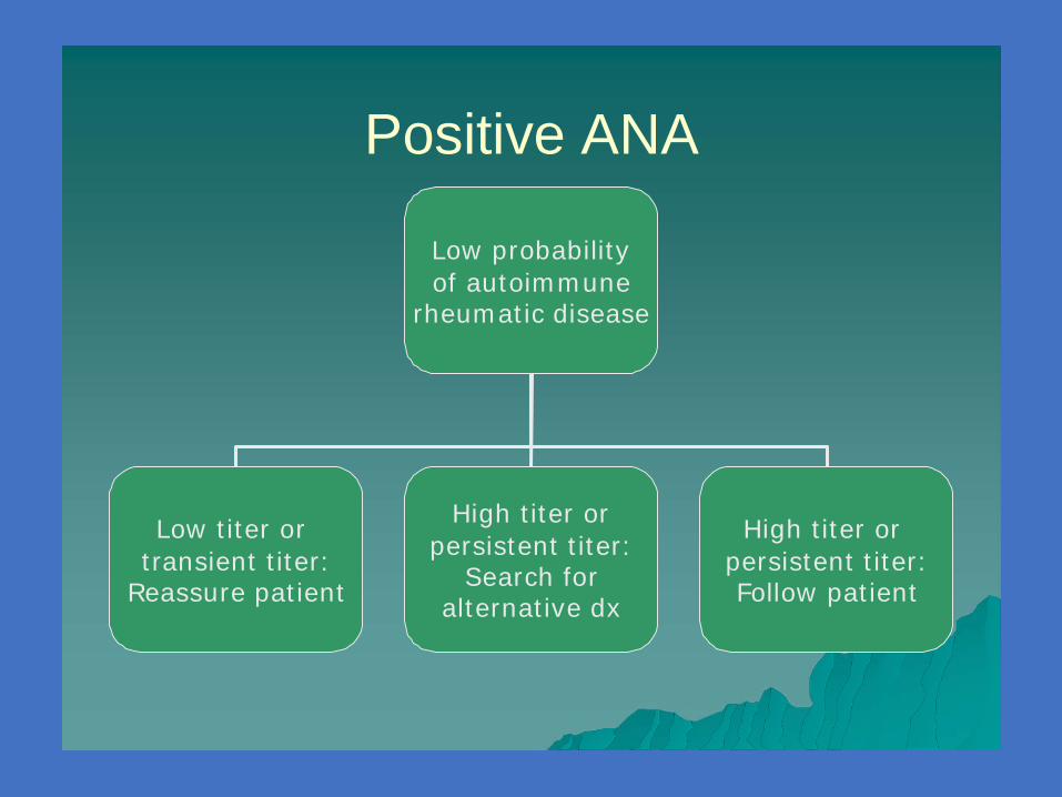
Positive ANA
Low probabilityof autoimmune
rheumatic disease
High titer orpersistent titer:
Search for alternative dx
Low titer ortransient titer:
Reassure patient
High titer orpersistent titer: Follow patient

Algorithm for the use of anti-nuclear antibodies (ANAs) in the diagnosis of connective tissue disorders
Kelley and Firestein's Textbook of Rheumatology, 10th ed. 2017

Autoantibodies Prevalence Comments
Anti-ds-DNA 70% activity of SLE and lupus nephritis. Level monitored in lupus nephritis
Anti-Sm 25% specific for SLE, correlation with disease activity and any particular lupus features is uncertain, alsoseen along with RNP antibody, common in blacks and Asians than whites
Anti-RNP 40% Defining autoantibody in mixed connective-tissue disease, not specific for SLE, Common in blacks than whites
Anti-Ro (SS-A) 30% Sjögrens syndrome, neonatal lupus, Congenitalheart block, photosensitiverash, subacute cutaneous lupus erythematosus, decreased risk of nephritis
Anti-La (SS-B) 10% associated with reduced risk of nephritis, Sjögrenssyndrome, neonatal lupus
Anti Histone antibody 70% SLE, Drug induced SLE
Anti erythrocyte antibody 60 Measured as direct Coombs test, Hemolysis
Anti Ribosomal antibody 20% Depression and Psychosis
Specific auto-antibodies in SLE


Antiphospholipid Antibody Syndrome
Diagnosis:Lupus anticoagulant (LAC) - antibodies that prolong
phospholipid-dependent coagulation reactions e.g.., APTTand/or DRVVT are prolonged and do not correct after equal mixwith normal plasma.
Anti-cardiolipin antibodies (aCL) - detected by ELISA (IgG or IgM or IgA)
Anti-β2-glycoprotein I antibody (Ig G, IgM, IgA)

Antiphospholipid Antibody Syndrome
Both LAC, ACL, AND Anti-β2-glycoprotein I antibodyshould be checked
• Both LA & aCL + in 60-70%• LA +, aCL neg 15-20%• aCL +, LA neg 15-20%
The strongest clinical associations have been seen with IgGaCL. IgM or IgA antibodies may be associated with thesyndrome.A positive aPL test should be confirmed by repeating in 6 to
8 weeks

Measurement of Complement
Disease associated with hypocomplementemia or deficiency of complement component
Monitor disease activity such as SLE
Measured by CH50, C3, C4 CH 50: Functional assay, checks classical pathway, can be false
positive C3 & C4: More accurate, measured by nephelometry Synthesized in liver, can be low in liver diseases Acute phase reactants, can be high in inflammatory states

Hypocomplementemia
• Systemic lupus erythematosus• Vasculitis
• Hypocomplementemic urticarial vasculitis• Polyarteritis nodosa (especially hepatitis B-
associated)• Glomerulonephritis
• Poststreptococcal• Membranoproliferative
• Cryoglobulinemia (types II and III)• Subacute bacterial endocarditis• Serum sickness• Inherited deficiency states

Clinical Syndromes Associated with Deficiencies of Components
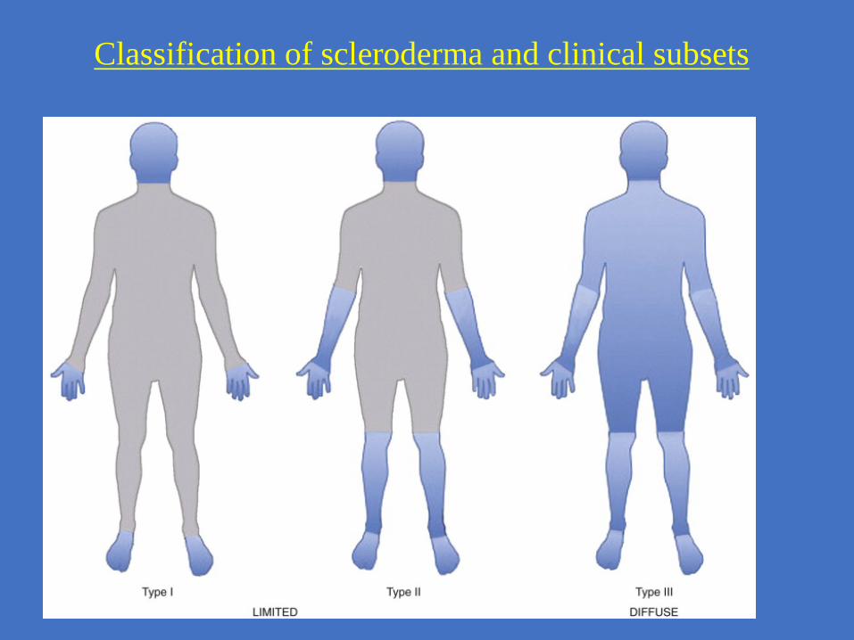
Classification of scleroderma and clinical subsets

Autoantibodies in Systemic Sclerosis
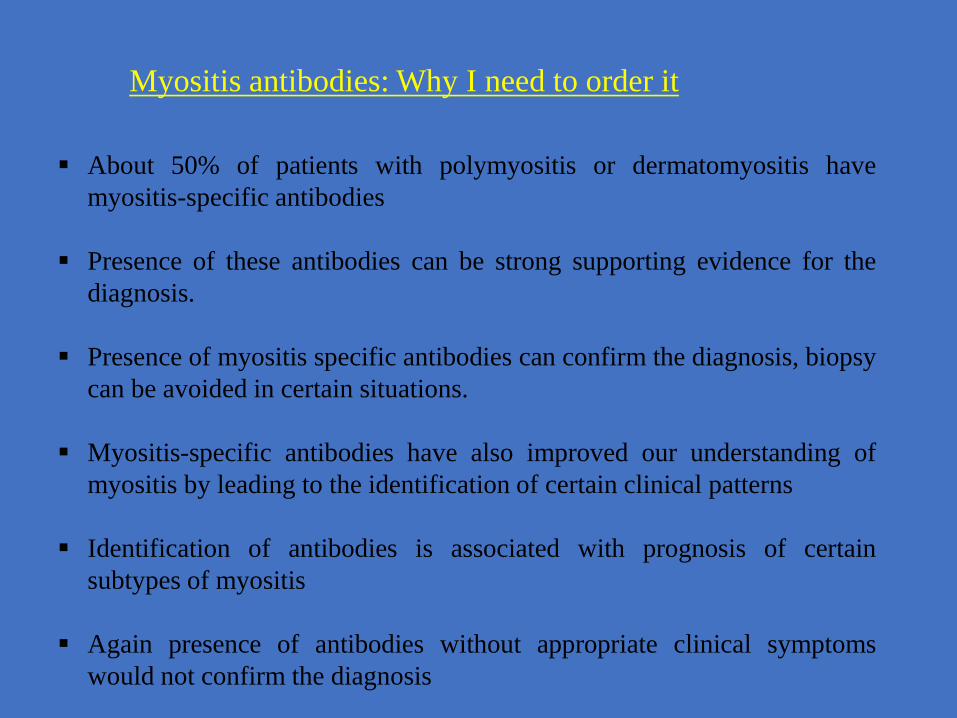
About 50% of patients with polymyositis or dermatomyositis havemyositis-specific antibodies
Presence of these antibodies can be strong supporting evidence for thediagnosis.
Presence of myositis specific antibodies can confirm the diagnosis, biopsycan be avoided in certain situations.
Myositis-specific antibodies have also improved our understanding ofmyositis by leading to the identification of certain clinical patterns
Identification of antibodies is associated with prognosis of certainsubtypes of myositis
Again presence of antibodies without appropriate clinical symptomswould not confirm the diagnosis
Myositis antibodies: Why I need to order it

Myositis Autoantibodies

Myositis Autoantibody Phenotypes Differ inClinical Presentation, Genetics and Prognosis
Anti-aminoacyl-tRNAsynthetases
Anti-SignalRecognition Particle
Anti-Mi-2: chromodomainhelicase DNA binding protein 4
Interstitial lung disease,Arthritis, Fevers,
Mechanic’s hands; DR375% 5-year survival
Classic Dermatomyositis,V-sign & shawl rashes,
Cuticular overgrowth; DR790% 5-year survival
Acute-onset PM, Severeweakness, Myalgias,
Myocarditis; DQA1*010425% 5-year survival 15

MSA/MAAs and clinical associations in adult myositis

Case # 3
30 year old male patient admitted to theRegional One Hospital with renal failureand necrotizing skin lesions.
Urine drug screen is + for cocaine
Bx of the kidney: Pauci-immune GN
The question is do I need to order serology and how it can be helpful??

Frequency of PR3- and MPO-ANCA specificity by clinical phenotypes
Arthritis Rheum. 2012 Oct; 64(10): 3452–3462.

Probability of relapse-free survival by ANCA specificity
All Kidney limited
MPA GPA
Arthritis Rheum. 2012 Oct; 64(10): 3452–3462.


ANCA: Types, Frequency, Target and Disease associations

+ ANCA in Non Vasculitis Diseases
Drugs: Cocaine, Levamisole, PTU, Minocycline
SLE, Felty’s syndrome, Rheumatoid Arthritis
IBD, Sclerosing cholangitis, autoimmune hepatitis
Infections:suppurative lung infections, endocarditis etc

Indirect Immunofluorescence (IIF) testing for ANCAs
Abelson A. Cleveland Clinic Journal of Medicine 2010


ANCA testing algorithm

Diagnostic Performance of Antineutrophil CytoplasmicAntibody Tests for Idiopathic Vasculitides
J Rheumatol 2001;28;1584-1590

Lab testing in RA

Normal labs do not r/o RA
Median ESR at presentation: 30ESR normal in 45%CRP normal in 33%All RF tests negative in 37%37% of patients had ESR < 28, normal CRP, or all negative RF tests
Sokka T, Pincus T. J Rheumatol 2009;36:1387
Observational study of 2370 patients

Rheumatoid FactorRF is an immunoglobulin that binds the Fc portion of another immunoglobulin

Cyclic Citrullinated Peptide Antibody(CCP Ab, ACPA Ab)
Smoking
Post translational modification of proteins

RF and CCP
Sensitivity Specificity
Anti-CCP 77% 97%
RF 74% 78%
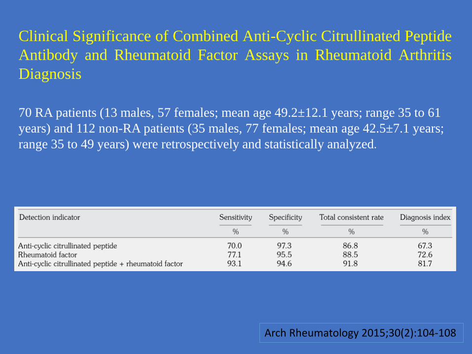
Arch Rheumatology 2015;30(2):104-108
Clinical Significance of Combined Anti-Cyclic Citrullinated PeptideAntibody and Rheumatoid Factor Assays in Rheumatoid ArthritisDiagnosis
70 RA patients (13 males, 57 females; mean age 49.2±12.1 years; range 35 to 61years) and 112 non-RA patients (35 males, 77 females; mean age 42.5±7.1 years;range 35 to 49 years) were retrospectively and statistically analyzed.
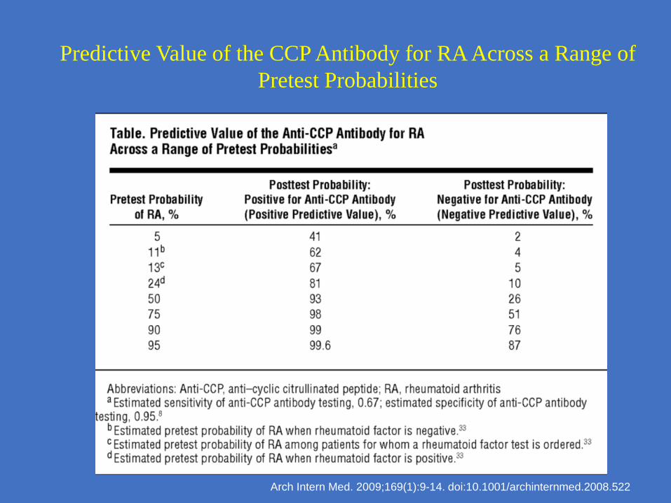
Arch Intern Med. 2009;169(1):9-14. doi:10.1001/archinternmed.2008.522
Predictive Value of the CCP Antibody for RA Across a Range of Pretest Probabilities

Rheumatoid+ Assists in diagnosis
Factor
– In a patient with suggestive findings(symmetric polyarthritis), presence increases the certainty of diagnosis, if other causes excluded
+ Assists in prognosis– High titer increases the progression to erosive
arthritis+ Assists in treatment decisions
– Warrants early DMARD use

Clinical associations of RF+ Rheumatoid arthritis (75-80%)+ Other rheumatic disease
–––––
Sjogren’s syndrome (~90%)SLE (15-20%) Sarcoidosis (~15%)Parvovirus arthropathy (~15%, Mixed cryoglobulinemia (95%)
transient)
+ Chronic infections–––
Chronic Hep COsteomyelitisBacterial endocarditis
+ Monoclonal IgM paraproteins+ Normal aging (present at low titer)

Specific Autoantibodies Precede the Symptoms of RA: A Study of Serial Measurements in Blood Donors
Arthritis & Rheumatism 2004; 380-386, 5 FEB 2004

Rheumatoid Factor (RF) 50% of patients with RA become positive for RF in first 6 months
85% become positive over the first 2 years, repeat testing advised.
Current detection methods cannot differentiate between naturally occurring,transiently induced, and RA-associated rheumatoid factor.
The levels are generally higher in RA than in many non-RA disorders, butsignificant overlap occurs
10% to 15% of RA patients remain seronegative for rheumatoid factorthroughout the disease course.
The level does not vary with the activity of RA. So serial testing is not advised
Seropositive (+ RF & CCP Ab), better response to Rituximab Therapy

ACPAs are present in nearly 20% of unaffected first-degree relatives and more than 10% of more distant relatives of RA patients.
ACPAs are also produced by synovial tissue B cells and can be detected in synovial fluid.
ACPAs are predictors of more aggressive disease marked by bone and cartilage destruction.
Accelerated atherosclerosis in RA, independent risk factor for ischemic heart disease.
In patients with early undifferentiated inflammatory arthritis, ACPAs are also predictive for individuals who will progress to RA.
CCP antibody (ACPA)
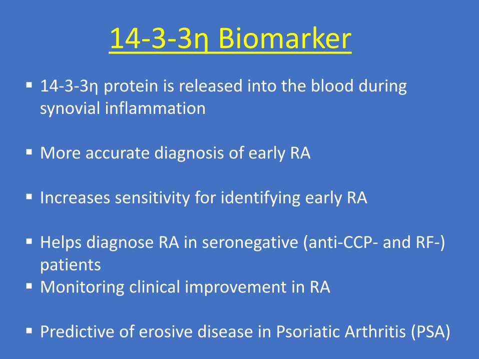
14-3-3η Biomarker 14-3-3η protein is released into the blood during
synovial inflammation
More accurate diagnosis of early RA
Increases sensitivity for identifying early RA
Helps diagnose RA in seronegative (anti-CCP- and RF-) patients Monitoring clinical improvement in RA
Predictive of erosive disease in Psoriatic Arthritis (PSA)

Conclusion
Musculoskeletal complaints are common in the general population
Prevalence of inflammatory rheumatic diseases are low
Serologic tests are not often highly diagnostic (not gold standard)
Positive predictive value of many rheumatologic tests is low when tests these are ordered indiscriminately