Embed Size (px)
Citation preview
Role of pro-inflammatory S100A9 protein in amyloid-neuroinflammatory cascade in Alzheimer’s disease and traumatic brain injury Chao Wang
Medical Biochemistry and Biophysics Umeå 2016

Responsible publisher under swedish law: the Dean of the Medical Faculty This work is protected by the Swedish Copyright Legislation (Act 1960:729) ISBN: 978-91-7601-547-6 ISSN: 0346-6612 New series nr 1838 Cover picture: Chao Wang designed for ADAM5 conference: Elektronisk version available at http://umu.diva-portal.org/ Printed by: VMC-KBC, Umeå University Umeå, Sweden 2016

致我的家人 To my family
ii
Table of Contents
ABSTRACT iii
LIST OF PAPERS v
ABBREVIATIONS vii
INTRODUCTION 1 1. Protein folding and misfolding 1 1.1. Proteins and structure 1 1.2. Protein folding, misfolding and amyloid formation 2 1.3. Kinetics of amyloid formation 3 2. Amyloid related diseases 4 2.1. General 4 2.2. AD 6 2.3. Traumatic brain injury (TBI) 7 2.4. TBI as a risk factor for AD 8 2.5. S100A9 protein 9 3. Methods used to study amyloids 11 3.1. Atomic force microscopy (AFM) 11 3.2. Amyloid specific molecular probes 12 3.2.1. Heptameric formic thiophene acetic acid (h-FTAA) 12 3.2.2. Antibody detection 13 3.2.2.1. Sequential immunohistochemistry 14
RESULTS AND DISCUSSION 16
CONCLUSIONS AND PERSPECTIVES 22
ACKNOWLEDGEMENTS 23
REFERENCES 26
iii
ABSTRACT
Background Traumatic brain injury (TBI) is a complex disease with a
spectrum of symptoms and disabilities. Over the past decade TBI has become
the focus of research due to growing epidemiological and clinical evidences
that TBI incidences are strong risk factors for Alzheimer’s disease (AD). Major
pathological hallmarks of AD are massive accumulations of amyloid-β peptide
(Aβ) toxic oligomers and plaques. Neuroinflammation is also considered as a
common denominator in AD and aging. The epidemiological and
experimental studies have supported that non-steroidal anti-inflammatory
drugs markedly reduce the age-related prevalence of AD and can slow amyloid
deposition by mechanisms that still remain elusive. S100A9 is a
multifunctional cytokine with diverse roles in the cell signaling pathways
associated with inflammation and cancers. A widespread expression of
S100A9 was also reported in many other ailments involving inflammatory
processes, such as AD, malaria, cerebral ischemia and TBI, implying that
S100A9 may be a universal biomarker of inflammation. The distinctive feature
of S100A9 compared to other pro-inflammatory cytokines is its ability to self-
assemble into amyloids, which may lead to the loss of its signaling functions
and acquired amyloid cytotoxicity, exceeding that of Aβ.
Methods S100A9 properties was studied under various ex vivo and in vitro
conditions. First, human and mouse tissues with TBI and AD were subjected
to microscopic, immunohistochemical and immunofluorescent techniques.
Then, aged mouse treated with native, oligomeric and fibrillary S100A9 was
also studied by using behavioral and neurochemical analysis. Moreover,
S100A9 was established as a biomarker of dementia progression and
compared with others such as Aβ42 and tau proteins, by studying
cerebrospinal fluid (CSF) samples from different stages of dementia. Finally,
in vitro experiments on S100A9 amyloidogenesis, co-aggregation with Aβ40
and Aβ42, digestion and cytotoxicity were also performed by using
spectroscopic, atomic force microscopy and cell biology methods.
iv
Results S100A9-driven amyloid-neuroinflammatory cascade serves as a link
between TBI and AD. We have found that S100A9 contributes to the plaque
formation and intraneuronal responses in AD, being a part of the amyloid-
neuroinflammatory cascade. In TBI we have found that extensive S100A9
neuronal production and amyloid self-assembly is triggered immediately after
injury, leading to apoptotic pathways and neuronal loss. S100A9 is an integral
component of both TBI precursor-plaques, formed prior to Aβ deposition, and
AD plaques, characterized by different degree of amyloid maturation,
indicating that all plaques are associated with inflammation. Both intra- and
extracellular amyloid-neuroinflammatory cascades are intertwined and
showed similar tendencies in human and mouse tissues in TBI and AD. Ex
vivo findings are further supported by in vitro experiments on S100A9
amyloidogenesis, digestion and cytotoxicity. Importantly, being highly
amyloidogenic itself, S100A9 can trigger and aggravate Aβ amyloid self-
assembly and significantly contribute to amyloid cytotoxicity. Moreover, the
CSF dynamics of S100A9 levels matches very closely the content of Aβ42 in AD,
vascular dementia and mild cognitive impairment due to AD, emphasizing the
involvement of S100A9 together with Aβ in the amyloid-neuroinflammatory
cascade in these ailments.
Conclusions The conclusions of this thesis is that the inflammatory
pathways and S100A9 specifically represent a potential target for the
therapeutic interventions during various post-TBI stages and far prior AD
development to halt and reverse these damaging processes.
v
LIST OF PAPERS
This thesis is based on the following articles
I. Wang, C., Klechikov, A.G., Gharibyan, A.L., Warmlander, S.K.,
Jarvet, J., Zhao, L., Jia, X., Narayana, V.K., Shankar, S.K., Olofsson,
A., et al. (2014). The role of pro-inflammatory S100A9 in Alzheimer's
disease amyloid-neuroinflammatory cascade. Acta neuropathologica
127, 507-522.
II. Gruden, M.A., Davydova, T.V., Wang, C., Narkevich, V.B., Fomina,
V.G., Kudrin, V.S., Morozova-Roche, L.A., and Sewell, R.D. (2016).
The misfolded pro-inflammatory protein S100A9 disrupts memory
via neurochemical remodelling instigating an Alzheimer's disease-
like cognitive deficit. Behavioural brain research 306, 106-116.
III. Horvath, I., Jia, X., Johansson, P., Wang, C., Moskalenko, R.,
Steinau, A., Forsgren, L., Wagberg, T., Svensson, J., Zetterberg, H., et
al. (2016). Pro-inflammatory S100A9 Protein as a Robust Biomarker
Differentiating Early Stages of Cognitive Impairment in Alzheimer's
Disease. ACS chemical neuroscience 7, 34-39.
IV. Wang, C., Iashchishyn, A.I., Nyström, S., Klementieva, O., Kara, J.,
Bengtsson, K.S.S., Foderà, V., Vetri, V., Sancataldo, G., Horvath, I.,
et al. S100A9-driven amyloid-neuroinflammatory cascade in
traumatic brain injury as a risk factor for Alzheimer’s disease.
Manuscript.
Contributions to the following articles, not included in the thesis,
were also made
I. Gruden, M.A., Davydova, T.V., Narkevich, V.B., Fomina, V.G., Wang,
C., Kudrin, V.S., Morozova-Roche, L.A., and Sewell, R.D. (2015).
Noradrenergic and serotonergic neurochemistry arising from
vi
intranasal inoculation with alpha-synuclein aggregates which incite
parkinsonian-like symptoms. Behavioural brain research 279, 191-
201.
II Gruden, M.A., Davydova, T.V., Narkevich, V.B., Fomina, V.G., Wang,
C., Kudrin, V.S., Morozova-Roche, L.A., and Sewell, R.D. (2014).
Intranasal administration of alpha-synuclein aggregates: a
Parkinson's disease model with behavioral and neurochemical
correlates. Behavioural brain research 263, 158-168.
vii
ABBREVIATIONS
Aβ amyloid-β AD Alzheimer's disease AFM atomic force microscopy CSF cerebrospinal fluid h-FTAA heptameric formic thiophene acetic acid H-tau total human tau MCI-AD mild cognitive impairment due to Alzheimer's disease NFTs neuronal neurofibrillary tangles P-tau tau phosphorylated at Thr181 SMCI stable mild cognitive impairment TBI traumatic brain injury ThT Thioflavin T VaD vascular dementia

1
INTRODUCTION
1. Protein folding and misfolding
1.1. Protein and structure
Proteins are molecules in the living organisms and perform multiple
functions, including facilitating biochemical reactions, transmitting signals,
transporting molecules, keeping the cell structures and storing amino acids.
In order to fulfil their functions, suitable folding status is very important,
which is dependent both on the surrounding environments where the protein
is produced and also on the sequences of amino acids in the polypeptide chain
(Anfinsen, 1973).
Essentially, 20 different amino acids make up all proteins on earth. By
changing the permutations and combinations of these amino acids in the
polypeptide chain, all the existing proteins can be produced. The spontaneous
transition from a disordered polypeptide chain to a functional protein with a
unique three-dimensional structure is defined as protein folding.
Proteins have four distinct levels of structure. The primary structure refers to
the linear sequence of amino acids in the polypeptide chain, being held by the
covalent bonds and determined by the specific gene sequences of the proteins;
the secondary structure refers to two main stable secondary structure
elements: α helix and β sheet. They are distinguished by patterns of hydrogen
bonds between the main-chain peptide groups. Meanwhile, some units do not
have a stable secondary structure. It was defined as random coil; those
secondary structures are further folded into a protein tertiary structure
(protein domain) by special tertiary interactions, such as hydrogen bonds,
hydrophobic interactions, Van der Waals interactions, salt bridges and
disulfide bonds; and finally, when several protein domains make up into a
complex, the quaternary structure is formed.
2
1.2. Protein folding, misfolding and amyloid formation
There are quite many molecular chaperones, folding catalysts involving to
control the correct protein folding. Alternatively complex degradation
pathways are involved to clean the misfolded and unfolded proteins. The
ability of a protein to maintain its corrected and functional folding status is
essential for its biological activity, while the unfolded and misfolded proteins
will lose their biological functions. In some cases, these misfolded proteins
may escape from the degradation system and further form into aggregates.
Amyloid is one of the specific types of aggregates.
Amyloid or amyloid fibrils are distinguished by cross-β-sheet structures which
are stabilized by hydrogen-bond interactions between groups in the
polypeptide backbone as well as the characteristic tinctorial properties. Diazo
dye ‒ Congo red will give a characteristic apple green birefringence under the
polarized light when it binds with amyloid fibrils (Klunk et al., 1989;
Steensma, 2001); the fluorescence intensity of the benzothiazole dye ‒
Thioflavin T (ThT) will increase significantly when it interacts with amyloid
fibrils (LeVine, 1993, 1999; Vassar and Culling, 1959).
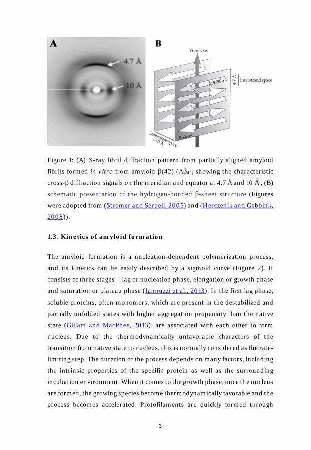
The amyloid cross-β-sheet structures have a distinctive X-ray diffraction
pattern which contains two predominate reflections. There are at 4.7 Å and 10
Å reflections, corresponding to the distance between the constituent β-strands
and the space between two layers of β-sheets, respectively (Aoki et al., 1978;
Eanes and Glenner, 1968; Glenner, 1980; Herczenik and Gebbink, 2008;
Stromer and Serpell, 2005) (Figure 1).
3
Figure 1: (A) X-ray fibril diffraction pattern from partially aligned amyloid
fibrils formed in vitro from amyloid-β(42) (Aβ42) showing the characteristic
cross-β diffraction signals on the meridian and equator at 4.7 Å and 10 Å , (B)
schematic presentation of the hydrogen-bonded β-sheet structure (Figures
were adopted from (Stromer and Serpell, 2005) and (Herczenik and Gebbink,
2008)).
1.3. Kinetics of amyloid formation
The amyloid formation is a nucleation-dependent polymerization process,
and its kinetics can be easily described by a sigmoid curve (Figure 2). It
consists of three stages – lag or nucleation phase, elongation or growth phase
and saturation or plateau phase (Iannuzzi et al., 2013). In the first lag phase,
soluble proteins, often monomers, which are present in the destabilized and
partially unfolded states with higher aggregation propensity than the native
state (Gillam and MacPhee, 2013), are associated with each other to form
nucleus. Due to the thermodynamically unfavorable characters of the
transition from native state to nucleus, this is normally considered as the rate-
limiting step. The duration of the process depends on many factors, including
the intrinsic properties of the specific protein as well as the surrounding
incubation environment. When it comes to the growth phase, once the nucleus
are formed, the growing species become thermodynamically favorable and the
process becomes accelerated. Protofilaments are quickly formed through

4
monomers or oligomers joining to the nuclei. Finally, during the last
saturation phase, amount of mature fibrils reach the highest level (Morozova-
Roche et al., 2000). The whole amyloid conversion from soluble peptide or
protein into mature fibrillar can be visualized by using different amyloid
specific florescence dyes, such as Congo red and ThT.
Figure 2: The kinetics of amyloid formation (Figure was adopted from
(Iannuzzi et al., 2013)).
2. Amyloid related diseases
2.1. General
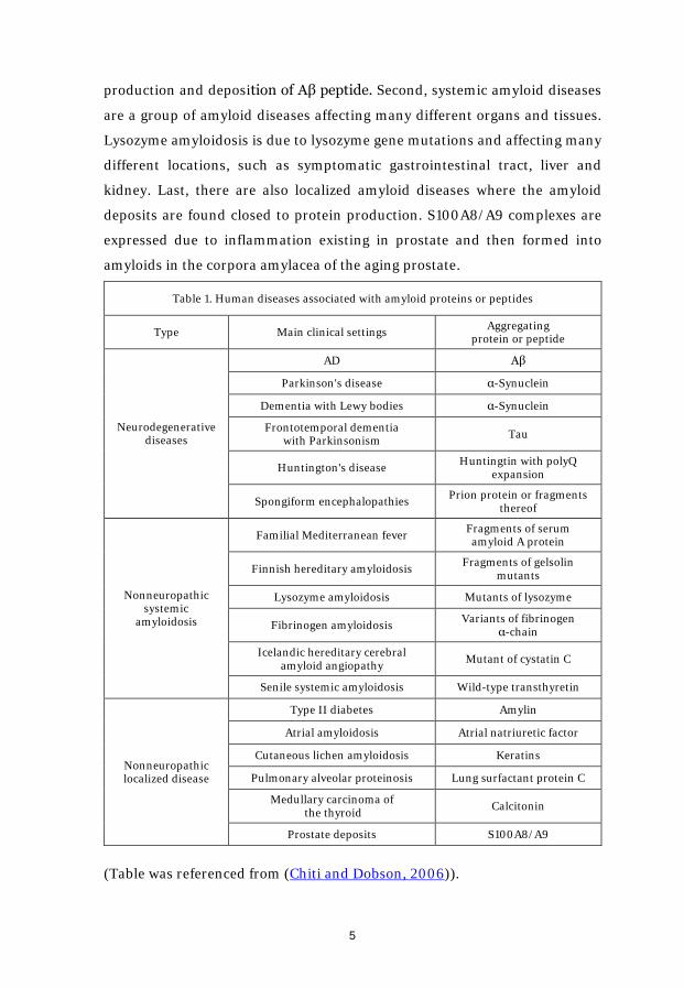
To date, large number of proteins and peptides are known to have ability to
form amyloids, causing multiple types of diseases in humans (See Table 1).
The localization of the amyloid deposits and protein/peptide involved are
largely determining the specific disease. They can be broadly distributed into
three clusters. First, neurodegenerative diseases are the most well-known
amyloid diseases, happening in the center nerve system. Alzheimer’s disease
(AD) is one of the common neurodegenerative diseases, driven by the
5
production and deposition of Aβ peptide. Second, systemic amyloid diseases
are a group of amyloid diseases affecting many different organs and tissues.
Lysozyme amyloidosis is due to lysozyme gene mutations and affecting many
different locations, such as symptomatic gastrointestinal tract, liver and
kidney. Last, there are also localized amyloid diseases where the amyloid
deposits are found closed to protein production. S100A8/A9 complexes are
expressed due to inflammation existing in prostate and then formed into
amyloids in the corpora amylacea of the aging prostate.
Table 1. Human diseases associated with amyloid proteins or peptides
Type Main clinical settings Aggregating protein or peptide
Neurodegenerative diseases
AD Aβ
Parkinson's disease α-Synuclein
Dementia with Lewy bodies α-Synuclein
Frontotemporal dementia with Parkinsonism Tau
Huntington's disease Huntingtin with polyQ expansion
Spongiform encephalopathies Prion protein or fragments thereof
Nonneuropathic systemic
amyloidosis
Familial Mediterranean fever Fragments of serum amyloid A protein
Finnish hereditary amyloidosis Fragments of gelsolin mutants
Lysozyme amyloidosis Mutants of lysozyme
Fibrinogen amyloidosis Variants of fibrinogen α-chain
Icelandic hereditary cerebral amyloid angiopathy Mutant of cystatin C
Senile systemic amyloidosis Wild-type transthyretin
Nonneuropathic localized disease
Type II diabetes Amylin
Atrial amyloidosis Atrial natriuretic factor
Cutaneous lichen amyloidosis Keratins
Pulmonary alveolar proteinosis Lung surfactant protein C
Medullary carcinoma of the thyroid Calcitonin
Prostate deposits S100A8/A9
(Table was referenced from (Chiti and Dobson, 2006)).
6
2.2. AD
AD is a progressive neurodegenerative disorder and the most common form
of dementia. The disease destroys the memory, language ability, causes the
difficulties with the activities and has a fetal consequences (Burns and Iliffe,
2009). According to the 2015 dementia report from the World Health
Organization, 47.5 million people have dementia and Alzheimer’s disease may
take up 60-70% of the cases.
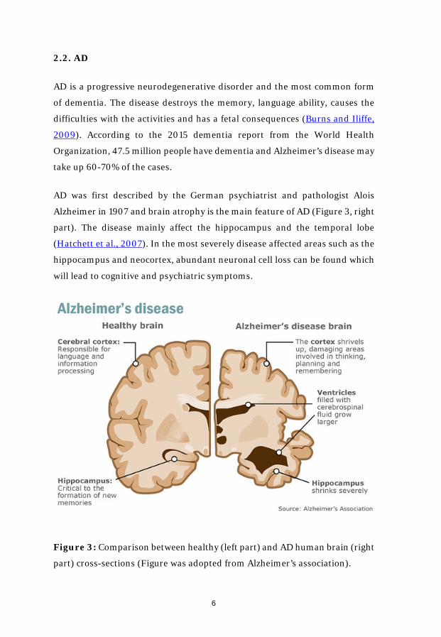
AD was first described by the German psychiatrist and pathologist Alois
Alzheimer in 1907 and brain atrophy is the main feature of AD (Figure 3, right
part). The disease mainly affect the hippocampus and the temporal lobe
(Hatchett et al., 2007). In the most severely disease affected areas such as the
hippocampus and neocortex, abundant neuronal cell loss can be found which
will lead to cognitive and psychiatric symptoms.
Figure 3: Comparison between healthy (left part) and AD human brain (right
part) cross-sections (Figure was adopted from Alzheimer’s association).
7
There are two well-known pathologic features of AD which are amyloid
plaques in the extracellular space as well as the intraneuronal neurofibrillary
tangles (NFTs). The major component of the plaques – Aβ peptide was
identified almost 30 years ago in 1984 (Glenner and Wong, 2012); meanwhile,
2 years later, the hyperphosphorylated tau was determined as the major
constituent of the NFTs (Grundke-Iqbal et al., 1986). It has been more than
one century passed since Dr. Alois Alzheimer described AD. However, there
are still many unknowns in front of us. No efficient therapy method was
developed to date and also no early diagnostics was established since the
pathological changes such as neuronal degeneration and profound
neuroinflammation have occurred 10 to 20 or even more years before the AD
symptoms are shown up (Holtzman et al., 2012).
2.3. Traumatic brain injury (TBI)
The term of TBI refers to “an insult to the brain caused by an external force
that may produce diminished or altered states of consciousness, which results
in impaired cognitive abilities or physical functioning” as given by National
Head Injury Foundation in 1988. From being a neglected disorder, over the
past decade TBI has become the focus of increasing attention due to frequent
injury incidents in modern society and sports. There are approximately
300,000 U.S. soldiers who have suffered TBI in Iraq and Afghanistan (Miller,
2012). The incidence rates of TBI are about 100/100,000 and 235/100,000 in
the USA and in Europe, respectively (Maas et al., 2008; Roozenbeek et al.,
2013). The real incidence rate should be even higher due to quite many
unrecorded mild TBI cases.
Importantly, TBI is a long-lasting pathological process, not only an simple
short event (Masel and DeWitt, 2010). As many as 15% of people with mild
TBI will live with the post-TBI symptoms lasting for one year or even more. It
initiates a chronic disease process by an initial injury event causing the
biochemical and cellular changes which in turn lead to chronic inflammatory
situation in the central nerve system or neuroinflammation (Morales et al.,
8
2014; Sivanandam and Thakur, 2012; Streit et al., 2004). Despite extensive
efforts to develop short and long-term neuroprotective strategies, these are
not yet satisfactory and a better understanding of underlying pathologies is
required to focus on the specific therapeutic targets.
2.4. TBI is a risk factor for AD
The amyloid cascade hypothesis considers the deposition of the Aβ peptides
as a central event of AD pathology. However, the reason of Aβ aberrant
accumulation in the central nerve system in the old age still remain
mysterious. Neuroinflammation, which is always connected with TBI, can
potentially serve as the cues and thus attracted increasing attentions
nowadays. More and more evidences show that in AD neuroinflammation is
not passive event activated by amyloid plaques or NFTs, but instead in some
extent, it promotes the pathological processes (Heneka et al., 2015).
Epidemiological studies found that long term use of non-steroidal anti-
inflammatory drugs in patients with rheumatoid arthritis produced a
protective effect against the development of the disease, delaying the onset of
the symptoms and reducing the risk of the disease occurrence (McGeer et al.,
1990). This is also supported by observations that important microglial-
expressed receptor genes, such as TREM2 and CD33 (Heneka et al., 2015;
Ulrich and Holtzman, 2016), act as strong factors promoting AD.
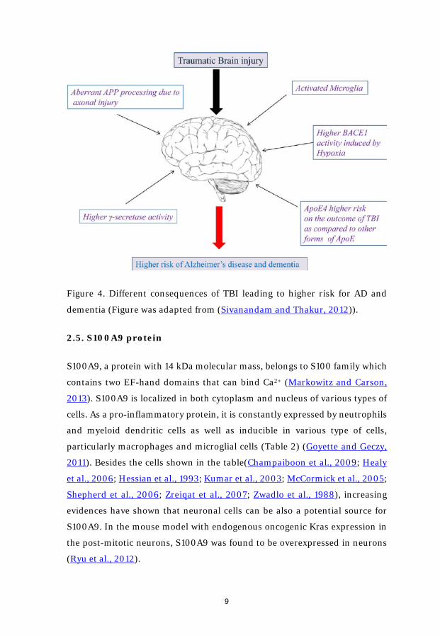
Epidemiologically TBI is a strong risk factor for AD (Figure 4) (Fleminger et
al., 2003; Magnoni and Brody, 2010). By examining the brain tissues from the
patients with the post-TBI survival times varying from 1 to 47 years, nearly
30% of them were found to possess Aβ plaques. Moreover, Aβ deposits can be
found in the TBI patient brain as early as few hours after the trauma
(Ikonomovic et al., 2004). After inducing trauma in the AD transgenic mice
that develop amyloid Aβ plaques, the accumulation of the plaques was
tremendously enhanced (Abrahamson et al., 2009; Breunig et al., 2013;
Hartman et al., 2002; Smith et al., 1998; Uryu et al., 2002).
9
Figure 4. Different consequences of TBI leading to higher risk for AD and
dementia (Figure was adapted from (Sivanandam and Thakur, 2012)).
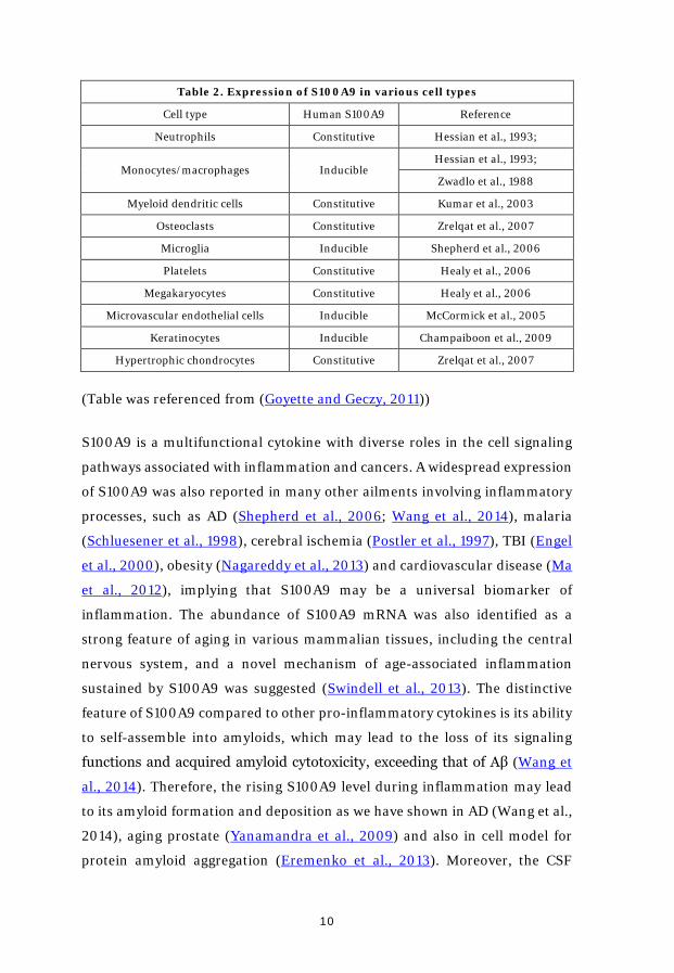
2.5. S100A9 protein
S100A9, a protein with 14 kDa molecular mass, belongs to S100 family which
contains two EF-hand domains that can bind Ca2+ (Markowitz and Carson,
2013). S100A9 is localized in both cytoplasm and nucleus of various types of
cells. As a pro-inflammatory protein, it is constantly expressed by neutrophils
and myeloid dendritic cells as well as inducible in various type of cells,
particularly macrophages and microglial cells (Table 2) (Goyette and Geczy,
2011). Besides the cells shown in the table(Champaiboon et al., 2009; Healy
et al., 2006; Hessian et al., 1993; Kumar et al., 2003; McCormick et al., 2005;
Shepherd et al., 2006; Zreiqat et al., 2007; Zwadlo et al., 1988), increasing
evidences have shown that neuronal cells can be also a potential source for
S100A9. In the mouse model with endogenous oncogenic Kras expression in
the post-mitotic neurons, S100A9 was found to be overexpressed in neurons
(Ryu et al., 2012).
10
Table 2. Expression of S100A9 in various cell types
Cell type Human S100A9 Reference
Neutrophils Constitutive Hessian et al., 1993;
Monocytes/macrophages Inducible Hessian et al., 1993;
Zwadlo et al., 1988
Myeloid dendritic cells Constitutive Kumar et al., 2003
Osteoclasts Constitutive Zrelqat et al., 2007
Microglia Inducible Shepherd et al., 2006
Platelets Constitutive Healy et al., 2006
Megakaryocytes Constitutive Healy et al., 2006
Microvascular endothelial cells Inducible McCormick et al., 2005
Keratinocytes Inducible Champaiboon et al., 2009
Hypertrophic chondrocytes Constitutive Zrelqat et al., 2007
(Table was referenced from (Goyette and Geczy, 2011))
S100A9 is a multifunctional cytokine with diverse roles in the cell signaling
pathways associated with inflammation and cancers. A widespread expression
of S100A9 was also reported in many other ailments involving inflammatory
processes, such as AD (Shepherd et al., 2006; Wang et al., 2014), malaria
(Schluesener et al., 1998), cerebral ischemia (Postler et al., 1997), TBI (Engel
et al., 2000), obesity (Nagareddy et al., 2013) and cardiovascular disease (Ma
et al., 2012), implying that S100A9 may be a universal biomarker of
inflammation. The abundance of S100A9 mRNA was also identified as a
strong feature of aging in various mammalian tissues, including the central
nervous system, and a novel mechanism of age-associated inflammation
sustained by S100A9 was suggested (Swindell et al., 2013). The distinctive
feature of S100A9 compared to other pro-inflammatory cytokines is its ability
to self-assemble into amyloids, which may lead to the loss of its signaling
functions and acquired amyloid cytotoxicity, exceeding that of Aβ (Wang et
al., 2014). Therefore, the rising S100A9 level during inflammation may lead
to its amyloid formation and deposition as we have shown in AD (Wang et al.,
2014), aging prostate (Yanamandra et al., 2009) and also in cell model for
protein amyloid aggregation (Eremenko et al., 2013). Moreover, the CSF
11
dynamics of S100A9 levels matches very closely the content of Aβ in AD,
vascular dementia and mild cognitive impairment (Horvath et al., 2016),
emphasizing the involvement of S100A9 together with Aβ in the amyloid-
neuroinflammatory cascade in these ailments. We suggested that S100A9 in
combination with Aβ and tau proteins can be used as a robust biomarker
differentiating early stages of cognitive impairment in AD (Horvath et al.,
2016). Interestingly, S100A9 knockdown attenuated memory impairment and
reduced amyloid plaque burden in an AD mouse model (Ha et al., 2010),
suggesting that S100A9 can be a prospective target for therapeutic
interventions.
3. Methods used to study amyloids
The investigation of amyloid species with extraordinary speed, high accuracy
and sensitivity is of great importance. Multiple techniques are developed and
widely used in our lab.
3.1. Atomic force microscopy (AFM)
Nowadays, AFM scanning is a well-developed and widely used technique to
visualize and even characterize the amyloid structures. Comparing with
scanning electron microscopy, both of them can provide a nanometer
resolution in the structure analysis. However, AFM does not required extra
pre-chemical treatment for the soft biological samples, it has ability to
characterize the physical properties of the structures, such as stiffness,
elasticity and adhesion. It can even work efficiently in the liquid conditions.
Thus, AFM scanning become more and more popular. In our lab, besides the
observation of amyloid structures on the surface of the mica (Figure 5A and
B), we have also developed the method to scan the human and mouse brain
tissues on the common glass slides which can provide higher resolution of
pathological structures such as precursor-plaques in the human TBI brain
tissues (Figure 5C and D).
12
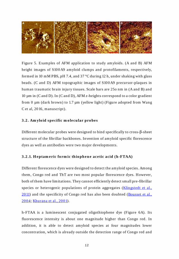
Figure 5. Examples of AFM application to study amyloids. (A and B) AFM
height images of S100A9 amyloid clumps and protofilaments, respectively,
formed in 10 mM PBS, pH 7.4, and 37 °C during 12 h, under shaking with glass
beads. (C and D) AFM topographic images of S100A9 precursor-plaques in
human traumatic brain injury tissues. Scale bars are 25o nm in (A and B) and
10 µm in (C and D). In (C and D), AFM z-heights correspond to a color gradient
from 0 µm (dark brown) to 1.7 µm (yellow light) (Figure adopted from Wang
C et al, 2016, manuscript).
3.2. Amyloid specific molecular probes
Different molecular probes were designed to bind specifically to cross-β-sheet
structure of the fibrillar backbones. Invention of amyloid specific florescence
dyes as well as antibodies were two major developments.
3.2.1. Heptameric formic thiophene acetic acid (h-FTAA)
Different florescence dyes were designed to detect the amyloid species. Among
them, Congo red and ThT are two most popular florescence dyes. However,
both of them have limitations. They cannot efficiently detect small pre-fibrillar
species or heterogenic populations of protein aggregates (Klingstedt et al.,
2011) and the specificity of Congo red has also been doubted (Bousset et al.,
2004; Khurana et al., 2001).
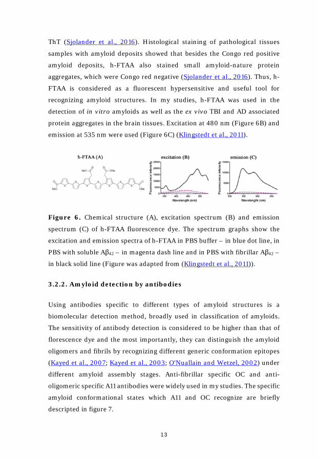
h-FTAA is a luminescent conjugated oligothiophene dye (Figure 6A). Its
fluorescence intensity is about one magnitude higher than Congo red. In
addition, it is able to detect amyloid species at four magnitudes lower
concentration, which is already outside the detection range of Congo red and
13
ThT (Sjolander et al., 2016). Histological staining of pathological tissues
samples with amyloid deposits showed that besides the Congo red positive
amyloid deposits, h-FTAA also stained small amyloid-nature protein
aggregates, which were Congo red negative (Sjolander et al., 2016). Thus, h-
FTAA is considered as a fluorescent hypersensitive and useful tool for
recognizing amyloid structures. In my studies, h-FTAA was used in the
detection of in vitro amyloids as well as the ex vivo TBI and AD associated
protein aggregates in the brain tissues. Excitation at 480 nm (Figure 6B) and
emission at 535 nm were used (Figure 6C) (Klingstedt et al., 2011).
Figure 6. Chemical structure (A), excitation spectrum (B) and emission
spectrum (C) of h-FTAA fluorescence dye. The spectrum graphs show the
excitation and emission spectra of h-FTAA in PBS buffer – in blue dot line, in
PBS with soluble Aβ42 – in magenta dash line and in PBS with fibrillar Aβ42 –
in black solid line (Figure was adapted from (Klingstedt et al., 2011)).
3.2.2. Amyloid detection by antibodies
Using antibodies specific to different types of amyloid structures is a
biomolecular detection method, broadly used in classification of amyloids.
The sensitivity of antibody detection is considered to be higher than that of
florescence dye and the most importantly, they can distinguish the amyloid
oligomers and fibrils by recognizing different generic conformation epitopes
(Kayed et al., 2007; Kayed et al., 2003; O'Nuallain and Wetzel, 2002) under
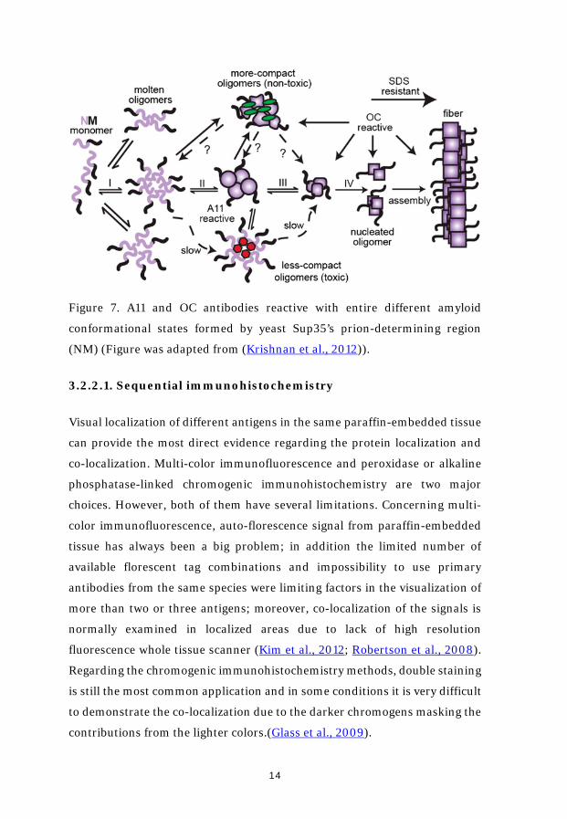
different amyloid assembly stages. Anti-fibrillar specific OC and anti-
oligomeric specific A11 antibodies were widely used in my studies. The specific
amyloid conformational states which A11 and OC recognize are briefly
descripted in figure 7.
14
Figure 7. A11 and OC antibodies reactive with entire different amyloid
conformational states formed by yeast Sup35’s prion-determining region
(NM) (Figure was adapted from (Krishnan et al., 2012)).
3.2.2.1. Sequential immunohistochemistry
Visual localization of different antigens in the same paraffin-embedded tissue
can provide the most direct evidence regarding the protein localization and
co-localization. Multi-color immunofluorescence and peroxidase or alkaline
phosphatase-linked chromogenic immunohistochemistry are two major
choices. However, both of them have several limitations. Concerning multi-
color immunofluorescence, auto-florescence signal from paraffin-embedded
tissue has always been a big problem; in addition the limited number of
available florescent tag combinations and impossibility to use primary
antibodies from the same species were limiting factors in the visualization of
more than two or three antigens; moreover, co-localization of the signals is
normally examined in localized areas due to lack of high resolution
fluorescence whole tissue scanner (Kim et al., 2012; Robertson et al., 2008).
Regarding the chromogenic immunohistochemistry methods, double staining
is still the most common application and in some conditions it is very difficult
to demonstrate the co-localization due to the darker chromogens masking the
contributions from the lighter colors.(Glass et al., 2009).
15
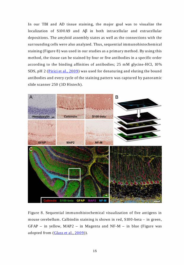
In our TBI and AD tissue staining, the major goal was to visualize the
localization of S100A9 and Aβ in both intracellular and extracellular
depositions. The amyloid assembly states as well as the connections with the
surrounding cells were also analyzed. Thus, sequential immunohistochemical
staining (Figure 8) was used in our studies as a primary method. By using this
method, the tissue can be stained by four or five antibodies in a specific order
according to the binding affinities of antibodies; 25 mM glycine-HCl, 10%
SDS, pH 2 (Pirici et al., 2009) was used for denaturing and eluting the bound
antibodies and every cycle of the staining pattern was captured by panoramic
slide scanner 250 (3D Histech).
Figure 8. Sequential immunohistochemical visualization of five antigens in
mouse cerebellum. Calbindin staining is shown in red, S100-beta – in green,
GFAP – in yellow, MAP2 – in Magenta and NF-M – in blue (Figure was
adopted from (Glass et al., 2009)).
16
RESULTS AND DISCUSSION
Paper I. The role of pro-inflammatory S100A9 in Alzheimer's
disease amyloid-neuroinflammatory cascade.
Here, by using sequential immunohistochemistry method, we have shown
that S100A9 is highly abundant in AD patients’ hippocampi and forms co-
aggregates with Aβ peptide in the amyloid plaques. These aggregates are
reactive with anti-fibrillar OC antibodies. By contrast, another well-known
brain inflammatory protein S100B was not detected in plaques, but in
surrounding amyloid plaque astrocytes. It is interesting to note that S100A8
protein, which was always forming hetero-complexes with S100A9 in many
diseases and tissues, was not found in the AD tissues. This indicates that only
S100A9 is contributed to AD plaque formation.
By using immunohistochemistry staining, we checked the brain tissues from
two TBI patients with survival time less than 72 h. Abundant S100A9
immunopositive plaques throughout the whole hippocampi and the
surrounding tissues were detected. These plaques were not reactive to Aβ, OC
and S100B antibodies, but instead, were reacted with A11 anti-amyloid
oligomeric antibodies. This indicates that S100A9 is not only rapidly secreted,
but also aggregates into plaques with amyloid nature within this very short
period.
The immunohistochemistry results also showed that S100A9 was
intraneuronally present in AD, TBI and aged brain tissues. It is important to
note that some immunopositive S100A9 staining was co-localized with Aβ
immunopositive staining pattern, which demonstrates that S100A9 and Aβ
can also co-aggregate within the neuronal cells.
By using in vitro amyloid formation and AFM scanning, we have
demonstrated that highly amyloidogenic S100A9 is able to form amyloid
structures as quickly as Aβ. In the physiological conditions in vitro (pH 7.4,
17
37 oC), S100A9 formed a plethora of amyloid complexes, including linear and
annular amyloid protofilaments as well as flexible fibrils. S100A9 and its
amyloids are also more hydrophobic than Aβ as shown by 1-
anilinonaphthalene-8-sulfonic acid binding assay which indicates that
S100A9 can be a good candidate for the role of plaque-forming protein, able
to sequestrate amyloid species on its sticky hydrophobic surfaces. Moreover,
S100A9 also readily co-aggregates with both Aβ40 and Aβ42 and promotes their
amyloid deposition, displaying smooth surfaces and significantly thicker and
loner fibrils. Therefore, the plaques of S100A9 rapidly developed in TBI brain,
potentially, can serve as the precursor template of AD amyloid plaques,
linking TBI and AD via the amyloid-neuroinflammatory cascade mechanism.
Cytotoxicity of the S100A9 amyloid species, formed individually as well as the
co-aggregates formed together with Aβ40 and Aβ42, were studied by using
WST-1 assay. The results demonstrated that S100A9 amyloid protofilaments
are cytotoxic and even more cytotoxic than the corresponding amyloid species
from Aβ40 and Aβ42. Co-aggregation of S100A9 with increasing concentrations
of Aβ40 and Aβ42 could reduce and even eliminate the S100A9 amyloid
cytotoxicity.
PAPER II. The misfolded pro-inflammatory protein S100A9
disrupts memory via neurochemical remodeling instigating an
Alzheimer's disease-like cognitive deficit.
Memory deficits are a common feature of aged people and AD patients.
Neuroinflammation and amyloidogenesis are considered as two contributory
factors involving in memory deficits.
In this study, dual properties of S100A9 protein as a pro-inflammatory and
amyloidogenic agent was explored in the passive avoidance memory task
along with neurochemical assays in the prefrontal cortex and hippocampus of
aged mice. S100A9 oligomers and fibrils were produced in vitro. Native
S100A9, S100A9 oligomers and fibrils and their combination were
18
intranasally administered over 14 days to the aged mice, followed by
behavioral and neurochemical analysis.
The results show both oligomers and fibrils evoked amnestic activity which
correlated with disrupted prefrontal cortical and hippocampal dopaminergic
neurochemistry. Meanwhile, the oligomer-fibril combination produced
similar but weaker neurochemistry to the fibrils administered alone but
without passive avoidance amnesia. Native S100A9 did not modify memory
task performance even though it generated a general and consistent decrease
in monoamine levels (DA, 5-HT and NA) and increased metabolic marker
ratios of DA and 5-HT turnover (DOPAC/DA, HVA/DA and 5-HIAA) in the
prefrontal cortex.
PAPER III. Pro-inflammatory S100A9 protein as a robust
biomarker differentiating early stages of cognitive impairment in
Alzheimer's disease.
In this study we focused on the detection in the CSF of S100A9 and compared
its contents with established AD biomarkers including Aβ42, total human tau
(H-tau) as well as tau phosphorylated at Thr181 (P-tau). Studied patients were
divided into five subgroups, including non-demented controls, stable mild
cognitive impairment (SMCI), mild cognitive impairment due to Alzheimer's
disease (MCI-AD), AD, and vascular dementia (VaD). Our findings have
interestingly revealed that S100A9 is involved in the disease pathology and
even already detectable as early as the SMCI stage.
By using immunohistochemistry staining of the brain tissues from a patient
diagnosed with SMCI, both Aβ42 and S100A9 immunopositive plaques were
detected. However, they were not co-localized. Both S100A9 and Aβ
immunopositive neurons were abundantly observed. The co-localization
pattern was also detected among the part of the immunopositive neurons. By
contrast, S100A8 was also not found in either extracellular plaques or
intracellular deposits at this stage of dementia. This again indicates that only
19
S100A9, but not S100A8, plays an important role in the amyloid-
neuroinflammatory cascade in AD.
By using dot-blot and enzyme-linked immunosorbent assay methods, the
precise level of S100A9 in the different subgroups was also studied. The
results revealed that the content of S100A9 was already significantly
decreased in the SMCI stage compared with the controls. In the MCI-AD
group, its level was further reduced, effectively reaching those characteristic
for AD individuals. AD and VaD groups gave similar S100A9 levels. It is
important to note that the level changes of Aβ42 followed exactly the same
trend as S100A9 throughout the whole dementia stages.
To the contrary, the CSF levels of H-tau and P-tau did not change in SMCI and
showed an opposite tendency compared with S100A9 and Aβ42 at later stages
of the dementia progression, but not in the subgroup with VaD.
PAPER IV. S100A9-driven amyloid-neuroinflammatory cascade in
traumatic brain injury as a risk factor for Alzheimer’s disease.
The connection between TBI and AD is the subject of current scrutiny, but still
remains unclear. For the first time we have presented here that the S100A9-
driven amyloid-neuroinflammatory cascade can link the amyloid and
inflammatory events triggered in TBI into a continuous process leading to AD.
By using immunohistochemistry, we have demonstrated that pro-
inflammatory cytokine and highly aggregation-prone protein S100A9 is highly
abundant both intra and extracellularly compared to Aβ in TBI, indicating that
S100A9, but not Aβ, may play a leading role in amyloid aggregation.
If previously the AD pathology was related to the appearance of Aβ plaques in
some TBI cases, we showed that in all studied young and old patients the
numbers of S100A9 precursor-plaques were overwhelmingly higher.
Following the dynamics of S100A9 and Aβ involvement in TBI, we showed
that both S100A9 and Aβ precursor-plaques were fresh lesions, not possessing
20
yet amyloid structures and their number reduced dramatically with increasing
post-TBI time. The remaining depositions, however, may represent the risk
for AD and pursuing this we examined the AD brain tissues in humans and
mouse model. In the human AD tissues we revealed two types of plaques:
S100A9 meso-plaques and S100A9-Aβ senile plaques. The former is the first
evidence of the amyloid plaques containing only S100A9. In the AD mouse
model the amyloid plaques were constituted of both S100A9 and Aβ, implying
that both polypeptides are essential for their formation.
By AFM, fluorescence and immunohistochemistry, we have demonstrated
that all plaques in TBI and AD are characterized by diffused depositions of
proteinaceous material spreading either from the center or condensing along
circumference, which reflect their common mechanisms of formation around
multiple initiation centers. Thus, a continuum of proteinaceous depositions
undergoing transformation and maturation during post-TBI and AD were
observed, which can be ranked from (a) non-amyloid S100A9 and Aβ
precursor-plaques in TBI to (b) S100A9 amyloid oligomeric meso-plaques and
(c) typical Aβ-S100A9 senile plaques in AD. Among them, the S100A9 meso-
plaques can be the most hazardous providing an abundant source of
neurotoxic amyloid oligomeric species.
In TBI hippocampi we have found that S100A9 is prevalent both in neurons
and microglial cells, but their responses occur on different time scales. If
neurons produce S100A9 immediately after injury, microglial cells – only in 4
day after post-TBI. With increasing post-TBI time the number of S100A9-
immunopositive neurons and microglial cells become similar, and they both
can sustain high S100A9 levels in the brain tissues. Remarkably, the same
pattern of neuronal and microglial production of S100A9 was observed in the
TBI mouse model, demonstrating that similar underlying cellular
mechanisms are involved in both humans and mice.
Graph-analysis revealed that there is a correlation between S100A9
production, oligomerization and activation of apoptotic markers such as Bax
21
and caspase-3 in neuronal cells. The probability or chance of intraneuronal
S100A9 oligomerization is 73%, and 60% of cells containing amyloid
oligomers have a chance to enter the apoptotic cascade manifested in caspase-
3 activation. This revealed the link between the S100A9 amyloid-
neuroinflammatory cascade and apoptotic pathways in TBI, which may lead
to tissue neurodegeneration.
Brain tissue acidification and fever were implicated in post-TBI, especially in
the cases with unfavorable neurological outcomes. We found that S100A9
amyloid self-assembly was significantly enhanced by acidification and rising
temperature, while proteinase K digestion of S100A9 amyloids was slowed
down, leading to the prolonged life-span of toxic species. The initial stage of
digestion produced an even larger population of short and cytotoxic S100A9
amyloids, which may lead to the secondary damage of the brain tissues. The
removal of calcium or addition of reducing agent increased the rate of S100A9
amyloid formation. All together the in vitro experiments demonstrated that
physiologically relevant environmental factors can regulate S100A9
amyloidogenicity, proteinase clearance and amyloid cytotoxicity.
22
CONCLUSIONS AND PERSPECTIVES
Here we presented compelling evidence for the critical role played by pro-
inflammatory cytokine S100A9 in the amyloid-neuroinflammatory cascade in
both TBI and AD. The S100A9 post-TBI responses were found to be similar in
humans and mice as well as further amyloid developments in the AD human
and mice tissues, implying that similar mechanisms underlie these
pathologies. Ex vivo findings in the human and mouse TBI and AD brain
tissues were supported by in vitro studies on the S100A9 amyloid formation,
proteinase digestion and cytotoxicity under various environmental and stress
conditions, demonstrating highly hazardous nature of the S100A9 amyloid
self-assembly process. Moreover, being highly amyloidogenic itself S100A9
can trigger and aggravate Aβ amyloid self-assembly and significantly
contribute to amyloid cytotoxicity. Therefore the S100A9-driven amyloid-
neuroinflammatory cascade may serve as a mechanistic link between TBI and
AD and TBI can be viewed as a precursor state for AD.
These findings present also an opportunity to target the inflammatory
pathways and S100A9 specifically in therapeutic interventions during various
post-TBI stages and far prior AD development to halt and reverse these
damaging processes. Moreover, S100A9, Aβ42 and tau proteins taken together
can be used as highly potent biomarkers able to differentiate accurately
various stages of dementia starting as early as SMCI, providing a high
accuracy, and also potentially differentiate AD from VaD. Our studies also
suggest that amyloid species of S100A9 create deleterious effects principally
on the dopaminergic system and this novel finding might be potentially
exploited during dementia management through a neuroprotective strategy.
23
ACKNOWLEDGEMENTS
Exactly four years study at the Department of Medical Biochemistry and
Biophysics, it was a very happy, busy fulfilling and contented life. I think all of
you in the department have crossed my path at one time or another, here or
there. I would like to warmly thank all of you, past and present.
Here comes the person I need to special mentioning:
First of all, I want to express my greatest gratefulness to my supervisor Prof.
Ludmilla Morozova-Roche for giving me the opportunity of conducting
my PhD education in your group and the constant encouragement, guidance
and invaluable support during the passing four years’ time. I am really lucky
and truly grateful to spend these years working in your lab under your
guidance.
I also would like to thank my co-supervisor Jonathan Gilthorpe for the
support, discussion and suggestion for the projects.
Many thanks to the current and past members of Prof. Ludmilla’s group:
Istvan H, being my colleague for more than two years, taught me quite a lot
such as protein purification, dot-blotting and DLS. You are really an erudite
biochemist. Really enjoy the discussion and collaboration with you. Wish you
all the best with your new life in Göteborg! Xueen J and your family,
introducing me into the AFM work, so many talking and discussion in and
after work. I will always remember Xian’s food. I have always considered she
is the best Chinese cooker in Umeå. Igor I, rescuing me from the AFM
calibration, function extension as well as the mathematic calculation, statistic
assay. You are really a very good biophysicist, computer-man, typesetting
experts and illustrator. Roman M and your wife, I will never forget that
tradition special chip fat. That’s my first time in my life eating fat meat. Also
special thanks to you and your wife for participating my wedding registration
in Stockholm City Hall. Kiran Y, never meet each other, but you really help

24
me quite a lot with my post-doc application with so many good suggestions.
Great appreciate and wish you and your family all the best. The former master
guiding by me, Hoa D, you were my first guiding master student and hope
you have a good life in Italy. John K, we spent only half a year in Ludmilla’s
group, but our communication is keeping. Sincerely hope you can find a PhD
position soon. Marie-sofia, unfortunately have not included any results from
your staining, but your name will be acknowledged in my second first-author
paper. Also, thanks to Lovisa, Jason, Jesper, Nasibh, Raza, for all the
help and good time in the lab.
Yue Shen and your family, both me and Binglei miss you and your family.
You introduced me to the department life, helped me everywhere from
experimental conducting, funding application and post-doc searching. Really
hope we have chance to meet in Vancouver.
Shaodong J and your family, always remember the first day you arrived
the department and the story with the unopened elevator. Truly happy for you
find the life suitable for you and settle down in a nice city.
ZiQing K and your family, really enjoy the time with you both in and after
work. So many talking, discussions, sports and hiking with you. Good luck
with your future life and work.
Kun S, I am starting to count the exact number of fish we received from you.
So many times conversations, hiking, playing poker as well as the splendor.
Clas, you are such helpful and happy friend! Congratulations for becoming a
father! Jenny, Anna, Ingrid, you are also great.
Anders Olofsson’s group, Irian and Kristoffer, when I started my PhD
study, you gave a lot kind help.
Rafil, you are really an expert in the protein purification and thanks a lot for
all your kind help.
25
Phong, Tohid and Thomas W, it is always interesting talking to you. By the
way, all of you should enjoy the weekend life. Remember how many times met
you in the labs during the weekends.
Olena R and Sushma, also thank you for giving me many suggestions and
help with my experiments.
Teaching labs, Farahnaz, Josefin, Yevgen, Mahsa, Khalil, Josefin,
Parham, Lars, Ulf, Jonna and Jani, special thanks to all of you.
Innebandy participates, it is really a lot fun with all of you!
Jikui Guan, you are a good researcher and teacher. Under your guiding, I
learned not only technics, but also the sprints in the research: scrupulous,
systematical and realistic.
Also give thanks to my Chinese friends, in Umeå, Sun Kun, Yongguang
Tong, Yaozong Li and Chaojun Tang, Huang Yang, Wanzhong Wang,
Xiaolian Gu, Yangbin M and Guangxiang Zang, Minde Wang, Lingyu
Meng, Shanshan Qian, Lingyan Shi and Zhihan Lv, Jianfeng Wang
and Linghua Zhou, Mingquan Liu, Zhao Wang, Wei Huang, Lingling
Gao, Chun Du, Zhiqiang Chen, Dengguo Wei and Yan Fang, Bin Yang
and many many others.
最后,我想用中文感谢我生命中最重要的一些人:我的父母,我的岳父母和
我的哥哥一家对我一直以来的支持和鼓励。现在我是我们家族里学位最高的
人啦 (好吧,截至 2016 年 9 月),哈哈!最后的最后,感谢我的妻子,韩冰磊,
是你默默的陪我走过了在于默奥的时光,是你给了我无数次的鼓励,是你熬
了许多的夜做我们出行的攻略,是你为了我学会了做菜……永远爱你!
26
REFERENCES Abrahamson, E.E., Ikonomovic, M.D., Dixon, C.E., and DeKosky, S.T. (2009). Simvastatin therapy prevents brain trauma-induced increases in beta-amyloid peptide levels. Annals of neurology 66, 407-414. Anfinsen, C.B. (1973). Principles that govern the folding of protein chains. Science 181, 223-230. Aoki, A., Metz, J., and Forssmann, W.G. (1978). Studies on the ultrastructure and permeability of the hemotrichorial placenta. II. Fetal capillaries and tracer administration into the fetal blood circulation. Cell and tissue research 192, 409-422. Bousset, L., Redeker, V., Decottignies, P., Dubois, S., Le Marechal, P., and Melki, R. (2004). Structural characterization of the fibrillar form of the yeast Saccharomyces cerevisiae prion Ure2p. Biochemistry 43, 5022-5032. Breunig, J.J., Guillot-Sestier, M.V., and Town, T. (2013). Brain injury, neuroinflammation and Alzheimer's disease. Frontiers in aging neuroscience 5, 26. Burns, A., and Iliffe, S. (2009). Alzheimer's disease. Bmj 338, b158. Champaiboon, C., Sappington, K.J., Guenther, B.D., Ross, K.F., and Herzberg, M.C. (2009). Calprotectin S100A9 calcium-binding loops I and II are essential for keratinocyte resistance to bacterial invasion. The Journal of biological chemistry 284, 7078-7090. Chiti, F., and Dobson, C.M. (2006). Protein misfolding, functional amyloid, and human disease. Annual review of biochemistry 75, 333-366. Eanes, E.D., and Glenner, G.G. (1968). X-ray diffraction studies on amyloid filaments. The journal of histochemistry and cytochemistry : official journal of the Histochemistry Society 16, 673-677. Engel, S., Schluesener, H., Mittelbronn, M., Seid, K., Adjodah, D., Wehner, H.D., and Meyermann, R. (2000). Dynamics of microglial activation after human traumatic brain injury are revealed by delayed expression of macrophage-related proteins MRP8 and MRP14. Acta Neuropathol. 100, 313-322. Eremenko, E., Ben-Zvi, A., Morozova-Roche, L.A., and Raveh, D. (2013). Aggregation of human S100A8 and S100A9 amyloidogenic proteins perturbs proteostasis in a yeast model. PloS one 8, e58218. Fleminger, S., Oliver, D.L., Lovestone, S., Rabe-Hesketh, S., and Giora, A. (2003). Head injury as a risk factor for Alzheimer's disease: the evidence 10 years on; a partial replication. Journal of neurology, neurosurgery, and psychiatry 74, 857-862. Gillam, J.E., and MacPhee, C.E. (2013). Modelling amyloid fibril formation kinetics: mechanisms of nucleation and growth. Journal of physics. Condensed matter : an Institute of Physics journal 25, 373101. Glass, G., Papin, J.A., and Mandell, J.W. (2009). SIMPLE: a sequential immunoperoxidase labeling and erasing method. The journal of histochemistry and cytochemistry : official journal of the Histochemistry Society 57, 899-905. Glenner, G.G. (1980). Amyloid deposits and amyloidosis. The beta-fibrilloses (first of two parts). The New England journal of medicine 302, 1283-1292.
27
Glenner, G.G., and Wong, C.W. (2012). Alzheimer's disease: initial report of the purification and characterization of a novel cerebrovascular amyloid protein. 1984. Biochemical and biophysical research communications 425, 534-539. Goyette, J., and Geczy, C.L. (2011). Inflammation-associated S100 proteins: new mechanisms that regulate function. Amino acids 41, 821-842. Grundke-Iqbal, I., Iqbal, K., Tung, Y.C., Quinlan, M., Wisniewski, H.M., and Binder, L.I. (1986). Abnormal phosphorylation of the microtubule-associated protein tau (tau) in Alzheimer cytoskeletal pathology. Proceedings of the National Academy of Sciences of the United States of America 83, 4913-4917. Ha, T.Y., Chang, K.A., Kim, J., Kim, H.S., Kim, S., Chong, Y.H., and Suh, Y.H. (2010). S100a9 knockdown decreases the memory impairment and the neuropathology in Tg2576 mice, AD animal model. PloS one 5, e8840. Hartman, R.E., Laurer, H., Longhi, L., Bales, K.R., Paul, S.M., McIntosh, T.K., and Holtzman, D.M. (2002). Apolipoprotein E4 influences amyloid deposition but not cell loss after traumatic brain injury in a mouse model of Alzheimer's disease. The Journal of neuroscience : the official journal of the Society for Neuroscience 22, 10083-10087. Hatchett, C.S., Tyler, S., Armstrong, D., Dawbarn, D., and Allen, S.J. (2007). Familial Alzheimer's disease presenilin 1 mutation M146V increases gamma secretase cutting of p75NTR in vitro. Brain research 1147, 248-255. Healy, A.M., Pickard, M.D., Pradhan, A.D., Wang, Y., Chen, Z., Croce, K., Sakuma, M., Shi, C., Zago, A.C., Garasic, J., et al. (2006). Platelet expression profiling and clinical validation of myeloid-related protein-14 as a novel determinant of cardiovascular events. Circulation 113, 2278-2284. Heneka, M.T., Carson, M.J., El Khoury, J., Landreth, G.E., Brosseron, F., Feinstein, D.L., Jacobs, A.H., Wyss-Coray, T., Vitorica, J., Ransohoff, R.M., et al. (2015). Neuroinflammation in Alzheimer's disease. The Lancet. Neurology 14, 388-405. Herczenik, E., and Gebbink, M.F. (2008). Molecular and cellular aspects of protein misfolding and disease. FASEB journal : official publication of the Federation of American Societies for Experimental Biology 22, 2115-2133. Hessian, P.A., Edgeworth, J., and Hogg, N. (1993). MRP-8 and MRP-14, two abundant Ca(2+)-binding proteins of neutrophils and monocytes. Journal of leukocyte biology 53, 197-204. Holtzman, D.M., Mandelkow, E., and Selkoe, D.J. (2012). Alzheimer disease in 2020. Cold Spring Harbor perspectives in medicine 2. Horvath, I., Jia, X., Johansson, P., Wang, C., Moskalenko, R., Steinau, A., Forsgren, L., Wagberg, T., Svensson, J., Zetterberg, H., and Morozova-Roche, L.A. (2016). Pro-inflammatory S100A9 Protein as a Robust Biomarker Differentiating Early Stages of Cognitive Impairment in Alzheimer's Disease. ACS chemical neuroscience 7, 34-39. Iannuzzi, C., Maritato, R., Irace, G., and Sirangelo, I. (2013). Misfolding and amyloid aggregation of apomyoglobin. International journal of molecular sciences 14, 14287-14300. Ikonomovic, M.D., Uryu, K., Abrahamson, E.E., Ciallella, J.R., Trojanowski, J.Q., Lee, V.M., Clark, R.S., Marion, D.W., Wisniewski, S.R., and DeKosky,
28
S.T. (2004). Alzheimer's pathology in human temporal cortex surgically excised after severe brain injury. Experimental neurology 190, 192-203. Kayed, R., Head, E., Sarsoza, F., Saing, T., Cotman, C.W., Necula, M., Margol, L., Wu, J., Breydo, L., Thompson, J.L., et al. (2007). Fibril specific, conformation dependent antibodies recognize a generic epitope common to amyloid fibrils and fibrillar oligomers that is absent in prefibrillar oligomers. Molecular neurodegeneration 2, 18. Kayed, R., Head, E., Thompson, J.L., McIntire, T.M., Milton, S.C., Cotman, C.W., and Glabe, C.G. (2003). Common structure of soluble amyloid oligomers implies common mechanism of pathogenesis. Science 300, 486-489. Khurana, R., Uversky, V.N., Nielsen, L., and Fink, A.L. (2001). Is Congo red an amyloid-specific dye? The Journal of biological chemistry 276, 22715-22721. Kim, M., Soontornniyomkij, V., Ji, B., and Zhou, X. (2012). System-wide immunohistochemical analysis of protein co-localization. PloS one 7, e32043. Klingstedt, T., Aslund, A., Simon, R.A., Johansson, L.B., Mason, J.J., Nystrom, S., Hammarstrom, P., and Nilsson, K.P. (2011). Synthesis of a library of oligothiophenes and their utilization as fluorescent ligands for spectral assignment of protein aggregates. Organic & biomolecular chemistry 9, 8356-8370. Klunk, W.E., Pettegrew, J.W., and Abraham, D.J. (1989). Quantitative evaluation of congo red binding to amyloid-like proteins with a beta-pleated sheet conformation. The journal of histochemistry and cytochemistry : official journal of the Histochemistry Society 37, 1273-1281. Krishnan, R., Goodman, J.L., Mukhopadhyay, S., Pacheco, C.D., Lemke, E.A., Deniz, A.A., and Lindquist, S. (2012). Conserved features of intermediates in amyloid assembly determine their benign or toxic states. Proceedings of the National Academy of Sciences of the United States of America 109, 11172-11177. Kumar, A., Steinkasserer, A., and Berchtold, S. (2003). Interleukin-10 influences the expression of MRP8 and MRP14 in human dendritic cells. International archives of allergy and immunology 132, 40-47. LeVine, H., 3rd (1993). Thioflavine T interaction with synthetic Alzheimer's disease beta-amyloid peptides: detection of amyloid aggregation in solution. Protein science : a publication of the Protein Society 2, 404-410. LeVine, H., 3rd (1999). Quantification of beta-sheet amyloid fibril structures with thioflavin T. Methods in enzymology 309, 274-284. Ma, L.P., Haugen, E., Ikemoto, M., Fujita, M., Terasaki, F., and Fu, M. (2012). S100A8/A9 complex as a new biomarker in prediction of mortality in elderly patients with severe heart failure. Int. J. Cardiol. 155, 26-32. Maas, A.I., Stocchetti, N., and Bullock, R. (2008). Moderate and severe traumatic brain injury in adults. The Lancet. Neurology 7, 728-741. Magnoni, S., and Brody, D.L. (2010). New perspectives on amyloid-beta dynamics after acute brain injury: moving between experimental approaches and studies in the human brain. Archives of neurology 67, 1068-1073. Markowitz, J., and Carson, W.E., 3rd (2013). Review of S100A9 biology and its role in cancer. Biochimica et biophysica acta 1835, 100-109.
29
Masel, B.E., and DeWitt, D.S. (2010). Traumatic brain injury: a disease process, not an event. Journal of neurotrauma 27, 1529-1540. McCormick, M.M., Rahimi, F., Bobryshev, Y.V., Gaus, K., Zreiqat, H., Cai, H., Lord, R.S., and Geczy, C.L. (2005). S100A8 and S100A9 in human arterial wall. Implications for atherogenesis. The Journal of biological chemistry 280, 41521-41529. McGeer, P.L., McGeer, E., Rogers, J., and Sibley, J. (1990). Anti-inflammatory drugs and Alzheimer disease. Lancet 335, 1037. Miller, G. (2012). Neuropathology. Blast injuries linked to neurodegeneration in veterans. Science 336, 790-791. Morales, I., Guzman-Martinez, L., Cerda-Troncoso, C., Farias, G.A., and Maccioni, R.B. (2014). Neuroinflammation in the pathogenesis of Alzheimer's disease. A rational framework for the search of novel therapeutic approaches. Frontiers in cellular neuroscience 8, 112. Morozova-Roche, L.A., Zurdo, J., Spencer, A., Noppe, W., Receveur, V., Archer, D.B., Joniau, M., and Dobson, C.M. (2000). Amyloid fibril formation and seeding by wild-type human lysozyme and its disease-related mutational variants. Journal of structural biology 130, 339-351. Nagareddy, P.R., Murphy, A.J., Stirzaker, R.A., Hu, Y., Yu, S., Miller, R.G., Ramkhelawon, B., Distel, E., Westerterp, M., Huang, L.S., et al. (2013). Hyperglycemia promotes myelopoiesis and impairs the resolution of atherosclerosis. Cell Metab 17, 695-708. O'Nuallain, B., and Wetzel, R. (2002). Conformational Abs recognizing a generic amyloid fibril epitope. Proceedings of the National Academy of Sciences of the United States of America 99, 1485-1490. Pirici, D., Mogoanta, L., Kumar-Singh, S., Pirici, I., Margaritescu, C., Simionescu, C., and Stanescu, R. (2009). Antibody elution method for multiple immunohistochemistry on primary antibodies raised in the same species and of the same subtype. The journal of histochemistry and cytochemistry : official journal of the Histochemistry Society 57, 567-575. Postler, E., Lehr, A., Schluesener, H., and Meyermann, R. (1997). Expression of the S-100 proteins MRP-8 and -14 in ischemic brain lesions. Glia 19, 27-34. Robertson, D., Savage, K., Reis-Filho, J.S., and Isacke, C.M. (2008). Multiple immunofluorescence labelling of formalin-fixed paraffin-embedded (FFPE) tissue. BMC cell biology 9, 13. Roozenbeek, B., Maas, A.I., and Menon, D.K. (2013). Changing patterns in the epidemiology of traumatic brain injury. Nature reviews. Neurology 9, 231-236. Ryu, M.J., Liu, Y., Zhong, X., Du, J., Peterson, N., Kong, G., Li, H., Wang, J., Salamat, S., Chang, Q., and Zhang, J. (2012). Oncogenic Kras expression in postmitotic neurons leads to S100A8-S100A9 protein overexpression and gliosis. The Journal of biological chemistry 287, 22948-22958. Schluesener, H.J., Kremsner, P.G., and Meyermann, R. (1998). Widespread expression of MRP8 and MRP14 in human cerebral malaria by microglial cells. Acta Neuropathol. 96, 575-580. Shepherd, C.E., Goyette, J., Utter, V., Rahimi, F., Yang, Z., Geczy, C.L., and Halliday, G.M. (2006). Inflammatory S100A9 and S100A12 proteins in Alzheimer's disease. Neurobiology of aging 27, 1554-1563.
30
Sivanandam, T.M., and Thakur, M.K. (2012). Traumatic brain injury: a risk factor for Alzheimer's disease. Neuroscience and biobehavioral reviews 36, 1376-1381. Sjolander, D., Rocken, C., Westermark, P., Westermark, G.T., Nilsson, K.P., and Hammarstrom, P. (2016). Establishing the fluorescent amyloid ligand h-FTAA for studying human tissues with systemic and localized amyloid. Amyloid : the international journal of experimental and clinical investigation : the official journal of the International Society of Amyloidosis 23, 98-108. Smith, D.H., Nakamura, M., McIntosh, T.K., Wang, J., Rodriguez, A., Chen, X.H., Raghupathi, R., Saatman, K.E., Clemens, J., Schmidt, M.L., et al. (1998). Brain trauma induces massive hippocampal neuron death linked to a surge in beta-amyloid levels in mice overexpressing mutant amyloid precursor protein. The American journal of pathology 153, 1005-1010. Steensma, D.P. (2001). "Congo" red: out of Africa? Archives of pathology & laboratory medicine 125, 250-252. Streit, W.J., Mrak, R.E., and Griffin, W.S. (2004). Microglia and neuroinflammation: a pathological perspective. Journal of neuroinflammation 1, 14. Stromer, T., and Serpell, L.C. (2005). Structure and morphology of the Alzheimer's amyloid fibril. Microscopy research and technique 67, 210-217. Swindell, W.R., Johnston, A., Xing, X., Little, A., Robichaud, P., Voorhees, J.J., Fisher, G., and Gudjonsson, J.E. (2013). Robust shifts in S100a9 expression with aging: a novel mechanism for chronic inflammation. Sci. Rep. 3, 1215. Ulrich, J.D., and Holtzman, D.M. (2016). TREM2 Function in Alzheimer's Disease and Neurodegeneration. ACS chemical neuroscience 7, 420-427. Uryu, K., Laurer, H., McIntosh, T., Pratico, D., Martinez, D., Leight, S., Lee, V.M., and Trojanowski, J.Q. (2002). Repetitive mild brain trauma accelerates Abeta deposition, lipid peroxidation, and cognitive impairment in a transgenic mouse model of Alzheimer amyloidosis. The Journal of neuroscience : the official journal of the Society for Neuroscience 22, 446-454. Wang, C., Klechikov, A.G., Gharibyan, A.L., Warmlander, S.K., Jarvet, J., Zhao, L., Jia, X., Narayana, V.K., Shankar, S.K., Olofsson, A., et al. (2014). The role of pro-inflammatory S100A9 in Alzheimer's disease amyloid-neuroinflammatory cascade. Acta neuropathologica 127, 507-522. Vassar, P.S., and Culling, C.F. (1959). Fluorescent stains, with special reference to amyloid and connective tissues. Archives of pathology 68, 487-498. Yanamandra, K., Alexeyev, O., Zamotin, V., Srivastava, V., Shchukarev, A., Brorsson, A.C., Tartaglia, G.G., Vogl, T., Kayed, R., Wingsle, G., et al. (2009). Amyloid formation by the pro-inflammatory S100A8/A9 proteins in the ageing prostate. PLoS One 4, e5562. Zreiqat, H., Howlett, C.R., Gronthos, S., Hume, D., and Geczy, C.L. (2007). S100A8/S100A9 and their association with cartilage and bone. Journal of molecular histology 38, 381-391. Zwadlo, G., Bruggen, J., Gerhards, G., Schlegel, R., and Sorg, C. (1988). Two calcium-binding proteins associated with specific stages of myeloid cell
31
differentiation are expressed by subsets of macrophages in inflammatory tissues. Clinical and experimental immunology 72, 510-515.


![The role of pro‑inflammatory S100A9 inAlzheimer’s disease ... · pH 7.4 to the required final concentration, determined by Bradford assay and by absorbance at 220 nm [2]. This](https://img.dokumen.tips/doc/110x75/5f175f9acd51e44516387e31/the-role-of-proainflammatory-s100a9-inalzheimeras-disease-ph-74-to-the.jpg)