Embed Size (px)
Citation preview

Brain Research, 167 (1979) 259-272 259 ~) Elsevier/North-Holland Biomedical Press
ROLE OF I N F E R I O R TEMPORAL CORTEX IN INTERHEMISPHERIC
TRANSFER*
LYNNE SEACORD, CHARLES G. GROSS** and MORTIMER MISHKIN
Princeton University, Princeton, N. J. 08540 and National hlstitute o f Mental Health, Bethesda, Md. 20014 (U.S.A.)
(Accepted August 31 st, 1978)
SUMMARY
Neurons in inferior temporal cortex of the rhesus monkey usually have large
receptive fields that extend well across the midline into both visual half-fields. The
responsiveness of these neurons to stimuli in the ipsilateral visual half-field depends on
the splenium and anterior commissure, the same pathways necessary for interhemi- spheric transfer of visual habits. Since inferior temporal neurons have the same trigger
features in both half-fields and are usually binocular, they may be the site of the inter-
hemispheric neural convergence that underlies interhemispheric transfer. If so, bilateral
removal of inferior temporal cortex should interfere with interhemispheric transfer
even when the commissures are intact. To test this, monkeys were trained on pattern
discriminations with one eye and then tested for transfer with the other eye. Five
experimental monkeys received bilateral inferior temporal lesions and, to restrict input
from each eye to one hemisphere, section of the optic chiasm. Ten controls received
either bilateral temporal lesions alone, chiasm section alone or remained unoperated.
Only the experimental animals showed impaired transfer. These results suggest that
inferior temporal neurons mediate interhemispheric transfer by providing perceptual
equivalence for patterns in the left and right visual fields, and, by implication, perhaps also for patterns in different parts of the same field.
INTRODUCTION
An object seen successively in the left and right visual fields is immediately re-
cognized as the same object. Yet the neural signals evoked by the object in the two
* Preliminary accounts of this study were presented at the 1975 Society for Neuroscience meeting, New York, N. y.19 and the Conference on Lateralization in the Nervous System, April, 1975, Rutgers University, N. j.10. ** Please send correspondence and proofs to: Dr. C. G. Gross, Dept. of Psychology, Princeton Uni- versity, Princeton, N. J. 08540, U.S.A.

260
locations travel to opposite hemispheres. How do neural events initiated in the separate hemispheres result in the same perceptual experience? A possible answer comes from a study of the role of inferior temporal cortex in the interhemispheric transfer of visual discrimination habits.
In primates, interhemispheric transfer of visual habits depends on the forebrain commissures, specifically on the splenium of the corpus callosum and the anterior commissure 3,6. That is, only if both these commissures have been sectioned and input from each eye restricted to one hemisphere by section of the optic chiasm will a visual discrimination learned through one eye have to be learned anew through the other eye. Since both the splenium and anterior commissure supply an interhemispheric input to inferior temporal cortex 1~,20, the commissurotomy results provide the first clue that the inferior temporal region may play a critical role in interhemispheric transfer.
A second clue is provided by the properties of inferior temporal neurons and the anatomical pathways that underlie them. Inferior temporal cortex is crucial for visual discrimination learning v,13, and its neurons respond exclusively to visual stimuli 7,H. They usually can be activated through either eye and have large receptive fields that always include the fovea and frequently extend well into both visual half-fields 11 . The trigger features for a given neuron, which are often highly specific, are the same through- out its receptive field even when the field is bilateral 11. The bilateral responsiveness of inferior temporal neurons depends on converging input from the left and right striate cortices is. That is, single inferior temporal neurons receive converging information about one visual half-field from the striate cortex in their own hemisphere and about the other visual half-field from the opposite striate cortex through the forebrain com- missures. Furthermore, just as both the splenium and anterior commissure must be cut to eliminate interhemispheric transfer of visual habits, both commissures must be cut to eliminate the bilateral responsiveness of inferior temporal neurons 9. This striking parallel suggests a functional relationship between the two sets of findings, namely, that the splenium and anterior commissure are critical for interhemispheric transfer pre- cisely because they provide inferior temporal neurons with converging input from the two visual half-fields.
If this hypothesis is correct, then bilateral removal of inferior temporal cortex should severely impair interhemispheric transfer even though the forebrain commissures remain intact. To test this prediction, we compared interocular transfer in four groups of monkeys. The experimental group received both a bilateral ablation of inferior temporal cortex and section of the optic chiasm. The purpose of the chiasm section was to restrict input from each eye to a single hemisphere so that interocular transfer would require interhemispheric transfer. The control groups consisted of animals with (a) the temporal ablation alone, (b) the chiasm section alone or (c) neither. From past findingsS, 6 the animals with chiasm section alone were expected to show approximately the same level of transfer as the normal animals. The animals with inferior temporal lesions alone were also expected to show normal transfer, since, with the chiasm intact, both eyes project to the same hemisphere and therefore interocular transfer does not necessitate interhemispheric transfer. Of course this control group with the temporal ablation alone, like the experimental group, was expected to show impaired discrimina-

261
tion learning with the first eye. According to the hypothesis, however, only the experi- mental group, with the combined temporal ablation and chiasm section, would show impaired transfer with the second eye.
METHODS
Subjects and surgery The subjects were 16 experimentally naive rhesus monkeys (15 Macaca mulatta
and I Macacafascicularis) weighing between 3 and 6 kg at the start of the study. They were divided into 4 groups. One group received a bilateral ablation of inferior temporal cortex and a midline section of the optic chiasm and is designated Group I T + C H or the 'experimental group' (animals 1-5). A second group, designated Group IT, re- ceived only a bilateral inferior temporal lesion (animals 6-9). The third group, de- signated Group CH, received only the midline section of the optic chiasm (animals 10-13). The final group, designated Group N, consisted of 3 unoperated animals (14- l 6), including the one Maeacafascicularis, no 16. Since the behavior of this animal was indistinguishable from that of the other normal animals on all measures, this species difference will not be considered further. Groups IT, CH and N are referred to collect- ively as the 'control groups'.
The animals were operated prior to the start of behavioral testing. Nembutal anesthesia (35 mg/kg) was used and the surgery was performed under aseptic conditions. For the inferior temporal lesion, the temporal muscle was retraced laterally, part of the temporal bone was removed, and a dural flap was turned. Anastomotic veins were co- agulated, and the cortex was removed with a small-gauge sucker. The intended lesion included the anterior bank of the ascending limb of the inferior occipital sulcus and extended 20 mm forward from this sulcus. The lesion extended dorsally to include the ventral bank of the superior temporal sulcus and ventrally to include the lateral bank of the occipitotemporal sulcus.
For the chiasm section, an area of frontal bone over the left orbit was removed, a dural flap was turned, and the left orbital cortex was raised slightly with a large curved brain spoon. The dorsal part of the chiasm was visualized through a surgical micro- scope, and the chiasm was then transected with microsurgical instruments.
Histological methods and results At the conclusion of the experiment, all the animals in Groups I T + C H and CH,
and two in Group IT (animals 6 and 9) were given a lethal anesthetic dose and perfused through the heart with 0.9 ~ saline followed by 10 ~ buffered neutral formalin. The brains were blocked in the coronal plane, sectioned at 33/~m and the sections stained with cresyl violet. Selected sections through the optic chiasm were processed with the Weil stain for myelinated fibers.
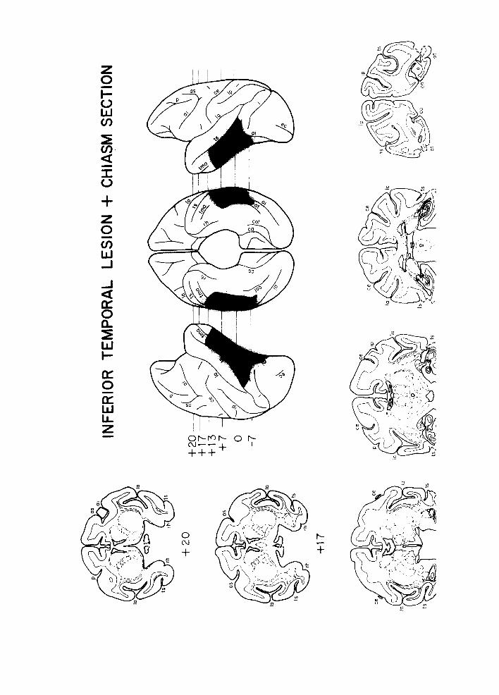
Lateral and ventral reconstructions made from the stained sections indicated that the ablations were as intended. Examination of the stained sections through the optic chiasm indicated that all the transections were complete. Reconstructions and cross- sections from a representative animal with inferior temporal lesion and section of the optic chiasm are shown in Fig. 1.
I~ ~' '
'-- ,,u
"j~..)~
Lo
INFE
RIO
R TE
MPO
RAL L
ESIO
N +
CHIA
SM SE
CTIO
N
~-17
(IS
OS
10
I
tS
13
1-20
F
17
F13
+7 0 -7
ce
~
4-20
ce
ce
ol

263
+ .... • ..-.- ,.., I , , ~ . . . . "--" . . . . ~ I l l l l l
I 2 5 4 5 6 T
Fig. 2. D e s c r i m i n a n d a u s e d in P r o b l e m s 1-7 . T h e pos i t ive o n e is s h o w n o n the left o f e a c h pair. T h e y are d r a w n to scale w i t h the m a x i m u m d i m e n s i o n 25 ram.
Apparatus Behavioral testing was conducted in an automated testing cage measuring
45 x 48 x 72 cm which was enclosed in a sound-attenuating and lightproof wooden
box. The cage was diffusely illuminated by a 25 W incandescent bulb located at the top
of the chamber. White noise (85 dB) and the hum of a ventilator fan masked extraneous
noises. At the center of the front wall were two circular lucite response keys, 33 mm in
diameter. These were mounted one above the other, 18 cm apart, with the lower key
positioned 30 cm above the chamber floor. The discriminanda were white patterns on a dark background, back-projected onto the keys by Industrial Electronic Engineers
Series 10 readout units (See Fig. 2). Directly below the keys at floor level was a recessed
container into which orange juice could be delivered by a solenoid-activated dipper
holding 0.25 cc. During juice delivery the container was illuminated from above by a
3-watt bulb. Solid-state programming and recording equipment was located in an ad-
jacent room.
Behavioral procedures At 3-5 weeks following surgery, the animals were trained to press whichever key
was illuminated. They were then trained with one eye and tested for transfer with the
other on two-choice pattern discrimination problems. Seven such problems were pre-
sented in succession. For each problem one eye was occluded and the animal trained to
a learning criterion, after which the occluder was transferred to the other eye and the
animal retrained to the same criterion. Then the next problem was presented for initial
acquisition and transfer. Half of the animals in each group learned Problems 1, 3, 4,
and 6 initially with their left eye occluded and Problems 2, 5, and 7 with their right eye
occluded. The reverse was the case for the other animals. The details of the procedure are given below.
Eye occluders. During behavioral testing, one of the animal's eyes was covered
with an opaque contact occluder which covered the cornea and fitted snugly under both
the upper and lower eyelids 14. The occluder was inserted while the animal was lightly
anesthetized with Ketaset (Ketamine Hydrochloride, 6 mg/kg). To avoid interference
Fig. l. Lateral and ventral reconstructions of and cross sections through the lesions of a typical animal with inferior temporal lesions and optic chiasm section. The lesions are shown in black in the reconstruc- tions and with heavy dashed lines on the cross-sections. The A-P stereotaxic coordinates are approxim- ate. ai, inferior arcuate sulcus; as, superior arcuate sulcus; ca, calcarine fissure; ce, central sulcus ; col, collateral sulcus; ec, ectocalcarine sulcus; 1, lunate sulcus, la, lateral fissure; oi, inferior occipital sulcus; or, occipitotemporal sulcus; p, principal sulcus; tma, anterior middle temporal sulcus; rh, rhinal fissure; ts, superior temporal sulcus.
264
with performance, the drug was administered no less than 8 h before a testing session, and no less than 4 h after.
The occluder was placed in one eye before acquisition of each problem. It re- mained in place until the animal reached the learning criterion, unless l0 days had elapsed. In that case, to minimize corneal damage, the occluder was removed for a three-day rest during which the animal was not trained. The occluder was then replaced in the same eye and training resumed until either criterion was reached or 10 days had again elapsed. On the day that the animal reached criterion, the occluder was removed and transferred to the other eye (except for 'Same-Eye' retests as described below). Training with the other eye began the following day. As before, the occluder remained in place for a maximum of ten consecutive days.
Each time an occluder was removed, the occluded eye was examined for signs of clouding, and minor infections were treated with Achromycin (Tetracyclin HCI in oil suspension). There was no relationship between the apparent state of the cornea and
performance. Learning. All monkeys were trained while water-deprived for 100 trials per day,
7 days a week. Both stimuli were presented on each trial, with the position of the positive stimulus randomized from trial to trial. A press on either key extinguished both stimuli, ending a trial. A press on the key displaying the positive stimulus resulted in delivery of the orange juice reward. The intertrial interval was 6 sec; a press on either key during the intertrial interval automatically restarted the interval. Training was continued until a criterion of 90 correct responses in a daily session was reached.
If after 600 trials on Problem 1, or after 1000 trials on Problems 2 to 7, an animal had not achieved 80 out of 100 correct responses in a daily session, a correction pro- cedure was instituted. In this procedure, following an error, the spatial location of the stimuli remained constant on succeeding trials until the animal responded correctly. A daily testing session consisted of 100 correct responses. When the animal had at- tained an average of 80 ~o correct responses in two successive testing sessions, the cor- rection procedure was discontinued and the standard procedure resumed.
Tests for interocular transfer. After an animal met the criterion on a problem with one eye, its occluder was transferred to the other eye and, on the following day, the animal was trained on the problem for 100 trials per day until it attained the original criterion of 90 correct responses in a testing session. The procedures for transfer testing were the same as those for learning, except that the correction procedure was not used.
Same eye retests. To determine whether any losses at the start of transfer testing could be due simply to instability in performance after criterion had been reached, animals were retested with the same eye that had been used for learning before being tested for interocular transfer on Problems 5, 6, and 7. On each of these last 3 problems, when an animal reached the learning criterion, the animal's occluder was removed but then replaced in the same eye. Starting the following day, the animal was trained for 100 trials per day until it regained the original criterion. On the next day, the animal
was tested for interocular transfer.

265
RESULTS
Learning As expected from previous studies v,13, the animals with inferior temporal lesions
(Group IT-l- CH and Group IT) made many more errors in attaining the learning crite- rion than the two groups without such lesions (Group CH and Group N). The mean errors scores per problem for these two pairs of groups were 451 and 97, respectively.
The variation in difficulty of the individual problems was so great, however, that the use of mean errors across the seven problems gives undue weight to the more difficult problems. For example, the mean errors on individual problems for the animals with temporal lesions ranged from 159 to 1278, and those of the animals without temporal
lesions, from 30 to 131. To obtain a less biased learning measure, all the animals were ranked on each problem and the mean rank order across the seven problems was com- puted for each animal. On this measure, the animals with temporal lesions were signif- icantly impaired relative to the animals without such lesions (P < 0.001 ; l-tailed Mann- Whitney U test for this and all subsequent comparisons). By contrast, there were no significant differences between the two groups of animals with temporal lesions or between the animals with chiasm section alone and the normal animals.
A similar pattern of impairment held for the individual problems; the animals with temporal lesions made more errors on each problem than those with intact cortex. This difference reached statistical significance on 5 of the 7 problems (Problems 1-3,
6 and 7, P .< 0.05). One of the two problems on which the animals with temporal lesions were not significantly impaired, Problem 5, involved the discrimination of two identical patterns that differed in orientation by 90 ° (See Fig. 2). Subsequent to this finding, we confirmed that inferior temporal lesions do not impair such discriminations, even when they are difficult s. The performance of the animals with chiasm section did not differ significantly from that of the normal animals on any problem. The performance of the
animals with combined temporal lesion and chiasm section did not differ significantly from that of the animals with temporal lesions alone except on Problem 5 (P < 0.02).
Interoeular transfer [nterocular transfer was assessed by 3 different measures. The first was the per cent
correct in the first 40 transfer trials, i.e., the first 40 training trials with the second eye. The second measure was the total number of errors required to attain the learning crite- rion with the second eye. The third measure was the savings ratio :
Learning errors - - transfer errors
Learning errors ÷ transfer errors,
which assesses transfer performance relative to initial learning. On all three measures, as predicted, only the experimental group, with combined inferior temporal lesion and section of optic chiasm, showed impaired interocular transfer.
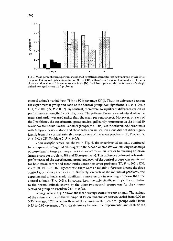
Initial transfer. The results on the first 40 transfer trials are illustrated in Fig. 3. The mean performance across problems for the animals with combined temporal and chiasm lesions varied from 49 ~ to 60 ~ correct (average, 55 ~) , whereas that of the
266
0 r~
0 0
I 0 0
90
80
70
6 0
50
'~'ii~ ill i IT CH IT+CH N
Fig. 3. Mean per cent correct performance in the first 40 trials of transfer testing by animals with infericr temporal lesions and optic chiasm section (IT + CH), with inferior temporal lesions alov.e (IT), with chiasm section alone (CH), and normal animals (N). Each bar represents the performance of a single animal averaged across the 7 problems.
control animals varied from 71 ~o to 92 ~o (average 83 %). Thus the difference between the experimental group and each of the control groups was significant (IT, P < 0.01 ; CH, P < 0.01 ; N, P < 0.02). By contrast, there were no significant differences in initial performance among the 3 control groups. The pattern of results was identical when the mean rank order was used rather than the mean per cent correct. Moreover, on each of the 7 problems, the experimental group made significantly more errors in the initial 40 trials than the animals in the 3 control groups (P < 0.05). On the other hand, the animals with temporal lesions alone and those with chiasm section alone did not differ signif- icantly from the normal animals except on one of the seven problems (IT, Problem 5, P -< 0.05; CH, Problem 2, P < 0.05).
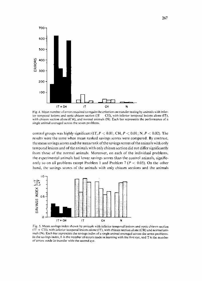
Total transfer errors. As shown in Fig. 4, the experimental animals continued to be impaired throughout training with the second or transfer eye, making an average of more than 10 times as many errors as the control animals prior to reaching criterion (mean errors per problem, 388 and 23, respectively). This difference between the transfer performance of the experimental group and each of the control groups was significant for both mean errors and mean ranks across the seven problems (IT, P < 0.01 ; CH, P < 0.01 ; N, P < 0.02). By contrast, there were no reliable differences among the three control groups on either measure. Similarly, on each of the individual problems, the experimental animals made significantly more errors in reaching criterion than the control animals (P ~< 0.01). By comparison, the only significant impairment relative to the normal animals shown by the other two control groups was for the chiasm- sectioned group on Problem 2 (P < @05).
Savings scores. Fig. 5 shows the mean savings scores for each animal. The savings of the animals with combined temporal lesion and chiasm section varied from 0.09 to 0.33 (average, 0.23), whereas those of the animals in the 3 control groups varied from 0.55 to 0.95 (average, 0.78): the difference between the experimental and each of the
267
700
600
500
03 4 0 0 - nr" 0
or" w 500-
200
100
IT + CH IT CH N
Fig. 4. Mean number of errors required to regain the criterion on transfer testing by animals with infer- ior temporal lesions and optic chiasm section (IT ÷ CH), with inferior temporal lesions alone (IT), with chiasm section alone (CH), and normal animals (N). Each bar represents the performance of a single animal averaged across the seven problems.
control groups was highly significant (IT, P < 0.01 ; CH, P < 0.01 ; N, P < 0.02). The
results were the same when mean ranked savings scores were compared. By contrast, the mean savings scores and the mean rank of the savings scores of the animals with only temporal lesions and of the animals with only chiasm section did not differ significantly from those of the normal animals. Moreover, on each of the individual problems, the experimental animals had lower savings scores than the control animals, signific- antly so on all problems except Problem 1 and Problem 7 (P < 0.05). On the other
hand, the savings scores of the animals with only chiasm sections and the animals
I0
I-- I ' - ,,I+, x w czl z_ o.s
z
if)
IT+CH
lii IT CH N
Fig. 5. Mean savings index shown by animals with inferior temporal lesions and optic chiasm section (IT 4 CH), with inferior temporal lesions alone (IT), with chiasm section alone (CH) and normal ani- mals (N). Each bar represents the savings index of a single animal averaged across the seven problems. In the savings index, L is the number of errors made in learning with the first eye, and T is the number of errors made in transfer with the second eye.

A f i"~'t: ~
j /
NORMAL
/i
INFERIOR TEMPORAL LESION
/ I
,'/ i i
CHIASM SECTION
D ~
INFERIOR TEMPORAL LESION PLUS CHIASM SECTION
Fig. 6. Diagrams showing how information from the left and right halves of space converge onto single neurons in striate and inferior temporal cortex, (A) in normal animals, (B) after chiasm section, (C) after inferior temporal lesions and (D) after combined inferior temporal lesions and chiasm section. Information from the left and right visual fields is shown by the letters L and R. The fields of the left and right eye are shown by solid and dashed circles, respectively, and information fromtheleft and right eye is shown by solid letters and dashed letters, respectively. The overlapping pairs of identical letters, represent convergence from corresponding hemiretinae. The letters joined by a plus sign represent con- vergence from non-corresponding hemiretinae. The dashed lines on the hemispheres delineate striate cortex (most posterior), eircumstriate cortex and inferior temporal cortex (most anterior) and the ap- proximate location of the inferior temporal lesions is shown in black. The arrows represent the flow of visual information, not specific monosynaptic pathways. The horizontal arrows represent theanterior commissure and the splenium of the corpus callosum. Note that convergence from the two eyes is absent only after combined inferior temporal lesion and chiasm section.

269
with only inferior temporal lesions did not differ from those of the normal animals on any problem.
Same-eye retests On Problems 5, 6 and 7, on which the animals were tested for same-eye retention
prior to transfer testing, retention was assessed with the same savings measure used to assess transfer. The mean savings score across all groups and problems was 0.97. There were no significant differences in same-eye retention among any of the groups on any problem.
DISCUSSION
Inferior temporal cortex and interhemispheric transfer When input from each eye was restricted to one hemisphere by midline section of
the optic chiasm, animals with bilateral lesions of the inferior temporal cortex showed severely impaired interocular transfer. By contrast, animals with either inferior tem- poral lesions alone or chiasm section alone showed normal interocular transfer. This pattern of results was repeated on seven different visual discrimination tests. The find- ings demonstrate that inferior temporal cortex is necessary for interhemispheric ex- change of the visual information used in transfer.
Fig. 6 illustrates the proposed pathways responsible for interocular transfer in each of the control groups and the absence of such pathways in the experimental group. In normal animals (Fig. 6A) and in animals with only inferior temporal lesions (Fig.6C) interocular transfer does not require interhemispheric transfer because the partial de- cussation of the optic tract provides each hemisphere with direct visual input from both eyes. After section of the optic chiasm, however, interocular transfer does require inter- hemispheric transfer, and in the case of animals with chiasm section only (Fig. 6B) such transfer is mediated by the projections of the splenium and anterior commissure onto inferior temporal cortex. This transfer can be interfered with in either of two ways. In previous experiments transfer was prevented by cutting the commissures themselves; in this experiment it was prevented by eliminating certain of their targets (Fig. 6D). In both cases, the animals were impaired in interocular transfer because they were de- prived of both (a) the direct binocular convergence onto striate neurons provided by the partial optic decussation and (b) the bilateral convergence onto inferior temporal neurons provided by the forebrain commissures.
It should be noted that in this formulation not only the splenium but also the anterior commissure, or at least some components of each, are considered to be visual pathways. Recognition of this characteristic of the anterior commissure has an import- ant implication. In the past it has often been supposed that interhemispheric transfer of a visual habit must involve the 'transfer of an engram' from one hemisphere to another 5. This was due in part to the known participation of the anterior commissure in the trans- fer phenomenon combined with the absence of any evidence regarding its visual prop- erties. Now that these visual characteristics of the anterior commissure have been ident- ified, however, it is clear that the explanation of interhemispheric transfer need not

270
invoke any such concept as engram transfer. Rather the known visual properties of inferior temporal cells and of the converging pathways that underlie them are sufficient to account for the phenomenon.
Although the experimental monkeys showed severe impairment in interhemis- pheric transfer, their average savings score of 0.23 was slightly higher than that reported for animals with complete forebrain commissurotomy. For example, an average inter- hemispheric savings score of 0.10 was obtained recently by Hamilton 12 for commissur- otomized animals trained on problems similar to those used here. The residual inter- hemispheric transfer in the animals of the present experiment as compared with com- missurotomized animals was presumably mediated by spared portions of the inferior temporal cortex, or by other visually responsive areas that receive commissural con- nections, or both. At the same time it must be emphasized that all of these areas combin- ed yielded only minimal interhemispheric transfer. A consideration of the properties of the commissurally connected visual areas outside the inferior temporal cortex suggests why they may be inadequate for the transfer function. These areas fall into two general classes. One class consists of the various circumstriate areas described by Zeki 21 for the rhesus monkey and by Allman and Kaas 1 for the owl monkey. Like inferior temporal cortex these areas have neurons with specific stimulus requirements. Unlike inferior temporal cortex, however, they are retinotopically organized and so their neurons do not have receptive fields that invariably include the fovea. Also, whereas the receptive fields of inferior temporal neurons are frequently large and bilateral, those of prestriate neurons seldom extend across the vertical meridian by more than a degree or two. Prestriate neurons may thus lack the receptive-field locus and size necessary for me- diating interhemispheric exchange of visual pattern information. It is interesting that in the cat the commissurally connected portions of visual areas 17, 18 and 19 are in fact not necessary for the interhemispheric transfer of pattern discriminationsL These areas, like prestriate cortex in the monkey, have small receptive fields and contain retinotopic representations of the contralateral visual field.
The second class of visually responsive areas that are commissurally connected include the inferior parietal lobulO 7, the frontal eye-fields 15, and the superior temporal polysensory area 4. Each of these areas has neurons with very large receptive fields that often extend even further into both visual half-fields than do the receptive fields of inferior temporal neurons. Unlike inferior temporal neurons, however, these cells typically do not require a stimulus of specific size, shape, orientation, or color to activate them nor are they particularly sensitive to foveal stimulation. Thus, the neurons in this class of commissurally connected visual areas may lack the stimulus selectivity needed for mediating the interhemispheric exchange of visual pattern information. In short, the reason that inferior temporal cortex may be uniquely required for interhemispheric transfer of visual discrimination habits is that it is the only visual area with neurons that combine a high degree of selectivity for stimulus quality with extensive, bilateral recept- ive fields.
Inferior temporal cortex and stimulus equivalence One of the central puzzles of perception is how an object can be perceived as the

271
same object no matter where it falls over a wide retinal area. The same stimulus in different retinal locations necessarily alters the activity of different populations of cells not only in the retina, but in the lateral geniculate body, the striate cortex, and in all the other retinotopic areas throughout the circumstriate belt. Yet, despite these widespread differences in the locus of neural activity, the stimulus is perceived as the same. Our interpretation of interocular transfer in the special case of the chiasm-sectioned monkey may also apply to this more general phenomenon of stimulus equivalence across retinal translation. Inferior temporal neurons respond similarly to a particular stimulus not only when it falls in the opposite hemiretinae of the two eyes, but wherever it falls within the central region of their large receptive fields. Presumably this is due to converging inputs onto single inferior temporal neurons from the cortical representations of differ- ent retinal loci within the hemisphere as well as between the hemispheres. Thus, inferior temporal neurons could mediate stimulus equivalence not only between the left and right visual fields, as in interhemispheric transfer, but also between the upper and lower fields, or between foveal and parafoveal fields, or indeed across any retinal translation. That is, the stimulus equivalence shown by inferior temporal cells within their large receptive fields may be the basis of the perceptual constancy across retinal translation experienced by both man and monkey.
The proposal that inferior temporal cortex provides a mechanism for stimulus equivalence across retinal translation has an important corollary. That is, it may be the loss of this mechani3m which is largely responsible for the impairment in pattern dis- crimination learning after inferior temporal ablation. During visual discrimination training, the discriminanda inevitably stimulate different retinal loci from fixation to fixation and from trial to trial. Stimulus equivalence across these variations in the locus of retinal stimulation must greatly facilitate learning, since it eliminates the need to learn the discrimination anew for each new retinal locus. Conversely, in the absence of such equivalence, the learning of a simple pattern discrimination would necessitate the learning of a large set of separate visual habits, one for each site of retinal excitation. The present experiment has demonstrated that at least with regard to two retinal loci
- - the left half of the left retina and the right half of the right re t ina - - a monkey without inferior temporal cortex does indeed have to learn the same pattern discrimination separately for each site. The effect has been referred to here as an impairment in trans- fer, as distinguished from learning. But if, as we have proposed, removal of inferior temporal cortex eliminates the mechanism of stimulus equivalence across retinal translation, then the learning impairment may itself be a form of transfer loss.
ACKNOWLEDGEMENT
We wish to thank David B. Bender for many helpful discussions, Robert Desi- mone and Charles Bruce for reading earlier drafts, Terry Martin and Vicky Ingalls for help with histology, and Barbara Pinkham for typing.
The National Science Foundation (BNS 75-23634) and the National Institutes of Health (M H-19420) provided financial support.

272
REFERENCES
l Allman,J.,Evolution of the visual system in the early primates, In J. M. SpragueandA. N. Epstein (Eds.), Progress in Psychobiology and Physiological Psychology, VoL 7, Academic Press, New York, 1979, pp. 17-123.
2 Berlucchi, G., Sprague, J. M., Lepore, F. and Mascetti, G. G., Effects of lesions of areas 17, 18 and 19 on interocular transfer of pattern discriminations in split-chiasm cats, Exp. Brain Res., 31 (1978) 275-297.
3 B~ack~P.andMyers~R.E.~Visua~functi~n~fthef~rebrainc~mmissuresinthechimpanzee~Science~ 146 (1964) 799-800.
4 Bruce, C., Desimone, R. and Gross, C. G., Large visual receptive fields in a potysensory area in the superior temporal sulcus of the macaque, Neurosci. Abstr., 3 (1977) 554.
5 Dory, R. W. and Nagr~o, N., Forebrain commissures and vision. In R. Jung (Ed.), Handbook ef Sensory Physiology, 1Iol. 1/11/3 B, Springer Verlag, Berlin, 1973, pp. 543-582.
6 Downer, J. L. de C., Interhemispheric integration in the visual system. In V. B. Mountcastle (Ed.), Interhemispheric Relations and Cerebral Dominance, John Hopkins Press, Baltimore, 1962, pp. 87-100.
7 Gross, C. G., Visual functions of inferotemporal cortex. In R. Jung (Ed.), Handbook of Sensory Physiology, VoL Fill3 B, Springer Verlag, Berlin, 1973, pp. 451-482.
8 Gross, C. G., Inferior temporal lesions do not impair discrimination of rotated patterns, J. comp. physiol. Psychol., 92 (1978) 1095-1109.
9 Gross, C. G., Bender, D. B. and Mishkin, M., Contributions of the corpus callosum and the anterior commissure to the visual activation of inferior temporal neurons, Brain Research, 13I (1977) 227- 239.
10 Gross, C. G. and Mishkin, M., The neural basis of stimulus equivalence across the retinal transla- tion. In S. Harnad, R. Doty, J. Jaynes, L. Goldstein and G. Krauthamer, (Eds.), Lateralization in the Nervous System, Academic Press, New York, 1977, pp. 109-122.
11 Gross, C. G., Rocha-Miranda, C. E. and Bender, D. B., Visual properties of neurons in inferotempo- ral cortex of the macaque, J. NeurophysioL, 35 (1972) 96-111.
12 Hamilton, C., Personal communication, 1978. 13 Mishkin, M., Cortical visual areas and their interaction. In A. G. Karczmar and J. C. Eccles (Eds.),
The Brain and Human Behavior, Springer Verlag, Berlin, 1972, pp. 187-208. 14 Mishkin, M., Gunkel, R. D. and Rosvold, H. E., Contact occluders: a method for restricting vision
in animals, Science, 129 (1959) 1220-1221. 15 Mohler, C. W., Goldberg, M. E. and Wurtz, R. H., Visual receptive fields of frontal eye field neu-
rons, Brain Research, 61 (1973) 385-389. 16 Pandya, D. N., Karol, E. A. and Heilbronn, D., The topographical distribution ofinterhemispheric
projections in the corpus callosum of the rhesuS monkey, Brain Research, 32 (1971) 31-43. 17 Robinson, D. L., Goldberg, M. E. and Stanton, G. B., Parietal association cortex in the primate:
Sensory mechanics and behavioral modulations, J. Neurophysiol., 41 (1978) 910-932. 18 Rocha-Miranda, C. E., Bender, D. B., Gross, C. G. and Mishkin, M., Visual activation of neurons
in inferotemporal cortex depends on striate cortex and the forebrain commissures, J. Neurophysiot,, 38 (1975) 475-491.
19 Seacord, L., Gross, C. G. and Mishkin, M., Role of inferior temporal cortex in perceptual equival- ence of stimuli in the left and right visual fields, Neurosci. Abstr., 1 (1975) 73.
20 Zeki, S. M.,Comparisonofthecorticaldegenerationinthevisualregionsofthetemporallobeofthe monkey following section of the anterior commissure and the splenium, J. comp. NeuroL, 148 (1973) 167-176.
21 Zeki, S. M., The mosaic organization of the visual cortex in the monkey. In R. Beltairs and E. G. Gray (Eds.), Essays on the Nervous System, Clarendon Press, Oxford, 1974, 327 ~ 343.