Embed Size (px)
Citation preview

Role of Humoral versus Cellular Responses Induced by aProtective Dengue Vaccine CandidateRaphael M. Zellweger1, Robyn Miller1, William E. Eddy1, Laura J. White2, Robert E. Johnston2,
Sujan Shresta1*
1 La Jolla Institute for Allergy & Immunology, La Jolla, California, United States of America, 2 Global Vaccines Inc., Research Triangle Park, North Carolina, United States of
America
Abstract
With 2.5 billion people at risk, dengue is a major emerging disease threat and an escalating public health problemworldwide. Dengue virus causes disease ranging from a self-limiting febrile illness (dengue fever) to the potentially fataldengue hemorrhagic fever/dengue shock syndrome. Severe dengue disease is associated with sub-protective levels ofantibody, which exacerbate disease upon re-infection. A dengue vaccine should generate protective immunity withoutincreasing severity of disease. To date, the determinants of vaccine-mediated protection against dengue remain unclear,and additional correlates of protection are urgently needed. Here, mice were immunized with viral replicon particlesexpressing the dengue envelope protein ectodomain to assess the relative contribution of humoral versus cellular immunityto protection. Vaccination with viral replicon particles provided robust protection against dengue challenge. Vaccine-induced humoral responses had the potential to either protect from or exacerbate dengue disease upon challenge, whereascellular immune responses were beneficial. This study explores the immunological basis of protection induced by a denguevaccine and suggests that a safe and efficient vaccine against dengue should trigger both arms of the immune system.
Citation: Zellweger RM, Miller R, Eddy WE, White LJ, Johnston RE, et al. (2013) Role of Humoral versus Cellular Responses Induced by a Protective Dengue VaccineCandidate. PLoS Pathog 9(10): e1003723. doi:10.1371/journal.ppat.1003723
Editor: Ron A. M. Fouchier, Erasmus Medical Center, Netherlands
Received June 5, 2013; Accepted August 28, 2013; Published October 31, 2013
Copyright: � 2013 Zellweger et al. This is an open-access article distributed under the terms of the Creative Commons Attribution License, which permitsunrestricted use, distribution, and reproduction in any medium, provided the original author and source are credited.
Funding: The project described was supported by NIH grants U54AI057517 from the Southeastern Regional Center of Excellence for Emerging Infectious andBiodefense to P.F. Sparling, R56 A1085063 (SS), U01 AI082185 (SS), and NIH Contract HHSN272200900042C (SS) and a LIAI Center for Infectious Disease ResearchFellowship to RMZ. The VRP vaccine was produced with the support of NIH grant 5U01-AI078060 (LJW). The funders had no role in study design, data collectionand analysis, decision to publish, or preparation of the manuscript.
Competing Interests: I have read the journal’s policy and have the following conflicts: REJ was a co-inventor of the VRP vectors, and will share in anyremuneration received by the University of North Carolina from the commercialization of this technology. He also holds an equity interest in AlphaVax, a privatelyheld company that holds commercial rights to the VRP vectors. LJW and REJ are inventors on a pending patent describing the immunogenic compositionscomprising alphavirus vectored dengue virus E protein antigens, including DENV2 E85 used in this study. LJW and REJ are currently employed at Global Vaccines,Inc. (GVI). GVI is a not-for-profit vaccine company. Therefore, its employees have no equity interest in the company. This does not alter our adherence to all PLoSPathogens policies on sharing data and materials.
* E-mail: [email protected]
Introduction
The four serotypes of dengue virus (DENV1-4) are mosquito-
borne and cause a spectrum of diseases ranging from a self-limiting
flu-like illness (dengue fever, DF) to the potentially lethal dengue
hemorrhagic fever/dengue shock syndrome (DHF/DSS) [1].
DENV is endemic in more than 100 countries [2] and 2.5 billion
people worldwide are at risk of infection, mostly in tropical and
subtropical regions [3]. It is estimated that 390 million cases of
DENV infection occur annually, of which 96 million are apparent,
500,000 are severe and 20,000 are fatal [4].
The more severe disease resulting from DENV infection, DHF/
DSS, usually occurs in individuals who have pre-existing dengue-
reactive antibodies (Abs), acquired either from a previous infection
with a heterologous DENV serotype or by passive transfer from
an immune mother in the case of infants [5]. Based on these
epidemiological observations, Halstead and colleagues hypothe-
sized that sub-protective levels of DENV-specific Abs may amplify
viral infection and thus exacerbate disease, a phenomenon termed
antibody-dependent enhancement of infection (ADE) [6,7]. We
and another group have recently confirmed this hypothesis
by demonstrating in mice that a sub-protective amount of
anti-DENV Abs can turn a mild illness into a lethal disease upon
infection with DENV [8,9].
The potential risk of ADE represents a major challenge
associated with the development of a safe vaccine against DENV
[2]. A vaccine that induces sub-protective levels of anti-DENV
Abs may not only be inefficient, but also potentially cause ADE-
mediated severe dengue disease upon infection. In addition,
despite the initial induction of a protective Ab response, the Ab
levels could wane and reach ADE-causing concentrations some
time after vaccination, as even protective anti-DENV Ab has the
ability to cause ADE at lower concentrations [9–11].
Detecting neutralizing Ab in vitro may not accurately correlate
with protection in vivo, as recently exemplified by the results of the
phase IIb clinical trial of the most advanced dengue-vaccine
candidate [12]. The vaccine candidate had only limited efficacy
despite induction of a balanced neutralizing Ab response to all
four serotypes. This infers the involvement of other branches of
the immune system in protection against DENV. The role of T
cells during re-infection is controversial, and often seen as minor
[2] or pathogenic [13]. Accordingly, it is commonly accepted
that the primary goal of dengue vaccination should be induction
of neutralizing Ab responses, and that vaccine-induced T cell
PLOS Pathogens | www.plospathogens.org 1 October 2013 | Volume 9 | Issue 10 | e1003723

responses likely play only a secondary role in protection. However,
there is a substantial lack of knowledge of the immune mechani-
sms involved in protection during successive DENV infections
[14,15]. Therefore, a better understanding of the relative role of
the humoral versus cellular components of a vaccine-induced
immune response to protection against dengue virus infection is
urgently needed. The goal of our study was to assess the relative
contribution of the humoral and cellular arms of the immune
system in protection mediated by a dengue-vaccine candidate.
Venezuelan equine encephalitis virus (VEE) is an alphavirus
that can be used as a vaccine expression vector in which the
genes coding for the structural proteins are replaced by one or
more transgenes [16]. The resulting viral replicon particles
(VRP) induce high level expression of the transgenes in a
single round of infection, but due to the absence of endogenous
structural proteins, do not propagate further in the host [16]. VRP
expressing HIV [17], influenza [18], or human cytomegalovirus
[19] immunogens have been used safely in phase I vaccine trials
in humans. VRP coding for DENV membrane (prM/M) and
envelope (E) proteins have been previously used to immunize mice
[20]. VRP immunization induced anti-DENV neutralizing Abs
and protected suckling mice from lethal intracranial DENV
challenge [20]. Recently, White and colleagues demonstrated that
immunization with VRPs expressing the DENV-E protein
ectodomain (E85-VRP) derived from each of the four DENV
serotypes induced balanced neutralizing Ab responses to all four
serotypes in mice and in macaques, and protected macaques from
infection with DENV (White, unpublished observations) [21].
In the present study, VRP expressing the DENV2-E protein
ectodomain (DENV2 E85-VRP) were used to immunize AG129
mice (type I and II IFN receptor-deficient mice in the 129/Sv
genetic background), followed by challenge with DENV serotype 2
(DENV2) to assess the relative contribution of the cellular versus
humoral components of a protective vaccine-induced immune
response against DENV. AG129 were used because DENV
replicates to high levels in these mice, and they represent the best-
characterized animal model of DENV infection in which DHF/
DSS-like disease can be induced. The results obtained using
AG129 mice were confirmed in an adoptive transfer system.
Wildtype (WT) mice were immunized with the DENV2 E85-VRP,
and, subsequently, DENV2 E85-VRP primed WT T cells, B cells
or serum were transferred into AG129 recipient mice prior to
challenge. This transfer system allowed us to assess the contribu-
tion of the cellular and humoral components the DENV2
E85-VRP-induced immune response generated in WT mice.
Due to their high sensitivity to DENV infection, the AG129
recipient mice served as a stringent challenge model to assess the
contribution of the transferred cells or serum from WT mice.
Two rounds of immunization with DENV2 E85-VRP efficiently
protected AG129 mice from challenge with DENV even when
disease-enhancing amounts of Abs were administered at the time
of challenge (ADE conditions). Mice were protected as early as
five days after the second immunization, and remained protected
for at least four weeks after immunization. Short-term protection
was mainly mediated by CD8+ T cells, whereas long-term
protection relied on CD8+ T cells to different degrees depending
on the immunization schedule. These results were confirmed in a
series of transfer experiments where T cells, B cells or serum from
DENV2 E85-VRP vaccinated WT mice were transferred into
AG129 mice before challenge. Transfer of DENV2 E85-VRP-
primed WT T cells into naıve AG129 mice reduced viral load
upon challenge with DENV. In contrast, transfer of DENV2 E85-
VRP-immune serum had the potential to increase viral load.
Transfer of DENV2 E85-VRP-primed WT B cells into naıve mice
either reduced or increased viral load depending on the number of
B cells transferred. These results demonstrate that, taken in
isolation and at certain concentrations, the humoral component of
a protective vaccine-induced immune response to DENV has the
potential to exacerbate dengue disease, whereas in all conditions
tested, the cellular immune response reduced viral load. This
implies that a safe and protective vaccine against DENV should
trigger both the cellular and humoral arms of the immune system
rather than relying exclusively on induction of DENV-specific
Abs.
Results
DENV2 E85-VRP immunization protects from DENVchallenge and induces DENV-specific antibody
To assess whether immunization with DENV2 E85-VRP would
protect AG129 mice (lacking type I and II IFN receptors) against
DENV, AG129 mice were immunized twice with DENV2 E85-
VRP prior to challenge with DENV. AG129 were used because
they are highly sensitive to DENV infection and can develop
DHF/DSS-like lethal disease upon infection, and are the best
characterized animal model of DENV infection to date. Using
these mice, we have previously demonstrated ADE in vivo and
confirmed that even a protective anti-DENV Ab can induce ADE
at sub-protective concentrations [9]. The viral strain used for
challenge in our study, S221, is a triple-plaque-purified clone
isolated from a mouse-passaged DENV2 strain and has been
previously described [22,23]. This strain was used to challenge the
mice in all our experiments, and for clarity will be referred to as
‘‘DENV’’ throughout the text. We have previously demonstrated
that at the S221 challenge dose of 56108 genomic equivalents
(GE), AG129 mice do not develop severe disease, but instead
manifest neurological symptoms between day 11–14 after infection
[9,22]. In contrast, in the presence of sub-neutralizing amounts of
anti-DENV Ab 2H2, antibody-mediated disease enhancement
occurs and the same dose of virus (56108 GE) causes elevated viral
RNA titers in the liver on day 3 and lethal DHF/DSS-like disease
by day 4–5 [9]. Therefore, 56108 GE was chosen as the challenge
Author Summary
Dengue virus is an escalating public health threat for over2.5 billion people worldwide. The disease caused bydengue virus ranges from mild (dengue fever) to lethal(dengue hemorrhagic fever, dengue shock syndrome). Todate, there is no cure or vaccine for dengue. One of thechallenges to developing a safe and efficient denguevaccine is that antibodies, usually induced by vaccines toprotect the host from re-infection, can increase theseverity of dengue disease if they are not present insufficient amounts to neutralize the virus. An efficientvaccine is urgently needed to slow down the progressionof dengue disease, but little is known about the way theimmune system protects the body against dengue re-infection. Using a protective vaccine candidate for dengue,the present study evaluates in mice the relative contribu-tion of T cells and antibodies to protection against dengue.We show that the antibody component of an immuneresponse that is overall protective had the ability, whenisolated from the other components of the immunesystem, to either decrease or increase viral burden,whereas T cells reduced viral burden in all situationstested. Our results suggest that vaccine developmentefforts should focus on approaches that induce both T celland antibody responses against dengue virus.
Humoral vs. Cellular Immunity in Dengue Vaccination
PLOS Pathogens | www.plospathogens.org 2 October 2013 | Volume 9 | Issue 10 | e1003723

dose for the present study, and we hypothesized that protective
vaccination should reduce liver RNA viral titers and prevent
neurological symptoms as well as death, whereas potential
enhancement would be reflected by elevated liver titers and acute
lethal disease around day 4–5.
AG129 mice were immunized with 16106 infectious units (IU)
of DENV2 E85-VRP either intraperitoneally (i.p.) or intra footpad
(i.f.) 14 and 5 days prior to challenge with 56108 GE DENV on
day 0. As non-vaccinated controls, two groups that were not
immunized were challenged with 56108 GE DENV: one group in
the presence of 15 mg of exogenous monoclonal anti-DENV Ab
2H2 given i.p. to cause Ab-mediated enhancement of infection
(‘‘ADE group’’) and another group in the presence of 15 mg of
C1.18, an isotype control Ab of irrelevant specificity (‘‘baseline
group’’). Viral RNA in the liver was quantified by qRT-PCR on
day 3 (figure 1A) and survival was monitored (figure 1B). The liver
was chosen because high viral RNA levels in the liver on day 3
correlate with increased severity of disease and decreased survival
[9]. Immunization through either the i.p. or i.f. route dramatically
reduced viral RNA levels in the liver (figure 1A) and prevented
death in 80% of immunized animals (figure 1B). As expected from
our previous work [9], the ADE group had approximately 10-
fold more viral RNA in the liver on day 3 and its survival was
decreased relative to the baseline group. These results demonstrate
that DENV2 E85-VRP immunization provides protection against
DENV infection and disease, as measured by liver DENV titer
and survival, respectively.
To start dissecting the immunological basis of this DENV2 E85-
VRP-induced protection, the induction of Abs following immu-
nization with DENV2 E85-VRP on day -14 and -5 was assessed in
the serum of AG129 mice 1 day before challenge with DENV.
Virus-specific serum IgG levels were measured by ELISA on
DENV-coated plates (figure 1C). Both i.p. and i.f. immunization
induced DENV-specific IgG, but the i.p. route induced a higher
Figure 1. DENV2 E85-VRP-immunization protects from DENV challenge. AG129 mice were immunized with 16106 IU DENV2 E85-VRP eitheri.f. (black circles) or i.p. (black triangles) 14 and 5 days prior to challenge with 56108 GE DENV. Two groups of mice were not immunized prior toinfection with DENV, one of which was infected in the presence of anti-DENV antibody 2H2, resulting in antibody-mediated enhancement of disease(black squares, ADE group) and the other one was infected in the presence of antibody C1.18 which is an isotype control of irrelevant specificity(white squares, baseline group). Viral RNA levels were measured in the liver 3 days after challenge (A) and survival was monitored (B). One day prior tochallenge, serum DENV-specific IgG were measured by ELISA on DENV-coated plates (C) and the neutralization capacity of the serum was determinedby PRNT50 (D). Each symbol depicts one mouse except in B where n = 4–5, and in C where each symbol represents the mean of 4 animals, P-valuesfrom two-tailed unpaired t-test with Welch’s correction, confidence interval 95% (A, C, D) or Gehan-Breslow-Wilcoxon test (B), *P#0.05, **P#0.01,***P#0.001. The dotted line represents the limit of detection of the assay. Samples with undetectable levels of DENV2 RNA are represented in grayunder the detection limit. As they have no numerical value, they were not taken into account to calculate the mean.doi:10.1371/journal.ppat.1003723.g001
Humoral vs. Cellular Immunity in Dengue Vaccination
PLOS Pathogens | www.plospathogens.org 3 October 2013 | Volume 9 | Issue 10 | e1003723

IgG response than the i.f. route. The virus-neutralizing capacity
of the serum was assessed by plaque reduction neutralization
test (PRNT50, figure 1D) and only i.p. immunization induced
detectable levels of neutralizing Abs. Therefore, the i.p. immuni-
zation route was chosen for subsequent experiments.
DENV2 E85-VRP immunization reduces viral load evenduring antibody-induced severe dengue disease
Next, to investigate whether immunization with DENV2 E85-
VRP could also protect from Ab-induced severe dengue disease,
AG129 mice were immunized with DENV2 E85-VRP as above.
Subsequently, immunized mice were challenged with 56108 GE
DENV in the presence (or absence) of exogenous anti-DENV Ab
2H2. Viral RNA levels were quantified in the liver on day 3 after
challenge (figure 2A). DENV2 E85-VRP immunization reduced
viral RNA levels to the same extent regardless of the presence or
absence of exogenous anti-DENV Ab. As seen in figure 1 and in
our previous studies [9], the viral RNA levels in the liver
were about 10-fold higher in the ADE group as compared to the
baseline group. To exclude a contribution from non-specific
immune responses elicited by the VRP vector, AG129 mice were
immunized with VRP expressing GFP (VRP-GFP) instead of the
DENV-E protein. DENV2 E85-VRP-immunization, but not
immunization with the non-specific VRP-GFP, reduced viral load
in the liver on day 3 after challenge (figure 2B), demonstrating
the specificity of the protection induced by DENV2 E85-VRP
immunization. Taken together, these data demonstrate that
immunization with DENV2 E85-VRP specifically reduces viral
load upon DENV challenge, even in the presence of exogenous Ab
(ADE).
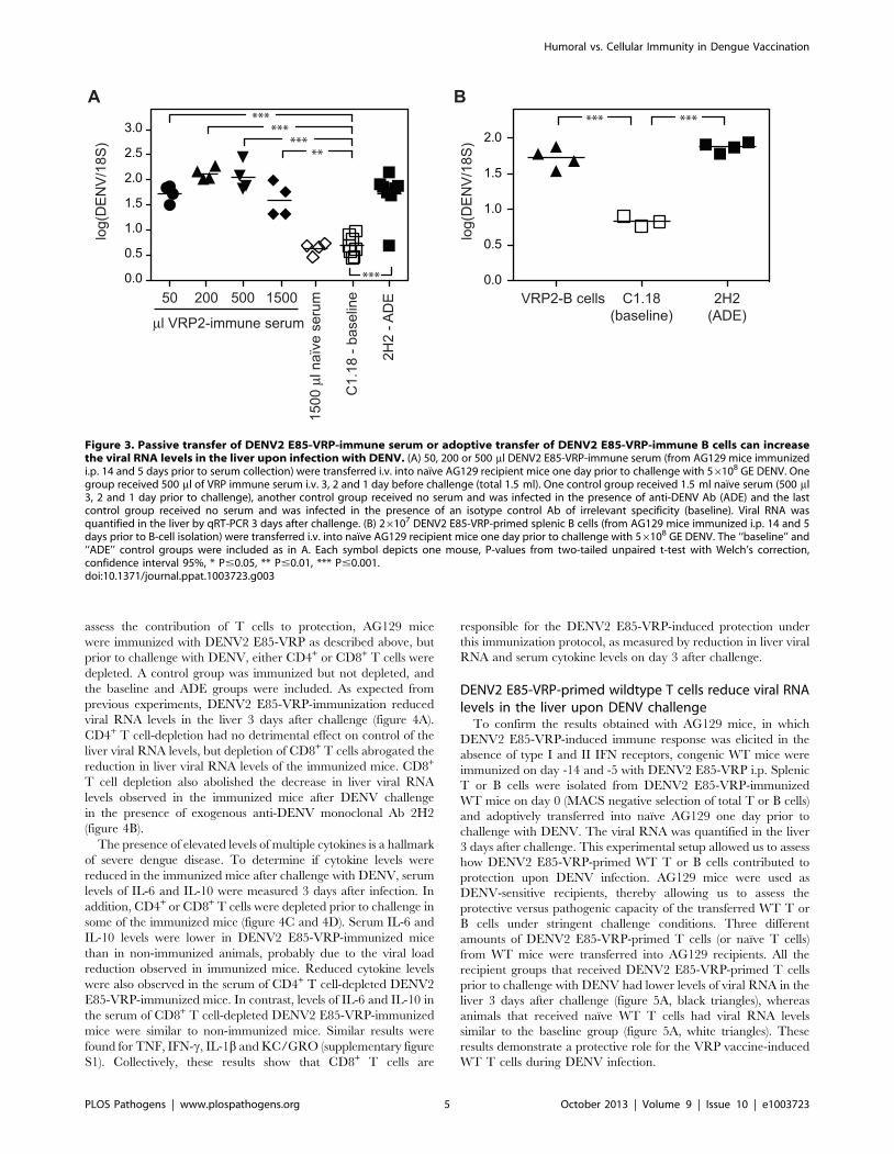
Passive transfer of serum from DENV2 E85-VRP-immunized mice prior to challenge can increase viralload upon infection
To assess whether the protective effect of the DENV2 E85-VRP
immunization was mediated by serum, 50 ml, 200 ml or 500 ml of
serum from AG129 mice immunized 14 and 5 days earlier with
16106 IU of DENV2 E85-VRP were injected i.v. into naıve
AG129 recipient mice one day prior to challenge with 56108
GE DENV. An additional group of recipients received a total
of 1500 ml of DENV2 E85-VRP-immune serum i.v. (500 ml on
day -3, -2 and -1). As controls, one group received 1500 ml naıve
serum (500 ml on day -3, -2, -1) and two groups received no
serum and were challenged either in the absence or presence of
exogenous anti-DENV Ab (baseline and ADE groups). Viral RNA
was quantified in the liver 3 days after challenge (figure 3A). Viral
RNA levels in the liver were significantly higher in all groups that
had received DENV2 E85-VRP-immune serum compared to the
baseline group. As transfer of naıve serum had no effect on the
viral load, we concluded that Ab present in the serum of
immunized mice had caused ADE. To confirm these results,
26107 B cells from AG129 mice immunized with DENV2 E85-
VRP 14 and 5 days earlier were adoptively transferred into naıve
AG129 recipients one day prior to challenge with DENV. Similar
to the ADE control animals, the mice that received DENV2 E85-
VRP-primed B cells prior to challenge had viral RNA levels in the
liver 3 days after challenge that were significantly higher than
baseline (figure 3B). Taken together, these experiments have
revealed that although immunization with DENV2 E85-VRP was
protective, the presence of either serum or B cells from DENV2
E85-VRP-immunized mice did not reduce viral load upon
challenge with DENV, but instead increased viral loads in the
liver.
CD8 T cells are required for protection afterimmunization with DENV2 E85-VRP 14 and 5 days priorto challenge
Thus far, our results have shown that immunization with
DENV2 E85-VRP can protect from DENV challenge, and even
prevent Ab-induced lethal dengue disease. With the chosen
immunization schedule (day -14 and -5), immune serum or
DENV2 E85-VRP-activated B cells had the potential, if
transferred into naıve recipients, to increase viral load in the liver
upon challenge. Therefore, we hypothesized that, using this
particular schedule and route of immunization, T cells could be
responsible for the DENV2 E85-VRP-mediated protection. To
Figure 2. DENV2 E85-VRP-immunization reduced viral load during antibody-mediated severe dengue disease. AG129 mice wereimmunized with 16106 IU DENV2 E85-VRP i.p. 14 and 5 days prior to challenge with 56108 GE DENV. The baseline and ADE groups were included asdescribed in figure 1. Viral RNA levels were measured in the liver 3 days after challenge. (A) One group of immunized mice was challenged in thepresence of exogenous anti-DENV antibody 2H2 (black diamonds), and another group of immunized mice was challenged in the presence of a non-specific isotype control C1.18 (black triangles). (B) One group of mice was immunized with 16106 IU VRP expressing GFP (VRP-GFP, white triangles)instead of the DENV-E ectodomain (DENV2 E85-VRP, black triangles). Each symbol depicts one mouse, P-values from two-tailed unpaired t-test withWelch’s correction, confidence interval 95%, * P#0.05, ** P#0.01, *** P#0.001. Dotted line and symbols in gray as described in figure 1.doi:10.1371/journal.ppat.1003723.g002
Humoral vs. Cellular Immunity in Dengue Vaccination
PLOS Pathogens | www.plospathogens.org 4 October 2013 | Volume 9 | Issue 10 | e1003723
assess the contribution of T cells to protection, AG129 mice
were immunized with DENV2 E85-VRP as described above, but
prior to challenge with DENV, either CD4+ or CD8+ T cells were
depleted. A control group was immunized but not depleted, and
the baseline and ADE groups were included. As expected from
previous experiments, DENV2 E85-VRP-immunization reduced
viral RNA levels in the liver 3 days after challenge (figure 4A).
CD4+ T cell-depletion had no detrimental effect on control of the
liver viral RNA levels, but depletion of CD8+ T cells abrogated the
reduction in liver viral RNA levels of the immunized mice. CD8+
T cell depletion also abolished the decrease in liver viral RNA
levels observed in the immunized mice after DENV challenge
in the presence of exogenous anti-DENV monoclonal Ab 2H2
(figure 4B).
The presence of elevated levels of multiple cytokines is a hallmark
of severe dengue disease. To determine if cytokine levels were
reduced in the immunized mice after challenge with DENV, serum
levels of IL-6 and IL-10 were measured 3 days after infection. In
addition, CD4+ or CD8+ T cells were depleted prior to challenge in
some of the immunized mice (figure 4C and 4D). Serum IL-6 and
IL-10 levels were lower in DENV2 E85-VRP-immunized mice
than in non-immunized animals, probably due to the viral load
reduction observed in immunized mice. Reduced cytokine levels
were also observed in the serum of CD4+ T cell-depleted DENV2
E85-VRP-immunized mice. In contrast, levels of IL-6 and IL-10 in
the serum of CD8+ T cell-depleted DENV2 E85-VRP-immunized
mice were similar to non-immunized mice. Similar results were
found for TNF, IFN-c, IL-1b and KC/GRO (supplementary figure
S1). Collectively, these results show that CD8+ T cells are
responsible for the DENV2 E85-VRP-induced protection under
this immunization protocol, as measured by reduction in liver viral
RNA and serum cytokine levels on day 3 after challenge.
DENV2 E85-VRP-primed wildtype T cells reduce viral RNAlevels in the liver upon DENV challenge
To confirm the results obtained with AG129 mice, in which
DENV2 E85-VRP-induced immune response was elicited in the
absence of type I and II IFN receptors, congenic WT mice were
immunized on day -14 and -5 with DENV2 E85-VRP i.p. Splenic
T or B cells were isolated from DENV2 E85-VRP-immunized
WT mice on day 0 (MACS negative selection of total T or B cells)
and adoptively transferred into naıve AG129 one day prior to
challenge with DENV. The viral RNA was quantified in the liver
3 days after challenge. This experimental setup allowed us to assess
how DENV2 E85-VRP-primed WT T or B cells contributed to
protection upon DENV infection. AG129 mice were used as
DENV-sensitive recipients, thereby allowing us to assess the
protective versus pathogenic capacity of the transferred WT T or
B cells under stringent challenge conditions. Three different
amounts of DENV2 E85-VRP-primed T cells (or naıve T cells)
from WT mice were transferred into AG129 recipients. All the
recipient groups that received DENV2 E85-VRP-primed T cells
prior to challenge with DENV had lower levels of viral RNA in the
liver 3 days after challenge (figure 5A, black triangles), whereas
animals that received naıve WT T cells had viral RNA levels
similar to the baseline group (figure 5A, white triangles). These
results demonstrate a protective role for the VRP vaccine-induced
WT T cells during DENV infection.
Figure 3. Passive transfer of DENV2 E85-VRP-immune serum or adoptive transfer of DENV2 E85-VRP-immune B cells can increasethe viral RNA levels in the liver upon infection with DENV. (A) 50, 200 or 500 ml DENV2 E85-VRP-immune serum (from AG129 mice immunizedi.p. 14 and 5 days prior to serum collection) were transferred i.v. into naıve AG129 recipient mice one day prior to challenge with 56108 GE DENV. Onegroup received 500 ml of VRP immune serum i.v. 3, 2 and 1 day before challenge (total 1.5 ml). One control group received 1.5 ml naıve serum (500 ml3, 2 and 1 day prior to challenge), another control group received no serum and was infected in the presence of anti-DENV Ab (ADE) and the lastcontrol group received no serum and was infected in the presence of an isotype control Ab of irrelevant specificity (baseline). Viral RNA wasquantified in the liver by qRT-PCR 3 days after challenge. (B) 26107 DENV2 E85-VRP-primed splenic B cells (from AG129 mice immunized i.p. 14 and 5days prior to B-cell isolation) were transferred i.v. into naıve AG129 recipient mice one day prior to challenge with 56108 GE DENV. The ‘‘baseline’’ and‘‘ADE’’ control groups were included as in A. Each symbol depicts one mouse, P-values from two-tailed unpaired t-test with Welch’s correction,confidence interval 95%, * P#0.05, ** P#0.01, *** P#0.001.doi:10.1371/journal.ppat.1003723.g003
Humoral vs. Cellular Immunity in Dengue Vaccination
PLOS Pathogens | www.plospathogens.org 5 October 2013 | Volume 9 | Issue 10 | e1003723

Transfer of DENV2 E85-VRP-primed WT B cells can reduceor increase the liver viral RNA levels depending on thenumber of B cells transferred; and DENV2 E85-VRP-immune WT serum can increase the liver viral RNA level
Three different numbers of DENV2 E85-VRP-primed B cells
from WT mice (26108, 46107 or 56106 B cells) were transferred
into naıve AG129 recipients one day prior to challenge. Transfer
of 26108 DENV2 E85-VRP-primed WT B cells reduced the liver
day 3 viral RNA levels in 3 out of 4 animals compared to the
animals that received no B cells (baseline), but this difference was
not statistically significant when the whole group was considered
(figure 5B). Transfer of 46107 DENV2 E85-VRP-primed WT B
cells caused a small but significant increase in the viral load in
the liver on day 3, likely via ADE. Transfer of 56106 B cells had
no effect on the viral load, probably due to the low amount of B
cells transferred. To verify that the transferred B cells produced
virus-specific Abs, DENV2-specific IgG levels were measured by
ELISA on DENV-virion-coated plates in serum obtained from
recipients 3 days after challenge (Figure 5C). No DENV2-specific
IgG was detected in the mice that had not received B cells and
were challenged with DENV on day 0 (either with or without
exogenous anti-DENV Ab). In the groups that received WT
DENV2 E85-VRP-primed B cells one day prior to challenge, the
amount of Ab detected 3 days after challenge was proportional to
the number of B cells transferred. DENV-specific IgG could not be
detected by ELISA in the group that received only 56106 WT
DENV2 E85-VRP-primed B cells, probably due to the low
amount of B cells transferred.
To confirm the results obtained from transfer of DENV2 E85-
VRP-primed WT B cells, we performed a passive transfer
experiment using DENV2 E85-VRP-immune serum from WT
Figure 4. Contribution of T cells to protection from DENV challenge after DENV2 E85-VRP-immunization. (A and B) AG129 mice wereimmunized with 16106 DENV2 E85-VRP i.p. 14 and 5 days prior to challenge with 56108 GE DENV. The ‘‘baseline’’ and ‘‘ADE’’ groups were included.Viral RNA was quantified in the liver 3 days post infection. (A) Prior to challenge, CD4+ T cells (black circles) or CD8+ T cells (black diamonds) weredepleted. This experiment was repeated twice with a total of 7–8 mice per group with similar results, one experiment is shown. (B) Mice werechallenged with 56108 GE DENV either in the absence (open symbols) or in the presence (black symbols) of exogenous anti-DENV antibody. Half ofthe mice were depleted of their CD8+ T cell population prior to challenge (diamonds). (C and D) IL-6 and IL-10 were measured in the serum 3 daysafter challenge with virus in mice either not immunized or immunized as described in A. In addition, CD4+ or CD8+ T cells were depleted as indicated.Each symbol depicts one mouse, P-values from two-tailed unpaired t-test with Welch’s correction, confidence interval 95%, * P#0.05, ** P#0.01, ***P#0.001. Dotted line and symbols in gray as described in figure 1.doi:10.1371/journal.ppat.1003723.g004
Humoral vs. Cellular Immunity in Dengue Vaccination
PLOS Pathogens | www.plospathogens.org 6 October 2013 | Volume 9 | Issue 10 | e1003723

Figure 5. DENV2 E85-VRP-immune T cells from WT mice can reduce viral RNA levels in mice whereas DENV2 E85-VRP-immuneserum from WT mice can enhance infection when transferred into AG129 mice. (A) WT129 mice were immunized on d-14 and d-5 with16106 IU DENV2 E85-VRP i.p. Splenic T cells were isolated on day 0 and adoptively transferred into naıve AG129 recipients one day prior to challengewith 56108 GE DENV. Control groups include the ‘‘baseline’’ and ‘‘ADE’’ groups, as well as groups receiving naıve T cells. Viral RNA was quantified inthe liver 3 days after challenge. (B, C) 26108, 46107 or 56106 B cells from WT mice immunized with DENV2 E85-VRP (d-14 and -5) as described abovewere transferred into naıve AG129 mice one day prior to challenge with DENV (as above). The ‘‘baseline’’ and ‘‘ADE’’ groups received no B cells. ViralRNA was quantified in the liver 3 days after challenge (B) and serum levels of DENV-IgG was measured by ELISA 3 days after challenge (C). (D) Serum(200 ml) from WT mice immunized with DENV2 E85-VRP (d-14 and -5) as described above was passively transferred into naıve AG129 mice one day
Humoral vs. Cellular Immunity in Dengue Vaccination
PLOS Pathogens | www.plospathogens.org 7 October 2013 | Volume 9 | Issue 10 | e1003723

mice. WT mice were immunized on d-14 and -5 with 16106 IU of
DENV2 E85-VRP i.p. followed by collection of serum on day 0
and transfer of the immune serum into naıve AG129 mice one day
prior to challenge with DENV. Viral RNA levels were quantified
in the liver 3 days after challenge (figure 5D). Mice that received
200 ml of DENV2 E85-VRP-immune WT serum had elevated
DENV RNA levels in the liver compared to mice that received
either naıve serum, or no serum (baseline).
Next, we assessed the effect of adoptively transferring DENV2
E85-VRP-primed WT T cells together with an enhancing amount
of DENV2 E85-VRP-immune WT serum into AG129 recipients.
WT mice were immunized with 16106 IU DENV2 E85-VRP i.p.
on days -14 and -5, and on day 0 serum was collected and total
splenic T cells were isolated by negative selection. 200 ml of
DENV2 E85-VRP-immune WT serum was transferred with or
without 46107 DENV2 E85-VRP-primed WT T cells into naıve
AG129 recipient mice one day prior to challenge with DENV2. As
shown in figure 5E, transfer of serum alone increased viral RNA
levels in the liver on day 3 after challenge, but co-transfer of T cells
and serum did not increase liver viral RNA levels.
Taken together, these data confirm the results obtained from
our studies using AG129 mice. Studies with both AG129 and WT
mice have demonstrated that DENV2 E85-VRP-immune T cells
can reduce viral load, whereas DENV2 E85-VRP-immune serum
induced by the day -14 and -5 immunization schedule, if taken
in isolation from other components of the immune system, can
increase viral load upon challenge.
DENV2 E85-VRP immunization confers long-termprotection against DENV challenge
We have shown that DENV2 E85-VRP-immunization confers
CD8+ T cell-mediated short-term protection against DENV
challenge. To examine whether DENV2 E85-VRP-immunization
can confer longer-term protection against DENV challenge,
AG129 mice were immunized twice with 16106 IU of DENV2
E85-VRP i.p. 9 days apart (as described in all experiments so far),
and mice were challenged with DENV 33 days after the second
immunization. Half of the immunized mice were depleted of
CD8+ T cells before challenge. Three days after challenge, viral
RNA levels in the liver were significantly lower in the immunized
mice compared to the non-immunized baseline group, and CD8-
depletion abrogated this decrease in viral load (figure 6A). Thus,
DENV2 E85-VRP immunization can provide CD8+ T cell-
dependent, long-term protection against DENV, as determined by
reduction of liver viral RNA titer 3 days post challenge in AG129
mice.
The time-interval between the two immunizationsmodulates the relative contribution of cellular andhumoral immunity to protection
Two immunizations with 16106 IU DENV2 E85-VRP i.p. 9
days apart provided CD8+ T cell-dependent long-term protection
against DENV challenge. However, the 9-day interval between
the two immunizations (as in all experiments so far) may not
be long enough to induce an optimal Ab response. Therefore,
we increased the time interval between the immunizations to
investigate whether this would modify the relative contribution of
cellular and humoral immunity to long-term protection.
AG129 mice were given two immunizations with 16106 IU of
DENV2 E85-VRP i.p. 28 or 9 days apart. Mice were challenged
with DENV 28 days after the second immunization and in each
group, some mice were depleted of their CD8+ or CD4+ T cells
prior to challenge. Viral RNA levels were measured in the liver 4
days after challenge (figure 6B, C). Livers were harvested 4 days
after challenge instead of 3 (as in the short-term experiments
performed thus far) because in the long-term experiments, mice
are older and heavier, and the viral load increase caused by
exogenous anti-DENV Ab (ADE) starts to be detectable 4 days
post infection (our unpublished observations). Baseline and ADE
groups were included as controls. All immunized and CD8+ T cell-
competent mice had lower viral RNA levels in the liver on day 4
compared to non-immunized mice (baseline) (figure 6B). CD8+ T
cell-depleted mice that were immunized 28 days apart also
contained lower viral RNA levels than the baseline group,
indicating minimal requirement for CD8+ T cells in protection
mediated by the 28-day apart immunization protocol, possibly due
to further maturation of the Ab response during the extended
interval. In contrast, CD8+ T cell-depleted mice that were
immunized 9 days apart had similar levels of viral RNA as
the baseline mice, indicating a critical role for CD8+ T cells in
protection induced by the 9-day apart immunization protocol.
This result suggests that reducing the time between immunizations
may increase the dependency on CD8+ T cells for protection,
possibly due to sub-optimal Ab maturation when the time between
immunizations is insufficient. Regardless of the immunization
protocol (28 or 9 days apart), depletion of CD4+ T cells prior to
challenge with DENV had only a minimal (and not significant)
effect on protection (figure 6C).
To determine if the different immunization schedules had an
influence on the induction of DENV-specific Ab, the serum of
mice immunized 28 days (d-61/-33) or 9 days (d-42/-33) apart was
analyzed 33 days after the second immunization (but before
challenge with DENV). No difference in DENV-specific IgG levels
was observed by ELISA (figure 6D), and neutralizing titers were
similar as determined by PRNT50 (figure 6E). Titers of DENV-
specific IgG1, IgG2a, IgG2b and IgG3 were also similar between
the two immunization protocols (figure 6F), suggesting that other
characteristics of the Ab response (e.g. epitope-specificity, affinity/
avidity, and biological activities such as ADCC and complement
fixation) induced by the extended immunization schedule likely
account for protection.
Discussion
In this study, alphavirus VRP expressing the DENV2-E protein
ectodomain (DENV2 E85-VRP) were used to immunize mice
prior to challenge with DENV in order to assess the relative
contribution of humoral and cellular immunity in a protective
vaccine-induced immune response to DENV. A better under-
standing of the determinants of protection after dengue vaccina-
tion is urgently needed [14], especially after the recent report of
the first phase IIb clinical trial of a tetravalent live-attenuated
dengue-vaccine candidate [12]. For reasons that remain unclear,
the phase IIb trial showed only limited efficacy of the vaccine
prior to challenge with DENV (as above). Viral RNA was quantified in the liver 3 days after challenge. (E) Serum (200 ml) from WT mice immunized withDENV2 E85-VRP (d-14 and -5) was transferred into naıve AG129 mice with or without 46107 T cells from WT mice immunized with DENV2 E85-VRP (d-14 and -5) one day prior to challenge with DENV2. Viral RNA was quantified in the liver 3 days after challenge. Each symbol depicts one mouse, exceptfor C where n = 4 for the groups receiving B cells and 2 for the groups receiving no B cells (base and ADE), P-values from two-tailed unpaired t-testwith Welch’s correction, confidence interval 95%, * P#0.05, ** P#0.01, *** P#0.001. The dotted line represents the limit of detection of the assay.doi:10.1371/journal.ppat.1003723.g005
Humoral vs. Cellular Immunity in Dengue Vaccination
PLOS Pathogens | www.plospathogens.org 8 October 2013 | Volume 9 | Issue 10 | e1003723
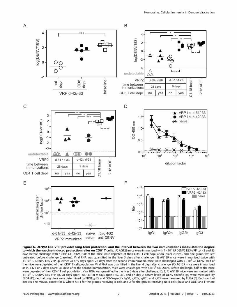
Figure 6. DENV2 E85-VRP provides long-term protection; and the interval between the two immunizations modulates the degreeto which the vaccine-induced protection relies on CD8+ T cells. (A) AG129 mice were immunized with 16106 IU DENV2 E85-VRP i.p. 42 and 33days before challenge with 56108 GE DENV. Half of the mice were depleted of their CD8+ T cell population (black circles), and one group was leftuntreated before challenge (baseline). Viral RNA was quantified in the liver 3 days after challenge. (B) AG129 mice were immunized twice with16106 IU DENV2 E85-VRP i.p. either 28 or 9 days apart. 28 days after the second immunization, mice were challenged with 56108 GE DENV. Half ofthe mice were depleted of their CD8+ T cell population. Viral RNA was quantified in the liver 4 days after challenge. (C) AG129 mice were immunizedas in B (28 or 9 days apart). 33 days after the second immunization, mice were challenged with 56108 GE DENV. Before challenge, half of the micewere depleted of their CD4+ T cell population. Viral RNA was quantified in the liver 3 days after challenge. (D, E, F) AG129 mice were immunized with16106 IU DENV2 E85-VRP i.p. 28 days apart (-61/-33) or 9 days apart (-42/-33), and on day 0, serum levels of DENV-specific IgG were measured byELISA (D), neutralizing titers were determined by PRNT50 (E), and DENV-specific IgG1, IgG2a, IgG2b and IgG3 were measured by ELISA (F). Each symboldepicts one mouse, except for D where n = 4 for the groups receiving B cells and 2 for the groups receiving no B cells (base and ADE) and F where
Humoral vs. Cellular Immunity in Dengue Vaccination
PLOS Pathogens | www.plospathogens.org 9 October 2013 | Volume 9 | Issue 10 | e1003723

despite induction of a balanced Ab response against all four
serotypes. Our study begins to address this lack of knowledge by
assessing the relative role of the humoral and cellular arms of a
protective vaccine-induced immune response.
Two immunizations with DENV2 E85-VRP (9 days apart, day -
14 and -5) dramatically reduced the viral RNA levels in the liver 3
days after challenge with DENV; viral RNA was undetectable
in some immunized animals. All immunized mice survived at least
20 days post challenge, and 80% survived without showing any
sign of disease until the experiment was terminated on day 60.
Immunization with DENV2 E85-VRP reduced the viral load
upon challenge even in the presence of sub-protective, enhancing
levels of exogenous anti-DENV Ab. A regimen of two immuni-
zations with DENV2 E85-VRP, either 9 or 28 days apart,
was protective up to 33 days after the second immunization,
the latest time point tested in this study. These findings are
remarkable considering that AG129 are highly susceptible to
DENV replication, and that a DENV dose as low as 56104 GE
(approximately 1 PFU) causes paralysis in 100% of the mice by
day 25 post-infection [22]. Thus, immunization with DENV2
E85-VRP confers robust protection and appears to mediate near-
sterilizing immunity.
T cell depletion and adoptive transfer experiments demonstrat-
ed that when mice were challenged 5 days after the second
immunization (immunization on day -14/-5), CD8+ T cells were
responsible for reducing the viral load upon challenge. When mice
were challenged more than 4 weeks after the second immuniza-
tion, protection appeared to be dependent on CD8+ T cells in
mice immunized 9 days apart, but independent of CD8+ T cells in
mice immunized 28 days apart. In all experiments performed,
DENV2 E85-VRP-primed T cells were protective as determined
by decreased viral RNA levels in the liver and reduced cytokine
levels 3 days after challenge. In contrast, transfer of DENV2 E85-
VRP-immune serum or DENV2 E85-VRP-immune B cells had
the potential to increase liver viral RNA levels. When T cells and
serum were co-transferred, no enhancement was observed.
The use of AG129 mice (lacking type I and II IFN receptors) in
this study is not ideal as the mechanisms by which DENV causes
disease in these mice may differ relative to WT animals. However,
the experiments described in this study would be impossible in WT
mice, as, to this day, there are no known DENV strains that
replicate and cause disease in WT mice. DENV inhibits IFN
signaling in humans but not in mice [24–26], possibly explaining
why WT mice are resistant to DENV infection and disease. The
AG129 mouse model of DENV infection is the most thoroughly
characterized animal model of DHF/DSS-like disease to date.
This mouse model recapitulates many key features of the human
disease including vascular leakage, cytokine release, high viremia,
low platelet counts and elevated hematocrit [8,9]. AG129 mice
have been used to demonstrate ADE [8,9] and to evaluate the
therapeutic efficacy of modified antibodies that no longer bind to
the Fcc-receptor [27].
The results obtained by immunization and challenge of AG129
mice were confirmed with transfer experiments in which DENV2
E85-VRP-immunized WT mouse T cells, B cells, or serum were
transferred into naıve AG129 recipients prior to challenge with
DENV. This experimental approach was chosen to assess under
stringent challenge conditions the efficacy of a DENV2 E85-VRP-
induced immune response generated in WT (i.e. IFN receptor
competent) animals. Additionally, the transfer approach allowed
us to separate the cellular and the humoral components of a
protective vaccine-induced immune response and evaluate their
relative contribution to protection versus disease enhancement.
The presence of DENV2 E85-VRP-primed WT T cells reduced
viral load upon challenge with DENV; whereas WT serum or
DENV2 E85-VRP-primed WT B cells had the potential to
increase viral RNA levels at certain concentrations. When present
in a sufficiently high concentration, DENV2 E85-VRP primed
WT B cells reduced viral load.
One of the challenges to developing a safe and efficient vaccine
against DENV is that a vaccine should simultaneously generate a
protective immune response against all 4 DENV serotypes. An
unbalanced immune response resulting in sub-protective levels of
Ab against one of the serotypes could potentially result in Ab-
mediated disease enhancement [2]. In theory, a vaccine-induced
Ab response that is initially protective could wane over time and
reach levels at which Ab exacerbates disease, as even neutralizing
Abs can cause ADE at lower, sub-protective concentrations [9–
11]. Although passive transfer of serum from DENV2 E85-VRP-
immune mice did not reduce viral load upon challenge in our
study, in another study, BALB/c females immunized with VRP
co-expressing DENV-E- and prM-protein could passively transfer
antibodies to their pups, which were subsequently protected from
a lethal intracranial challenge with DENV2 strain NGC [20]. It is
also important to note that in our long-term experiments, two
immunizations with DENV2 E85-VRP 28 days apart reduced
DENV viral RNA titer in the liver upon challenge in the absence
of CD8+ or CD4+ T cells. This strongly suggests that vaccination
with DENV2 E85-VRP can readily induce a protective antibody
response in AG129 mice when another immunization schedule is
chosen. In our study, T cell responses were beneficial for the host
and co-transfer of immune T cells together with immune serum
abrogated the immune serum-mediated enhancement. Therefore,
we propose that a vaccine triggering both the humoral and the
cellular arms of the immune system may be more efficient and
safer than a vaccine relying exclusively on the induction of Ab.
DENV2 E85-VRP induced a highly protective T cell response
although the DENV E-protein is not a major T cell target during
natural DENV infection [28–30]. A likely explanation for the
protective T cell responses elicited by the VRP vaccination is the
excellent ability of VRPs to induce CD8+ T cell responses [31].
VRPs efficiently target and activate dendritic cells [32] and are
known to induce both humoral and cellular immunity to the
encoded transgene [31]. In addition, VRPs not encoding a
transgene have potent adjuvant ability when co-injected with
antigen [33] and strongly amplify the CD8+ T cell responses to the
co-delivered antigen [34]. During a natural infection with DENV,
the T cell response to non-structural antigens is dominant [30,35]
and may outcompete the response to structural proteins. As the
DENV2 E85-VRP does not code for DENV non-structural
proteins, the T cell response to the E-protein may be higher than
what would be expected after natural infection.
The relative contribution of CD8+ T cells to the VRP vaccine-
mediated protection in our murine model was influenced by the
interval between immunizations. In the experiments where mice
were challenged four weeks after the second immunization, two
doses of DENV2 E85-VRP given 9 days apart protected via the
induction of CD8+ T cells, whereas if the doses were given 28 days
apart, CD8+ T cells were unnecessary for the vaccine-induced
protection. The interval of 9 days between the two immunizations
n = 5 for experimental groups, naıve serum was included as a negative control, P-values from two-tailed unpaired t-test with Welch’s correction,confidence interval 95%, * P#0.05, ** P#0.01, *** P#0.001. Dotted line and symbols in gray as described in figure 1.doi:10.1371/journal.ppat.1003723.g006
Humoral vs. Cellular Immunity in Dengue Vaccination
PLOS Pathogens | www.plospathogens.org 10 October 2013 | Volume 9 | Issue 10 | e1003723
may not induce an Ab response that is sufficient to protect from
challenge in the absence of CD8+ T cells. However, the 28-day
interval immunization schedule provides protection even in the
absence of CD8+ T cells, possibly through a more complete
maturation of humoral immunity. This suggests that a robust
CD8+ T cell response may be crucial for protection if the Ab
response is insufficient and/or inefficient. Both protocols induced
similar levels of anti-DENV Ab responses, as measured by ELISA
and PRNT50, but only the 28-day protocol protected from
challenge in the absence of CD8+ T cells. Therefore, the presence
of neutralizing Abs in the serum before challenge (as measured by
PRNT50) did not correlate with protection in vivo. Similarly,
immunization with DENV2 E85-VRP via the i.p. or the i.f. routes
induced short-term protection when mice were challenged, but
only the i.p. route induced neutralizing Ab responses. These results
support the emerging notion that measuring neutralizing Abs by
PRNT50 may not accurately predict the efficacy of a vaccine
against DENV [36–38], and highlights the urgent need for further
investigation into the correlates of protection against DENV.
Based on our results, the lack of induction of a robust anti-
DENV T cell response may be a potential explanation for the
recent results of a phase IIb clinical trial of a live attenuated
tetravalent dengue vaccine showing low efficacy against DENV2
despite the induction of DENV2-specific neutralizing Abs [12].
The vaccine used in the clinical trial consisted of a yellow fever
backbone, and therefore did not contain DENV non-structural
proteins, the major targets of CD8+ T cells during natural
infection in humans. The cellular responses were possibly skewed
towards the yellow fever backbone non-structural proteins. Unlike
the VRPs used in our study, which induced a protective cellular
response to the E-protein, the live attenuated vaccine used in the
phase IIb trial may not have triggered a strong T cell response to
the DENV E proteins. The protective T cell responses to DENV
E-protein observed in our study may be the direct result of the
remarkable ability of the VRPs to trigger CD8+ T cell responses to
the encoded transgene.
Cellular immunity during DENV infection is often perceived as
minimally protective [2], or potentially pathogenic [13], although
a protective role for T cells has been demonstrated previously in
mouse models of DENV infection [35,39]. The present study
investigated the contribution of different amounts of homologous
T cells to protection. Our results showed that the presence of
homologous T cells was always beneficial to the host, and did not
increase the severity of disease or the viral load. Further studies are
now needed to determine whether heterologous T cells behave
similarly to homologous T cells, or whether they contribute to
pathogenesis. In addition, the mechanisms by which CD8+ T cells
mediate protection after DENV2 E85-VRP vaccination need to be
clarified.
Our model is well suited to investigate the relative contribution
of humoral and cellular immunity to protection or pathogenesis
after vaccination or during sequential infections with DENV. Our
approach makes it possible to isolate serum, B cells and/or T cells
and assess their respective roles in vivo, either alone or in
combination. However, extrapolation to dengue vaccination in
humans should be done with great caution, as is the case with any
finding made using an animal model. Our findings thus delineate
areas that deserve thorough exploration in future human studies.
They also highlight the need to take a comprehensive approach
that considers the roles of both humoral and cellular immunity in
order to tackle the challenges posed by the development of a
dengue vaccine [36].
In summary, despite several dengue vaccine candidates in phase
I, II and III clinical trials, little is known about the immunological
mechanisms of protection (or potential enhancement) after dengue
vaccination. This study starts to explore the mechanisms of dengue
vaccine-mediated protection or enhancement by examining the
relative contribution of the humoral and cellular arms of the
immune system during a protective vaccine-induced immune
response to DENV. Our results demonstrate that the humoral
component of a protective vaccine-induced immune response to
DENV had the potential, when taken in isolation from other
components of the immune system, to reduce or increase viral load
upon challenge, whereas cellular immunity, alone or in combina-
tion with humoral immunity, was always beneficial to the host.
These findings suggest that the role of T cells in the context of
DENV vaccination should not be ignored, and that a safe and
efficient vaccine against DENV should ideally trigger both arms of
the immune system.
Materials and Methods
Ethics statementThis study was carried out in strict accordance with the
recommendations in the Guide for the Care and Use of
Laboratory Animals of the National Institutes of Health, the US
Public Health Service Policy on Humane Care and Use of
Laboratory Animals, and the Association for Assessment and
Accreditation of Laboratory Animal Care International (AAA-
LAC). All experimental procedures were approved and performed
according to the guidelines set by the La Jolla Institute for Allergy
and Immunology Animal Care and Use Committee (protocol
number AP-28SS1-0809).
Mouse experiments129/Sv mice deficient in type I and II interferon receptors
(AG129, originally obtained from Dr. Skip Virgin at Washington
University in St. Louis) and wild-type 129/Sv mice (purchased
from Taconic) were housed under SPF conditions at the La Jolla
Institute for Allergy and Immunology (LIAI). For all experiments,
sex-matched 5 to 6 week-old mice were used. For challenge
experiments, mice were infected intravenously (via the tail vein)
with 56108 GE of DENV serotype 2, strain S221 diluted in a total
volume of 200 ml PBS with 10% FCS. For survival studies, mice
were sacrificed when moribund or at the first signs of paralysis.
Viral stocks productionDENV strain S221 (serotype 2) is a triple plaque-purified clone
from the D2S10 quasi-species population [23]. S221 was amplified
in C6/36 cells (purchased from ATCC) cultured at 28uC in
Leibovitz’s L-15 medium (Gibco) supplemented with penicillin,
streptomycin, HEPES, and 10% FBS as previously described [40].
Genomic equivalents (GE) were quantified by real-time qRT-PCR
as previously described [40]. Based on a standard baby hamster
kidney cell (BHK-21) plaque assay, there are approximately 56104
GE/PFU for S221.
Antibodies2H2 is an IgG2a reactive for the prM/M protein of DENV,
serotypes 1–4 (IgG2a anti-DENV1-4 prM). 2H2 hybridoma was
purchased from ATCC and grown in PFHM-II (Gibco) with
penicillin (100 U/ml), streptomycin (100 mg/ml) and 55 mM b-
mercaptoethanol in CELLine CL1000 bioreactors (Wilson Wolf
Manufacturing Corporation). Antibody was purified using protein
G-coupled resin according to the manufacturer’s instructions
(Pierce), dialyzed against PBS, concentrated, and sterile filtered
prior to use in experiments. The purity of Ab preparations was
verified by SDS-PAGE and binding to DENV was assessed by
Humoral vs. Cellular Immunity in Dengue Vaccination
PLOS Pathogens | www.plospathogens.org 11 October 2013 | Volume 9 | Issue 10 | e1003723

ELISA. Protein content was quantified using a BCA protein assay
kit (Pierce). Antibody C1.18, mouse IgG2a isotype control, was
purchased from BioXCell.
Viral RNA quantificationTissues were collected into RNAlater (Qiagen) and subsequently
homogenized as described previously [40]. RNA was isolated
and DENV and relative 18S were quantified using real-time
qRT-PCR as described previously [40].
T cell depletionsT cell-depleting antibodies 2.43 (IgG2b anti-mouse CD8),
GK1.5 (IgG2b anti-mouse CD4) and the isotype control LTF2
(IgG2b) were purchased from BioXCell. CD8+ or CD4+ T cells
were depleted by administering 250 mg of 2.43 or GK1.5 antibody
intraperitoneally in 200 ml total volume in PBS 3 days and 1 day
before challenge with virus.
DENV ELISASucrose-purified DENV2 strain S221 was used to coat 96-
well plates overnight at 4uC (109 GE per well in 50 ml 0.1 M
NaHCO3). Next, virus on plates was UV-inactivated and plates
were blocked with 100 ml Blocker Casein in PBS (Thermo
Scientific, 1 hour, room temperature). Blocking solution was
flicked off and serum was titrated 1:3 over 7 titration steps,
starting with an initial dilution of 1 in 30 in PBS. Serum dilutions
were incubated 1.5 hr at room temperature, followed by washing
of the plates three times with 0.05% (v/v) Tween 20 (Sigma) in
PBS (Gibco). Bound antibody was detected using a 1:5000 dilution
of HRP-conjugated goat anti-mouse IgG, IgG1, IgG2a, IgG2b
or IgG3 antibodies (Jackson Immunoresearch) and TMB
(eBioscience). Results are reported as a plot of absorption (OD
450 nm) versus dilution. Alternatively, in the ELISA measuring
the DENV-specific IgG1, IgG2a, IgG2b, IgG3, the titer was
reported as the greatest dilution with an absorption higher than
twice the background absorption.
Neutralizing activity of serum (plaque reductionneutralization test 50%, PRNT50)
One day prior to infection, a total of 16105 BHK cells were
seeded in each well of standard 24-well plates (Costar) in 1 ml of
medium (16105 cells/ml cell suspension in MEM supplemented
with 10% FCS, 10 mM HEPES Buffer, 100 U/ml Penicillin,
100 mg/ml Streptomycin, all from Gibco). On the day of the assay,
20 ml of serum was diluted in 80 ml culture medium and
subsequently titrated 1:2 over 6 titration steps. 50 ml of each
serum dilution was mixed with 50 ml of DENV2 (7.56107 GE/ml
in medium) and incubated 1 hr at 4uC. Each serum/virus mix
(total volume of 100 ml) was used to infect BHK cells in one well of
the 24-well plates seeded the day before. The medium was
carefully removed from the monolayer of cells prior to infection,
and 100 ml of the serum/virus mix was applied to the monolayer
of cells in each well. For each serum sample, the dilutions tested
were therefore 1:10, 1:20, 1:40, 1:80, 1:160 and 1:320. Plate were
subsequently incubated for 1 hour at 37uC (with 5% CO2). After
1 hour, the infection medium was removed and each well
was overlaid with 1 ml of culture medium (as described above)
containing 1% (w/v) Carboxymethylcellulose (CMC) Medium
Viscosity (Sigma). Following incubation of the plates for 3 days at
37uC (with 5% CO2), cells were fixed with 1 ml per well of a 4%
buffered formalin solution (Fisher Scientific) for 30 minutes at
room temperature. Overlay/formalin mix was decanted, the cells
were washed 3 times with PBS, and the cell layer was stained with
1% crystal violet (Fisher Scientific) in 20% ethanol. Plaques were
counted and the highest dilution neutralizing 50% of plaques or
more was reported as neutralizing titer (PRNT50). As a positive
control, 10 mg of the DENV-neutralizing monoclonal Ab 4G2
(IgG2a anti-DENV1-4 E) was used and titrated 1:2 like the serum
samples.
Adoptive transfersDonor spleens were homogenized through 70 mm strainers and
the desired cell populations were isolated by MACS negative
selection (depletion of non-target cells, keeping the desired po-
pulation untouched) using the Pan B Cell Isolation Kit or Pan T
Cell Isolation Kit II from Miltenyi Biotech. Procedures were
performed according to the manufacturer’s instructions. Cells
were enriched to over 90% purity (T cells) and 80% purity (B cells)
as determined by flow cytometry. Cells were administered
intravenously into recipient mice one day prior to challenge with
DENV.
Cytokine measurementsCytokine levels in the serum were measured using the mouse
pro-inflammatory 7-plex base kit (IFN-c, IL-1b, IL-6, IL-10, IL-
12p70, KC/GRO/CINC, TNF) from MDS Meso Scale Discov-
ery according to the manufacturer’s instructions.
Supporting Information
Figure S1 AG129 mice were immunized with 16106 DENV2
E85-DENV2 E85-VRP i.p. 14 and 5 days prior to challenge with
56108 GE DENV. Some groups were depleted of their CD4+ or
CD8+ T cell populations as indicated. Baseline and ADE groups
were included. Three days after challenge, levels of TNF (A), IFN-
c (B), IL-1b (C) and KC (D) were measured in the serum. Each
symbol depicts one mouse, P-values from two-tailed unpaired t-test
with Welch’s correction, confidence interval 95%, * P#0.05, **
P#0.01, *** P#0.001.
(EPS)
Author Contributions
Conceived and designed the experiments: RMZ SS LJW REJ. Performed
the experiments: RMZ RM WEE. Analyzed the data: RMZ RM WEE.
Contributed reagents/materials/analysis tools: LJW REJ. Wrote the paper:
RMZ SS.
References
1. Halstead SB (2007) Dengue. Lancet 370: 1644–1652.
2. Murphy BR, Whitehead SS (2011) Immune response to dengue virus and
prospects for a vaccine. Annu Rev Immunol 29: 587–619.
3. Guzman MG, Halstead SB, Artsob H, Buchy P, Farrar J, et al. (2010) Dengue: a
continuing global threat. Nat Rev Microbiol 8: S7–16.
4. Bhatt S, Gething PW, Brady OJ, Messina JP, Farlow AW, et al. (2013) The
global distribution and burden of dengue. Nature 496: 504–7.
5. Halstead SB (1982) Immune enhancement of viral infection. Prog Allergy 31:
301–364.
6. Halstead SB (2003) Neutralization and antibody-dependent enhancement of
dengue viruses. Adv Virus Res 60: 421–467.
7. Halstead SB, Nimmannitya S, Cohen SN (1970) Observations related
to pathogenesis of dengue hemorrhagic fever. IV. Relation of disease
severity to antibody response and virus recovered. Yale J Biol Med 42: 311–
328.
8. Balsitis SJ, Williams KL, Lachica R, Flores D, Kyle JL, et al. (2010) Lethal
antibody enhancement of dengue disease in mice is prevented by Fc
modification. PLoS Pathog 6: e1000790.
Humoral vs. Cellular Immunity in Dengue Vaccination
PLOS Pathogens | www.plospathogens.org 12 October 2013 | Volume 9 | Issue 10 | e1003723

9. Zellweger RM, Prestwood TR, Shresta S (2010) Enhanced infection of liver
sinusoidal endothelial cells in a mouse model of antibody-induced severe denguedisease. Cell Host Microbe 7: 128–139.
10. Mehlhop E, Ansarah-Sobrinho C, Johnson S, Engle M, Fremont DH, et al.
(2007) Complement protein C1q inhibits antibody-dependent enhancement offlavivirus infection in an IgG subclass-specific manner. Cell Host Microbe 2:
417–426.11. Pierson TC, Xu Q, Nelson S, Oliphant T, Nybakken GE, et al. (2007) The
stoichiometry of antibody-mediated neutralization and enhancement of West
Nile virus infection. Cell Host Microbe 1: 135–145.12. Sabchareon A, Wallace D, Sirivichayakul C, Limkittikul K, Chanthavanich P, et
al. (2012) Protective efficacy of the recombinant, live-attenuated, CYDtetravalent dengue vaccine in Thai schoolchildren: a randomised, controlled
phase 2b trial. Lancet 380: 1559–1567.13. Mathew A, Rothman AL (2008) Understanding the contribution of cellular
immunity to dengue disease pathogenesis. Immunol Rev 225: 300–313.
14. Halstead SB (2012) Dengue vaccine development: a 75% solution? Lancet 380:1535–1536.
15. Wahala WM, Silva AM (2011) The human antibody response to dengue virusinfection. Viruses 3: 2374–2395.
16. Pushko P, Parker M, Ludwig GV, Davis NL, Johnston RE, et al. (1997)
Replicon-helper systems from attenuated Venezuelan equine encephalitis virus:expression of heterologous genes in vitro and immunization against heterologous
pathogens in vivo. Virology 239: 389–401.17. Wecker M, Gilbert P, Russell N, Hural J, Allen M, et al. (2012) Phase I safety
and immunogenicity evaluations of an alphavirus replicon HIV-1 subtype C gagvaccine in healthy HIV-1-uninfected adults. Clin Vaccine Immunol 19: 1651–
1660.
18. Hubby B, Talarico T, Maughan M, Reap EA, Berglund P, et al. (2007)Development and preclinical evaluation of an alphavirus replicon vaccine for
influenza. Vaccine 25: 8180–8189.19. Bernstein DI, Reap EA, Katen K, Watson A, Smith K, et al. (2009)
Randomized, double-blind, Phase 1 trial of an alphavirus replicon vaccine for
cytomegalovirus in CMV seronegative adult volunteers. Vaccine 28: 484–493.20. White LJ, Parsons MM, Whitmore AC, Williams BM, de Silva A, et al. (2007)
An immunogenic and protective alphavirus replicon particle-based denguevaccine overcomes maternal antibody interference in weanling mice. J Virol 81:
10329–10339.21. White LJ, Sariol CA, Mattocks MD, Wahala WM, Yingsiwaphat V, et al. (2013)
An alphavirus vector based tetravalent dengue vaccine induces a rapid and
protective immune response in macaques that differs qualitatively fromimmunity induced by live virus infection. J Virol 87: 3409–24.
22. Prestwood TR, Morar MM, Zellweger RM, Miller R, May MM, et al. (2012)Gamma Interferon (IFN-gamma) Receptor Restricts Systemic Dengue Virus
Replication and Prevents Paralysis in IFN-alpha/beta Receptor-Deficient Mice.
J Virol 86: 12561–12570.23. Shresta S, Sharar KL, Prigozhin DM, Beatty PR, Harris E (2006) Murine model
for dengue virus-induced lethal disease with increased vascular permeability.J Virol 80: 10208–10217.
24. Aguirre S, Maestre AM, Pagni S, Patel JR, Savage T, et al. (2012) DENV
inhibits type I IFN production in infected cells by cleaving human STING. PLoS
Pathog 8: e1002934.
25. Ashour J, Morrison J, Laurent-Rolle M, Belicha-Villanueva A, Plumlee CR, et
al. (2010) Mouse STAT2 restricts early dengue virus replication. Cell Host
Microbe 8: 410–421.
26. Yu CY, Chang TH, Liang JJ, Chiang RL, Lee YL, et al. (2012) Dengue virus
targets the adaptor protein MITA to subvert host innate immunity. PLoS Pathog
8: e1002780.
27. Williams KL, Sukupolvi-Petty S, Beltramello M, Johnson S, Sallusto F, et al.
(2013) Therapeutic Efficacy of Antibodies Lacking FcgammaR against Lethal
Dengue Virus Infection Is Due to Neutralizing Potency and Blocking of
Enhancing Antibodies. PLoS Pathog 9: e1003157.
28. Duangchinda T, Dejnirattisai W, Vasanawathana S, Limpitikul W, Tangth-
awornchaikul N, et al. (2010) Immunodominant T-cell responses to dengue virus
NS3 are associated with DHF. Proc Natl Acad Sci U S A 107: 16922–16927.
29. Rothman AL (2003) Immunology and immunopathogenesis of dengue disease.
Adv Virus Res 60: 397–419.
30. Weiskopf D, Yauch LE, Angelo MA, John DV, Greenbaum JA, et al. (2011)
Insights into HLA-restricted T cell responses in a novel mouse model of dengue
virus infection point toward new implications for vaccine design. J Immunol 187:
4268–4279.
31. Rayner JO, Dryga SA, Kamrud KI (2002) Alphavirus vectors and vaccination.
Rev Med Virol 12: 279–296.
32. Tonkin DR, Whitmore A, Johnston RE, Barro M (2012) Infected dendritic cells
are sufficient to mediate the adjuvant activity generated by Venezuelan equine
encephalitis virus replicon particles. Vaccine 30: 4532–4542.
33. Thompson JM, Whitmore AC, Konopka JL, Collier ML, Richmond EM, et al.
(2006) Mucosal and systemic adjuvant activity of alphavirus replicon particles.
Proc Natl Acad Sci U S A 103: 3722–3727.
34. Thompson JM, Whitmore AC, Staats HF, Johnston RE (2008) Alphavirus
replicon particles acting as adjuvants promote CD8+ T cell responses to co-
delivered antigen. Vaccine 26: 4267–4275.
35. Yauch LE, Zellweger RM, Kotturi MF, Qutubuddin A, Sidney J, et al. (2009) A
protective role for dengue virus-specific CD8+ T cells. J Immunol 182: 4865–
4873.
36. (2012) Defeating dengue: a challenge for a vaccine. Nat Med 18: 1622–1623.
37. Durbin AP, Whitehead SS (2013) The Dengue Human Challenge Model: Has
the Time Come to Accept This Challenge? J Infect Dis 207: 697–9.
38. Whitehead SS, Blaney JE, Durbin AP, Murphy BR (2007) Prospects for a
dengue virus vaccine. Nat Rev Microbiol 5: 518–528.
39. Kyle JL, Balsitis SJ, Zhang L, Beatty PR, Harris E (2008) Antibodies play a
greater role than immune cells in heterologous protection against secondary
dengue virus infection in a mouse model. Virology 380: 296–303.
40. Prestwood TR, Prigozhin DM, Sharar KL, Zellweger RM, Shresta S (2008) A
mouse-passaged dengue virus strain with reduced affinity for heparan sulfate
causes severe disease in mice by establishing increased systemic viral loads.
J Virol 82: 8411–8421.
Humoral vs. Cellular Immunity in Dengue Vaccination
PLOS Pathogens | www.plospathogens.org 13 October 2013 | Volume 9 | Issue 10 | e1003723