Embed Size (px)
Citation preview

Role of EBUS in mediastinal staging of lung cancer
-Dr. Nandakishore Baikunje

Overview of the seminar
• Introduction
• Endosonography to stage the mediastinum
• Technical aspects of EBUS-TBNA for mediastinal staging
• Comparison of staging methods
• Suitability of EBUS-TBNA for sub typing of NSCLC
• Adequacy for multiple tumor genotyping
• Staging algorithm

Introduction
• Treatment of choice for patients with localizednon small cell lung cancer (NSCLC) who haveno evidence of mediastinal nodal metastasis-Pulmonary resection
• Patients with contra lateral mediastinal lymphnode metastases (N3)- should not be operated
• Treatment of patients with NSCLC metastasesrestricted to the ipsilateral mediastinal (N2)lymph nodes: controversial

• -(1)Definitive concomitant chemoradiotherapy
- (2) Neoadjuvant therapy followed by surgeryif mediastinal lymph nodes become free oftumor (nodal downstaging)
• In the absence of extrathoracic, contralaterallung, or pleural cavity metastasis, assessmentof the mediastinum becomes crucial foroperable patients with resectable NSCLC
CG121 Lung Cancer. NICE guidelines

Role of CT
• Lymph nodes with a short axis diameter ofless than 10 mm in axial CT scans –more likelyto be benign than are enlarged nodes
• Roughly 40% of all nodes deemed to bemalignant by CT are benign and 20% of nodesdeemed to be benign by CT are malignant
• CT has moderate test characteristics, with asensitivity of 51% (95% CI 47–54) and aspecificity of 86% (95% CI 84–88)
Silvestri GA et al.Noninvasive staging of NSCLC.CHEST 2007

Role of PET
• FDG PET assessment of mediastinal lymph nodesin NSCLC has a sensitivity of 74% (69–79) and aspecificity of 85% (82–88)
• Combination of node size and metaboliccharacteristics provided by integrated FDG PET CT-improved accuracy of staging by better anatomiclocalization of FDG hotspots
• For most patients, integrated FDG PET CT doesnot eliminate the need for invasive testing
Gould et al. Ann Intern Med 2003

• In patients with small peripheral tumorswithout enlarged or FDG-avid hilar ormediastinal lymph nodes- <6% mediastinalnodal metastasis
• Most clinicians use these criteria to rule outmediastinal involvement and proceed withthoracotomy
Detterbeck FC et al. Chest 2007
Which patients with resectable lung cancer require sampling of mediastinal
nodes?• 1)Abnormal mediastinum by imaging: Mediastinal
lymph nodes suspected of containing metastases onthe basis of either size (short axis ≥10 mm) or FDGuptake. Nodal metastasis in this group ranges from50% to 80%
• 2)Normal mediastinum by imaging:small mediastinallymph nodes without increased FDG uptake-still has a6–30% prevalence of mediastinal metastases-in thepresence of a centrally located primary tumor,enlarged or FDG-avid hilar lymph nodes, or a primarytumor and lymph nodes that are not FDG avid
Hishida T et al. Thorax 2008

Thoroughness of mediastinal staging
• Detterbeck and coworkers have classified 3levels:
• Level A staging (complete sampling) is definedas sampling of each visible lymph node ineach station (1, 2R, 2L, 3, 4R, 4L, and 7) usingat least three passes per node or rapid on-sitecytologic examination (ROSE)
Detterbeck F et al,CHEST;2010

• Level B staging (systematic assessment) requiressampling nodes in each station using at leastthree passes per node or ROSE at stations 2R, 2L,4R, 4L, and 7
• Both complete (Level A) and systematic (Level B)staging require sampling stations 5 and 6 if a leftupper lobe tumor is present
• Invasive staging of stations 5 and 6 may requiretransthoracic needle aspiration or a surgicalapproach

• Level C staging (selective assessment) isdefined by aspiration of at least one abnormallymph node (>1 cm by CT or ultrasound) orfewer than three passes and no ROSE
• Systematic surgical mediastinal staging has asuperior accuracy when compared withselective approaches, although the effect onoverall survival is less clear
Detterbeck FC et al. Sem Th Ca Vas Sur;2007

Surgical staging of the mediastinum
• Most accurate method to establish the clinical Nstage
• For more than 50 years, this procedure has beendone by cervical mediastinoscopy
• Ideally 5(at least 3) mediastinal nodal stations—paratracheal left (stations 2L and 4L),paratrachealright (stations 2R and 4R), and subcarinal (station7)—should be examined, with at least one nodebiopsy sample taken from each station unlessnone are present after dissection
• A 2007 systematic review of surgicalmediastinoscopy in more than 6500 patients withNSCLC reported a sensitivity of 78% (76–79) anda negative predictive value of 88% (86–88)
• If CT or FDG PET suggest enlarged or FDG-avidlymph nodes outside the range of a cervicalmediastinoscopy—targeted surgical staging withvideo-assisted thoracoscopy or parasternalmediastinotomy (Chamberlain procedure) can beused
Detterbeck FC et al:Chest 2007

Endosonography to stage the mediastinum
• Endoscopic ultrasound-guided fine-needleaspiration (EUS-FNA) and endobronchialultrasound with real-time guided transbronchialneedle aspiration (EBUS-TBNA) enable accurateand systematic assessment of mediastinal lymphnodes
• In a meta-analysis by Micames andcolleagues,which included 18 studies and 1201patients, EUS-FNA had a pooled sensitivity of 83%(95% CI 78–87) and a specificity of 97% (96–98)

• Eight studies restricted to patients who hadenlarged mediastinal nodes according to CThad a pooled sensitivity of 90% (84–94).
• In patients without enlarged mediastinallymph nodes (four studies) sensitivity was 58%(39–75)
Micames CG et al. CHEST 2007

• In a metaanalysis by Gu and colleagues,including 11 studies and 1299 patients, EBUS-TBNA had a sensitivity of 93% (91–94) and aspecificity of 100% (99–100)
• The sensitivity in patients selected on thebasis of positive CT or PET results was 94%(93–96), which was better than that in thesubgroup without any selection, who have asensitivity of 76% (65–85)
Gu P et al. Eur J Cancer 2009

• A meta-analysis by Adams and colleagues,which included ten studies of EBUS-TBNA,reported a sensitivity of 88% (79–94)
• One study including 20 patients was also usedin the meta-analysis of Gu and colleagues
Adams K et al. Thorax 2009

Noninvasive and invasive staging modalities prior to thoracotomy
CHEST 2015;147(5):1401-12
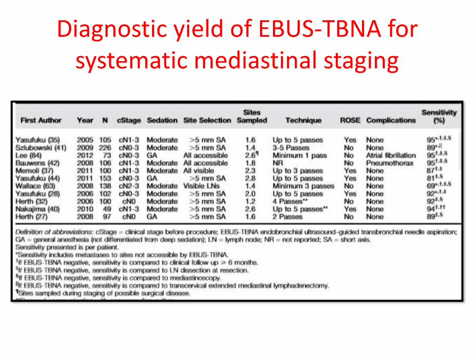
Diagnostic yield of EBUS-TBNA for systematic mediastinal staging

Technique of EBUS-TBNA
• Lymph node to be aspirated is visualized underultrasound, generally using a 7.5 MHz transducer
• A saline-filled balloon may be used to enhancethe ultrasound image
• Images may be frozen and measurement of alymph node performed
• Verification of the presence of vasculature withinthe ultrasound field may be confirmed usingDoppler mode

• The sheath is advanced under visualization. Theneedle may then be safely inserted into the lymphnode (accounting for the 30-degree angle between theneedle and transducer)
• The stylet is used to remove bronchial epithelial cells.Suction may be applied
• Aspirated material is then smeared onto glass slides forfixation and evaluation. Additionally, material forcreation of cell block, microbiologic evaluation, andflow cytometry may be collected and may be helpfulwhen evaluating for alternative or concurrentdiagnoses
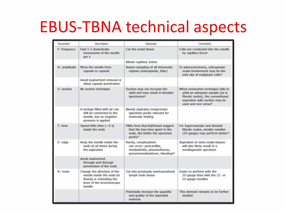
EBUS-TBNA technical aspects

Where to start lymph node sampling?
• Sample the highest station lymph nodesfirst:N3→N2→N1
• Each station should be considered for possibleneedle aspiration, regardless of PET avidity
• Herth and colleagues identified previouslyunsuspected malignancy in 9 of 97 patients bysampling lymph nodes <1 cm in short axis onCT and without FDG uptake in patients ofNSCLC
Herth et al,CHEST 2008

Single needle or different needles?
• A single EBUS-TBNA needle may be used toperform the staging procedure if the N3 nodesare sampled first, followed by the N2 lymphnode stations and the N1 lymph node stations
• Alternatively, a different needle may be usedfor each station, although this approachincreases the cost of the procedure
Herth FJF,Sem Res Cr Cr Med;2011Yusufuku K,CHEST;2006
USG characteristics of malignant nodes
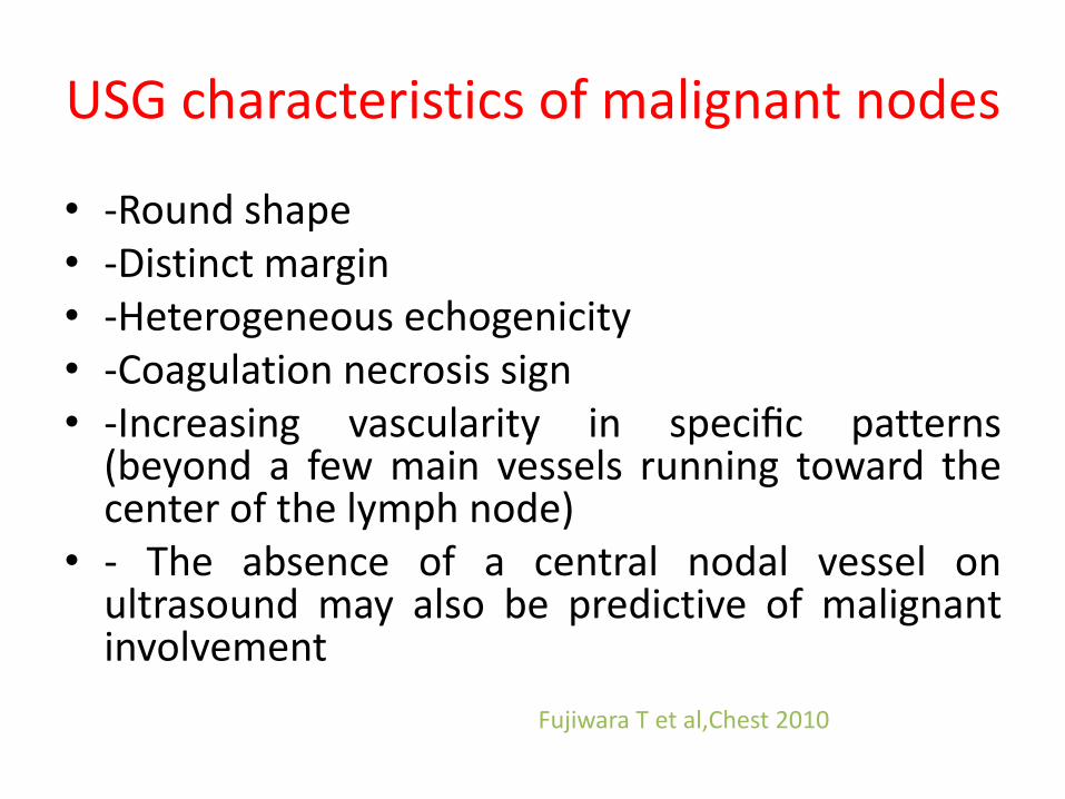
• -Round shape• -Distinct margin• -Heterogeneous echogenicity• -Coagulation necrosis sign• -Increasing vascularity in specific patterns
(beyond a few main vessels running toward thecenter of the lymph node)
• - The absence of a central nodal vessel onultrasound may also be predictive of malignantinvolvement
Fujiwara T et al,Chest 2010

21-g or 22-g?
• A multicenter retrospective comparison of yieldby needle size did not report significantdifferences, although use of the 21-gauge needlewas associated with fewer passes when ROSE wasused
• In a well designed prospective analysis, the twodifferent needles were each used to sample thesame lymph nodes from 33 patients. There wereno differences in diagnostic yield; however, the21-gauge needle resulted in better preservationof histologic structure.
Yarmus LB,Chest ;2013Nakajima T et al,Respirology;2011

Comparison of staging techniques

Methods
• Patients were included if they had known orsuspected lung cancer on the basis of a lungor mediastinal abnormality on CT and if theyhad no pathologically proven extrathoracicmetastases
• Consecutive patients who met the studycriteria were included
• First patient was enrolled on November 18,2004, and the last on October 30, 2006
• CT and PET were performed separately in allpatients before invasive staging
• Lymph nodes were considered enlarged if theshort-axis diameter was 1 cm or greater asmeasured by CT
• PET activity was classified by the standarduptake value and considered positive if thevalue was 2 or greater

TBNA, EBUS-FNA, and EUS-FNA Staging
• TBNA, EBUS-FNA, and EUS-FNA wereperformed as a single combined procedurewith the patient under conscious sedation
• Bronchoscopic TBNA was performed first,followed immediately by EBUS-FNA
• EUS FNA was performed immediately afterTBNA and EBUS-FNA
• All procedures were performed blinded to theresults of the other

• Performed with topical oropharyngeal anestheticand appropriate sedation
• TBNA, with at least 3 fine needle aspiration(FNA) passes, was performed at regions withenlarged lymph nodes on chest CT
• In EBUS, visible lymph nodes, regardless of size,were sampled using FNA. If more than 1 lymphnode was present in a specific location, thelargest lymph node was sampled
• ROSE was not available

Demographic characteristics of study participants(N=138)

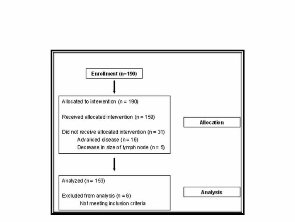
Flow of patients through the study

Final Histological results

Estimated sensitivities and NPVs

Selected comparisons of sensitivities

Locations of malignant lymph nodes detected by each procedure in
patients with NSCLC(n=68)

Subgroup analysis
• Four nonmutually exclusive subgroups werepredefined to determine whether a singleprocedure would be adequate for diagnosis incertain patients, as defined by the location of theabnormal lymph nodes
• Subgroup 1 (“EUS suited”) was defined aspatients who presented with a PET-positivesubcarinal node or in whom CT showed anenlarged lymph node in a subaortic, subcarinal,paraesophageal ,or pulmonary ligament location

• In this subgroup of 54 patients, EUS-FNA wasnot significantly more sensitive than EBUS-FNA (estimated sensitivities,75% [15/20] vs.70% [14/20], respectively)
• The combination of EUS-FNA plus EBUS-FNAhad a sensitivity of 100%
• The NPVs of EUS FNA, EBUS-FNA, and EUS plusEBUS were 87% (34/39), 85% (34/40), and100% (34/34), respectively

• Subgroup2(“EBUS suited”) was defined as patients whopresented with a PET-positive subcarinal node or withan enlarged lymph node in an upper paratracheal ,lower paratracheal, or subcarinal location
• In this subgroup of 74 patients, EBUS-FNA was moresensitive than EUS-FNA (estimated sensitivities, 76%[22/29] vs. 69% [20/29], respectively)
• Both were less sensitive than the combination (100%[29/ 29])
• NPVs of EUS-FNA, EBUS-FNA, and EUS plus EBUS were83% (45/54), 87% (45/52), and 100% (45/ 45),respectively
• Subgroup 3(“bronchoscopy suited”) was definedas patients who presented with a PET-positivesubcarinal node or an enlarged lymph node in thesubcarinal location
• In this subgroup of 50 patients, the estimatedsensitivity of TBNA (47% [9/19]) was lower thanthose of EUS-FNA (74% [14/19]), EBUS-FNA (68%[13/19]), and EUS plus EBUS (100%)
• NPVs were 76%(31/41) for TBNA, 86%(31/36) forEUS-FNA, 84% (31/37) for EBUS-FNA, and 100%(31/ 31) for EUS plus EBUS

• Subgroup 4 (“CT- and PET-negative mediastinum”) wasdefined as patients who had negative results by CT andPET; 60 study participants met this criterion
• In this subgroup, TBNA had low estimatedsensitivity(17%[2/12]),whereas the estimatedsensitivities of EUS-FNA, EBUS-FNA, and EUS plus EBUSwere 67% (8/12), 50% (6/12), and 75% (9/ 12),respectively
• NPVs were 83% (48/58) for TBNA, 92%(48/52) for EUSFNA, 89% (48/54) for EBUS-FNA, and 94% (48/51) forEUS plus EBUS

Comparison of EBUS-TBNA with mediastinoscopy
J Thorac Cardiovas Surg 2011:142:1393-400

• Prospective, controlled trial in patients withconfirmed or suspected NSCLC who required amediastinoscopy as part of their staginginvestigations to determine suitability forresection
• All patients underwent CECT chest+upperabdomen
• PET was available for patients who wereeligible to undergo PET scan

• Under GA, all patients underwent EBUS-TBNAfollowed by standard cervical mediastinoscopyin same setting
• Each patient served as his/her own control
• Surgeon was blinded for pathologic findings ofEBUS-TBNA
• Both EBUS-TBNA and mediastinoscopy wereperformed in all patients even if EBUS-TBNAyielded N2 or N3 disease

• If there was no evidence of N2 or N3 disease onEBUS-TBNA or mediastinoscopy samples, patientsunderwent thoracotomy, pulmonary resection,and mediastinal lymphadenectomy at the samesetting or at a different time
• EBUS-TBNA: Convex probe EBUS was used toperform EBUS-TBNA.CP-EBUS was integrated witha convex transducer(7.5 MHz) that scans parallelto the insertion direction of the bronchoscope

• A dedicated 22-gauge needle was used toperform all EBUS-TBNA procedures
• Smears were air dried and fixed in modifiedCarnoy’s solution. The air dried smears werestained with a modified Field’s stain andevaluated by an on-site cytopathologist toconfirm “adequate” cell material
• Adequate cell material was defined as sufficientmaterial for a specific diagnosis or the presenceof lymphocytes on the specimen
• If adequate tissue was not identified by rapidon-site evaluation(ROSE) after 5 passes, thebiopsy of that site was terminated
• Contralateral lymph nodes were sampled firstfollowed by midline or ipsilateral lymph nodes
• Where multiple nodes were seen, mostsuspicious node in each group was targeted
• Suspicious nodes were defined as round, welldemarcated and echo poor

• Different needles were used for differentlymph node station to prevent crosscontamination
• Localization of lymph nodes was describedaccording to the 7th TNM classification of lungcancer
• EBUS-TBNA was performed for all lymphnodes greater than 5 mm in CT short-axisdiameter or suspicious lymph nodes on EBUS

Patient characteristics


LN stations biopsied by EBUS-TBNA and mediastinoscopy

Lymph node staging based on different modalities

Agreement in mediastinal LN staging between EBUS-TBNA and
mediastinoscopy

• Both EBUS-TBNA and mediastinoscopy wereincorrect in 4 patients
• 1 patient had metastasis located in station 4Rand 3 patients had metastasis in station 5 or6,which were out of reach of both EBUS-TBNAand mediastinoscopy
• Mediastinoscopy incorrectly staged themediastinum in 7 patients and EBUS-TBNAcorrectly diagnosed these patients with N2(n=5)or N3(n=2) disease

• On the other hand, EBUS-TBNA incorrectlystaged 6 patients and mediastinoscopycorrectly staged these patients with N2(n=5)or N3(n=1) disease
• 6 patients understaged by EBUS-TBNAincluded metastases in lymph node stationsnot sampled by EBUS-TBNA(station 2 R) in 2patients and micro metastases in 4 patients(stations 4R,4L,7)

• Majority of patients had clinical N0 disease onchest CT or PET scan (n=90, 59%), with a normalmediastinum by CT imaging criteria
• This contributes to the sensitivity of 81% inassessing the mediastinum by EBUS-TBNA,because sensitivity is related to the underlyingprevalence of N2/N3 disease
• Majority of instances of inadequate sampling byEBUS-TBNA were in lymph nodes less than 5 mmin short axis. None of these inadequate samplingshad metastases on final pathology

Study limitations
• EBUS-TBNA performed under GA through an ETtube in majority of cases. This might contribute tothe high diagnostic yield . However, stations 2Rand 2 L were sometimes difficult to assessbecause of the presence of ET tube
• A cytopathologist was always present for ROSEfor EBUS-TBNA. Because not all centers have theresources to perform ROSE, the results may notbe generalizable to all settings

• Present study shows that EBUS-TBNA canreplace mediastinoscopy for accurate stagingof mediastinum in potentially resectable lungcancer
• EBUS-TBNA avoids an incision, is morecomfortable for the patient and enablesmediastinal reassessment

J Thorac Oncol. 2013;8: 630–636

• All consecutive patients considered candidatesfor surgical treatment of NSCLC, who hadundergone either primary staging or restagingafter neodjuvant chemo- or chemo-radiotherapy with EBUS, EUS, or combinedEBUS with EUS (CUS) with FNA and cytologicalstudy of the aspirated specimen at a Polandhospital from January 1, 2007 to December31, 2010, were included

Primary staging
• Always started with CT (and PET/ CT onselective patients)
• Patients with M1 disease discovered on CT orPET/CT were excluded from further staging
• CT or PET/CT was followed by EBUS, EUS, orCUS. The choice of the particular endoscopicprocedure was decided by the endoscopist,based on the localization of the suspectednodes

• In case of positive results (discovery of metastaticmediastinal nodes), the patients were referredfor neoadjuvant chemo- or chemo-radiotherapy,depending on the opinion of the oncologist
• In case of negative results, the patientsunderwent TEMLA
• All patients with negative TEMLA underwentthoracotomy for lung resection andintraoperative systematic exploration for anyresidual mediastinal lymph nodes
• Thoracotomy with pulmonary resection andsystematic nodal dissection supplementingthe previous TEMLA was the final test for themediastinal nodal staging
• In case of positive results (discovery ofmetastatic mediastinal nodes) the patientswere referred for neoadjuvant chemo- orchemoradiotherapy, depending on the opinionof the oncologist

• In patients with partial or complete responseafter neoadjuvant therapy, restaging with theimaging studies and EBUS/EUS was performed
• Patients who were considered operable andhad no evidence of persistent N2/N3 nodesunderwent pulmonary resection andsystematic nodal dissection

Primary staging of patients with NSCLC

• Number of nodes biopsied with EBUS, EUS,and CUS, and the number of nodes removedon TEMLA for the primary staging andrestaging were calculated
• Diagnostic results of EBUS/EUS werecompared with the results of TEMLA forprimary staging and for restaging

Results
• PET/CT was performed on 78 patients. Distantmetastases were discovered in nine patients(11.5%)
• Sensitivity of PET/CT was 54%, specificity 78%,positive predictive value (PPV) 37%, and negativepredictive value (NPV) 87%
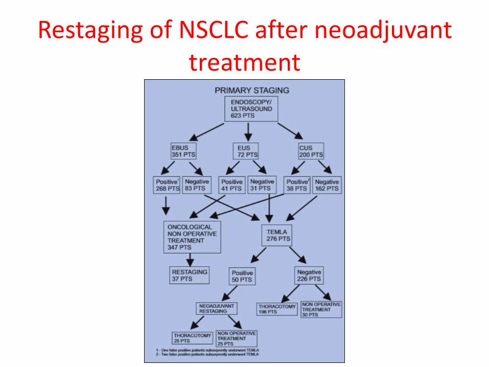
• The endoscopic ultrasound staging wasperformed on 623 patients: EBUS in 351, EUS in72, and CUS in 200 patients. There was nomortality or morbidity after EBUS and EUS

• Mean number of nodes biopsied in the staginggroup -2.1 (range, 1–3) during EBUS, 2.4 (range,1–4) during EUS, and 3.7 (range, 2–5) during CUS
• TEMLA preceded by negative EBUS/EUS-performed on 276 patients
• Mean number of nodes removed during TEMLAfor the primary staging - 32.8 (range, 8–77)
• One patient died after TEMLA (30-day in-hospitalmortality 0.4%) from myocardial infarction
• Morbidity after TEMLA- 7.2%

• TEMLA led to the discovery of metastaticnodes in 50 patients, including 43 patientswith N2 involvement and seven patients withN3 metastases
• There were 31 patients with single-levelinvolvement (29 patients with N2 and 2patients with N3) and 19 patients withmultilevel metastatic involvement (14 with N2and 5 with N3)

• 226 patients were considered candidates forprimary surgery. 30 were not operated for variousreasons
• 189 pulmonary resections with systematiclymphadenectomy and seven exploratorythoracotomies (3.6%) in the primary staginggroup (operability 196 of 226, 86.7%;resectability 189 of 196, 96.4%)
• 2 patients died after resection(mortality 2 of196,1%)
• After thoracotomy, residual N2 nodes omittedduring previous TEMLA were found in twopatients (single station 8 node in 1 patient,single station 5 nodes in the other)
• There were 88 patients in the restagingendoscopy group, including 32 patients whounderwent EBUS, six patients who underwentEUS, and 50 patients who underwent CUS

• Mean number of nodes biopsied in therestaging group was 2.1 (range, 1–3) duringEBUS, 2.4 (range, 1–4) during EUS, and 3.7(range, 2–5) during CUS
• TEMLA was performed for restaging in 78patients
• Mean number of nodes removed duringTEMLA in the restaging group was 27.9 (range,10–46)

• There were 14 patients with N2 involvementand one with N3 disease
• Patients with no mediastinal nodalinvolvement were regarded as candidates forsurgery

Comparison of diagnostic yield of EBUS/EUS and TEMLA for Primary
Staging of NSCLC
Restaging of NSCLC after neoadjuvant treatment

Comparison of Diagnostic Yields ofEBUS/EUS and TEMLA for Restaging
of NSCLC after NeoadjuvantTreatment

• The results of TEMLA were significantly betterthan those of EBUS and EUS, despite significantlylower prevalence of metastatic nodes in TEMLAgroups, both for primary staging and restaging
• Prevalence of the metastatic mediastinal nodesin the TEMLA group for primary staging andrestaging were 18.4% and 19.2%, respectively,which indicated the number of metastatic nodesomitted during previous EBUS/EUS

• Mean number of biopsied nodes on EBUS,EUS, and CUS were 2.1, 2.4, and 3.7,respectively in comparison with 32.8 and 27.9mean number of nodes removed with thesurrounding mediastinal fatty tissue duringTEMLA at staging and restaging, respectively
CHEST 2014; 146(2):389-3 97

Study design
• This single-center prospective study wasconducted in patients with potentiallyresectable NSCLC
• All subjects underwent EBUS, EUS, and SMS aswell as a CT scan of the chest and upperabdomen and PET-CT scan prior to enrollment


Study interventions
• All procedures took place in the operating room undergeneral anesthesia
• Through a LMA, flexible videobronchoscopy was usedto survey the airway. EBUS was then performed with alinear puncture echoendobronchoscope (BF-UC180F;Olympus America, Inc)
• All accessible LN stations were examined, andstandard SMS LN stations were biopsied by fine-needleaspiration with a 22-gauge needle under real-timeEBUS guidance
• Other suspicious LN stations based on CT scan, PET-CTscan, or EBUS were also biopsied

• LMA was removed, and patients underwentorotracheal intubation with a single-lumenendotracheal tube
• EUS was then performed with the same techniqueused for EBUS (EUS linear scope GF-UC140P-AL5[Olympus America, Inc] and EUS 22-gauge needle
• In addition to mediastinal LN stations, the celiac axisLNs, liver, and bilateral adrenal glands were evaluatedand biopsied if found to be abnormal
• A minimum of two needle passes was performed intoeach LN station

• Rapid-on-site cytologic examination of EBUS/EUSspecimens was not performed
• EBUS and EUS were immediately followed byCM. An attempt to biopsy stations 4R, 4L, and 7was made in all patients. Stations 1, 2R, and 2Lwere biopsied selectively based on clinicalsuspicion (CT scan, PET-CT scan, and surgicalevaluation)
• Patients with isolated mediastinal adenopathy inthe level 5 or 6 position underwent CM followedby left-sided AM (Chamberlain procedure)

• Order of LN biopsy for EBUS, EUS, andmediastinoscopy was from the highest-level station tothe lowest-level station to avoid cross-contaminationof lower-level stations and avoid upstaging
• All medically acceptable patients with negativemediastinal staging underwent anatomic pulmonaryresection performed during a separate operation
• Systematic mediastinal LN sampling or dissection wasperformed at the time of pulmonary resection

Flow of study participant selection

Interprocedural agreement between Endosonographic and surgical staging

• EBUS, EUS, combined EBUS/EUS, and SMSsampled a mean of 2.2, 1.7, 3.9, and 3.1 LNstations, respectively
• Prevalence of N2/N3 disease was 32% (53 of166 patients)
• There were 5 patients in whom the SMSprocedure yielded positive results for N2disease and the endosonographic mediastinalstaging procedure findings were negative

Secondary outcomes
EBUS EUS CombinedEBUS/EUS
SMS
Negative predictive
value
90% (95% CI, 0.83-0.95)
90% (0.83-0.95)
92% (0.85-0.96)
89% (0.82-0.94)
Diagnostic accuracy
90% (0.83-0.95)
89% (0.82-0.94)
91% (0.84-0.96)
89% (0.82-0.94)

Adverse events
• Major adverse events occurring during SMS were tracheal injuryrequiring muscle flap coverage (n=1), external jugular vein injuryrequiring vessel ligation (n=1), left sided recurrent nerve injuryresulting in vocal cord paralysis (n=1), and left -sided vocal cordparesis that recovered after 4 months (n=1)
• Major adverse events occurring during EBUS were left sidedmainstem bronchus laceration requiring surgical repair (n=1) andmassive hemoptysis controlled with endoscopic interventions (n=1)
• Major adverse events occurring during EBUS were left sidedmainstem bronchus laceration requiring surgical repair (n=1) andmassive hemoptysis controlled with endoscopic interventions (n =1). There were no major adverse events during EUS

Conclusions from the trial
• In patients with potentially resectable NSCLC, thecombined EBUS/EUS procedure is sensitive andaccurate
• Endosonography leads to improved staging comparedwith SMS because it allows for the biopsy of LNs andmetastases not attainable with SMS techniques
• The combined EBUS/EUS procedure can replace SMS inpatients with potentially resectable NSCLC
• Negative results of a combined EBUS/EUS procedure inthe preoperative evaluation of potentially resectablelung cancer do not require confirmation with surgicalstaging
J Thorac Oncol. 2015;10: 331–337

Inclusion criteria
• (1) Histologically proven NSCLC
• (2) a suspicion of N2 or N3 lymph nodemetastasis on chest CT or PET/CT scans [at leastone of three criteria had to be met, and thesewere: (a) enlarged (short-axis diameter 1 cm ormore) mediastinal node(s), (b) FDG uptake bymediastinal node(s), and/or (c) FDG uptake by N1node(s)]
• (3) the subject was a candidate for curativesurgery.

Study design
• Each patient underwent EBUS-TBNA followed bymediastinoscopy. Thoracic surgeons andpathologists were blinded to the EBUS-TBNA data
• However, if N3 disease was confirmed by EBUS-TBNA at any nodal station examined that wasinaccessible by mediastinoscopy, the latterprocedure was cancelled and the EBUS-TBNAresults were reported because performance ofmediastinoscopy was not ethically justifiable
• Mediastinoscopy was performed within 3 weeksof EBUS-TBNA

• EBUS-TBNA and biopsies were conducted using aconvex probe-EBUS bronchoscope (BF-UC260F-OL8;Olympus, Tokyo, Japan) and a 22-gauge needle
• Interventions were conducted with local anesthesiathrough nebulization with lidocaine and conscioussedation using midazolam
• Each visible station was sampled systematically. If astation had multiple lymph nodes on EBUS, lymphnodes were chosen based on the size and FDG uptake
• ROSE was not available
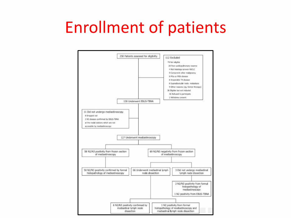
Enrollment of patients

Clinical characteristics of the patients(n=138)

Mediastinal nodal stations in 127 patients who underwent both EBUS-
TBNA and mediastinoscopy


Am J Respir Crit Care Med Vol 185, Iss. 12, pp 1316–1322, 2012

Methods
• Consecutive patients with suspected NSCLCunderwent EBUS-TBNA between January 2009and March 2011 across five centers in the UnitedKingdom
• Interpretation of the EBUS-TBNA specimens wasperformed by the local pathologist
• Classification of NSCLC was based onmorphological appearances (H&E stain), andimmunostaining was performed if clinicallyindicated and if the sample was sufficient

• EGFR mutations were detected using DNAsequencing techniques, and patients wereconsidered to be positive for EGFR mutation if 1of 29 EGFR mutations was detected bypolymerase chain reaction–based assays
• Primary endpoint was the proportion of patientswith NSCLC undergoing EBUS-TBNA in whom itwas possible to subtype the lung cancer
• Coprimary endpoint was the proportion ofsamples that was suitable for EGFR testing asdetermined by the local testing center

Baseline characteristics of patients
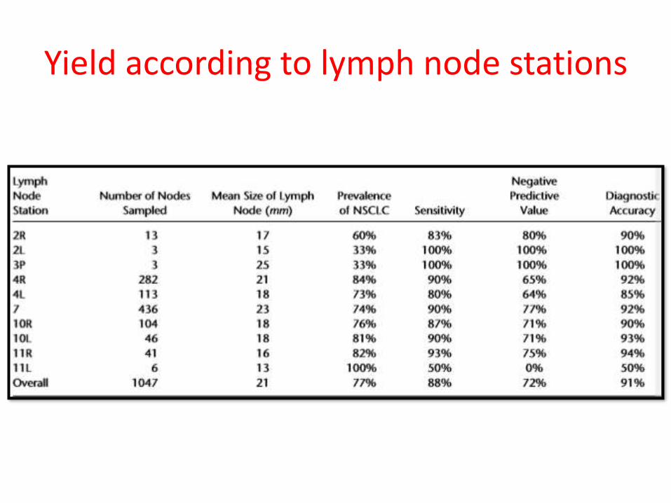
Yield according to lymph node stations

Flowchart of patients

Factors to predict NSCLC-NOS

J Thorac Oncol. 2013;8: 1438–1444

Patient selection
• Patients with a diagnosis of lung cancer and whosetumors were genotyped for at least EGFR mutationswere retrospectively identified through an ongoingInstitutional Review Board–approved protocol at BethIsrael Deaconess Medical Center
• Patients and tumor pairs were excluded if genotypingof at least EGFR mutation, KRAS mutation, and ALKtranslocations were not performed
• There were 207 patient-tumor specimens that weresubmitted for these multiple tumor genotypetechniques between 2007 and 2012

EBUS Technique and Tumor Collection with TBNA
• The CP-EBUS bronchoscope used for tissueacquisition was a 7.5 MHz Olympus fitted withcolor Doppler ultrasound capability
• A 21-gauge needle was used to obtain TBNAsamples. Two to eight passes (usually 3passes) per lymph node were obtained
• Out of 207 patient-tumor pairs that wereincluded in the cohort, 42 samples(20.2%)were obtained from EBUS-TBNA

Baseline patient and tumor characteristics


Success and failure rates of genotype tests

Failed Specimens Using CP-EBUS–Derived Nodal Tissue
• Insufficient tumor cells in the cell block specimen• In CP-EBUS–derived nodal tissues that were successful 17
of 19 (89.4%) had 100 cells or more whereas in failure casesonly two of five (40%) had 100 cells or more (p = 0.042)
• Other possible characteristics- the size of the nodal tissuebiopsied, the location of the node, the number of passesper lymph node, use of touch preparation for rapid on-siteevaluation, presence of extensive desmoplastic stromalresponse, and number of slides cut from the paraffin blockused for immunohistochemical and ancillary studies-werenot significantly different

TAKE HOME MESSAGE