Embed Size (px)
Citation preview

Washington University School of MedicineDigital Commons@Becker
Open Access Publications
2013
Role of choline deficiency in the fatty liverphenotype of mice fed a low protein, very lowcarbohydrate ketogenic dietRebecca C. SchugarWashington University School of Medicine in St. Louis
Xiaojing HuangWashington University School of Medicine in St. Louis
Ashley R. MollWashington University School of Medicine in St. Louis
Elizabeth M. BruntWashington University School of Medicine in St. Louis
Peter A. CrawfordWashington University School of Medicine in St. Louis
Follow this and additional works at: http://digitalcommons.wustl.edu/open_access_pubs
This Open Access Publication is brought to you for free and open access by Digital Commons@Becker. It has been accepted for inclusion in OpenAccess Publications by an authorized administrator of Digital Commons@Becker. For more information, please contact [email protected].
Recommended CitationSchugar, Rebecca C.; Huang, Xiaojing; Moll, Ashley R.; Brunt, Elizabeth M.; and Crawford, Peter A., ,"Role of choline deficiency inthe fatty liver phenotype of mice fed a low protein, very low carbohydrate ketogenic diet." PLoS One.8,8. e74806. (2013).http://digitalcommons.wustl.edu/open_access_pubs/1724

Role of Choline Deficiency in the Fatty Liver Phenotype ofMice Fed a Low Protein, Very Low CarbohydrateKetogenic DietRebecca C. Schugar1, Xiaojing Huang1, Ashley R. Moll1, Elizabeth M. Brunt2, Peter A. Crawford1,3*
1 Department of Medicine, Center for Cardiovascular Research, Washington University, St. Louis, Missouri, United States of America, 2 Department ofPathology and Immunology, Washington University, St. Louis, Missouri, United States of America, 3 Department of Genetics, Washington University, St. Louis,Missouri, United States of America
Abstract
Though widely employed for clinical intervention in obesity, metabolic syndrome, seizure disorders and otherneurodegenerative diseases, the mechanisms through which low carbohydrate ketogenic diets exert theirameliorative effects still remain to be elucidated. Rodent models have been used to identify the metabolic andphysiologic alterations provoked by ketogenic diets. A commonly used rodent ketogenic diet (Bio-Serv F3666) that isvery high in fat (~94% kcal), very low in carbohydrate (~1% kcal), low in protein (~5% kcal), and choline restricted(~300 mg/kg) provokes robust ketosis and weight loss in mice, but through unknown mechanisms, also causessignificant hepatic steatosis, inflammation, and cellular injury. To understand the independent and synergistic roles ofprotein restriction and choline deficiency on the pleiotropic effects of rodent ketogenic diets, we studied four customdiets that differ only in protein (5% kcal vs. 10% kcal) and choline contents (300 mg/kg vs. 5 g/kg). C57BL/6J micemaintained on the two 5% kcal protein diets induced the most significant ketoses, which was only partially diminishedby choline replacement. Choline restriction in the setting of 10% kcal protein also caused moderate ketosis andhepatic fat accumulation, which were again attenuated when choline was replete. Key effects of the 5% kcal proteindiet – weight loss, hepatic fat accumulation, and mitochondrial ultrastructural disarray and bioenergetic dysfunction –were mitigated by choline repletion. These studies indicate that synergistic effects of protein restriction and cholinedeficiency influence integrated metabolism and hepatic pathology in mice when nutritional fat content is very high,and support the consideration of dietary choline content in ketogenic diet studies in rodents to limit hepaticmitochondrial dysfunction and fat accumulation.
Citation: Schugar RC, Huang X, Moll AR, Brunt EM, Crawford PA (2013) Role of Choline Deficiency in the Fatty Liver Phenotype of Mice Fed a LowProtein, Very Low Carbohydrate Ketogenic Diet. PLoS ONE 8(8): e74806. doi:10.1371/journal.pone.0074806
Editor: Marcia B. Aguila, State University of Rio de Janeiro, Biomedical Center, Institute of Biology, Brazil
Received April 10, 2013; Accepted August 6, 2013; Published August 29, 2013
Copyright: © 2013 Schugar et al. This is an open-access article distributed under the terms of the Creative Commons Attribution License, which permitsunrestricted use, distribution, and reproduction in any medium, provided the original author and source are credited.
Funding: This work was supported in part by NIH grants HL007275 (a training grant that supports RCS), DK091538 (to PAC), and a grant from theDiabetic Cardiovascular Disease Center at Washington University (to PAC), the David and Deborah Winston Fellowship in Diabetic CardiovascularDisease (to RCS), the Diabetic Research Center (DK020579), and the Nutrition and Obesity Research Center (DK056341) at Washington UniversitySchool of Medicine. The funders had no role in study design, data collection and analysis, decision to publish, or preparation of the manuscript.
Competing interests: The authors have declared that no competing interests exist.
* E-mail: [email protected]
Introduction
Ketogenic diets confer a multitude of beneficial effects onhealth in experimental and clinical settings, including weightloss in obesity and the amelioration of metabolic syndrome[1–5], anticonvulsant activity [6,7], autism [8],neurodegenerative diseases including Parkinson’s andAlzheimer’s [9–11], cardiomyopathy [12], and they showpromise as potential adjunctive therapy for cancers [13,14].However, the mechanisms through which ketogenic dietstransduce their pleiotropic effects, and the long-termphysiological and metabolic alterations that may occur duringadherence to ketogenic diets, are incompletely understood. In
particular, it is not clear whether beneficial effects of ketogenicdiets are related to the generation and/or metabolism of ketonebodies, or whether the low carbohydrate content itself supportsa salutary metabolic and/or endocrine milieu [7]. Rodents havebeen used extensively to determine the physiological andmetabolic responses to ketogenic diets [15–21]. However,because diet-induced ketonemia in rodents requires markeddiminution of both carbohydrates and protein content, trulyketogenic diets for rodents consist of a macronutrient balancethat humans do not ingest [22]. A widely used[8,12,15,16,18–21,23–25] micronutrient-supplementedketogenic diet for rodents, Bio-Serv F3666, is very high in fat(93.3% kcal), very low in carbohydrate (1.8%), and relatively
PLOS ONE | www.plosone.org 1 August 2013 | Volume 8 | Issue 8 | e74806

depleted of protein (4.7%). This diet provokes ketonemia,weight loss, and induces a hepatic gene expression signatureconsistent with decreased de novo lipogenesis and increasedfatty acid oxidation [15,16]. C57BL/6J mice maintained on thisketogenic diet become lean, euglycemic, ketotic,hypoinsulinemic, and glucose intolerant [15,21]. Additionally,mice fed this ketogenic diet exhibit a distinctive nonalcoholicfatty liver disease (NAFLD) profile including micro- andmacrovesicular steatosis with hepatocellular injury and repair.The macronutrient composition that induces this histologicalsignature is atypical for human NAFLD, which is commonlyassociated with increased carbohydrate intake and activatedde novo lipogenesis [16,26,27]. In fact, low carbohydrate dietsin humans may improve NAFLD [26–28].
To establish safe and effective therapeutic nutritionalapproaches for diseases that may be responsive to low-carbohydrate diets, it will ultimately be important to understandthe driver mechanisms responsible for favorable responses,and whether these responses and their underlying mechanismscan be nutritionally dissociated from concomitantly-triggeredpathophysiological responses. A potential contributor to theliver fat accumulation and injury that are provoked by Bio-Serv
F3666 [16,21], despite its salutary effects in other organsystems [8,12,20], is protein restriction. In fact, Bio-Serv F3666diet may mimic a subset of the systemic and hepatic sequelaeof malnutrition attributable to very low protein ingestion inhuman patients with kwashiorkor [29]. An additionalprospective explanation is the influence of choline restriction[30]. While fully replete rodent diets are supplemented tocontain ≥ 2 g/kg choline, Bio-Serv F3666 is not supplemented,and therefore contains only ~300 mg/kg from naturally-derivedfat sources. In this study, we examined the roles of dietaryprotein and choline contents on the systemic and hepaticderangements that occur in the setting of very high fat, very lowcarbohydrate diets. Using Bio-Serv F3666 as a reference diet,we designed a very low carbohydrate, very low protein, andcholine restricted (VLP/C-) diet and three additional diets whichdiffer only in total protein [10% kcal (low protein, LP) vs. 5%kcal (very low protein, VLP)] and choline content [replete (C+)vs. restricted (C-)] to elucidate the contributing mechanisticroles of each component in the onset and progression of thehepatic pathology observed in ketogenic diet-fed mice.
Table 1. Macronutrient composition of mouse diets used in this study.
DietVendor (CatalogNumber) Carb % kcal Fat % kcal
Protein %kcal
Choline g/kg (degree ofrestriction)
Methionine g/kg(degree of restriction)
Cholesterolmg/kg kcal/g
Standard Chow Lab Diet (5053) 62.1 13.2 24.6 2.0 (N/A) 7.0 (N/A) 141 3.4
LP/C+ Harlan Teklad (110291) 1.4 88.9 9.7 5.3 (0%) 7.4 (0%) 2500 7.3
LP/C- Harlan Teklad (110406) 1.4 89.0 9.6 0.3 (88%) 4.4 (37%) 2500 7.2
VLP/C+ Harlan Teklad (110634) 1.3 94.1 4.6 5.3 (0%) 3.7 (47%) 2500 7.6
VLP/C- Harlan Teklad (110633) 1.3 94.1 4.6 0.3 (>90%*) 2.2 (82%*) 2500 7.6
All diets are irradiated (to sterilize) before feeding. Values represent percentage of total kcal. Choline added to diets as choline chloride (Lab Diet) or choline bitartrate(Harlan Teklad). Methionine content is calculated based on the amino acid composition of casein, plus additional elemental methionine (for VLP/C+ and LP/C+). * Correctedfor diminished mass of diet consumed.doi: 10.1371/journal.pone.0074806.t001
Table 2. Serum parameters after 6 weeks on diets.
Diet βOHB, mM Glucose, mg/dL Insulin, ng/mL HOMA-IR FFA, mM TAG, mg/dLStandard Chow 0.10±0.03 121.6±3.7 1.10±0.14 8.2±1.1 1.24±0.10 70.3±4.5LP/C+ 0.44±0.03a 212.2±15.2aa 0.90±0.16 11.7±2.4 0.36±0.08aaa 67.3±3.1LP/C- 0.60±0.08a 175.8±11.6a 0.70±0.10 8.6±1.8 0.30±0.06 aaa 57.3±3.8VLP/C+ 0.80±0.11a,b 172.5±8.9a 0.62±0.12 7.1±1.5 0.48±0.06 aaa 85.7±8.5VLP/C- 1.46±0.34aaa,bb 119.0±15.8b,c 0.37±0.04 aa,b 2.9±0.5a,b 0.64±0.05 aaa,b 95.0±10.7 bb
HOMA-IR, homeostatic model assessment of insulin resistanceFFA, non-esterified(free) fatty acidsTAG, triacylglycerolsa p≤0.05, aa p≤0.01, aaa p≤0.001 (one-way ANOVA) compared to chowb p≤0.05, bb p≤0.01 (one-way ANOVA) attributable to decrease in protein content (from 10% kcal to 5% kcal) at a fixed choline contentc p≤0.05 (one-way ANOVA) attributable to restriction in choline content (from 5.3 g/kg to 0.3 g/kg) at 5% kcal proteindoi: 10.1371/journal.pone.0074806.t002
Choline and Ketogenic-Diet Induced Fatty Liver
PLOS ONE | www.plosone.org 2 August 2013 | Volume 8 | Issue 8 | e74806

Methods
Animals and dietsC57BL/6J wild-type mice were maintained on Lab Diet
(5053) ad libitum and autoclaved water on cedar chip beddingat 22°C. Lights were off between 1800 and 0600. Beginning atthe age of 6 weeks male mice were maintained for 6 weeks onone of the following very high fat, very low carbohydrate(ketogenic) paste diets, whose contents are summarized inTable 1: 1) low protein, choline replete (LP/C+, Harlan-TekladTD.110291), 2) low protein, choline restricted (LP/C-, Harlan-Teklad TD.110406), 3) very low protein, choline replete (VLP/C+, Harlan-Teklad TD.110634), or 4) very low protein, cholinerestricted (VLP/C-, Harlan-Teklad TD.110633). Diets weresterilized prior to shipment, stored at 4°C, and administeredevery 2-3 days in autoclaved glass dishes on the cagebottoms. Consumption of diets was determined between weeks2 and 4 of the administration period, and was performed byweighing paste diet mass prior to and after 48 h of feeding.Age-matched cohorts of mice were maintained on the standardlow-fat, polysaccharide-rich chow pellets (Lab Diet 5053) toserve as controls. Consistent with the historically-used Bio-
Serv F3666 diet, the contributing fat sources of all fourketogenic diets are lard and milk fat; casein is the proteinsource, and sucrose is the minimal carbohydrate source (~1%kcal). All diets were supplemented with vitamin mix AIN-76 andmineral mix AIN-76A. The distribution of fatty acids among allfour diets is similar, with 46-52% originating from saturated,38-41% from monounsaturated, and 7-17% frompolyunsaturated fatty acids. Choline replete diets aresupplemented with 5.0 g/kg supplied as choline bitartrate. Allfour diets are soy meal (phytoestrogen) free. All experimentswere performed after protocol approval by the Animal StudiesCommittee at Washington University.
Metabolite and insulin measurementsSerum samples were acquired from animals that had been
fasted for 4 h on fresh cedar chip bedding. Serum glucose, freefatty acids (FFA), β-hydroxybutyrate (βOHB), triacylglycerols(TAG), and insulin concentrations were measured aspreviously described [18]. Hepatic TAG, using a Folch extractof liver and biochemical quantification, were quantified aspreviously described [24]. Serum alanine aminotransferase
Figure 1. Metabolic parameters of mice maintained on very high fat, low protein, very low carbohydrate diets. (A) Bodyweight responses to 6 weeks of maintenance on the experimental paste diets, compared to chow controls. n=10-15 mice/group. a,p≤0.001; b, p≤0.001; c, p≤0.01. See end of this legend for description of the individual comparisons depicted by each letter. (B)Caloric consumption of diet, normalized per mouse, between weeks two and four of the 6 weeks of maintenance on the diets.n=5-10 mice/group. a, p≤0.001; c, p≤0.05. (C) Caloric consumption of diet, normalized per gram of body weight (BW). n=5-10 mice/group. a, p≤0.001; b, p≤0.01. (D) Percent adiposity after 6 weeks on each of the diets. n=5-10 mice/group. a, p≤0.01; c, p≤0.05. Forall panels, data are presented as means±SEM. a, significantly different compared to chow; b, significant difference attributable todecrease in protein content (from 10% kcal to 5% kcal) at a fixed choline content; c, significant difference attributable to restriction incholine content (from 5.3 g/kg to 0.3 g/kg) at a fixed protein content; by 1-way ANOVA with Tukey’s post hoc testing.doi: 10.1371/journal.pone.0074806.g001
Choline and Ketogenic-Diet Induced Fatty Liver
PLOS ONE | www.plosone.org 3 August 2013 | Volume 8 | Issue 8 | e74806

Figure 2. Roles of varying protein and choline nutrientcontents in very high fat, low protein, very lowcarbohydrate diet-induced hepatic steatosis. (A) Liverweight/body weight ratios after 6 weeks on the diets. n=5-10mice/group. c, p≤0.05 for LP/C- and p≤0.01 for VLP/C-. Seeend of this legend for description of the individual comparisonsdepicted by each letter. (B) Biochemical quantification ofhepatic TAG content, normalized to liver mass. n=5-10 mice/group. a, p≤0.05 for LP/C- and p≤0.01 for VLP/C-; b, p≤0.05; c,p≤0.05 for LP/C- and p≤0.01 for VLP/C-. (C) Serum alanineaminotransferase (ALT) concentrations. n=4-6 mice/group. c,p≤0.05. Data are presented as means±SEM. a, significantdifference compared to chow; b, significant differenceattributable to decrease in protein content (from 10% kcal to5% kcal) at a fixed choline content; c, significant differenceattributable to restriction in choline content (from 5.3 g/kg to 0.3g/kg) at a fixed protein content; by 1-way ANOVA with Tukey’spost hoc testing.doi: 10.1371/journal.pone.0074806.g002
(ALT) was measured using an assay from Teco Diagnostics,according to manufacturer’s instructions.
Measurements of body compositionPercent body fat was determined in awake animals using an
EchoMRI instrument (Echo Medical Systems, Houston, TX).
HistologyFor hematoxylin and eosin stains, livers (n=3 separate
specimens from three different animals/diet condition) werecollected from mice immediately post-sacrifice and fixed in 10%neutral buffered formalin. Tissue was embedded in paraffin,microtome-sectioned, stained, imaged; low power images werephotographed using standard methods, while higher powerimages were acquired at 0.5 μm slice thickness using a ZeissLSM 700 confocal microscope and Zeiss Zen software. ForF4/80 immunostains (n=3 stained sections, derived from threeseparate animals/diet condition), rat anti-F4/80 (Abcam,Cambridge MA) was incubated for 1 h at room temperature onliver cryosections (diluted 1:100 in 5% bovine serum albumin(BSA) in phosphate buffered saline (PBS), followed by AlexaFluor 594 conjugated goat anti-rat IgG (Invitrogen, 1:300 in 5%BSA/PBS), and counterstained with 4',6-diamidino-2-phenylindole (DAPI, 40 ng/mL in PBS for 5 min) as previouslydescribed [16]. All slides were examined and images wereacquired at 0.5 μm slice thickness using a Zeiss LSM 700confocal microscope and Zeiss Zen software. Using ImageJ,F4/80+ cells were quantified and normalized per 100 nuclei ineach field; on average 350 DAPI positive nuclei were quantifiedin each 20X field.
Transmission electron microscopy (TEM)Livers (n=2 separate specimens from two different animals/
diet condition) were fixed in a modified Karnovsky’s fixative of3% glutaraldehyde and 1% paraformaldehyde in 0.1M sodiumcacodylate buffer for at least 24 h, followed by a post-fix in 2%osmium tetroxide in 0.1 M sodium cacodylate buffer for 1 hprior to en bloc staining with 2% aqueous uranyl acetate for 30min, dehydration in graded ethanols and embedding inPolyBed 812 (Polysciences, Hatfield,PA). Tissue blocks weresectioned at 90 nm thickness, then stained with Venable’s leadcitrate and viewed with a JEOL model 1200EX electronmicroscope. Digital images were acquired using the AMTAdvantage HR (Advanced Microscopy Techniques, Danvers,MA) high definition charge-coupled device, 1.3 megapixel TEMcamera.
Measurements of hepatic very low density lipoprotein(VLDL) secretion in vivo
Mice were fasted overnight on fresh cedar chip bedding withfree access to drinking water. The following morning, micewere injected with 500 mg/kg Triton WR-1339 (tyloxapol,Sigma) [31], and blood was collected at 0, 2, 4 and 6 h. Tomeasure hepatic VLDL secretion, serum TAG levels weremeasured at each time point as described above.
Mitochondrial genome copy number. gDNA was isolatedfrom liver of mice on all diets using a DNeasy Blood & Tissue
Choline and Ketogenic-Diet Induced Fatty Liver
PLOS ONE | www.plosone.org 4 August 2013 | Volume 8 | Issue 8 | e74806

Kit (Qiagen). Genome copy number was quantified using real-time quantitative PCR using the ΔΔCt approach as described[18], normalized using primers against nuclear Rpl32 (F: 5’-CCTCTGGTGAAGCCCAAGATC; R: 5’-TCTGGGTTTCCGCCAGTTT) and mitochondrial genomeprimers against mt-Atp6 (F: 5’- GCCATTCCACTATGAGCTG;R: 5’-GTTCCTTGTGGAAGGAAGTG); mt-Cox1 (F: 5’-GACTCCTACCACCATCATTTC; R: 5’-AGGTGGGTAGACTGTTCAT); mt-Cox3 (F: 5’-
GGATTCTTCTGAGCGTTCTATC; R: 5’-GGGACTTCTAGAGGGTTAAGT); mt-Cytb (F: 5’-CCTTCATACCTCAAAGCAACGA; R: 5’-GATAAGTAGGTTGGCTACTAGGATTCAGT).
Mitochondrial respirationWhole livers were excised, washed, and suspended in
mitochondrial isolation medium (MIM; 300 mM sucrose, 0.2mM EDTA, 10 mM HEPES, pH 7.4) containing BSA (1 mg/mL).
Figure 3. Intrahepatic triglyceride content and hepatic histopathology in mice fed very high fat, low protein, very lowcarbohydrate diets. Hepatic histology in mice maintained for 6 weeks on (A–C) standard chow; (D–F) LP/C+ diet, which causedvery small amounts of mixed large and small droplet steatosis in hepatocytes restricted to zone 2 (a representative example of zone2 is displayed in panel F), and no inflammation; (G–I) LP/C- diet, which caused mixed large and small droplet macrovesicularsteatosis in a zone 2 distribution (a representative example of zone 2 is displayed in panel I); (J–L) VLP/C+ diet, which exhibitedsmall lipid droplets only at higher power; (M–O) VLP/C- diet, which caused diffuse steatosis that is predominantly small andmicrovesicular with some macrovesicular droplets. Numerous clusters of inflammatory cells, some of which are likely associatedwith necrotic hepatocytes, were observed. Livers of mice fed both VLP/C+ (P) and VLP/C- (Q) exhibit inflammatory foci (arrows). (R)Only in livers from VLP/C--fed were mitotic figures observed (arrow). Scale bars: (A, B, D, E, G, H, J, K, M, N, lower power imagestaken with standard light microscopy, original magnification at 10X or 20X), 100 μm; (C, F, I, L, O, higher power images taken withconfocal microscopy, original magnification at 80X), 10 μm; (P, Q, R, medium power images taken with standard light microscopy,original magnification at 40X), 50 μm.doi: 10.1371/journal.pone.0074806.g003
Choline and Ketogenic-Diet Induced Fatty Liver
PLOS ONE | www.plosone.org 5 August 2013 | Volume 8 | Issue 8 | e74806

The samples were rapidly homogenized on ice by using aGlas-Col dounce homogenizer, and centrifuged at 600g for 10min at 4°C. The resulting supernatant, which containedmitochondria, was spun at 8,000g for 15 min at 4°C, thesupernatant discarded, the mitochondrial pellet resuspended in10 mL of ice-cold MIM-BSA, and the sample centrifuged againat 8,000g for 15 min at 4°C. The pellet was briefly washed inice-cold MIM and resuspended in 75 μL of ice-cold MIM (pH7.2) per 100 mg of liver. Total protein was quantified byBradford assay (Bio-Rad). Respiration was quantified at 37°Cusing a water-jacketed Clark Electrode (HansatechInstruments) under conditions described previously [32].Briefly, 0.5 mg of mitochondria were added to 1 mL ofrespiration buffer [125 mM KCl, 20 mM HEPES, 3 mM Mg-acetate, 0.4 mM EGTA, 0.3 mM dithiothreitol (DTT), 5 mMKH2PO4, 0.2% BSA, pH 7.1] containing one of four substratecombinations: (i) 20 μM palmitoyl-L-carnitine and 5 mM malate,(ii) 5 mM succinate and 10 μM rotenone, (iii) 100 μMduroquinol and 10 μM rotenone, or (iv) 0.5 mM N,N,N′,N′-Tetramethyl-p-phenylenediamine (TMPD), 2 mM ascorbate,and 10 μM rotenone. Duroquinol was prepared using a methodpreviously described [33]. Briefly, duroquinone was reducedusing KBH4 under acidic pH and anoxic atmosphere. Thesolubility of oxygen in the respiration buffer at 37°C was 235nmol O2 per mL. Following measurement of basal (state 2)respiration, ADP was added to the respiration chamber at aconcentration of 1 mM in respiration buffer, and maximal (state3) respiration quantified. Thereafter, state 4 (F1F0 ATPase-independent) respiration was measured by adding 1 μg/mLoligomycin (Sigma) to inhibit ATP synthase. Mitochondrialuncoupling was measured following the addition of 5 μMcarbonyl cyanide 4-(trifluoromethoxy) phenylhydrazone (FCCP)where applicable.
Statistical analysesAnalyses were performed with GraphPad software (Prism,
San Diego, CA), using tests described within the text and figurelegends.
Results
Comparison of choline replete and choline restricted,low- and very low protein diets on body compositionand baseline metabolic parameters
To determine the effects of choline and protein content onsystemic and hepatic metabolism in the setting of very high fat(≥89% kcal), very low carbohydrate (~1% kcal) macronutrientcomposition, cohorts of 6 week-old C57BL/6J mice, all withmean starting weights between 20.0 and 21.0 g, were fed oneof four custom paste diets for 6 weeks, defined as (1) lowprotein (~10% kcal), choline replete (LP/C+), (2) low protein,choline restricted (LP/C-), (3) very low protein (~5% kcal),choline replete (VLP/C+), and (4) very low protein, cholinerestricted (VLP/C-), and we compared responses of thesegroups of mice to a cohort maintained on standard chow (Table1). The VLP/C- diet was formulated to replicate Bio-ServF3666, a diet that we and others previously studied because itinduces ketosis and weight loss in murine models[8,12,15,16,18–21,23–25]. Bio-Serv F3666 also causes hepaticsteatosis, hepatocellular injury, and hepatic macrophageaccumulation in C57BL/6J wild-type mice [16].
As previously observed in mice maintained on Bio-ServF3666, but unlike mice maintained on standard chow or theother three experimental diets, mice maintained on VLP/C- dietfor 6 weeks failed to gain weight (Figure 1A). While micemaintained on VLP/C+ gained weight over the 6 week period,the extent of weight gain was ~60% less than that of micemaintained on standard chow, LP/C+, or LP/C-. Differences in
Figure 4. Hepatic macrophage density in mice fed very high fat, low protein, very low carbohydrate diets. (A) Confocalimages of F4/80+ macrophages (scale bars, 50 μm) and (B) quantification of F4/80+ macrophages normalized to the number ofDAPI-stained nuclei from liver sections of mice maintained on the indicated diets for 6 weeks. Data are presented as means±SEM.n=3 mice/group with n=3 20X fields quantified per section/mouse.doi: 10.1371/journal.pone.0074806.g004
Choline and Ketogenic-Diet Induced Fatty Liver
PLOS ONE | www.plosone.org 6 August 2013 | Volume 8 | Issue 8 | e74806
weight gain between VLP/C+ and VLP/C- fed cohorts may bepartially attributable to diminished caloric consumption by micefed VLP/C- (Figure 1B), but normalizing caloric consumption tobody weight revealed no differences between these two dietcohorts (Figure 1C). Independent of whether caloricconsumption was normalized per animal or to body mass,caloric intake was increased in all cohorts of mice fed thesepaste diets, compared to chow-fed mice. Together, theseobservations are consistent with increased energy expenditureof mice maintained on these very high fat diets (compared tostandard chow), which was observed in studies of micemaintained on Bio-Serv F3666 [15,21]. Irrespective of cholinecontent, LP diet-ingesting cohorts both gained significantlymore weight than mice in the VLP-ingesting cohorts, indicatingthat increased protein content of these two diets supportsweight gain [34]. Percent adiposity, measured by MRI, wascommensurate with the chow-fed cohort for mice maintained
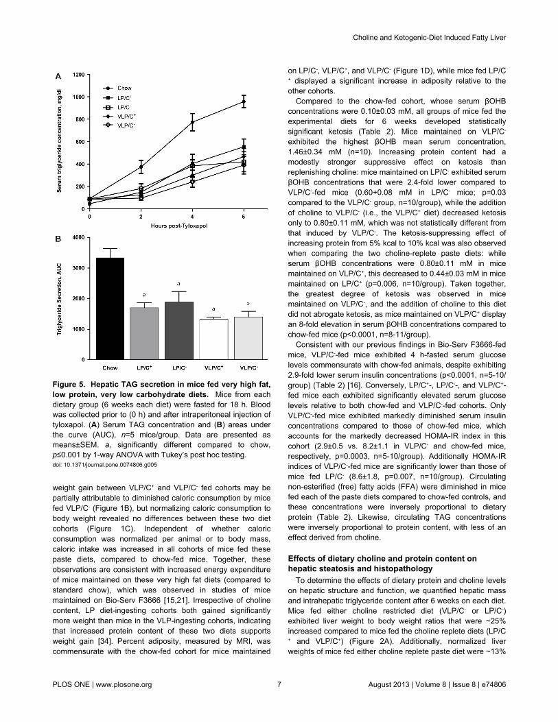
Figure 5. Hepatic TAG secretion in mice fed very high fat,low protein, very low carbohydrate diets. Mice from eachdietary group (6 weeks each diet) were fasted for 18 h. Bloodwas collected prior to (0 h) and after intraperitoneal injection oftyloxapol. (A) Serum TAG concentration and (B) areas underthe curve (AUC), n=5 mice/group. Data are presented asmeans±SEM. a, significantly different compared to chow,p≤0.001 by 1-way ANOVA with Tukey’s post hoc testing.doi: 10.1371/journal.pone.0074806.g005
on LP/C-, VLP/C+, and VLP/C- (Figure 1D), while mice fed LP/C+ displayed a significant increase in adiposity relative to theother cohorts.
Compared to the chow-fed cohort, whose serum βOHBconcentrations were 0.10±0.03 mM, all groups of mice fed theexperimental diets for 6 weeks developed statisticallysignificant ketosis (Table 2). Mice maintained on VLP/C-
exhibited the highest βOHB mean serum concentration,1.46±0.34 mM (n=10). Increasing protein content had amodestly stronger suppressive effect on ketosis thanreplenishing choline: mice maintained on LP/C- exhibited serumβOHB concentrations that were 2.4-fold lower compared toVLP/C--fed mice (0.60+0.08 mM in LP/C- mice; p=0.03compared to the VLP/C- group, n=10/group), while the additionof choline to VLP/C- (i.e., the VLP/C+ diet) decreased ketosisonly to 0.80±0.11 mM, which was not statistically different fromthat induced by VLP/C-. The ketosis-suppressing effect ofincreasing protein from 5% kcal to 10% kcal was also observedwhen comparing the two choline-replete paste diets: whileserum βOHB concentrations were 0.80±0.11 mM in micemaintained on VLP/C+, this decreased to 0.44±0.03 mM in micemaintained on LP/C+ (p=0.006, n=10/group). Taken together,the greatest degree of ketosis was observed in micemaintained on VLP/C-, and the addition of choline to this dietdid not abrogate ketosis, as mice maintained on VLP/C+ displayan 8-fold elevation in serum βOHB concentrations compared tochow-fed mice (p<0.0001, n=8-11/group).
Consistent with our previous findings in Bio-Serv F3666-fedmice, VLP/C--fed mice exhibited 4 h-fasted serum glucoselevels commensurate with chow-fed animals, despite exhibiting2.9-fold lower serum insulin concentrations (p<0.0001, n=5-10/group) (Table 2) [16]. Conversely, LP/C+-, LP/C--, and VLP/C+-fed mice each exhibited significantly elevated serum glucoselevels relative to both chow-fed and VLP/C--fed cohorts. OnlyVLP/C--fed mice exhibited markedly diminished serum insulinconcentrations compared to those of chow-fed mice, whichaccounts for the markedly decreased HOMA-IR index in thiscohort (2.9±0.5 vs. 8.2±1.1 in VLP/C- and chow-fed mice,respectively, p=0.0003, n=5-10/group). Additionally HOMA-IRindices of VLP/C--fed mice are significantly lower than those ofmice fed LP/C- (8.6±1.8, p=0.007, n=10/group). Circulatingnon-esterified (free) fatty acids (FFA) were diminished in micefed each of the paste diets compared to chow-fed controls, andthese concentrations were inversely proportional to dietaryprotein (Table 2). Likewise, circulating TAG concentrationswere inversely proportional to protein content, with less of aneffect derived from choline.
Effects of dietary choline and protein content onhepatic steatosis and histopathology
To determine the effects of dietary protein and choline levelson hepatic structure and function, we quantified hepatic massand intrahepatic triglyceride content after 6 weeks on each diet.Mice fed either choline restricted diet (VLP/C- or LP/C-)exhibited liver weight to body weight ratios that were ~25%increased compared to mice fed the choline replete diets (LP/C+ and VLP/C+) (Figure 2A). Additionally, normalized liverweights of mice fed either choline replete paste diet were ~13%
Choline and Ketogenic-Diet Induced Fatty Liver
PLOS ONE | www.plosone.org 7 August 2013 | Volume 8 | Issue 8 | e74806

smaller compared to chow-fed mice. Hepatic TAG content inmice maintained on VLP/C- (21.5±4.2 mg/g tissue) wasmarkedly higher than that of mice from any of the other pastediet cohorts (Figure 2B). Hepatic TAG content in micemaintained on the LP/C- diet was significantly higher than thoseof mice maintained on LP/C+, VLP/C+, or chow control,indicating that choline restriction in the context of a very highfat diet predisposes to increased hepatic TAG content and thatthis is exacerbated by severe protein limitation. Serum alanineALT activities were significantly elevated in both cohortsmaintained on the choline restricted diets, independent ofprotein content (Figure 2C).
To determine the histopathological changes that accompanyhepatic lipid accumulation in mice maintained on theseketogenic diets, liver sections from cohorts maintained on eachof the diets were stained with hematoxylin and eosin. Liversections from chow-fed animals displayed normal architectureand were devoid of steatosis and inflammatory cell infiltration(Figure 3A–C). Livers from mice maintained on LP/C+ exhibitedvery small amounts of mixed large and small droplet steatosisin hepatocytes located exclusively in acinar zone 2; periportal
and perivenular hepatocytes were virtually free of steatosis(Figure 3D–F). Mice fed the choline-restricted counterpart,LP/C-, displayed a marked increase in mixed large and smalldroplet macrovesicular steatosis in a zone 2 distribution,between the portal tracts and terminal hepatic venules (Figure3G–I).
Low power images from livers from mice fed VLP/C+ wereunremarkable, with no signs of steatosis (Figure 3J-K), whilehigher power images revealed scant small droplet steatosis inhepatocytes in acinar zone 2 (Figure 3L). Conversely, liversfrom mice maintained on VLP/C- diet displayed diffuse andextensive steatosis (Figure 3M–O), primarily of small dropletmacrovesicular and microvesicular morphologies. Diffuseactivation of sinusoid-lining cells, characterized by increasednumbers of intrasinusoidal nuclei, and occasional acidophilbodies were also noted, but no zonal necrosis was observed.While differing markedly in hepatic lipid content, inflammatoryfoci were readily observed in higher power images of hepaticsections from both VLP/C+- and VLP/C--fed mice (Figure 3P-Q). Mitotic figures, an additional sign of hepatocellular injury,were observed uniquely in livers from VLP/C--fed mice (Figure
Figure 6. Abnormal mitochondrial ultrastructure in mice fed a choline restricted, very high fat, low protein, very lowcarbohydrate diet. (A) Transmission electron micrograph of hepatocytes from mice maintained for 6 weeks on standard chowreveals normal mitochondrial structure. Arrows, mitochondria. (B) Higher power image of mitochondria from mice maintained onstandard chow, demonstrating normal cristae. (C) Hepatocyte mitochondria from livers of mice maintained on VLP/C+ exhibitednormal cristae folding and evident double membranes. Sparse microvesicular lipid droplets were also evident (white circularstructure). Arrows, mitochondria; arrowhead; autophagosome. (D) Higher power image of hepatocyte mitochondria from micemaintained on VLP/C+, showing morphology of the cristae. (E) VLP/C- diet induces massive hepatocyte steatosis (note large circularpale fat droplets), and swollen mitochondria with disorganized and dilated cristae. Hepatocyte nucleus is on the right side of theimage. Arrows, mitochondria. (F) Higher power image of hepatocyte mitochondria from mice maintained on VLP/C-, with dilatedcristae (arrow) and an autophagosome (arrowhead). (G) High power image of hepatocyte mitochondria from mice maintained onVLP/C-, with dilated cristae (arrows). Scale bars, 500 nm (A–F), 100 nm (G).doi: 10.1371/journal.pone.0074806.g006
Choline and Ketogenic-Diet Induced Fatty Liver
PLOS ONE | www.plosone.org 8 August 2013 | Volume 8 | Issue 8 | e74806

3R). While immunostaining for F4/80+ macrophages revealedmodest differences in macrophage density (per total number ofcells observed), none of these differences was statisticallysignificant (Figure 4). Moreover, abundances of the mRNAsencoding neither the macrophage marker CD68 nor theinflammatory maker TNFα differed among the diets (data notshown). While the total macrophage density trended lower inmice fed VLP/C-, F4/80+ cell clusters were observed in liverssections from VLP/C--fed mice, but not in hepatic sections frommice fed the other diets (Figure 4A).
Very low carbohydrate/very high fat diets impair VLDLsecretion
Choline deficiency has been linked to impaired VLDLpackaging and secretion in the setting of high carbohydratediets [31]. Because choline is an essential nutrient required forthe formation of phosphatidylcholine (PC), and PC biosynthesisis required for the formation of VLDL particles for egress ofTAG from the liver [35], we determined whether the increasedhepatic TAG content of mice maintained on these cholinerestricted diets was attributable to impaired VLDL secretion byquantifying serum TAG content after overnight-fasted mice hadbeen administered the lipoprotein lipase inhibitor tyloxapol.Surprisingly, while all groups demonstrate significantly impairedVLDL secretion relative to chow-fed control mice, nodifferences among the four paste diet-fed cohorts wereobserved (Figure 5).
Figure 7. Relative hepatic mitochondrial genome contentin mice fed very low protein and carbohydrate, very highfat diets. Quantification of mitochondrial genome copy number(relative abundance) by qPCR using purified liver gDNA frommice maintained on the diets for 6 weeks. Data are presentedas means±SEM; n=4-5/group, *p≤0.05 by 1-way ANOVA withTukey’s post hoc testing.doi: 10.1371/journal.pone.0074806.g007
Influence of dietary choline on mitochondrial structure,number, and function in very low protein andcarbohydrate, very high fat diets
Among the paste diet cohorts, mice maintained on the VLP/C+ and VLP/C- diets exhibited the greatest difference inintrahepatic fat content, both histologically and biochemically,despite similar VLDL secretion capacity and equal caloriccontents of protein, carbohydrate, and fat. Because adequatecholine content is essential for normal mitochondrial structureand function in other macronutrient contexts [36–40], wedetermined whether choline restriction in the VLP dietarycontext was associated with abnormalities of mitochondrialultrastructure. As expected, transmission electron microscopyof liver sections from chow-fed mice revealed that hepatocytemitochondria exhibit smooth outer and inner membranes withtightly-organized cristae (Figure 6A-B). Most hepatocytemitochondria of VLP/C+-fed mice also exhibited normalultrastructure, although a small subset of hepatocytemitochondria lacked organized cristae (Figure 6C-D).Moreover, sparse microvesicular lipid droplets were evident inhepatocytes of VLP/C+-fed mice, a feature that was notobserved in hematoxylin and eosin-stained sections for lightmicroscopy. Unlike hepatocyte mitochondria in chow-fed andVLP/C+-fed mice, all hepatocyte mitochondria of VLP/C--fedmice exhibited swollen and disorganized cristae, and manylacked definitive cristae entirely (Figure 6E–G). Additionally, thespatial organization of mitochondria within the cytoplasm wasmarkedly disrupted by the presence of extensive lipid dropletsin hepatocytes of VLP/C--fed mice.
To further probe whether impairments in mitochondrialfunction contribute to the extensive lipid accumulation observedin mice fed the VLP/C- diet, we quantified the effects of cholinecontent in the VLP dietary contexts on hepatic mitochondrialDNA content. Relative abundance of the mitochondrial genome(mtDNA), normalized to the nuclear genome (nDNA) wasquantified by qPCR using isolated liver genomic DNA fromlivers of chow-, VLP/C+, and VLP/C--fed mice. Primers targetingmt-Atp6, mt-Cox1, mt-Cox3, and mt-Cytb were independentlyemployed as reporters of mtDNA relative abundance,normalized against the nDNA gene Rpl32 (Figure 7). Whilevariation in the normalized mtDNA content was observed,depending on the mtDNA gene quantified, livers of mice fedboth VLP/C+ and VLP/C- exhibited statistically significantdiminutions in mtDNA content, relative to chow-fed controlwhen using mt-Cox3 as a marker, with similar trends observedusing the other three mtDNA markers.
To determine if the marked diminution in hepatic lipid contentin VLP/C+-fed mice could be linked to alterations inmitochondrial bioenergetics, mitochondria were isolated fromlivers of mice maintained for 6 weeks on chow, VLP/C+, orVLP/C- diets, and respiration studies were performed.Substrates palmitoyl-L-carnitine + malate (which donateelectrons to Complexes I and II of the electron transport chain),succinate + rotenone (rotenone is a Complex I inhibitor,allowing electrons from succinate to be delivered exclusively toComplex II), duroquinol (which selectively donates an electronto Complex III), and TMPD (which selectively donates anelectron to Complex IV) + ascorbate (an antioxidant that
Choline and Ketogenic-Diet Induced Fatty Liver
PLOS ONE | www.plosone.org 9 August 2013 | Volume 8 | Issue 8 | e74806

Figure 8. Respiration studies of hepatic mitochondria isolated from mice fed very low protein and carbohydrate, very highfat diets. (A) Respiration rates in the basal leak condition (state 2), ADP-stimulated condition (state 3), F1F0-ATPase independentcondition (state 4), and uncoupled condition in hepatic mitochondria isolated from chow-fed, VLP/C+-fed, and VLP/C--fed (for 6weeks) mice using palmitoyl-L-carnitine and malate as substrates. n=4 mice/group. (B) Relative respiratory ratios of basal leak(state 2/state 3), respiratory control (RCR, state 3/state 4), and coupling control (CCR, state 4/uncoupled), derived from panel A. (C)Respiration rates in states 2-4 and while uncoupled in hepatic mitochondria isolated from chow-, VLP/C+, and VLP/C--fed mice thatrespired using the Complex II-electron donor substrate succinate in the presence of rotenone (Complex I activity inhibitor). n=9mice/group. (D) Relative respiratory ratios of state 2/state 3, state 3/state 4, and state 4/uncoupled, derived from panel C. (E)Respiration rates in states 2-4 in hepatic mitochondria isolated from chow-, VLP/C+, and VLP/C--fed mice that respired using theComplex III-donor substrate duroquinol plus rotenone. n=9 mice/group. (F) Relative respiratory ratios of state 2/state 3, state 3/state4, and state 4/uncoupled, derived from panel E. (G) Respiration rates in states 2-4 in hepatic mitochondria isolated from chow-,VLP/C+, and VLP/C--fed mice that respired using the Complex IV-donor substrate combination TMPD/ascorbate, plus rotenone. n=9mice/group. (H) Relative respiratory ratios of state 2/state 3, state 3/state 4, and state 4/uncoupled, derived from panel G. Data arepresented as means±SEM. *p≤0.05; **p≤0.01 by 1-way ANOVA with Tukey’s post hoc testing.doi: 10.1371/journal.pone.0074806.g008
Choline and Ketogenic-Diet Induced Fatty Liver
PLOS ONE | www.plosone.org 10 August 2013 | Volume 8 | Issue 8 | e74806

preserves TMPD’s reduced state) were independently used toprobe bioenergetic function.
Oxygen consumption in state 2 (basal proton leak) wassignificantly increased in hepatic mitochondria isolated fromVPL/C--fed mice that were tested using palmitoyl-L-carnitine +malate, compared to mitochondria prepared from livers ofchow-fed mice (Figure 8A). However, while trends towardincreased respiratory rates were observed, no significantdifferences among state 3 (ADP-stimulated), state 4 (F1F0 ATP-synthase inhibited), or uncoupled respiration (provoked byaddition of the ionophore FCCP) were observed among thediets using palmitoyl-L-carnitine + malate as substrates (Figure8A). Likewise there were no significant differences among theratios of states 2/3 (basal leak/stimulated ratio), 3/4(Respiratory Control Ratio, RCR), or state 4/uncoupled(Coupling Control Ratio, CCR) (Figure 8B). A trend towarddecreased CCR was observed in the VLP/C- group.
In the presence of succinate + rotenone, hepaticmitochondria from VLP/C+-fed mice exhibited significantlyincreased ADP-stimulated (state 3) oxygen consumption (Fig.8C), which likely accounts for a significantly diminished relativebasal leak ratio (Fig. 8D), compared to chow-fed mice.
Figure 9. Influence of choline restriction in integratedhepatic mitochondrial fatty acyl-CoA metabolism. Red textand red arrows highlight hepatic processes known to beimpaired by administration of experimental choline deficientdiets. Unlike previous observations using lower fat cholinedeficient versus replete formulations, in this study, cholinerepletion in ~90% kcal fat diets did not alter triacylglycerol(TAG) secretion as VLDL. However, choline restriction in a~90% kcal fat diet was associated with mitochondrial structuraland functional abnormalities, which were linked to liver fataccumulation and injury. CPT1, carnitine palmitoyltransferase1; TCAC, tricarboxylic acid cycle; ETC, electron transportchain; ATP, adenosine triphosphate.doi: 10.1371/journal.pone.0074806.g009
Duroquinol-stimulated respiration revealed that Complex III-associated basal leak and oligomycin-inhibited respiration wereincreased selectively in hepatic mitochondria isolated fromVLP/C--fed mice (Fig. 8E-F). Finally, respiratory rates inmitochondria stimulated with TMPD + ascorbate revealed thathepatic mitochondria from VLP/C--fed mice exhibited significantimpairment in ADP-stimulated oxygen consumption, comparedto hepatic mitochondria from chow-fed control mice (Fig. 8G),and consequently, significantly diminished RCR compared tomitochondria from chow-fed animals (Fig. 8H). Using TMPD,mitochondria from VLP/C--fed mice also exhibited a strongtrend toward decreased RCR, when compared to hepaticmitochondria from VLP/C+-fed mice (p=0.06, n=9/group). Takentogether, these results indicate that choline restriction in thesetting of a high fat, very low carbohydrate diet contributes toabnormal hepatic mitochondrial function, potentially impairingterminal oxidation of fatty acids and contributing to hepatic TAGaccumulation. It is important to note that the deficiencies ofmitochondrial coupling and efficiency exhibited by isolatedhepatocyte mitochondria from VLP/C--fed mice likelyunderestimate in vivo bioenergetic dysfunction, because anenriched population of relatively intact mitochondria is assayedusing readily accessible experimental substrates. Thereforeintegration of the mitochondrial structure and disordered spatialorganization with these respiration studies suggestssignificantly impaired mitochondrial function in hepatocytes ofVLP/C--fed mice.
Discussion
The driver mechanisms for beneficial effects of lowcarbohydrate ketogenic diets still remain to be elucidated, andthe prospective roles of these diets in preventing orameliorating human NAFLD are not defined. A commonly usedrodent high fat, and very low protein, very low carbohydrateketogenic diet, Bio-Serv F3666, has been employed tomeasure the effects of the ketogenic nutrient milieu onintegrated metabolic homeostasis, and has also been used tomitigate abnormal phenotypes in the nervous andcardiovascular systems in mutant mouse strains. However, thisdiet also induces hepatic steatosis, inflammation andhepatocyte injury and repair in mice. To determine theunderlying nutritional determinants of these adverse effects, wereplicated this formulation by generating the VLP/C- diet, whichrecapitulated the integrated metabolic and hepatichistopathological responses to Bio-Serv F3666 in C57BL/6Jmice, and then tested three additional diets that systematicallyvaried protein and choline content in VLP/C-. We observed thatwhile protein and choline restriction synergistically contribute tothe integrated metabolic phenotypes provoked by a high fat,very low carbohydrate diet in mice, choline restriction in thissetting stimulated hepatic fat accumulation to a greater extentthan protein restriction. Replenishment of choline in the VLPdiet (i.e., comparing VLP/C- to VLP/C+), markedly diminishedhepatic fat accumulation, while increasing dietary proteincontent to 10% kcal in the setting of restricted choline(comparing VLP/C- to LP/C-) exhibited a smaller mitigatingeffect on hepatic steatosis. Adding protein to a choline-replete
Choline and Ketogenic-Diet Induced Fatty Liver
PLOS ONE | www.plosone.org 11 August 2013 | Volume 8 | Issue 8 | e74806

diet (comparing VLP/C+ to LP/C+) exerted marginal additionaleffect on hepatic histopathology. While subtle variations wereevident in histopathological inflammation and in serologicalevidence of hepatocellular injury (elevated ALT) among micemaintained on the four paste diets, significant differences in thehepatic density of F4/80+ macrophages after 6 weeks on thediets were not observed. We previously demonstrated thatmice maintained on Bio-Serv F3666 (most similar to VLP/C-)exhibit an increased density of hepatic F4/80+ macrophagesafter 12 weeks on the diet [16]. Prolonged exposure to thesehigh fat diets likely exacerbates hepatic inflammation throughmechanisms summarized elsewhere [26]. Only VLP/C-, whichcaused the greatest degree of fat accumulation, inducedhistopathological evidence of hepatocyte regeneration at the 6week time point, suggesting that this diet provokes the greatestdegree of hepatocellular injury. Taken together, these findingssuggest that inflammation and hepatocellular injury are likelysecondary to the extent and duration of the metabolicabnormalities imposed by the nutrient contents.
Experiments in rodents have revealed molecularunderpinnings of liver fat accumulation using methionine andcholine deficient (MCD) diets [30,41], and diminished cholineintake has also been associated with a more aggressiveNAFLD course in humans [42]. The ability of experimentalMCD diets to trigger steatosis and liver injury is facilitated byabundant mono- or disaccharides in the diet, becausereplacing these simple carbohydrates with starch markedlyameliorates the adverse histopathological effects of the MCDdiet, a portion of which is hypothesized to occur throughincreased de novo lipogenesis [31,43]. Our results indicate thatcholine restriction also triggers hepatic steatosis andhepatocellular injury during a nutritional state in which themediators of de novo lipogenesis are transcriptionally silenceddue to very high fat content in the diet and diminishedcirculating insulin concentrations. The metabolism of cholinehas been mechanistically linked in mice to VLDL packagingand secretion through its direct contribution to PC synthesis[35]. Genetic models indicate that metabolic procession ofcholine through the phosphatidylethanolamine (PE) N-methyltransferase pathway, which synthesizes methionine fromhomocysteine and choline-derived methyl groups, is also animportant contributor to VLDL secretion [35,44]. However, inour experiments, while hepatic VLDL secretion was impaired inmice maintained on all the paste diets, no additive effect ofcholine restriction was observed in these macronutrientcontexts. It is therefore unlikely that an impairment of VLDLsecretion explains the marked fatty liver phenotype of VLP/C--fed mice.
Mitochondrial dysfunction is a known contributor to NAFLDpathogenesis [26,45]. Thus, we assessed hepaticmitochondrial structure and function in mice fed these very highfat diets. In mice maintained on VLP/C- diet, we observedchaotic mitochondrial ultrastructure, including swollen cristaeand many degenerating mitochondria. Choline repletion in theVLP diet was associated with improved mitochondrialultrastructure and respiratory coupling and capacity in isolatedmitochondria. However, choline repletion in the VLP diet didnot correct the reduction in mitochondrial DNA content.
Nonetheless, choline repletion may partially mitigatemitochondrial dysfunction by contributing to PC synthesis. PCis a major constituent of all cellular membranes, includingmitochondria [36,37]. Though the exact phospholipidcomposition of membranes varies among experimentalcontexts, PC and PE constitute approximately 40% and 30% ofthe mitochondrial membranes, respectively [46]. Experimentalcholine deficiency in lower fat diet formulations than thosetested here causes a decreased ratio of total hepatic PC/PE[44,47–49] and hepatic mitochondrial dysfunction [38–40].Furthermore, NAFLD patients have a reduction of the totalhepatic PC/PE ratio and impaired hepatocyte membraneintegrity [44,50]. Maintenance of mitochondrial functionrequires homeostatic membrane phospholipid composition tocoordinate surface charge, stereoelectronic relationships, andmembrane dynamics that each effect membrane function andstructural integrity, critical determinants of mitochondrialelectron transport and oxidative phosphorylation [51–53].
Adaptation to a diet with ~90% kcal derived from fat requiresintegration of hepatic mitochondrial fatty acyl-CoA transport,esterification and storage in neutral pools, lipoprotein secretion,and lipolysis, with mitochondrial β-oxidation, ketogenesis,tricarboxylic acid (TCA) cycle, electron transport, and oxidativephosphorylation (Figure 9), and impairment of hepaticmitochondrial fatty acid oxidation contributes to liver fataccumulation [54]. Effective procession of fatty acids throughintegrated mitochondrial pathways also stimulatesgluconeogenesis [55,56]. Of the four paste diets tested,however, only VLP/C--fed mice were not hyperglycemic.Moreover, basal hepatic glucose production of mice maintainedon the VLP/C- parent diet Bio-Serv F3666 is normal, despitedelivering > 90% kcal as fat, and a poor hepatic suppressiveresponse to exogenous insulin [21]. A likely contributor torelative hypoglycemia in VLP/C- and Bio-Serv F3666-fed miceis hepatic mitochondrial dysfunction that prevents adequatelymatched fatty acyl-CoA mitochondrial uptake and terminaloxidation. Differences in extrahepatic glucose disposal couldalso contribute to relative hypoglycemia in VLP/C- mice, butglucose excursions among the four paste diets were notsignificantly different (data not shown). Hyperglycemia in micefed LP/C-, despite steatosis and likely hepatic mitochondrialdysfunction, is multifactorial but is most likely attributable toadditional dietary glucogenic amino acids that supportgluconeogenesis after conversion to TCA cycle intermediates.
Choline deficient diets provoke defects in carnitinepalmitoyltransferase (CPT 1) dependent fatty acyl-CoAtransport [57], palmitate oxidation [57], electron transport [58],and as a result, gluconeogenesis [59,60] (Figure 9).Conversely, ketogenesis is not directly impaired by cholinedeficiency: both the fate-committing ketogenic enzyme,mitochondrial hydroxymethylglutaryl-CoA synthase (HMGCS2),and the enzyme catalyzing the subsequent ketogenic reaction,hydroxymethylglutaryl-CoA lyase, are soluble mitochondrialmatrix enzymes that are influenced only indirectly bydysfunctional mitochondrial membrane structure and function.The results of these experiments strongly suggest that therobust ketosis in VLP/C- mice reports the relative fraction offatty acyl-CoAs that successfully complete hepatic β-oxidation,
Choline and Ketogenic-Diet Induced Fatty Liver
PLOS ONE | www.plosone.org 12 August 2013 | Volume 8 | Issue 8 | e74806

and not those that fail to gain entry into mitochondria or thosethat cannot complete terminal oxidation. Therefore, comparedto their choline restricted counterparts, ketosis was lesspronounced in mice fed the very high fat, choline replete dietsin part because integrated hepatic mitochondrial functionsupports mitochondrial acyl-CoA uptake and terminal oxidation.Conversely, relative suppression of ketosis in mice fed cholinerestricted LP/C-, compared to mice fed VLP/C-, is attributable to(i) the sensitivity in rodents to amino acid-mediated insulinsecretion [22,61], which in turn suppresses ketogenesis [62],and (ii) the modest ability of amino acid-derived gluconeogenicmetabolites to suppress ketogenesis [63].
The independent contributions of choline and methioninedeficiencies to abnormal hepatic metabolism and pathology inmice are incompletely understood because studies performedheretofore suggest macronutrient context-dependent effects ofeach of these micronutrients. In the context of carbohydrate-enriched diets, selective choline deficiency drives hepaticsteatosis, while selective methionine deficiency has beenlinked to hepatic inflammation, oxidative stress andhepatocellular injury [64]. Conversely, mice fed a high fat (60%kcal), 26% kcal carbohydrate diet are protected against hepaticTAG accumulation, and do not exhibit overt inflammation orhepatocellular injury by methionine restriction [65]. Becausethresholds for methionine deficiency-induced hepatocellularinjury are unknown and are highly dependent on macronutrientdistribution, these studies were not designed to definitivelysegregate the independent roles of methionine and cholinerestriction in the observed hepatic phenotypes. Nonetheless,this study supports the supplementation of choline in rodentexperiments that are targeted to understand the physiologicaladaptations and responses to diet-induced ketosis. Theseexperiments also demonstrate that nutritional induction ofketosis by a very high fat, very low carbohydrate diet in micecan occur in the context of maintained weight andhyperglycemia. Therefore, experimental use of VLP/C+ dietmay allow mechanistic partitioning of the relative roles ofglycemia versus ketosis in experimental conditions that areameliorated by ketogenic diets.
An additional limitation of this study is that formal methods ofhepatic biometry and stereology were not used forhistopathological quantification of hepatocyte and fat dropletsizes [66], and for histopathology, relatively small numbers ofanimals were analyzed for each group. Nonetheless, hepatichistopathological characteristics were highly uniform amonganimals within each diet condition, and complementary formalbiochemical quantification of hepatic fat was performed in
larger numbers of animals. Moreover, effects of each of the fivediets studied were determined in at least three separatecohorts of mice that were matched for age, strain, sex, andbody weight at the time of initiation of each of the study diets.Analyses of histopathology, serum and hepatic biochemistry,and mitochondrial function were performed among thesereplicating cohorts to confirm reproducibility of the results.Future experiments will incorporate formal histopathologicalquantifications that reveal the mechanistic determinantsrelating these nutritional states to hepatic lipid droplet size andabundance.
In summary, these experiments demonstrate that the uniquesignature of hepatic lipid accumulation, inflammation, andcellular and mitochondrial injury induced in mice maintained ona very high fat, protein-restricted, very low carbohydrate,ketogenic diet can be linked to the adverse effects of cholinedeficiency on hepatic mitochondrial structure and function. Thefindings also underscore the concept that ketosis does notnecessarily report effective hepatic fatty acid oxidation whensupply overwhelms relative mitochondrial capacity. In setting ofthe VLP/C- diet, ketogenesis provides a spill-over pathway thatonly partially compensates for inadequate fatty acid catabolism.However, it is important to note that while incompletely studied,ketogenic diets in humans are unlikely to cause mitochondrialdysfunction or fatty liver. Nonetheless, attentiveness to cholinecontent, and its downstream effects on membrane phospholipidcomposition and function are important considerations whendesigning and measuring the physiological effects of lowcarbohydrate diets, which ultimately will require integration ofbiochemical approaches to quantify compartment-specificphospholipidomics with measurements of mitochondrialfunction, biogenesis, and autophagic turnover.
Acknowledgements
The authors thank Jessica Flowers (Harlan-Teklad), AbhinavDiwan, and David Cotter for helpful discussions, Joan Avery inthe Center for Cardiovascular Research for assistance withconfocal microscopy, the Department of Pathology andImmunology Research Electron Microscopy Core, and LauraKyro for assistance with graphics.
Author Contributions
Conceived and designed the experiments: RCS XH PAC.Performed the experiments: RCS XH ARM. Analyzed the data:RCS XH EMB PAC. Wrote the manuscript: RCS PAC.
References
1. Foster GD, Wyatt HR, Hill JO, Makris AP, Rosenbaum DL et al. (2010)Weight and metabolic outcomes after 2 years on a low-carbohydrateversus low-fat diet: a randomized trial. Ann Intern Med 153: 147-157.doi:10.7326/0003-4819-153-3-201008030-00005. PubMed: 20679559.
2. Shai I, Schwarzfuchs D, Henkin Y, Shahar DR, Witkow S et al. (2008)Weight loss with a low-carbohydrate, Mediterranean, or low-fat diet. NEngl J Med 359: 229-241. doi:10.1056/NEJMoa0708681. PubMed:18635428.
3. Kirk JK, Graves DE, Craven TE, Lipkin EW, Austin M et al. (2008)Restricted-carbohydrate diets in patients with type 2 diabetes: a meta-
analysis. J Am Diet Assoc 108: 91-100. doi:10.1016/j.jada.2008.06.251.PubMed: 18155993.
4. Veech RL (2004) The therapeutic implications of ketone bodies: theeffects of ketone bodies in pathological conditions: ketosis, ketogenicdiet, redox states, insulin resistance, and mitochondrial metabolism.Prostaglandins Leukot Essent Fatty Acids 70: 309-319. doi:10.1016/j.plefa.2003.09.007. PubMed: 14769489.
5. Westman EC, Feinman RD, Mavropoulos JC, Vernon MC, Volek JS etal. (2007) Low-carbohydrate nutrition and metabolism. Am J Clin Nutr86: 276-284. PubMed: 17684196.
Choline and Ketogenic-Diet Induced Fatty Liver
PLOS ONE | www.plosone.org 13 August 2013 | Volume 8 | Issue 8 | e74806

6. Freeman JM, Kossoff EH (2010) Ketosis and the ketogenic diet, 2010:advances in treating epilepsy and other disorders. Adv Pediatr 57:315-329. doi:10.1016/j.yapd.2010.08.003. PubMed: 21056745.
7. Danial NN, Hartman AL, Stafstrom CE, Thio LL (2013) How Does theKetogenic Diet Work?: Four Potential Mechanisms. J Child NeurolEpub ahead of print. PubMed: 23670253.
8. Ruskin DN, Svedova J, Cote JL, Sandau U, Rho JM et al. (2013)Ketogenic Diet Improves Core Symptoms of Autism in BTBR Mice.PLOS ONE 8: e65021. doi:10.1371/journal.pone.0065021. PubMed:23755170.
9. Maalouf M, Rho JM, Mattson MP (2009) The neuroprotective propertiesof calorie restriction, the ketogenic diet, and ketone bodies. Brain ResRev 59: 293-315. doi:10.1016/j.brainresrev.2008.09.002. PubMed:18845187.
10. Yao J, Chen S, Mao Z, Cadenas E, Brinton RD (2011) 2-Deoxy-D-glucose treatment induces ketogenesis, sustains mitochondrialfunction, and reduces pathology in female mouse model of Alzheimer’sdisease. PLOS ONE 6: e21788. doi:10.1371/journal.pone.0021788.PubMed: 21747957.
11. Yang X, Cheng B (2010) Neuroprotective and anti-inflammatoryactivities of ketogenic diet on MPTP-induced neurotoxicity. J MolNeurosci 42: 145-153. doi:10.1007/s12031-010-9336-y. PubMed:20333481.
12. Krebs P, Fan W, Chen YH, Tobita K, Downes MR et al. (2011) Lethalmitochondrial cardiomyopathy in a hypomorphic Med30 mouse mutantis ameliorated by ketogenic diet. Proc Natl Acad Sci U S A 108:19678-19682. doi:10.1073/pnas.1117835108. PubMed: 22106289.
13. Seyfried TN, Kiebish MA, Marsh J, Shelton LM, Huysentruyt LC et al.(2011) Metabolic management of brain cancer. Biochim Biophys Acta1807: 577-594. doi:10.1016/j.bbabio.2010.08.009. PubMed: 20804725.
14. Poff AM, Ari C, Seyfried TN, D’Agostino DP. (2013) The ketogenic dietand hyperbaric oxygen therapy prolong survival in mice with systemicmetastatic cancer. PLOS ONE 8: e65522. doi:10.1371/journal.pone.0065522. PubMed: 23755243.
15. Kennedy AR, Pissios P, Otu H, Roberson R, Xue B et al. (2007) A high-fat, ketogenic diet induces a unique metabolic state in mice. Am JPhysiol Endocrinol Metab 292: E1724-E1739. doi:10.1152/ajpendo.00717.2006. PubMed: 17299079.
16. Garbow JR, Doherty JM, Schugar RC, Travers S, Weber ML et al.(2011) Hepatic steatosis, inflammation, and ER stress in micemaintained long-term on a very low carbohydrate ketogenic diet. AmerJ Physiol GI/Liver 300: G956-G967.
17. Badman MK, Pissios P, Kennedy AR, Koukos G, Flier JS et al. (2007)Hepatic fibroblast growth factor 21 is regulated by PPARalpha and is akey mediator of hepatic lipid metabolism in ketotic states. Cell Metab 5:426-437. doi:10.1016/j.cmet.2007.05.002. PubMed: 17550778.
18. Wentz AE, d’Avignon DA, Weber ML, Cotter DG, Doherty JM et al.(2010) Adaptation of myocardial substrate metabolism to a ketogenicnutrient environment. J Biol Chem 285: 24447-24456. doi:10.1074/jbc.M110.100651. PubMed: 20529848.
19. Srivastava S, Baxa U, Niu G, Chen X, Lv R (2013) A ketogenic dietincreases brown adipose tissue mitochondrial proteins and UCP1levels in mice. IUBMB Life 65: 58-66. doi:10.1002/iub.1102. PubMed:23233333.
20. Badman MK, Kennedy AR, Adams AC, Pissios P, Maratos-Flier E(2009) A Very Low Carbohydrate Ketogenic Diet Improves GlucoseTolerance in ob/ob Mice Independent of Weight Loss. Am J PhysiolEndocrinol Metab 297: E1197-E1204. doi:10.1152/ajpendo.00357.2009. PubMed: 19738035.
21. Jornayvaz FR, Jurczak MJ, Lee HY, Birkenfeld AL, Frederick DW et al.(2010) A high-fat, ketogenic diet causes hepatic insulin resistance inmice despite increasing energy expenditure and preventing weightgain. Am J Physiol Endocrinol Metab 299: E808-E815. doi:10.1152/ajpendo.00361.2010. PubMed: 20807839.
22. Bielohuby M, Menhofer D, Kirchner H, Stoehr BJ, Müller TD et al.(2011) Induction of ketosis in rats fed low-carbohydrate, high fat dietsdepends on the relative abundance of dietary fat and protein. Am JPhysiol Endocrinol Metab 300: E65–E76. doi:10.1152/ajpendo.00478.2010. PubMed: 20943751.
23. Badman MK, Koester A, Flier JS, Kharitonenkov A, Maratos-Flier E(2009) Fibroblast Growth Factor 21-Deficient Mice DemonstrateImpaired Adaptation to Ketosis. Endocrinology 150: 4931-4940. doi:10.1210/en.2009-0532. PubMed: 19819944.
24. Crawford PA, Crowley JR, Sambandam N, Muegge BD, Costello EK etal. (2009) Regulation of myocardial ketone body metabolism by the gutmicrobiota during nutrient deprivation. Proc Natl Acad Sci U S A 106:11276-11281. doi:10.1073/pnas.0902366106. PubMed: 19549860.
25. Thio LL, Erbayat-Altay E, Rensing N, Yamada KA (2006) Leptincontributes to slower weight gain in juvenile rodents on a ketogenic
diet. Pediatr Res 60: 413-417. doi:10.1203/01.pdr.0000238244.54610.27. PubMed: 16940251.
26. Fabbrini E, Sullivan S, Klein S (2010) Obesity and nonalcoholic fattyliver disease: biochemical, metabolic, and clinical implications.Hepatology 51: 679-689. doi:10.1002/hep.23280. PubMed: 20041406.
27. Browning JD, Baker JA, Rogers T, Davis J, Satapati S et al. (2011)Short-term weight loss and hepatic triglyceride reduction: evidence of ametabolic advantage with dietary carbohydrate restriction. Am J ClinNutr 93: 1048-1052. doi:10.3945/ajcn.110.007674. PubMed: 21367948.
28. Pérez-Guisado J, Muñoz-Serrano A (2011) The effect of the SpanishKetogenic Mediterranean Diet on nonalcoholic fatty liver disease: a pilotstudy. J Med Food 14: 677-680. doi:10.1089/jmf.2011.0075. PubMed:21688989.
29. Flores H, Pak N, Maccioni A, Monckeberg F (1970) Lipid transport inkwashiorkor. Br J Nutr 24: 1005-1011. doi:10.1079/BJN19700103.PubMed: 5484721.
30. Maher JJ (2011) New insights from rodent models of fatty liver disease.Antioxid Redox Signal 15: 535-550. doi:10.1089/ars.2010.3749.PubMed: 21126212.
31. Pickens MK, Yan JS, Ng RK, Ogata H, Grenert JP et al. (2009) Dietarysucrose is essential to the development of liver injury in the methionine-choline-deficient model of steatohepatitis. J Lipid Res 50: 2072-2082.doi:10.1194/jlr.M900022-JLR200. PubMed: 19295183.
32. Lehman JJ, Boudina S, Banke NH, Sambandam N, Han X et al. (2008)The transcriptional coactivator PGC-1alpha is essential for maximaland efficient cardiac mitochondrial fatty acid oxidation and lipidhomeostasis. Am J Physiol Heart Circ Physiol 295: H185-H196. doi:10.1152/ajpheart.00081.2008. PubMed: 18487436.
33. Shiemke AK, Cook SA, Miley T, Singleton P (1995) Detergentsolubilization of membrane-bound methane monooxygenase requiresplastoquinol analogs as electron donors. Arch Biochem Biophys 321:421-428. doi:10.1006/abbi.1995.1413. PubMed: 7646068.
34. Goettsch M (1960) Comparative protein requirement of the rat andmouse for growth, reproduction and lactation using casein diets. J Nutr70: 307-312.
35. Corbin KD, Zeisel SH (2012) Choline metabolism provides novelinsights into nonalcoholic fatty liver disease and its progression. CurrOpin Gastroenterol 28: 159-165. doi:10.1097/MOG.0b013e32834e7b4b. PubMed: 22134222.
36. Daum G (1985) Lipids of mitochondria. Biochim Biophys Acta 822:1-42. doi:10.1016/0304-4157(85)90002-4. PubMed: 2408671.
37. Colbeau A, Nachbaur J, Vignais PM (1971) Enzymic characterizationand lipid composition of rat liver subcellular membranes. BiochimBiophys Acta 249: 462-492. doi:10.1016/0005-2736(71)90123-4.PubMed: 5134192.
38. Oliveira CP, Coelho AM, Barbeiro HV, Lima VM, Soriano F et al. (2006)Liver mitochondrial dysfunction and oxidative stress in thepathogenesis of experimental nonalcoholic fatty liver disease. Braz JMed Biol Res 39: 189-194. PubMed: 16470305.
39. Petrosillo G, Portincasa P, Grattagliano I, Casanova G, Matera M et al.(2007) Mitochondrial dysfunction in rat with nonalcoholic fatty liverInvolvement of complex I, reactive oxygen species and cardiolipin.Biochim Biophys Acta 1767: 1260-1267. doi:10.1016/j.bbabio.2007.07.011. PubMed: 17900521.
40. Romestaing C, Piquet MA, Letexier D, Rey B, Mourier A et al. (2008)Mitochondrial adaptations to steatohepatitis induced by a methionine-and choline-deficient diet. Am J Physiol Endocrinol Metab 294: E110-E119. PubMed: 17986629.
41. Handler P, Dubin IN (1946) The significance of fatty infiltration in thedevelopment of hepatic cirrhosis due to choline deficiency. J Nutr 31:141-159. PubMed: 21015910.
42. Guerrerio AL, Colvin RM, Schwartz AK, Molleston JP, Murray KF et al.(2012) Choline intake in a large cohort of patients with nonalcoholicfatty liver disease. Am J Clin Nutr 95: 892-900. doi:10.3945/ajcn.111.020156. PubMed: 22338037.
43. Pickens MK, Ogata H, Soon RK, Grenert JP, Maher JJ (2010) Dietaryfructose exacerbates hepatocellular injury when incorporated into amethionine-choline-deficient diet. Liver Int 30: 1229-1239. doi:10.1111/j.1478-3231.2010.02285.x. PubMed: 20536716.
44. Li Z, Agellon LB, Allen TM, Umeda M, Jewell L et al. (2006) The ratio ofphosphatidylcholine to phosphatidylethanolamine influences membraneintegrity and steatohepatitis. Cell Metab 3: 321-331. doi:10.1016/j.cmet.2006.03.007. PubMed: 16679290.
45. Sunny NE, Parks EJ, Browning JD, Burgess SC (2011) ExcessiveHepatic Mitochondrial TCA Cycle and Gluconeogenesis in Humanswith Nonalcoholic Fatty Liver Disease. Cell Metab 14: 804-810. doi:10.1016/j.cmet.2011.11.004. PubMed: 22152305.
Choline and Ketogenic-Diet Induced Fatty Liver
PLOS ONE | www.plosone.org 14 August 2013 | Volume 8 | Issue 8 | e74806

46. Osman C, Voelker DR, Langer T (2011) Making heads or tails ofphospholipids in mitochondria. J Cell Biol 192: 7-16. doi:10.1083/JCB1926OIA7. PubMed: 21220505.
47. McMahon KE, Farrell PM (1986) Effect of choline deficiency on lungphospholipid concentrations in the rat. J Nutr 116: 936-943. PubMed:3755165.
48. Mas E, Danjoux M, Garcia V, Carpentier S, Segui B et al. (2009) IL-6deficiency attenuates murine diet-induced non-alcoholic steatohepatitis.PLOS ONE 4: e7929.
49. Sánchez-Garrido MA, Chico Y, González R, Ranchal I, González-RubioS et al. (2009) Interleukin-6 is associated with liver lipid homeostasisbut not with cell death in experimental hepatic steatosis. Innate Immun15: 337-349. doi:10.1177/1753425909104900. PubMed: 19710104.
50. Arendt BM, Ma DW, Simons B, Noureldin SA, Therapondos G et al.(2013) Nonalcoholic fatty liver disease is associated with lower hepaticand erythrocyte ratios of phosphatidylcholine tophosphatidylethanolamine. Appl Physiol Nutr Metab 38: 334-340. doi:10.1139/apnm-2012-0261. PubMed: 23537027.
51. Tasseva G, Bai HD, Davidescu M, Haromy A, Michelakis E et al. (2013)Phosphatidylethanolamine deficiency in Mammalian mitochondriaimpairs oxidative phosphorylation and alters mitochondrial morphology.J Biol Chem 288: 4158-4173. doi:10.1074/jbc.M112.434183. PubMed:23250747.
52. Zick M, Rabl R, Reichert AS (2009) Cristae formation-linkingultrastructure and function of mitochondria. Biochim Biophys Acta 1793:5-19. doi:10.1016/j.bbamcr.2008.06.013. PubMed: 18620004.
53. Paumard P, Vaillier J, Coulary B, Schaeffer J, Soubannier V et al.(2002) The ATP synthase is involved in generating mitochondrialcristae morphology. EMBO J 21: 221-230. doi:10.1093/emboj/21.3.221.PubMed: 11823415.
54. An J, Muoio DM, Shiota M, Fujimoto Y, Cline GW et al. (2004) Hepaticexpression of malonyl-CoA decarboxylase reverses muscle, liver andwhole-animal insulin resistance. Nat Med 10: 268-274. doi:10.1038/nm995. PubMed: 14770177.
55. Williamson JR, Scholz R, Browning ET (1969) Control mechanisms ofgluconeogenesis and ketogenesis. II. Interactions between fatty acidoxidation and the citric acid cycle in perfused rat liver. J Biol Chem 244:4617-4627. PubMed: 5808508.
56. Williamson JR, Browning ET, Scholz R (1969) Control mechanisms ofgluconeogenesis and ketogenesis. I. Effects of oleate on
gluconeogenesis in perfused rat liver. J Biol Chem 244: 4607-4616.PubMed: 4390110.
57. Serviddio G, Giudetti AM, Bellanti F, Priore P, Rollo T et al. (2011)Oxidation of hepatic carnitine palmitoyl transferase-I (CPT-I) impairsfatty acid beta-oxidation in rats fed a methionine-choline deficient diet.PLOS ONE 6: e24084. doi:10.1371/journal.pone.0024084. PubMed:21909411.
58. Teodoro JS, Rolo AP, Duarte FV, Simões AM, Palmeira CM (2008)Differential alterations in mitochondrial function induced by a choline-deficient diet: understanding fatty liver disease progression.Mitochondrion 8: 367-376. doi:10.1016/j.mito.2008.07.008. PubMed:18765303.
59. Raubenheimer PJ, Nyirenda MJ, Walker BR (2006) A choline-deficientdiet exacerbates fatty liver but attenuates insulin resistance andglucose intolerance in mice fed a high-fat diet. Diabetes 55: 2015-2020.doi:10.2337/db06-0097. PubMed: 16804070.
60. Wu G, Zhang L, Li T, Lopaschuk G, Vance DE et al. (2012) CholineDeficiency Attenuates Body Weight Gain and Improves GlucoseTolerance in ob/ob Mice. J Obes, 2012: 319172. PubMed: 22778916
61. MacDonald MJ, Fahien LA, Brown LJ, Hasan NM, Buss JD et al. (2005)Perspective: emerging evidence for signaling roles of mitochondrialanaplerotic products in insulin secretion. Am J Physiol EndocrinolMetab 288: E1-15. PubMed: 15585595.
62. Cotter DG, Schugar RC, Crawford PA (2013) Ketone body metabolismand cardiovascular disease. Am J Physiol Heart Circ Physiol 304:H1060-H1076. doi:10.1152/ajpheart.00646.2012. PubMed: 23396451.
63. Krebs HA, Hems R (1970) Fatty acid metabolism in the perfused ratliver. Biochem J 119: 525-533. PubMed: 5500312.
64. Caballero F, Fernández A, Matías N, Martínez L, Fucho R et al. (2010)Specific contribution of methionine and choline in nutritionalnonalcoholic steatohepatitis: impact on mitochondrial S-adenosyl-L-methionine and glutathione. J Biol Chem 285: 18528-18536. doi:10.1074/jbc.M109.099333. PubMed: 20395294.
65. Ables GP, Perrone CE, Orentreich D, Orentreich N (2012) Methionine-Restricted C57BL/6J Mice Are Resistant to Diet-Induced Obesity andInsulin Resistance but Have Low Bone Density. PLOS ONE 7: e51357.doi:10.1371/journal.pone.0051357. PubMed: 23236485.
66. Aguila MB, Pinheiro Ada R, Parente LB, Mandarim-de-Lacerda CA(2003) Dietary effect of different high-fat diet on rat liver stereology.Liver Int 23: 363-370. doi:10.1034/j.1478-3231.2003.00858.x. PubMed:14708898.
Choline and Ketogenic-Diet Induced Fatty Liver
PLOS ONE | www.plosone.org 15 August 2013 | Volume 8 | Issue 8 | e74806


![Research Paper extends lifespan and delays tumorigenesis ...€¦ · Research Paper 3 [32] whereas p53-deficiency resulted in pro-inflammatory phenotype [33, 34]. Noteworthy, the](https://img.dokumen.tips/doc/110x75/5f12253a4e2350741512f060/research-paper-extends-lifespan-and-delays-tumorigenesis-research-paper-3-32.jpg)


