Embed Size (px)
Citation preview
REVIEW Open Access
Role of chaperone-mediated autophagy inthe pathophysiology including pulmonarydisordersYusuke Hosaka†, Jun Araya*†, Yu Fujita and Kazuyoshi Kuwano
Abstract
Autophagy is a highly conserved mechanism of delivering cytoplasmic components for lysosomal degradation.Among the three major autophagic pathways, chaperone-mediated autophagy (CMA) is primarily characterized byits selective nature of protein degradation, which is mediated by heat shock cognate 71 kDa protein (HSC70: alsoknown as HSPA8) recognition of the KFERQ peptide motif in target proteins. Lysosome-associated membraneprotein type 2A (LAMP2A) is responsible for substrate binding and internalization to lysosomes, and thus, thelysosomal expression level of LAMP2A is a rate-limiting factor for CMA. Recent advances have uncovered not onlyphysiological but also pathological role of CMA in multiple organs, including neurodegenerative disorders, kidneydiseases, liver diseases, heart diseases, and cancers through the accumulation of unwanted proteins or increaseddegradation of target proteins with concomitant metabolic alterations resulting from CMA malfunction. Withrespect to pulmonary disorders, the involvement of CMA has been demonstrated in lung cancer and chronicobstructive pulmonary disease (COPD) pathogenesis through regulating apoptosis. Further understanding of CMAmachinery may shed light on the molecular mechanisms of refractory disorders and lead to novel treatmentmodalities through CMA modulation.
Keywords: Autophagy, Chaperone-mediated autophagy, Chronic obstructive pulmonary disease, Lung cancer
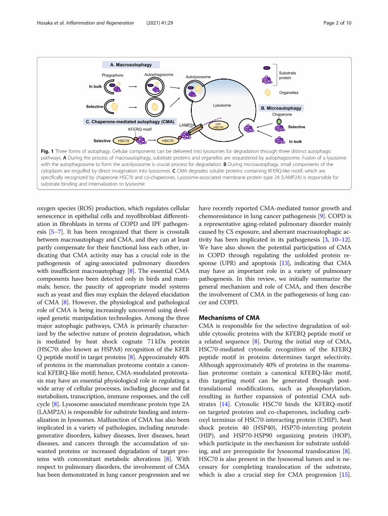
BackgroundAutophagy is a highly conserved mechanism of deliver-ing cytoplasmic components for lysosomal degradationto maintain the homeostatic balance between the syn-thesis, degradation, and recycling of cellular proteinsand organelles [1]. Three forms of distinct autophagyhave been identified: chaperon-mediated autophagy(CMA), microautophagy, and macroautophagy [1] (Fig.1). CMA is a type of selective autophagy for the lyso-somal degradation of proteins with the KFERQ peptidemotif. Microautophagy requires small components ofthe cytoplasm to be engulfed by direct invagination intolysosomes. During the process of macroautophagy,
substrate proteins and organelles are sequestered by theautophagosome. The fusion of a lysosome with theautophagosome to form the autolysosome is a crucialprocess for degradation [1]. Because macroautophagy isthe best-characterized form of autophagy, recent studiesof the molecular mechanisms and pathophysiological ef-fects of autophagy have mainly focused on macroauto-phagy. We have reported pathogenic involvement ofmacroautophagy in idiopathic pulmonary fibrosis (IPF),a form of progressive fibrosing interstitial pneumoniaand in chronic obstructive pulmonary disease (COPD),which is characterized by progressive airflow limitationmainly caused by cigarette smoke (CS) exposure [2, 3].Both IPF and COPD are aging-associated pulmonarydisorders and the lysosomal function declines with aging[4]. Insufficient macroautophagy, including mitochondria-selective mitophagy enhances mitochondrial reactive
© The Author(s). 2021 Open Access This article is licensed under a Creative Commons Attribution 4.0 International License,which permits use, sharing, adaptation, distribution and reproduction in any medium or format, as long as you giveappropriate credit to the original author(s) and the source, provide a link to the Creative Commons licence, and indicate ifchanges were made. The images or other third party material in this article are included in the article's Creative Commonslicence, unless indicated otherwise in a credit line to the material. If material is not included in the article's Creative Commonslicence and your intended use is not permitted by statutory regulation or exceeds the permitted use, you will need to obtainpermission directly from the copyright holder. To view a copy of this licence, visit http://creativecommons.org/licenses/by/4.0/.
* Correspondence: [email protected]†Yusuke Hosaka and Jun Araya contributed equally to this work.Division of Respiratory Diseases, Department of Internal Medicine, The JikeiUniversity School of Medicine, 3-25-8 Nishi-shimbashi, Minato-ku, Tokyo105-8461, Japan
Inflammation and RegenerationHosaka et al. Inflammation and Regeneration (2021) 41:29 https://doi.org/10.1186/s41232-021-00180-9
oxygen species (ROS) production, which regulates cellularsenescence in epithelial cells and myofibroblast differenti-ation in fibroblasts in terms of COPD and IPF pathogen-esis [5–7]. It has been recognized that there is crosstalkbetween macroautophagy and CMA, and they can at leastpartly compensate for their functional loss each other, in-dicating that CMA activity may has a crucial role in thepathogenesis of aging-associated pulmonary disorderswith insufficient macroautophagy [8]. The essential CMAcomponents have been detected only in birds and mam-mals; hence, the paucity of appropriate model systemssuch as yeast and flies may explain the delayed elucidationof CMA [8]. However, the physiological and pathologicalrole of CMA is being increasingly uncovered using devel-oped genetic manipulation technologies. Among the threemajor autophagic pathways, CMA is primarily character-ized by the selective nature of protein degradation, whichis mediated by heat shock cognate 71 kDa protein(HSC70: also known as HSPA8) recognition of the KFERQ peptide motif in target proteins [8]. Approximately 40%of proteins in the mammalian proteome contain a canon-ical KFERQ-like motif; hence, CMA-modulated proteosta-sis may have an essential physiological role in regulating awide array of cellular processes, including glucose and fatmetabolism, transcription, immune responses, and the cellcycle [8]. Lysosome-associated membrane protein type 2A(LAMP2A) is responsible for substrate binding and intern-alization in lysosomes. Malfunction of CMA has also beenimplicated in a variety of pathologies, including neurode-generative disorders, kidney diseases, liver diseases, heartdiseases, and cancers through the accumulation of un-wanted proteins or increased degradation of target pro-teins with concomitant metabolic alterations [8]. Withrespect to pulmonary disorders, the involvement of CMAhas been demonstrated in lung cancer progression and we
have recently reported CMA-mediated tumor growth andchemoresistance in lung cancer pathogenesis [9]. COPD isa representative aging-related pulmonary disorder mainlycaused by CS exposure, and aberrant macroautophagic ac-tivity has been implicated in its pathogenesis [3, 10–12].We have also shown the potential participation of CMAin COPD through regulating the unfolded protein re-sponse (UPR) and apoptosis [13], indicating that CMAmay have an important role in a variety of pulmonarypathogenesis. In this review, we initially summarize thegeneral mechanism and role of CMA, and then describethe involvement of CMA in the pathogenesis of lung can-cer and COPD.
Mechanisms of CMACMA is responsible for the selective degradation of sol-uble cytosolic proteins with the KFERQ peptide motif ora related sequence [8]. During the initial step of CMA,HSC70-mediated cytosolic recognition of the KFERQpeptide motif in proteins determines target selectivity.Although approximately 40% of proteins in the mamma-lian proteome contain a canonical KFERQ-like motif,this targeting motif can be generated through post-translational modifications, such as phosphorylation,resulting in further expansion of potential CMA sub-strates [14]. Cytosolic HSC70 binds the KFERQ-motifon targeted proteins and co-chaperones, including carb-oxyl terminus of HSC70-interacting protein (CHIP), heatshock protein 40 (HSP40), HSP70-intercting protein(HIP), and HSP70-HSP90 organizing protein (HOP),which participate in the mechanism for substrate unfold-ing, and are prerequisite for lysosomal translocation [8].HSC70 is also present in the lysosomal lumen and is ne-cessary for completing translocation of the substrate,which is also a crucial step for CMA progression [15].
Fig. 1 Three forms of autophagy. Cellular components can be delivered into lysosomes for degradation through three distinct autophagicpathways. A During the process of macroautophagy, substrate proteins and organelles are sequestered by autophagosome. Fusion of a lysosomewith the autophagosome to form the autolysosome is crucial process for degradation. B During microautophagy, small components of thecytoplasm are engulfed by direct invagination into lysosomes. C CMA degrades soluble proteins containing KFERQ-like motif, which arespecifically recognized by chaperone HSC70 and co-chaperones. Lysosome-associated membrane protein type 2A (LAMP2A) is responsible forsubstrate binding and internalization to lysosome
Hosaka et al. Inflammation and Regeneration (2021) 41:29 Page 2 of 10

The role of HSC70 in autophagy is not restricted toCMA but is also linked to chaperone-assisted selectiveautophagy (a type of selective macroautophagy forubiquitin-positive protein aggregates) [16] and microau-tophagy [17], suggesting the potential implication ofHSC70 in conducting all type of autophagic processes[8].LAMP2 is a lysosomal component that is indispens-
able for completing CMA [8]. Among the three isoformsof LAMP2 (LAMP2A, 2B, and 2C), LAMP2A is the onlyisoform that is necessary for the CMA machinery.LAMP2A is responsible for both substrate binding andinternalization to lysosomes. The cytosolic tail domainof LAMP2A is crucially required for binding to substratecomplexes containing HSC70 [18]. Multimerization ofLAMP2A to form a 700 kDa protein complex, a trans-membrane protein channel is an essential process forsubstrate translocation into the lumen [19]. The lyso-somal expression level of LAMP2A is considered to bethe rate-limiting factor for CMA activity and can be al-tered by synthesis, degradation, and redistribution [8].Oxidative stress and genotoxic damage have been shownto induce de novo synthesis of LAMP2A [20, 21], indi-cating a protective role for CMA in responding to cellstress conditions. Although the detailed mechanisms fortranscriptional upregulation of LAMP2A remain uncer-tain, the involvement of nuclear factor of activated Tcells (NFAT1) has been reported in the transcriptionalupregulation of LAMP2A in T cells [22]. Nrf2 is a mas-ter transcription factor that orchestrates the antioxidantdefense system via expression of a wide array of antioxi-dant enzymes for redox homeostasis and cell survival inresponse to oxidative stress [23]. It has been reportedthat forced Nrf2 expression prevents CMA decline andhas a neuroprotective effect in a mouse model of Parkin-son’s disease [24]. A recent paper has demonstrated thatNrf2 upregulates LAMP2A expression levels by bindingto the LAMP2A gene [25], which may at least partly ex-plain the mechanism for CMA activation during oxida-tive stress. We have also detected the participation ofNrf2-mediated LAMP2A expression in cigarette smoke(CS)-induced CMA activation [13]. Alternatively, in-crease in LAMP2A levels is also mediated through alter-ations of protein stability in starvation conditions. Anextension of half-life of LAMP2A from 36 h to 72 h hasbeen reported in cultured hepatocytes and fibroblasts inresponse to prolonged starvation [26].In addition to basal CMA activity, CMA is upregulated
in response to a wide array of stressors, including starva-tion, oxidative stress, genotoxic stress, hypoxia, and radi-ation, indicating the presence of fine-tuning mechanismsfor CMA [8]. Actually, there are several signaling path-ways regulating CMA activity, including the calcineurin-NFAT pathway in CMA activation in T cells, PARα
signaling in CMA inhibition, and the TOR complex2(TORC2)-AKT1-PHLPP1 axis in CMA inhibition [8].However, involvement of pathway-mediated regulationof CMA activity in both physiological and pathologicalsettings remains uncertain especially in pulmonarydisorders.
Physiological roles of CMACMA substrates with a targeting KFERQ motif includeapproximately 40% of proteins in the mammalian prote-ome and potential CMA substrates can be further gener-ated by post-translational modifications; hence, it is notsurprising that CMA is responsible for a wide array ofphysiological processes for maintaining cellular function(Fig. 2). CMA is activated by a variety of stressors andhas an essential role in preserving proteostasis. Impair-ment of CMA activity results in the accumulation of oxi-dized and aggregated proteins [27]. In the mannersimilar to macroautophagy, CMA activation is consid-ered to be a part of the first line of defense againststress-induced aggregation of damaged and misfoldedproteins to improve cellular resistance to proteotoxicity[8]. Starvation can activate both macroautophagy andCMA, but the timing of activation is different. Initially,macroautophagy is activated and CMA is subsequentlyupregulated and sustained in condition of prolonged nu-trient deprivation. CMA is mainly responsible for re-plenishment of intracellular amino acids for maintainingprotein synthesis and gluconeogenesis. Furthermore, ithas been reported that CMA participates in the regula-tion of glucose and lipid metabolism through timely se-lective degradation of key enzymes in these pathways interms of selective proteome remodeling, indicating thatCMA may have a dominant role in controlling metabolicpathways and cellular energetics [8]. The majority ofglycolytic enzymes are selective targets for CMA degrad-ation, especially in conditions of starvation. Proteins thatparticipate in lipid metabolism, including lipogenesis en-zymes and lipid droplet coat proteins, have also beendemonstrated to be selective targets for CMA degrad-ation. CMA may maintain intracellular lipid levels byregulating both lipogenesis and lipolysis [8].CMA is involved in controlling the cell cycle through
the degradation of target proteins associated with cellcycle regulation. Checkpoint kinase 1 (CHK1) activationis involved in both normal and DNA damage-inducedcell cycle arrest and CHK1 is a CMA substrate [28, 29].Hypoxia-inducible factor-1 subunit α (HIF-1α) is a CMAsubstrate and is responsible for cell cycle arrest as anadaptive response to oxygen deprivation [30]. HIF-1α-mediated cell cycle arrest is linked to increased expres-sion levels of the cyclin-dependent kinase (CDK) inhibi-tors p21 and p27. MYC is a transcription factor and canstimulate cell cycle progression through a variety of
Hosaka et al. Inflammation and Regeneration (2021) 41:29 Page 3 of 10

mechanisms, including cyclins, CDK, and E2F transcrip-tion factors [29, 31]. It has been demonstrated thatCMA regulates MYC expression levels via an indirectmechanism [29]. Accordingly, CMA may regulate bothcell cycle arrest and progression through the fine-tuningof degradation or accumulation of those target proteins,which is governed by specific stimuli and conditions.It has been reported that CMA activity is a critical de-
terminant of pluripotency of embryonic stem cells. Con-trolled low baseline CMA activity promotes self-renewal,but upregulation enhances differentiation through regu-lating intracellular α-ketoglutarate level, which modulatehistone and DNA methylation [32]. Furthermore, a re-cent study has shown a pivotal role for CMA in main-taining hematopoietic stem-cell (HSC) function throughprotein quality control and appropriate energetic supplyover a lifespan; aging-linked pathological decreases inCMA activity are associated with the functional declineof HSC capacity for self-renewal and multilineage re-population, which can be restored by genetic or pharma-cological activation of CMA [33]. It is likely that bothquality control by degrading unwanted oxidized proteinand appropriate energetic supply by regulating metabolicenzymes are necessary for CMA-mediated maintenanceof cell stemness.CMA is also involved in the regulation of inflamma-
tion and immune responses through a variety of mecha-nisms. Nuclear factor-κB (NF-κB), a representativeproinflammatory transcription factor, can be activatedby CMA-mediated degradation of NF-κB inhibitor-α(IκBα) [34]. CMA regulates CD4+ T cell activationthrough selective degradation of negative regulators of Tcell response signaling [22]. The cyclic GMP-AMP syn-thase (cGAS)-STING pathway, which provides an innate
immune antiviral response can be controlled by CMAespecially in the late phase of infection. Deactivation ofthe cGAS-STING pathway is induced by CMA-mediateddegradation of desumoylated STING to ensure optimalactivation [35].Taken together, CMA plays an essential role in physio-
logical processes, including proteostasis, metabolic path-ways, cellular energetics, the cell cycle, cell stemness,and immune responses. CMA activity is decreased alongwith aging, which can be attributed to the lower stabilityof LAMP2A at the lysosomal membrane conferred by al-terations of lipid composition [8]. Although there iscross-talk between macroautophagy and CMA to com-pensate for the functional impairment of each [36], theirfunctional redundancy is not sufficient for complete re-covery. Both CMA and macroautophagy functionally de-cline during aging, suggesting that CMA malfunctionmay play a pivotal role in disease pathogenesis, particu-larly associated with aging.
Pathogenic implication of CMACMA malfunction has been widely implicated in aging-associated pathologies, including neurodegenerative dis-orders, metabolic disorders, and cancers [37]. ReducedCMA is implicated in Parkinson’s disease (PD) patho-genesis through the inability to degrade mutant alpha-synuclein [38]. There are several CMA substrates linkedto PD development, including PARK7, leucine-rich re-peat serine/threonine-protein kinase 2 (LRRK2), andubiquitin carboxyl-terminal hydrolase isozyme L1(UCHL1) [8]. CMA malfunction has also been impli-cated in the pathogenesis of other neurodegenerativedisorders, including Alzheimer’s disease, frontotemporallobar degeneration, amyotrophic lateral sclerosis, and
Fig. 2 Physiological roles of CMA. CMA has an essential role in physiological processes, including proteostasis, cellular energetics, cell cycle, cellsurvival, cell stemness, immune responses, and metabolic pathways
Hosaka et al. Inflammation and Regeneration (2021) 41:29 Page 4 of 10

Huntington’s disease [8]. Impaired degradation of CMAsubstrates is caused by pathogenic variants, which aretargeted to lysosomes but fail to degrade and can inhibitCMA, resulting in the accumulation of toxic aggregates.Pathogenic variants may also diminish CMA activity byaffecting lysosome biogenesis; thus, the accumulation ofvariant proteins at the lysosomal surface may furtherdisrupt CMA-mediated proteostasis [8]. ReducedLAMP2A protein levels have been demonstrated in bothfamilial and idiopathic PD patients’ brains [39], suggest-ing the existence of a variety of CMA substrates andmechanisms for CMA impairment associated with thedevelopment of neurodegenerative disorders.Because CMA has a pivotal role in glucose and lipid
metabolism, the involvement of CMA malfunction inmetabolic disorders has been reported [40]. The poten-tial involvement of reduced CMA has been demon-strated in both non-alcoholic fatty liver disease andalcoholic liver disease [41, 42]. Experimental blockage ofCMA induces metabolic dysregulation in the liver,resulting in hepatic glycogen depletion and hepatostea-tosis [43]. CMA is suppressed in the renal cortex duringacute diabetes mellitus, resulting in the accumulation ofproteins with KFERQ motifs, including paired box 2(PAX2) and glyceraldehyde 3-phosphate dehydrogenase(GAPDH). PAX2 is a transcription factor that regulatesepithelial cell differentiation of the fetal kidney and ur-eter [40]. Thus, the accumulation of specific proteins byimpaired CMA may be causally associated with the de-velopment of diabetic-induced renal hypertrophy [44].Mucolipidosis type IV (MLIV) is a lysosomal storage dis-order caused by mutations of transient receptor poten-tial mucolipin-1 (TRPML1), which is a member of theTRP cation channel gene family and localized to lyso-somes [45]. CMA is defective in fibroblasts isolated fromMLIV patients. Increased levels of oxidized proteins inMLIV fibroblasts compared to control fibroblasts impli-cate deficient CMA in the pathogenesis of MLIV devel-opment. Protein interactions between TRPML1 andHSC70 as well as TRPML1 and HSP40 suggest thatTRPML1 may have a regulatory role in CMA activityand may explain the mechanism for deficient CMA inMLIV [45].It has been reported that CMA activity is upregulated
in most cancer cell lines and increased LAMP2A expres-sion levels are observed in a wide array of human tu-mors [8]. CMA inhibition suppresses cell survival andtumorigenicity, indicating that CMA activation is in-volved in the mechanisms of tumor progression. Thesurvival benefit for cancer cells can be at least partly at-tributed to CMA-mediated degradation of damaged pro-teins for maintaining proteostasis, resulting in enhancedresistance to oxidative stress and DNA damage [8]. Ithas been reported that CMA prevents apoptosis and
contribute to resistance to oxaliplatin in hepatocellularcarcinoma by degrading the apoptosis trigger cyclin D1[46] and to irradiation by degrading HMGB1 [47]. Deg-radation of the acetyltransferase p300/CBP by CMAconfers resistance to 5-fluorouracil in colorectal cancer[48]. CMA-mediated degradation of glycolytic enzymesis linked to the Warburg effect, which is the metabolicshift to glycolysis, an essential energy source for tumorgrowth and proliferation [49]. Accordingly, it is likelythat inhibition of CMA has anticancer effects by increas-ing chemosensitivity via enhanced damage-induced celldeath. Actually, we have recently reported that CMA isinvolved in the chemoresistance in lung cancer cell lines[9]. In line with macroautophagy, it is important to notethat CMA has an anti-oncogenic role in non-transformed cells in physiological condition. CMA is in-volved in the mechanisms for DNA repair and may pre-vent malignant transformation by maintaining genomestability [8]. CMA may prevent cellular transformationby accelerating the proteasomal degradation of MYC[50], indicating that aging-associated functional loss ofCMA may contribute to malignant transformation.Hence, the anti- and pro-oncogenic effects of CMA areprobably numerous and depend on the types and stagesof cancer development [8, 51].Although potential participation of CMA in the patho-
geneses of a variety of disorders, including heart, liver,and kidney diseases has been demonstrated, among pul-monary diseases, only lung cancer and COPD have beenreported to be associated with CMA malfunction [8].
CMA in lung cancer pathogenesisUpregulation of the CMA pathway is associated withpositive modulation of cancer cell survival and growth[49]. However, CMA in non-tumorigenic cells has ananti-tumor functions, preventing malignant transform-ation [50]. COPD is recognized to be a major risk factorfor lung cancer development, and both macroautophagyand CMA may have inhibitory role in malignant trans-formation [52]. Accordingly, reduced autophagy can be apart of the mechanisms for higher frequency of lungcancer development in COPD patients. However, thecausal link between CMA and lung cancer developmentremains to be established. In contrast, CMA becomeshighly active to sustain important pro-oncogenic func-tions after malignant transformation. It has been re-ported that LAMP2A expression, a surrogate for CMAactivity in human tumors, is elevated in many human tu-mors, including gastric cancer, colon cancer, breast can-cer, and non-small cell lung cancer (NSCLC) [53].Indeed, CMA-induced degradation of misfolded nuclearreceptor corepressor (NCOR) proteins has an importantrole in the neutralization of ER stress in NSCLC [54].CMA-mediated stabilization of MCL1, a pro-survival
Hosaka et al. Inflammation and Regeneration (2021) 41:29 Page 5 of 10

protein stabilization, has been demonstrated to contrib-ute to survival in NSCLC cell lines [55]. Furthermore,CMA-mediated degradation of damaged proteins mayconfer resistance to chemotherapeutic agents-mediatedoxidative stress and DNA damage [8].We examined whether CMA modulates the response
to platinum-based chemotherapy by regulating apoptoticsignaling in NSCLC. In line with previous findings, im-munohistochemical analysis revealed significantly higherLAMP2A expression levels in NSCLC compared to nor-mal lung samples and high expression levels of LAMP2Awere significantly associated with poor relapse free sur-vival [9]. Furthermore, LAMP2A expression correlateswith responses of NSCLC patients to platinum-basedchemotherapy. In in vitro experiments using NSCLC celllines, CMA blockage suppresses cell proliferation and in-creases sensitivity to chemotherapeutic drugs throughenhancing intrinsic apoptosis signaling [9]. In vivo can-cer xenograft models using NSCLC cell lines withLAMP2A knockdown show reduced tumorigenic abilityand increased sensitivity to cisplatin treatment [9]. Un-derstanding the precise function of CMA in NSCLCmay allow for the use of LAMP2A as a biomarker forpredicting patient response to platinum-based chemo-therapy (Fig. 3) and further assist in the development ofnew therapeutic strategies against chemoresistantNSCLC.
CMA in COPD pathogenesisCOPD, which is mainly caused by CS exposure, is aleading cause of death worldwide [56]. COPD is a repre-sentative aging-associated pulmonary disease character-ized by progressive airflow limitation, which isprogressive even after smoking cessation and aggressivemedical intervention. Accumulating evidence indicatesthat oxidative stress may have an essential role in COPD
development [57–59]. CS induces oxidative modifica-tions to a variety of lung macromolecules including pro-teins, resulting in the accumulation of damaged andmisfolded proteins with a concomitantly enhanced un-folded protein response (UPR) [60, 61]. UPR is generallya cytoprotective mechanism, but also induces apoptosisduring excessive ER stress [62]. A recent paper showsthat UPR actives CMA via p38-mediated phosphoryl-ation of LAMP2A, indicating that CMA-regulated pro-teostasis may have an essential role in COPDpathogenesis with increased UPR [63]. In addition, aninhibitory role for CMA in CS extract (CSE)-inducedepithelial cell apoptosis has been reported for immortal-ized BEAS-2B bronchial epithelial cells [64]. Our in vitroexperiments have shown that CSE induces CMA activa-tion of LAMP2A expression through Nrf2-regulatedtransactivation [13]. CMA inhibition enhances the UPR,accompanied by increased apoptosis in response to CSEexposure, which is clearly reversed by LAMP2A overex-pression in human bronchial epithelial cells (HBECs), in-dicating functional crosstalk between UPR and CMAduring CSE exposure [13]. Among UPR proteins, CHOPexpression is responsible for CS-induced and CMA-regulated apoptosis in HBECs. Compared with never-smokers and non-COPD smokers, reduced Nrf2 andLAMP2A expression levels have been demonstrated inairway epithelial cells in COPD lungs by immunohisto-chemical evaluation. Both Nrf2 and LAMP2A expressionlevels are significantly reduced in HBECs isolated fromCOPD patients and there is a positive correlation be-tween Nrf2 and LAMP2A expression levels are detected.LAMP2A expression levels in HBECs are significantlycorrelated with pulmonary function tests, indicating thatimpaired CMA modulated by Nrf2 may be causally asso-ciated with COPD development through enhanced UPR-mediated apoptosis in lung epithelial cells (Fig. 4).
Fig. 3 Elevated LAMP2A in chemoresistance in NSCLC. Elevated LAMP2A is associated with resistance to platinum-based chemotherapy throughanti-apoptotic property of CMA, resulting in cancer progression
Hosaka et al. Inflammation and Regeneration (2021) 41:29 Page 6 of 10
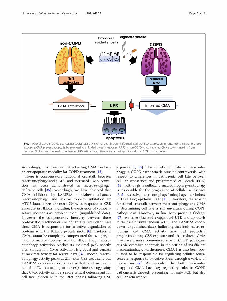
Accordingly, it is plausible that activating CMA can be aan antiapoptotic modality for COPD treatment [13].There is compensatory functional crosstalk between
macroautophagy and CMA, and increased CMA activa-tion has been demonstrated in macroautophagy-deficient cells [36]. Accordingly, we have observed thatCMA inhibition by LAMP2A knockdown enhancesmacroautophagy, and macroautophagy inhibition byATG5 knockdown enhances CMA, in response to CSEexposure in HBECs, indicating the existence of compen-satory mechanisms between them (unpublished data).However, the compensatory interplay between theseproteostatic machineries are not totally redundant, andsince CMA is responsible for selective degradation ofproteins with the KFERQ peptide motif [8], insufficientCMA cannot be completely compensated for by upregu-lation of macroautophagy. Additionally, although macro-autophagy activation reaches its maximal peak shortlyafter stimulation, CMA activation is gradual and persistsat maximal activity for several days [37]. Indeed, macro-autophagy activity peaks at 24 h after CSE treatment, butLAMP2A expression levels peak at 48 h and are main-tained at 72 h according to our experiments, suggestingthat CMA activity can be a more critical determinant forcell fate, especially in the later phases following CSE
exposure [3, 13]. The activity and role of macroauto-phagy in COPD pathogenesis remains controversial withrespect to differences in pathogenic cell fate betweencellular senescence and programmed cell death (PCD)[65]. Although insufficient macroautophagy/mitophagyis responsible for the progression of cellular senescence[3, 5], excessive macroautophagy/ mitophagy may inducePCD in lung epithelial cells [11]. Therefore, the role offunctional crosstalk between macroautophagy and CMAin determining cell fate is still uncertain during COPDpathogenesis. However, in line with previous findings[27], we have observed exaggerated UPR and apoptosisin the case of simultaneous ATG5 and LAMP2A knock-down (unpublished data), indicating that both macroau-tophagy and CMA activity have cell protectiveproperties during CSE exposure and that reduced CMAmay have a more pronounced role in COPD pathogen-esis via excessive apoptosis in the setting of insufficientmacroautophagy. Furthermore, CMA has also been pos-tulated to be responsible for regulating cellular senes-cence in response to oxidative stress through a variety ofmechanism [66]. We speculate that both macroauto-phagy and CMA have key regulatory roles in COPDpathogenesis through preventing not only PCD but alsocellular senescence.
Fig. 4 Role of CMA in COPD pathogenesis. CMA activity is enhanced through Nrf2-mediated LAMP2A expression in response to cigarette smokeexposure. CMA prevent apoptosis by attenuating unfolded protein response (UPR) in non-COPD lung. Impaired CMA activity resulting fromreduced Nrf2 expression leads to enhanced UPR with concomitantly enhanced apoptosis during COPD pathogenesis
Hosaka et al. Inflammation and Regeneration (2021) 41:29 Page 7 of 10
We have recently reported the involvement of in-creased ferroptosis, a form of regulated necrosis associ-ated with lipid peroxidation, in COPD pathogenesis [67].Intriguingly, CMA may have an inconsistent role inregulating ferroptosis. CMA may activate ferroptosis viaglutathione peroxidase 4 (GPX4) degradation but con-versely may also prevent ferroptosis by increasing gluta-thione (GSH) levels [68, 69]. Thus, the precise role ofCMA in regulating ferroptosis with respect to COPDpathogenesis remains uncertain.
ConclusionsRecent research advances have shed light on both themolecular mechanisms and physiological and patho-logical roles of CMA. A growing body of evidence impli-cates CMA in the pathogenesis of a wide array ofdiseases in multiple organs, and CMA regulation is a po-tential therapeutic target. Although there is no estab-lished agent for modifying CMA activity for clinicalapplication, recent study has shown the potential thera-peutic efficacy of specific CMA activation by retinoicacid receptor alpha (RARα) antagonists for mouse modelof Parkinson disease [70]. Accordingly, CMA activationby using these antagonists should be studied as a poten-tial therapeutic approach for COPD. With respect topulmonary disorders, the involvement of CMA has beendemonstrated only in lung cancer and COPD pathogen-esis through modulating apoptosis. Due to the canonicalrole of CMA in cell physiology, its participation shouldbe examined in other common pulmonary disorders, in-cluding interstitial pneumonia, bronchial asthma, andlung infectious diseases in terms of regulating proteosta-sis, metabolic pathways, and immune responses.
AbbreviationsATG5: Autophagy related 5; CBP: Creb-binding protein; CDK: Cyclin-dependent kinase; CSE: Cigarette smoke extract; cGAS: Cyclic GMP-AMP syn-thase; CHIP: Carboxyl terminus of HSC70-interacting protein;CHK1: Checkpoint kinase 1; CMA: Chaperone-mediated autophagy;COPD: Chronic obstructive pulmonary disease; ER: Endoplasmic reticulum;GAPDH: Glyceraldehyde 3-phosphate dehydrogenase; GPX4: Glutathioneperoxidase 4; HBEC: Human bronchial epithelial cell; HIF-1α: Hypoxia-inducible factor-1 subunit α; HIP: HSP70-interacting protein; HMGB1: Highmobility group box-1 protein; HOP: HSP70-HSP90 organizing protein;HSC: Hematopoietic stem-cell; HSC70: Heat shock cognate 71 kDa protein;HSP40: Heat shock protein 40; LAMP2A: Lysosomal-associated membraneprotein type 2A; LRRK2: Leucine-rich repeat serine/threonine-protein kinase 2;MCL1: Myeloid cell leukemia sequence 1; MLIV: Mucolipidosis type IV;NCOR: Nuclear receptor corepressor; NFAT1: Nuclear factor of activated Tcells 1; NF-κB: Nuclear factor-κB; Nrf2: Nuclear factor-erythroid 2-related factor2; NSCLC: Non-small cell lung cancer; PARK7: Parkinson disease protein 7;PAX2: Paired box 2; PCD: Programmed cell death; PD: Parkinson’s disease;PHLPP1: PH domain and leucine rich repeat protein phosphatase 1;PPARα: Peroxisome proliferator-activated receptor-alpha; STING: Stimulator ofinterferon genes; TORC2: Target of rapamycin complex-2; TRPML1: Transientreceptor potential mucolipin-1; UPR: Unfolded protein response
AcknowledgementsWe wish to thank Steven R, PhD from FORTE Science Communications(www.forte-science.co.jp) for editing a draft of this manuscript.
Authors’ contributionsY.H., J.A., and Y.F. wrote the manuscript. K.K. supervised the project. Theauthor(s) read and approved the final manuscript.
FundingNot applicable
Availability of data and materialsNot applicable
Declarations
Ethics approval and consent to participateNot applicable
Consent for publicationAll authors have read the manuscript and approved its submission.
Competing interestsThe authors declare no conflicts of interest associated with this manuscript.
Received: 17 June 2021 Accepted: 8 September 2021
References1. Araya J, Hara H, Kuwano K. Autophagy in the pathogenesis of pulmonary
disease. Intern Med. 2013;52(20):2295–303. https://doi.org/10.2169/internalmedicine.52.1118.
2. Araya J, Kojima J, Takasaka N, Ito S, Fujii S, Hara H, et al. Insufficientautophagy in idiopathic pulmonary fibrosis. Am J Physiol Lung Cell MolPhysiol. 2013;304(1):L56–69. https://doi.org/10.1152/ajplung.00213.2012.
3. Fujii S, Hara H, Araya J, Takasaka N, Kojima J, Ito S, et al. Insufficientautophagy promotes bronchial epithelial cell senescence in chronicobstructive pulmonary disease. Oncoimmunology. 2012;1(5):630–41. https://doi.org/10.4161/onci.20297.
4. Carmona-Gutierrez D, Hughes AL, Madeo F, Ruckenstuhl C. The crucialimpact of lysosomes in aging and longevity. Ageing Res Rev. 2016;32:2–12.https://doi.org/10.1016/j.arr.2016.04.009.
5. Ito S, Araya J, Kurita Y, Kobayashi K, Takasaka N, Yoshida M, et al. PARK2-mediated mitophagy is involved in regulation of HBEC senescence in COPDpathogenesis. Autophagy. 2015;11(3):547–59. https://doi.org/10.1080/15548627.2015.1017190.
6. Kobayashi K, Araya J, Minagawa S, Hara H, Saito N, Kadota T, et al.Involvement of PARK2-mediated mitophagy in idiopathic pulmonary fibrosispathogenesis. J Immunol. 2016;197(2):504–16. https://doi.org/10.4049/jimmunol.1600265.
7. Tsubouchi K, Araya J, Kuwano K. PINK1-PARK2-mediated mitophagy inCOPD and IPF pathogeneses. Inflamm Regen. 2018;38(1):18. https://doi.org/10.1186/s41232-018-0077-6.
8. Kaushik S, Cuervo AM. The coming of age of chaperone-mediatedautophagy. Nat Rev Mol Cell Biol. 2018;19(6):365–81. https://doi.org/10.1038/s41580-018-0001-6.
9. Ichikawa A, Fujita Y, Hosaka Y, Kadota T, Ito A, Yagishita S, et al. Chaperone-mediated autophagy receptor modulates tumor growth andchemoresistance in non-small cell lung cancer. Cancer Sci. 2020;111(11):4154–65. https://doi.org/10.1111/cas.14629.
10. Chen ZH, Kim HP, Sciurba FC, Lee SJ, Feghali-Bostwick C, Stolz DB, et al. Egr-1 regulates autophagy in cigarette smoke-induced chronic obstructivepulmonary disease. PLoS One. 2008;3(10):e3316. https://doi.org/10.1371/journal.pone.0003316.
11. Mizumura K, Cloonan SM, Nakahira K, Bhashyam AR, Cervo M, Kitada T, et al.Mitophagy-dependent necroptosis contributes to the pathogenesis ofCOPD. J Clin Invest. 2014;124(9):3987–4003. https://doi.org/10.1172/JCI74985.
12. Araya J, Tsubouchi K, Sato N, Ito S, Minagawa S, Hara H, et al. PRKN-regulated mitophagy and cellular senescence during COPD pathogenesis.Autophagy. 2019;15(3):510–26. https://doi.org/10.1080/15548627.2018.1532259.
13. Hosaka Y, Araya J, Fujita Y, Kadota T, Tsubouchi K, Yoshida M, et al.Chaperone-mediated autophagy suppresses apoptosis via regulation of theunfolded protein response during chronic obstructive pulmonary disease
Hosaka et al. Inflammation and Regeneration (2021) 41:29 Page 8 of 10

pathogenesis. J Immunol. 2020;205(5):1256–67. https://doi.org/10.4049/jimmunol.2000132.
14. Quintavalle C, Di Costanzo S, Zanca C, Tasset I, Fraldi A, Incoronato M, et al.Phosphorylation-regulated degradation of the tumor-suppressor form ofPED by chaperone-mediated autophagy in lung cancer cells. J Cell Physiol.2014;229(10):1359–68. https://doi.org/10.1002/jcp.24569.
15. Cuervo AM, Dice JF, Knecht E. A population of rat liver lysosomesresponsible for the selective uptake and degradation of cytosolic proteins. JBiol Chem. 1997;272(9):5606–15. https://doi.org/10.1074/jbc.272.9.5606.
16. Arndt V, Dick N, Tawo R, Dreiseidler M, Wenzel D, Hesse M, et al.Chaperone-assisted selective autophagy is essential for musclemaintenance. Curr Biol. 2010;20(2):143–8. https://doi.org/10.1016/j.cub.2009.11.022.
17. Sahu R, Kaushik S, Clement CC, Cannizzo ES, Scharf B, Follenzi A, et al.Microautophagy of cytosolic proteins by late endosomes. Dev Cell. 2011;20(1):131–9. https://doi.org/10.1016/j.devcel.2010.12.003.
18. Cuervo AM, Dice JF. A receptor for the selective uptake and degradation ofproteins by lysosomes. Science. 1996;273(5274):501–3. https://doi.org/10.1126/science.273.5274.501.
19. Bandyopadhyay U, Sridhar S, Kaushik S, Kiffin R, Cuervo AM. Identification ofregulators of chaperone-mediated autophagy. Mol Cell. 2010;39(4):535–47.https://doi.org/10.1016/j.molcel.2010.08.004.
20. Kiffin R, Christian C, Knecht E, Cuervo AM. Activation of chaperone-mediatedautophagy during oxidative stress. Mol Biol Cell. 2004;15(11):4829–40.https://doi.org/10.1091/mbc.e04-06-0477.
21. Park C, Suh Y, Cuervo AM. Regulated degradation of Chk1 by chaperone-mediated autophagy in response to DNA damage. Nat Commun. 2015;6(1):6823. https://doi.org/10.1038/ncomms7823.
22. Valdor R, Mocholi E, Botbol Y, Guerrero-Ros I, Chandra D, Koga H, et al.Chaperone-mediated autophagy regulates T cell responses throughtargeted degradation of negative regulators of T cell activation. NatImmunol. 2014;15(11):1046–54. https://doi.org/10.1038/ni.3003.
23. Zhao H, Eguchi S, Alam A, Ma D. The role of nuclear factor-erythroid 2related factor 2 (Nrf-2) in the protection against lung injury. Am J PhysiolLung Cell Mol Physiol. 2017;312(2):L155–L62. https://doi.org/10.1152/ajplung.00449.2016.
24. Gan L, Vargas MR, Johnson DA, Johnson JA. Astrocyte-specificoverexpression of Nrf2 delays motor pathology and synuclein aggregationthroughout the CNS in the alpha-synuclein mutant (A53T) mouse model. JNeurosci. 2012;32(49):17775–87. https://doi.org/10.1523/JNEUROSCI.3049-12.2012.
25. Pajares M, Rojo AI, Arias E, Diaz-Carretero A, Cuervo AM, Cuadrado A.Transcription factor NFE2L2/NRF2 modulates chaperone-mediatedautophagy through the regulation of LAMP2A. Autophagy. 2018;14(8):1310–22. https://doi.org/10.1080/15548627.2018.1474992.
26. Cuervo AM, Mann L, Bonten EJ, d'Azzo A, Dice JF. Cathepsin Aregulates chaperone-mediated autophagy through cleavage of thelysosomal receptor. EMBO J. 2003;22(1):47–59. https://doi.org/10.1093/emboj/cdg002.
27. Massey AC, Kaushik S, Sovak G, Kiffin R, Cuervo AM. Consequences of theselective blockage of chaperone-mediated autophagy. Proc Natl Acad Sci US A. 2006;103(15):5805–10. https://doi.org/10.1073/pnas.0507436103.
28. Patil M, Pabla N, Dong Z. Checkpoint kinase 1 in DNA damage responseand cell cycle regulation. Cell Mol Life Sci. 2013;70(21):4009–21. https://doi.org/10.1007/s00018-013-1307-3.
29. Andrade-Tomaz M, de Souza I, Rocha CRR, Gomes LR. The role ofchaperone-mediated autophagy in cell cycle control and its implications incancer. Cells. 2020;9(9):2140. https://doi.org/10.3390/cells9092140.
30. Hubbi ME, Hu H, Kshitiz, Ahmed I, Levchenko A, Semenza GL. Chaperone-mediated autophagy targets hypoxia-inducible factor-1alpha (HIF-1alpha)for lysosomal degradation. J Biol Chem. 2013;288(15):10703-10714, DOI:https://doi.org/10.1074/jbc.M112.414771.
31. Garcia-Gutierrez L, Delgado MD, Leon J. MYC oncogene contributions torelease of cell cycle brakes. Genes (Basel). 2019;10(3):244. https://doi.org/10.3390/genes10030244.
32. Xu Y, Zhang Y, Garcia-Canaveras JC, Guo L, Kan M, Yu S, et al. Chaperone-mediated autophagy regulates the pluripotency of embryonic stem cells.Science. 2020;369(6502):397–403. https://doi.org/10.1126/science.abb4467.
33. Dong S, Wang Q, Kao YR, Diaz A, Tasset I, Kaushik S, et al. Chaperone-mediated autophagy sustains haematopoietic stem-cell function. Nature.2021;591(7848):117–23. https://doi.org/10.1038/s41586-020-03129-z.
34. Cuervo AM, Hu W, Lim B, Dice JF. IkappaB is a substrate for a selectivepathway of lysosomal proteolysis. Mol Biol Cell. 1998;9(8):1995–2010. https://doi.org/10.1091/mbc.9.8.1995.
35. Hu MM, Yang Q, Xie XQ, Liao CY, Lin H, Liu TT, et al. Sumoylation promotesthe stability of the DNA sensor cGAS and the adaptor STING to regulate thekinetics of response to DNA virus. Immunity. 2016;45(3):555–69. https://doi.org/10.1016/j.immuni.2016.08.014.
36. Kaushik S, Massey AC, Mizushima N, Cuervo AM. Constitutive activation ofchaperone-mediated autophagy in cells with impaired macroautophagy.Mol Biol Cell. 2008;19(5):2179–92. https://doi.org/10.1091/mbc.e07-11-1155.
37. Cuervo AM, Wong E. Chaperone-mediated autophagy: roles in disease andaging. Cell Res. 2014;24(1):92–104. https://doi.org/10.1038/cr.2013.153.
38. Cuervo AM, Stefanis L, Fredenburg R, Lansbury PT, Sulzer D. Impaireddegradation of mutant alpha-synuclein by chaperone-mediated autophagy.Science. 2004;305(5688):1292–5. https://doi.org/10.1126/science.1101738.
39. Alvarez-Erviti L, Rodriguez-Oroz MC, Cooper JM, Caballero C, Ferrer I, ObesoJA, et al. Chaperone-mediated autophagy markers in Parkinson diseasebrains. Arch Neurol. 2010;67(12):1464–72. https://doi.org/10.1001/archneurol.2010.198.
40. Liao Z, Wang B, Liu W, Xu Q, Hou L, Song J, et al. Dysfunction ofchaperone-mediated autophagy in human diseases. Mol Cell Biochem.2021;476(3):1439–54. https://doi.org/10.1007/s11010-020-04006-z.
41. You Y, Li WZ, Zhang S, Hu B, Li YX, Li HD, et al. SNX10 mediates alcohol-induced liver injury and steatosis by regulating the activation of chaperone-mediated autophagy. J Hepatol. 2018;69(1):129–41. https://doi.org/10.1016/j.jhep.2018.01.038.
42. Ma SY, Sun KS, Zhang M, Zhou X, Zheng XH, Tian SY, et al. Disruption ofPlin5 degradation by CMA causes lipid homeostasis imbalance in NAFLD.Liver Int. 2020;40(10):2427–38. https://doi.org/10.1111/liv.14492.
43. Schneider JL, Suh Y, Cuervo AM. Deficient chaperone-mediated autophagyin liver leads to metabolic dysregulation. Cell Metab. 2014;20(3):417–32.https://doi.org/10.1016/j.cmet.2014.06.009.
44. Sooparb S, Price SR, Shaoguang J, Franch HA. Suppression of chaperone-mediated autophagy in the renal cortex during acute diabetes mellitus.Kidney Int. 2004;65(6):2135–44. https://doi.org/10.1111/j.1523-1755.2004.00639.x.
45. Venugopal B, Mesires NT, Kennedy JC, Curcio-Morelli C, Laplante JM, DiceJF, et al. Chaperone-mediated autophagy is defective in mucolipidosis typeIV. J Cell Physiol. 2009;219(2):344–53. https://doi.org/10.1002/jcp.21676.
46. Guo B, Li L, Guo J, Liu A, Wu J, Wang H, et al. M2 tumor-associatedmacrophages produce interleukin-17 to suppress oxaliplatin-inducedapoptosis in hepatocellular carcinoma. Oncotarget. 2017;8(27):44465–76.https://doi.org/10.18632/oncotarget.17973.
47. Wu JH, Guo JP, Shi J, Wang H, Li LL, Guo B, et al. CMA down-regulates p53expression through degradation of HMGB1 protein to inhibit irradiation-triggered apoptosis in hepatocellular carcinoma. World J Gastroenterol.2017;23(13):2308–17. https://doi.org/10.3748/wjg.v23.i13.2308.
48. Du C, Huang D, Peng Y, Yao Y, Zhao Y, Yang Y, et al. 5-Fluorouracil targetshistone acetyltransferases p300/CBP in the treatment of colorectal cancer.Cancer Lett. 2017;400:183–93. https://doi.org/10.1016/j.canlet.2017.04.033.
49. Kon M, Kiffin R, Koga H, Chapochnick J, Macian F, Varticovski L, et al.Chaperone-mediated autophagy is required for tumor growth. Sci TranslMed. 2011;3(109):109ra17.
50. Gomes LR, Menck CFM, Cuervo AM. Chaperone-mediated autophagyprevents cellular transformation by regulating MYC proteasomaldegradation. Autophagy. 2017;13(5):928–40. https://doi.org/10.1080/15548627.2017.1293767.
51. Arias E, Cuervo AM. Pros and cons of chaperone-mediated autophagy incancer biology. Trends Endocrinol Metab. 2020;31(1):53–66. https://doi.org/10.1016/j.tem.2019.09.007.
52. White E. Deconvoluting the context-dependent role for autophagy incancer. Nat Rev Cancer. 2012;12(6):401–10. https://doi.org/10.1038/nrc3262.
53. Zhou J, Yang J, Fan X, Hu S, Zhou F, Dong J, et al. Chaperone-mediatedautophagy regulates proliferation by targeting RND3 in gastric cancer.Autophagy. 2016;12(3):515–28. https://doi.org/10.1080/15548627.2015.1136770.
54. Ali AB, Nin DS, Tam J, Khan M. Role of chaperone mediated autophagy(CMA) in the degradation of misfolded N-CoR protein in non-small cell lungcancer (NSCLC) cells. PLoS One. 2011;6(9):e25268. https://doi.org/10.1371/journal.pone.0025268.
55. Suzuki J, Nakajima W, Suzuki H, Asano Y, Tanaka N. Chaperone-mediatedautophagy promotes lung cancer cell survival through selective stabilization
Hosaka et al. Inflammation and Regeneration (2021) 41:29 Page 9 of 10
of the pro-survival protein, MCL1. Biochem Biophys Res Commun. 2017;482(4):1334–40. https://doi.org/10.1016/j.bbrc.2016.12.037.
56. Barnes PJ. Senescence in COPD and its comorbidities. Annu Rev Physiol.2017;79(1):517–39. https://doi.org/10.1146/annurev-physiol-022516-034314.
57. Yamada K, Asai K, Nagayasu F, Sato K, Ijiri N, Yoshii N, et al. Impaired nuclearfactor erythroid 2-related factor 2 expression increases apoptosis of airwayepithelial cells in patients with chronic obstructive pulmonary disease dueto cigarette smoking. BMC Pulm Med. 2016;16(1):27. https://doi.org/10.1186/s12890-016-0189-1.
58. Saito N, Araya J, Ito S, Tsubouchi K, Minagawa S, Hara H, et al. Involvementof lamin B1 reduction in accelerated cellular senescence during chronicobstructive pulmonary disease pathogenesis. J Immunol. 2019;202(5):1428–40. https://doi.org/10.4049/jimmunol.1801293.
59. Hara H, Araya J, Takasaka N, Fujii S, Kojima J, Yumino Y, et al. Involvement ofcreatine kinase B in cigarette smoke-induced bronchial epithelial cellsenescence. Am J Respir Cell Mol Biol. 2012;46(3):306–12. https://doi.org/10.1165/rcmb.2011-0214OC.
60. Kelsen SG, Duan X, Ji R, Perez O, Liu C, Merali S. Cigarette smoke induces anunfolded protein response in the human lung: a proteomic approach. Am JRespir Cell Mol Biol. 2008;38(5):541–50. https://doi.org/10.1165/rcmb.2007-0221OC.
61. Jorgensen E, Stinson A, Shan L, Yang J, Gietl D, Albino AP. Cigarette smokeinduces endoplasmic reticulum stress and the unfolded protein response innormal and malignant human lung cells. BMC Cancer. 2008;8(1):229. https://doi.org/10.1186/1471-2407-8-229.
62. Kelsen SG. The unfolded protein response in chronic obstructive pulmonarydisease. Ann Am Thorac Soc. 2016;13(Suppl 2):S138–45.
63. Li W, Zhu J, Dou J, She H, Tao K, Xu H, et al. Phosphorylation of LAMP2A byp38 MAPK couples ER stress to chaperone-mediated autophagy. NatCommun. 2017;8(1):1763. https://doi.org/10.1038/s41467-017-01609-x.
64. Lee CH, Lee KH, Jang AH, Yoo CG. The impact of autophagy on thecigarette smoke extract-induced apoptosis of bronchial epithelial cells.Tuberc Respir Dis (Seoul). 2017;80(1):83–9. https://doi.org/10.4046/trd.2017.80.1.83.
65. Mizumura K, Cloonan S, Choi ME, Hashimoto S, Nakahira K, Ryter SW, et al.Autophagy: friend or foe in lung disease? Ann Am Thorac Soc. 2016;13(Suppl 1):S40–7.
66. Moreno-Blas D, Gorostieta-Salas E, Castro-Obregon S. Connectingchaperone-mediated autophagy dysfunction to cellular senescence. AgeingRes Rev. 2018;41:34–41. https://doi.org/10.1016/j.arr.2017.11.001.
67. Yoshida M, Minagawa S, Araya J, Sakamoto T, Hara H, Tsubouchi K, et al.Involvement of cigarette smoke-induced epithelial cell ferroptosis in COPDpathogenesis. Nat Commun. 2019;10(1):3145. https://doi.org/10.1038/s41467-019-10991-7.
68. Wu Z, Geng Y, Lu X, Shi Y, Wu G, Zhang M, et al. Chaperone-mediatedautophagy is involved in the execution of ferroptosis. Proc Natl Acad Sci US A. 2019;116(8):2996–3005. https://doi.org/10.1073/pnas.1819728116.
69. Lee JJ, Ishihara K, Notomi S, Efstathiou NE, Ueta T, Maidana D, et al.Lysosome-associated membrane protein-2 deficiency increases the risk ofreactive oxygen species-induced ferroptosis in retinal pigment epithelialcells. Biochem Biophys Res Commun. 2020;521(2):414–9. https://doi.org/10.1016/j.bbrc.2019.10.138.
70. Ho PW, Leung CT, Liu H, Pang SY, Lam CS, Xian J, et al. Age-dependentaccumulation of oligomeric SNCA/alpha-synuclein from impaireddegradation in mutant LRRK2 knockin mouse model of Parkinson disease:role for therapeutic activation of chaperone-mediated autophagy (CMA).Autophagy. 2020;16(2):347–70. https://doi.org/10.1080/15548627.2019.1603545.
Publisher’s NoteSpringer Nature remains neutral with regard to jurisdictional claims inpublished maps and institutional affiliations.
Hosaka et al. Inflammation and Regeneration (2021) 41:29 Page 10 of 10