Embed Size (px)
Citation preview
Spring 2013
Getting Physical With Corneal Structure
Annual School of Medicine Gala Casts a Bright Future
Corneal Surgeon Shines in His Dream Job
Discovery Eye Foundation’s Gift of Sight
In This Issue
www.eye.uci.edu/spring
Thank you for supporting the Shine The Light Campaign
Gifts of $25,000 and above received since January 1, 2012, to support construction of the Gavin Herbert Eye Institute:
Paul and Nancy Arentsen Arnold and Mabel Beckman Foundation
William and Anne Brownstein Karen and Bruce Cahill Carol and Budge Collins J. Stuart Cumming, MD David & Julianna Pyott Foundation Discovery Eye Foundation Susy and David Endicott Yvonne and Handel Evans Josephine D. Troy Gleis Sylvia and Ronald Hartman, MD Gavin and Ninetta S. Herbert Josephine Herbert Gleis Foundation Richard P. Kratz, MD, DSci Kelly and Jim Mazzo Schoellerman Foundation Helen and Jacob Shaham The Robert M. Sinskey Foundation The Rosso Family Foundation
Gifts of $25,000 and above received since January 1, 2012, for research:
Arnold and Mabel Beckman Foundation
ARVO Foundation for Eye Research Branna & Irv Sisenwein Charitable Foundation
Cystinosis Research Foundation Discovery Eye Foundation Josephine Herbert Gleis Foundation Geneva M. Matlock, MD Polly and Michael Smith
For more information about the Gavin Herbert Eye Institute, please call (949) 824-0091.
Affecting approximately 1 in every 2,000 people, keratoconus is a
degenerative eye disease marked by a thinning of the cornea, which causes the cornea to bulge forward. The condition is associated with allergies, physically rubbing the eye, genetics and Down’s syndrome. Keratoconus symptoms usually present during the teenage years and progress through the mid-30s, and vision becomes progressively blurry with increasing astigmatism and nearsightedness. Spectacle prescriptions can change as rapidly as every two months and get to the point that they no longer help in improving vision.
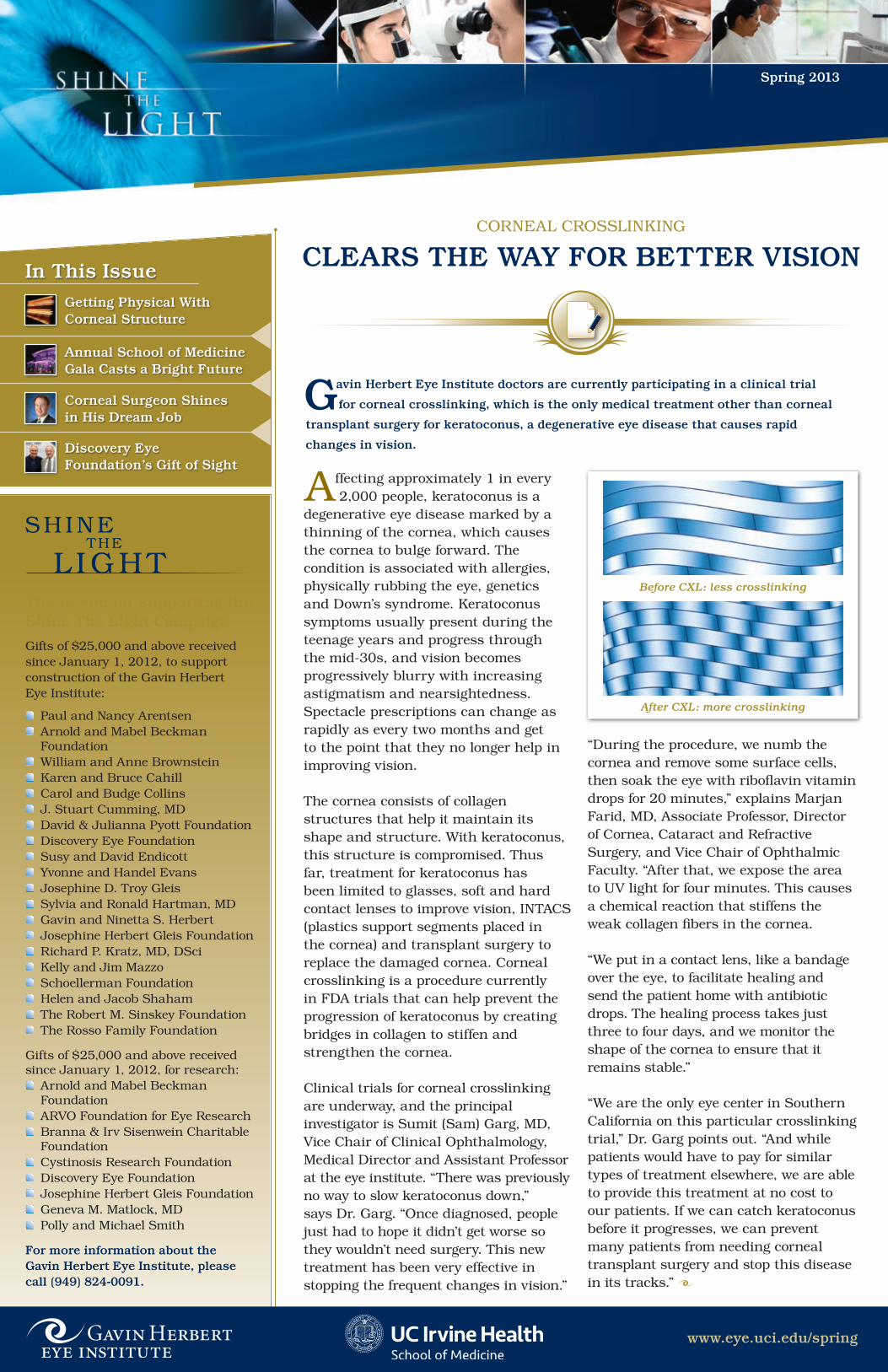
The cornea consists of collagen structures that help it maintain its shape and structure. With keratoconus, this structure is compromised. Thus far, treatment for keratoconus has been limited to glasses, soft and hard contact lenses to improve vision, INTACS (plastics support segments placed in the cornea) and transplant surgery to replace the damaged cornea. Corneal crosslinking is a procedure currently in FDA trials that can help prevent the progression of keratoconus by creating bridges in collagen to stiffen and strengthen the cornea.
Clinical trials for corneal crosslinking are underway, and the principal investigator is Sumit (Sam) Garg, MD, Vice Chair of Clinical Ophthalmology, Medical Director and Assistant Professor at the eye institute. “There was previously no way to slow keratoconus down,” says Dr. Garg. “Once diagnosed, people just had to hope it didn’t get worse so they wouldn’t need surgery. This new treatment has been very effective in stopping the frequent changes in vision.”
“During the procedure, we numb the cornea and remove some surface cells, then soak the eye with riboflavin vitamin drops for 20 minutes,” explains Marjan Farid, MD, Associate Professor, Director of Cornea, Cataract and Refractive Surgery, and Vice Chair of Ophthalmic Faculty. “After that, we expose the area to UV light for four minutes. This causes a chemical reaction that stiffens the weak collagen fibers in the cornea.
“We put in a contact lens, like a bandage over the eye, to facilitate healing and send the patient home with antibiotic drops. The healing process takes just three to four days, and we monitor the shape of the cornea to ensure that it remains stable.”
“We are the only eye center in Southern California on this particular crosslinking trial,” Dr. Garg points out. “And while patients would have to pay for similar types of treatment elsewhere, we are able to provide this treatment at no cost to our patients. If we can catch keratoconus before it progresses, we can prevent many patients from needing corneal transplant surgery and stop this disease in its tracks.”
CORNEAl CROSSlINKING
CLEArS THE WAy For BETTEr VISIon
Gavin Herbert Eye Institute doctors are currently participating in a clinical trial
for corneal crosslinking, which is the only medical treatment other than corneal
transplant surgery for keratoconus, a degenerative eye disease that causes rapid
changes in vision.
SHINETHE
l IGHT
After CXL: more crosslinking
Before CXL: less crosslinking

As the Gavin Herbert Eye Institute building nears completion, we begin a series of “firsts” (first patients seen in the Beckman Center for Clinical Care, first major surgeries performed on the Irvine campus at the Argyros Surgical Center) and “lasts” (last resident and fellow graduation prior to the opening of our state-of-the-art education center). We are working hard to prepare for our opening day of patient care on September 16. So this fall, please make plans to join us in celebrating the world-class institute we have built together, as a community truly committed to advancing eye care.
Sincerely,
Roger Steinert, MDChair, Department of Ophthalmology
For more information on how you can support GHEI, please contact Janice Briggs, Executive Development Director, Health Advancement, at (949) 824-0091.
RESEARCH FOCUS:
A SITE To SEE
GETTInG PHySICAL WITH CornEAL STruCTurE
The cornea serves as the eye’s protective outer layer, shielding
its delicate inner components from physical and chemical dangers. It also determines how light enters the eye, known as refraction. If something is wrong with the shape of the cornea, such as flattening or bulges caused by astigmatism or keratoconus, vision can be adversely affected in several ways—including blurriness and distortions.
“We don’t currently know why or how these changes in the cornea occur,” says researcher Moritz Winkler, a PhD candidate in biomedical engineering at the Gavin Herbert Eye Institute. “The goal of my research is to deeply understand the structure of the cornea. Just as bridges are engineered to withstand high loads for long periods of time, we are looking at how the arrangement of collagen structures in the cornea affects its strength. What are the different patterns of the structures? What do they mean for biomechanics and corneal stiffness? How does this correlate to a perfect or imperfect corneal shape?”
Looking for AnswersAs a child growing up in Germany,
Winkler was fascinated by how the math equations of physics could be used to describe the world around us. Intent on turning this interest into a career, he studied physics at the University of Heidelberg, where he decided to apply his knowledge of physics to medicine.
“Much of physics is very abstract and theoretical, dealing with particles. However, it can take decades or longer to see any breakthroughs,” says Winkler. “I chose to specialize in medical physics because it is closer to real-world problems. Diseases need to be cured. Helping to find a better way to visualize
and treat eye diseases is very rewarding.”
An expert in using ultra-fast laser pulses and optics to produce three-dimensional images of microscopic specimens, Winkler was invited by James Jester, PhD, to join his corneal research lab at the institute. “The cornea is very well suited to this kind of imaging because it is
transparent,” explains Winkler. “The collagen structures we study are less than 1/1000th of an inch in diameter. We use a very high-powered microscope along with a laser that can emit tens of millions of short pulses each second to capture numerous images of tiny bits of the overall structure of the cornea or an area of interest. Then, we use software to combine the images into a 3D mosaic that we can zoom in and out of to see a section in focus or the overall picture.
“Processing that used to take a month can now be accomplished in a week, thanks to software our lab has written
www.eye.uci.edu/spring
Moritz Winkler uses 3D imaging to study the collagen structure of the cornea.
Watch the building being constructed in real time with the EarthCam, which can be found
on our website: www.eye.uci.edu/spring

that can look at hundreds of thousands of fibers per hour and then stitch all of those images together. This allows us to get 3D numbers. In science, numbers are worth a thousand pictures. We can also use a transducer to ‘poke’ the cornea to record how the structures respond to external forces.”
While his research started with human corneas, Winkler found that this did not give him enough information to determine how structure and biomechanics affect the shape of the cornea. His solution was to study animal corneas as well. “In the last year and a half, I have studied at least a dozen different species: dogs, rabbits,
great white and Atlantic sharp nose sharks, cats, sea lions, alligators, bearded dragons, salamanders, frogs and fish—including salmon, mahi-mahi and sturgeon.
“Once we have a model of corneal biomechanics and how shape is controlled, we can apply this knowledge to predicting and simulating corneal surgery. Then, surgeons could know precisely how and where to make cuts, allowing natural forces in the eye to take care of the rest. We could optimize corneal procedures, including crosslinking. In addition, we would better understand why conditions like keratoconus and corneal scarring occur.”
In anticipation of the new eye care facility opening this fall, Winkler looks forward to increased opportunities for collaboration between Gavin Herbert Eye Institute clinicians and researchers toward new eye therapies. “It’s a great advantage to us as researchers to have access to world-class clinicians that we can talk to and bounce ideas off when we have questions. We look at the eye from different angles—I see the cornea from a physics and engineering viewpoint, and they see how their patients are affected. With their help, I can apply my discoveries to the real world through contributing to sight-saving eye treatments.”
These 3D reconstructions show differences in the corneal structures of great white sharks (A), red-tailed hawks (B) and humans (C).
FACuLTy MEMBErS
To ConTACT FACuLTy MEMBErS or To MAkE An APPoInTMEnT, CALL (949) 824-2020 (IrVInE) or (714) 456-7183 (orAnGE)
Lbachir BenMohamed, PhD
Anand Bhatt, MD
Donald J. Brown, PhD
robert Wade Crow, MD
Marjan Farid, MD Vice Chair of Ophthalmic Faculty
Sumit (Sam) Garg, MD Vice Chair of Clinical Ophthalmology
James V. Jester, PhD Jack H. Skirball Endowed Chair
Tibor Juhasz, PhD
Maria Cristina kenney, MD, PhD
Henry klassen, MD, PhD
Baruch kuppermann, MD, PhDVice Chair of Clinical Research
robert W. Lingua, MD
Linda S. Lippa, MD
Stephanie Lu, MD
Donald S. Minckler, MDOphthalmic Pathology
Sameh Mosaed, MD
Anthony B. nesburn, MDVice Chair of Research
Jennifer Simpson, MD
roger F. Steinert, MDIrving H. Leopold Professor and
Chair of Ophthalmology Professor of Biomedical Engineering Director, Gavin Herbert Eye Institute
Jeremiah Tao, MD
Matthew Wade, MD
Steven L. Wechsler, PhD
“The goal of my research is to deeply understand the structure of the cornea. Just as bridges are engineered to withstand high loads for long periods of time,
we are looking at how the arrangement of collagen structures in the cornea affects its strength.”
— Moritz Winkler

Early detection of diseases and proper identification of infections
can make a big difference in treatment options, clinical care and recovery times for patients. This is particularly true for diseases of the cornea. However, there are times when corneal conditions can be difficult to correctly diagnose.
“If you don’t know what’s going on in the eye, it can make it hard to fix the problem,” says Sumit (Sam) Garg, MD, Vice Chair of Clinical Ophthalmology, Medical Director, and Assistant Professor of Cataract, Corneal and Refractive Surgery at the Gavin Herbert Eye Institute. “Strong infections left untreated can replicate and destroy the cells of the cornea. This can lead to irreparable damage, sometimes requiring a corneal transplant to remove the infection. Visual rehabilitation may be needed.
“Also, many corneal diseases are progressive or degenerative, which requires periodic monitoring. Some of these conditions are inherited, meaning they could be passed down to children. At the institute, we use a variety of cutting-edge imaging techniques to make diagnostic and therapeutic choices for our patients.”
While a healthy cornea has a smooth, rounded curve, conditions such as astigmatism, keratoconus and complications after surgery can actually alter the shape of the cornea. This, in turn, can often create visual distortions and blurriness requiring new glasses and contact lenses, or even surgery to restore vision.
What are some of the corneal tests available, and what are they used for?In order to determine the exact shape of the cornea, a device known as a topographer is used. At the institute, the physicians have several makes of topographers (each with its own benefits) to aid in diagnostic and therapeutic decision-making for treatments such as crosslinking or transplant surgery. Measuring corneal thickness, which is called corneal pachymetry, may also be used prior to lASIK, corneal, cataract and glaucoma surgery.
The topographer is vital for the management of naturally occurring
or post-operative astigmatism. One such topographer is the Pentacam®. This device has a rotating camera that creates a 3D image of the eye, which is then used to make topographic maps that display the high and low areas of the cornea.
www.eye.uci.edu
EDUCATION FOCUS:
CornEAL CLInICAL TESTInG
www.eye.uci.edu/spring
“At the institute, we use a variety of cutting-edge imaging techniques to make
diagnostic and therapeutic choices for our patients.”— Sumit (Sam) Garg, MD
Topography of the cornea produced by a Pentacam showing an irregular cornea.
An OCT (optical coherence tomography) scan taken after corneal transplant surgery
where a thin graft was attached.

If a patient with keratoconus, a progressive thinning disorder of the cornea, needs a femtosecond laser keratoplasty (a specialized laser-assisted corneal transplant procedure pioneered at the institute), they may also receive an OCT, or optical coherence tomography scan. Similar to an ultrasound, an OCT uses light instead of sound to create a detailed 3D image so the ophthalmologist can check the corneal structure for changes or abnormalities. An OCT may also be used for monitoring the cornea after surgery.
Slit lamp photography is another form of imaging that is typically used on the front portion of the eye, where the cornea is located. It can be used to document foreign bodies and infections, or to monitor healing after eye surgery. In addition, the pictures can be used to explain to patients what is going on in their eye.
If a patient has an eye infection that doesn’t respond to antibiotics, it may be a fungal or protozoan infection rather than bacterial. To find the
right treatment, the infection must be identified. In this case, the ophthalmologist may use confocal microscopy, a technique that provides a non-contact microscopic view of the cornea without the need for a biopsy.
“These and other corneal tests help us to look at specific layers of the eye when we are diagnosing and treating patients,” explains Dr. Garg. “We have many of the newest versions of these imaging instruments at the institute. By keeping at the forefront of corneal imaging technology, we provide our patients with the highest standard in eye care available.”
Make an appointment todayTo learn more details about corneal clinical testing and other state-of-the-art ophthalmic services, and for dates and times of upcoming patient education seminars, visit our website at www.eye.uci.edu/spring. To schedule an appointment with a corneal specialist, please contact the Gavin Herbert Eye Institute at (949) 824-2020.
UC IRVINE SCHOOl OF MEDICINE
“CrEATIVITy AnD MAGIC” GALA CASTS A BrIGHT FuTurE
Confocal microscopy showing a fungal corneal infection.
Slit lamp photography of sutures from a corneal transplant.
The new building was lit with rotating colors to complement the gala’s theme.
(Left to Right) Dr. Roger Steinert, Gavin Herbert
and James Mazzo were in attendance to proudly support
UC Irvine research and medical education programs.
Derek Paravicini, a blind and autistic musician, brought down the house
with a standing ovation.

Born and raised in Salt lake City, Utah, Matthew Wade, MD, pictured
a career in engineering and physics when he graduated from Brigham Young University with an applied physics degree. It wasn’t long before he decided to change his career and become a doctor. “My first job was located in a basement laboratory where I worked alone, and I thought about how great it would be to work with people instead,” explains Dr. Wade. “I really enjoy seeing smiles on my patients’ faces. It puts a smile on my face, too.”
After receiving his medical degree from George Washington University, Dr. Wade completed a clinical research fellowship at the National Institutes of Health. He came to the Gavin Herbert Eye Institute as a resident and also completed a corneal and refractive surgery fellowship. “I was excited to be trained by Dr. Roger Steinert, one of the brightest refractive surgeons in the world,” recalls Dr. Wade, who has since joined the institute staff as Assistant Clinical Professor.
“The people I work with—and what I get to work on—make this my dream job,” says Dr. Wade. “There is a real camaraderie among the faculty. What
I love about the Gavin Herbert Eye Institute is that everyone is a world- class specialist, and also a wonderful human being who really cares about their patients.”
Primarily a corneal surgeon who helps patients with complications involving cataracts, corneal disorders and astigmatism, Dr. Wade also conducts research on treatments such as laser corneal transplants. He uses an ultra-fast femtosecond laser to make an
interlocking pattern on the cornea that results in quicker healing times and better vision after transplant surgery. In addition, he instructs nine residents and two corneal fellows at the institute and, along with other members of the faculty, provides eye screenings at community clinics as a volunteer through the Illumination Foundation. “It’s
www.eye.uci.edu/spring
CornEAL SurGEon SHInES In HIS DrEAM JoBlIGHT ATTRACTS lIGHT:
Matthew Wade, MD, performs eye screenings as an Illumination Foundation volunteer.
“What I love about the Gavin Herbert Eye Institute is that
everyone is a world-class specialist,
and also a wonderful human being who really cares about their patients.”
— Matthew Wade, MD
wonderful to have the chance to give back by serving the Orange County community,” says Dr. Wade.
As the new eye institute building nears completion, Dr. Wade looks forward to the opportunities it will bring. “We will be right across the parking lot from our basic science researchers. That alone will allow for crosspollination and partnering on projects to improve vision for our patients in Orange County and all over the world.”

Since 1984, optometrist Paul Blaze, OD, has served as a volunteer
contact lens (Cl) instructor for the UC Irvine ophthalmology and Gavin
Herbert Eye Institute residents. He has been in practice in Huntington Beach for over 25 years, focusing on contact lens fittings for post-surgical
and keratoconus patients. Over his career, Dr. Blaze helped to design one of the first successful soft Cls for the correction of astigmatism and was involved in the early phases of research for extended wear soft Cls.
“The use of topographic imaging and the diagnostic fitting of hard contact lenses can often benefit patients with irregularly shaped corneas,” explains Dr. Blaze. “These patients cannot see well with glasses, and their best option is often a rigid contact lens
to maximize their corrected vision. Helping them to see clearly again is very gratifying.”
Dr. Blaze works with Gavin Herbert Eye Institute residents on patients who need specialty Cl fitting. “After the resident presents a patient’s case history, we use a diagnostic contact lens fitting set along with a special dye to observe and analyze the lenses on the eye. Then, we walk through the process of deciding which lens design will help the patient to obtain the best vision correction.
“It has been a pleasure to be involved with these talented ophthalmology residents over the years,” says Dr. Blaze. “Their enthusiasm to learn is infectious, even while discussing the particulars of contact lenses. They keep me on my toes and make me feel young.”
oPToMETrIST HELPS rESIDEnTS SEE ConTACTS In A DIFFErEnT LIGHT
BUIlDING THE FUTURE:
“What I love about the Gavin Herbert Eye Institute is that
everyone is a world-class specialist,
and also a wonderful human being who really cares about their patients.”
Paul Blaze, OD, helps eye institute residents with therapeutic contact lens consultations.
CHArITABLE GIVInG THAT kEEPS on GIVInGThe gift of sight is one that the Gavin Herbert Eye Institute strives to give to each of its patients through world-class care and innovative research. But you don’t have to be one of the institute’s specially trained ophthalmologists to be a part of this precious gift.
New tax laws could make you responsible for paying higher taxes this year. One way to reduce your income and capital gains taxes is to make a charitable gift to the institute. Congress reauthorized IRA rollover for 2013, which means that if you are 70½ or older, you can make a charitable gift of up to $100,000 from your IRA. Your gift will qualify for your 2013 required minimum distribution, and you will not have to pay federal income tax on the amount given from your IRA to charity.
To make an IRA rollover gift, contact your IRA custodian and request an amount to be transferred to the institute. Your gift could be $1,000, $10,000, $50,000 or even $100,000. Join us in our mission to provide quality eye care for years to come. To learn more about how you can redirect your unneeded IRA income or other ways to donate to the Gavin Herbert Eye Institute, please contact Roland Ho in the UC Irvine Planned Giving department at (949) 824-6454 or email [email protected].
MAkE A GIFT oF SIGHT
Through your generous support, the Gavin Herbert Eye Institute will continue to develop new therapies and cures for blindness. Your gifts advance eye research, help retain and attract doctors and build state-of-the-art eye care facilities.
Visit www.eye.uci.edu/spring to see a motion picture of the Gavin Herbert Eye Institute and for more information. To make a donation, go to the “Campaign overview” page in the Philanthropy section. At the bottom of the page, click on “To make an online donation” and follow the instructions. you can also contact Janice Briggs, Executive Development Director, Health Advancement, at (949) 824-0091.
Thank you for helping us find cures for blindness and improve vision for people in orange County, the nation and the world.

T he Discovery Eye Foundation (DEF) has supported the efforts of the
Gavin Herbert Eye Institute (GHEI) for over a decade. One of the institute’s
largest contributors and private supporters, DEF most recently donated $1 million to establish GHEI’s Discovery Center for Eye Research, plus an additional $2 million to help complete the new GHEI facility. According to DEF Board of Directors Chairman Jack Schoellerman, “The goal was to provide a home for this world-class eye institute, to be the place where the research we have funded will be translated into treatments.”
Schoellerman, who has keratoconus and received a corneal transplant performed by Dr. Roger Steinert just last year, will serve as Chairman of the Board for the Discovery Center for Eye Research. An active supporter of research on new eye treatments since the 1980s,
Schoellerman recently made a personal gift toward the GHEI building. The facility’s Healing Garden will be named in his honor.
DEF’s dual missions are to provide support for people with retinal and corneal diseases and to provide funds for cutting-edge research that seeks to treat and cure these conditions. DEF conducts patient education seminars across the country and serves as a parent foundation for both the National Keratoconus Foundation and the Macular Degeneration Partnership, helping to run websites with forums and resources for patients and their family members. For more information on the Discovery Eye Foundation, please visit discoveryeye.org.
SUSTAINING THE VISION:
DISCoVEry EyE FounDATIon MAkES $3 MILLIon GIFT
NONPROFIT ORG. U.S. POSTAGE
PAIDSanta Ana, CA
Permit No. 1106
101 Academy Way, Suite 120
Irvine, CA 92617-3953
RETURN SERVICE REQUESTED
The Gavin Herbert Eye Institute Ribbon Cutting Ceremony for
the new facility will be held on Wednesday, September 11, 2013.
Join us for “site seeing” as we celebrate this momentous event!
For more information, please contact Janice Briggs, Executive
Development Director, Health Advancement, at 949-824-0091
The 7th Annual Gavin Herbert Eye Institute Colloquium will be
hosted at the newly opened facility October 11–12, 2013.
Focusing on Advances and Controversies, the symposium
will bring together eye surgeons, clinicians and researchers
from around the world to discuss innovative new technologies
and treatments in eye care. For more information, visit
http://www.ghei.uci.edu/colloquium.html.
www.eye.uci.edu
Download a QR reader from the app store and scan the QR code with your smartphone to watch a video of exciting developments.
UPCOMING EVENTS
Jack Schoellerman, Chairman (left), and Anthony Nesburn, MD, GHEI Vice Chair of Research and President/Medical Director,
of the Discovery Eye Foundation.