Embed Size (px)
Citation preview

Seediscussions,stats,andauthorprofilesforthispublicationat:https://www.researchgate.net/publication/277780571
RobustNeuroprostheticControlfromtheStrokePerilesionalCortex
ArticleinTheJournalofNeuroscience:TheOfficialJournaloftheSocietyforNeuroscience·June2015
DOI:10.1523/JNEUROSCI.5007-14.2015·Source:PubMed
CITATIONS
8
READS
47
7authors,including:
TanujGulati
UniversityofCalifornia,SanFrancisco
8PUBLICATIONS76CITATIONS
SEEPROFILE
seokjoonWon
UniversityofCalifornia,SanFrancisco
60PUBLICATIONS1,930CITATIONS
SEEPROFILE
DhakshinSRamanathan
UniversityofCalifornia,SanFrancisco
8PUBLICATIONS280CITATIONS
SEEPROFILE
KaruneshGanguly
UniversityofCalifornia,SanFrancisco
51PUBLICATIONS1,719CITATIONS
SEEPROFILE
AllcontentfollowingthispagewasuploadedbyseokjoonWonon08June2016.
Theuserhasrequestedenhancementofthedownloadedfile.Allin-textreferencesunderlinedinblueareaddedtotheoriginaldocument
andarelinkedtopublicationsonResearchGate,lettingyouaccessandreadthemimmediately.

Systems/Circuits
Robust Neuroprosthetic Control from the Stroke PerilesionalCortex
X Tanuj Gulati,1,3 Seok Joon Won,1,3 Dhakshin S. Ramanathan,1,2,4 Chelsea C. Wong,1,3 Anitha Bodepudi,1,3
X Raymond A. Swanson,1,3 and Karunesh Ganguly1,3
1Neurology and Rehabilitation Service and 2Department of Psychiatry, San Francisco Veterans Affairs Medical Center, San Francisco, California 94158,3Department of Neurology, University of California San Francisco, San Francisco, California 94158, and 4Department of Psychiatry, University of CaliforniaSan Francisco, San Francisco, California 94143
Intracortical brain–machine interfaces (BMIs) may eventually restore function in those with motor disability after stroke. However,current research into the development of intracortical BMIs has focused on subjects with largely intact cortical structures, such as thosewith spinal cord injury. Although the stroke perilesional cortex (PLC) has been hypothesized as a potential site for a BMI, it remainsunclear whether the injured motor cortical network can support neuroprosthetic control directly. Using chronic electrophysiologicalrecordings in a rat stroke model, we demonstrate here the PLC’s capacity for neuroprosthetic control and physiological plasticity. Weinitially found that the perilesional network demonstrated abnormally increased slow oscillations that also modulated neural firing.Despite these striking abnormalities, neurons in the perilesional network could be modulated volitionally to learn neuroprostheticcontrol. The rate of learning was surprisingly similar regardless of the electrode distance from the stroke site and was not significantlydifferent from intact animals. Moreover, neurons achieved similar task-related modulation and, as an ensemble, formed cell assemblieswith learning. Such control was even achieved in animals with poor motor recovery, suggesting that neuroprosthetic control is possibleeven in the absence of motor recovery. Interestingly, achieving successful control also reduced locking to abnormal oscillations signifi-cantly. Our results thus suggest that, despite the disrupted connectivity in the PLC, it may serve as an effective target for neuroprostheticcontrol in those with poor motor recovery after stroke.
Key words: brain–machine interface; electrophysiology; plasticity; stroke
IntroductionResearch into the development of intracortical brain–machineinterfaces (BMIs) has flourished over the past decade, leading todemonstrations of direct neural control of prosthetic devices inreal-time through modulation of neural signals (Chapin et al., 1999;Serruya et al., 2002; Taylor et al., 2002; Carmena et al., 2003; Musal-lam et al., 2004; Hochberg et al., 2006; Santhanam et al., 2006;Jarosiewicz et al., 2008; Moritz et al., 2008; Velliste et al., 2008; Gan-guly et al., 2009; Hochberg et al., 2012; Collinger et al., 2013). Task-dependent volitional modulation of the activity of selectedneurons is also closely tied to the concept of BMIs (Fetz, 1969;Ganguly and Carmena, 2009; Koralek et al., 2010; Ganguly et al.,
2011). Together, this body of work has also indicated that neuralplasticity likely plays an important role in achieving stable neu-roprosthetic control (Taylor et al., 2002; Jarosiewicz et al., 2008;Moritz et al., 2008; Ganguly and Carmena, 2009; Koralek et al.,2010; Arduin et al., 2013; Gulati et al., 2014). Importantly, thisresearch into the development of intracortical BMIs has primar-ily focused on subjects with intact cortical structures.
In contrast, the development of BMIs specifically compatiblewith stroke (Langhorne et al., 2011; Norrving and Kissela, 2013)or other forms of direct cortical injury requires a greater under-standing of the neurophysiology of the injured neural network(Nudo et al., 1996; Ward, 2004; Dancause, 2006; Cramer, 2008;Murphy and Corbett, 2009). Although both the perilesionalcortex (PLC) surrounding the injury (Daly and Wolpaw, 2008;Guggenmos et al., 2013) and motor areas in the unaffected hemi-sphere are possible targets of neural interfaces after stroke, moreattention has been placed on the unaffected contralateral hemi-sphere (Bundy et al., 2012). This interest may have been fueled byfindings of an increased role of the contralesional hemisphere inmovements of the ipsilateral limb after stroke (Brinkman andKuypers, 1973; Dancause, 2006; Hummel and Cohen, 2006).However, the PLC is an important site of reorganization afterstroke (Nudo and Milliken, 1996; Brown et al., 2010); moreover,task-specific rehabilitation is also known to have synergistic ef-fects on perilesional cortical plasticity (Nudo et al., 1996; Bier-
Received Dec. 9, 2014; revised April 29, 2015; accepted May 4, 2015.Author contributions: T.G., S.-J.W., R.S., and K.G. designed research; T.G., S.-J.W., C.C.W., and K.G. performed
research; T.G., S.-J.W., D.S.R., C.C.W., R.A.S., and K.G. contributed unpublished reagents/analytic tools; T.G. and A.B.analyzed data; T.G. and K.G. wrote the paper.
This work is supported by the Department of Veterans Affairs (Grant B6674 to K.G.), the Burroughs WellcomeFund (Grant 1009855 to K.G.), the American Heart/Stroke Association (Grant 0875016N to K.G.), and by start-upfunds from the SFVAMC, NCIRE, and UCSF Department of Neurology; an Advanced Research Fellowship from the VAto D.R.
The authors declare no competing financial interests.Correspondence should be addressed to Karunesh Ganguly, Neurology and Rehabilitation Service, San
Francisco Veterans Affairs Medical Center, 1700 Owens Street, San Francisco, CA 94158. E-mail:[email protected].
DOI:10.1523/JNEUROSCI.5007-14.2015Copyright © 2015 the authors 0270-6474/15/358653-09$15.00/0
The Journal of Neuroscience, June 3, 2015 • 35(22):8653– 8661 • 8653
naskie and Corbett, 2001; Ramanathan et al., 2006). It remainsunknown whether the impaired local structural and functionalconnectivity present after stroke (Nudo et al., 1996; Ward, 2004;Dancause, 2006; Cramer, 2008; Murphy and Corbett, 2009; Gug-genmos et al., 2013) can support neuroprosthetic control. Byadapting chronic electrophysiological recordings to a rodentstroke model, we investigated the electrophysiological character-istics of the injured PLC and whether its spiking activity can bemodulated volitionally to control an artificial actuator. We alsomeasured directly the physiological plasticity induced by neuro-prosthetic learning in the poststroke PLC.
Materials and MethodsAnimals and surgeryExperiments were approved by the Institutional Animal Care and UseCommittee at the San Francisco Veterans Affairs Medical Center. Weused 13 adult Long–Evans male rats (�8 weeks old). Animals were keptunder controlled temperature and a 12 h light: 12 h dark cycle with lightson at 6:00 A.M. Probes were implanted during a recovery surgery per-formed under isofluorane (1–3%) anesthesia after induction with ket-amine [2 mg/kg body weight (b.w.)] and xylazine (1 mg/kg b.w.).Atropine sulfate was also administered before anesthesia (0.02 mg/kgb.w.). In eight rats, a focal photothromobotic stroke was induced in theupper limb primary motor cortex area (M1). For this, unilateral femoralvein cannulation was done before craniotomy and an intravenous cath-eter was placed for rose-bengal dye injection. After this, the head wasfixed in the stereotaxic frame and a craniotomy was performed. Strokewas induced via illumination with white light (KL-500) using a fiberoptic cable for 15 min. We used a 3 mm aperture for stroke induction(centered in the M1 area based on stereotactic coordinates) and coveredthe remaining cortical area with a custom mask to prevent light penetra-tion. After induction, a probe was implanted immediately in PLC withone of the outermost rows of the array implanted immediately proximalto the stroke site (see Fig. 1A). The postoperative recovery regimen in-cluded administration of buprenorphine at 0.02 mg/kg b.w and meloxi-cam at 0.2 mg/kg b.w. Dexamethasone at 0.5 mg/kg b.w. andTrimethoprim sulfadiazine at 15 mg/kg b.w. were also administeredpostoperatively for 5 d. All animals were allowed to recover for 5 d beforestart of experiments.
ElectrophysiologyWe recorded extracellular neural activity using tungsten microwire elec-trode arrays (MEAs, n � 13 rats; Tucker-Davis Technologies). We usedeither 16- or 32-channel arrays (33 �m polyimide-coated tungsten mi-crowire arrays). Arrays were lowered down to 1500 –1800 �m in M1 (1–3mm anterior to bregma and 2– 4 mm lateral from midline or immediatelyproximal to the stroke site). Final localization of depth was based onquality of recordings across the array at the time of implantation. Werecorded spike and LFP activity using a 128-channel TDT-RZ2 system(Tucker-Davies Technologies). Spike data were sampled at 24414 Hz andLFP data at 1018 Hz. ZIF-clip-based analog headstages with a unity gainand high impedance (�1 G�) were used. Only clearly identifiable unitswith stable waveforms and a high signal-to-noise ratio (SNR) were usedfor these experiments. To calculate the SNR, we used following equation:
SNR �A
2 � SDnoise
Where A is the peak-to-peak voltage of the averaged spike waveform andSDnoise is the SD of the noise (Suner et al., 2005). Unit clusters with SNR�5 were used for subsequent analysis. A total of 258 cells were recordedfrom all the rats. After the above online sorting, we also used standardoffline cluster cutting methods in TDT’s OpenSorter software to confirmquality of recordings. Behavior-related timestamps (i.e., trial onset, trialcompletion) were sent to the RZ2 analog input channel using an Arduinodigital board and synchronized to neural data.
BehaviorBefore surgery, animals were acclimated and then trained in a reach tograsp single pellet task after determination of handedness. After the per-
formance plateaued (typically �75% accuracy), animals underwent ei-ther control implantation in M1 (n � 5) or in the PLC (n � 8). Afterrecovery, animals were allowed to rest for a few days before the start ofexperimental sessions. Animals were also acclimated to a custom Plexi-glas behavioral box for the BMI task. Five of the stroke rats underwentBMI training; three others served as the no BMI training control group.Animals were then water scheduled such that water from the feeding tubewas available in a randomized fashion while in the behavioral box. Wealso tested their reach performance on the reach to grasp task. During thisperiod, we closely monitored the animals and ensured that body weightsdid not drop below 95% of the initial weight. Reach sessions were typi-cally conducted in the morning (consisting of 25 trials). This was thenfollowed by the brain-control task for a period of 1–2 h. Recorded neuraldata were entered in real time to custom routines in MATLAB (TheMathWorks). These then served as control signals for the angular veloc-ity of the feeding tube. The rats typically performed �100 –200 trialsduring these sessions.
Neural control of the feeding tubeDuring the direct neural control training sessions, we selected well iso-lated neurons and allowed their spiking activity to control the angularvelocity of the feeding tube. We binned the spiking activity into 100 msbins. We then established a mean firing rate for each neuron over a 3–5min baseline period. The mean firing rate was then subtracted from itscurrent firing rate at all times. To test the capacity for neuroprostheticcontrol, the specific transform used was as follows:
�v � C � �W� � r1 � W � r2
Where �v was the angular velocity of the feeding tube, and W� � �1 andW � 1, r1 and r2 were the firing rates of the units chosen. In bothcases, C was a fixed constant that scaled the firing rates to angular veloc-ity. The animals were then allowed to control the feeding tube via mod-ulation of neural activity. In some stroke rats, for initial sessions, onlyW� was used to get the rats used to PLC volitional modulation. Datafrom these sessions are not presented here. The tube started at the sameposition at the onset of each trial. The calculated angular velocity wasadded to the previous angular position at each time step (100 ms). Dur-ing each trial, the angular position could range from 45 to �180 de-grees. If the tube stayed in the “target zone” for a period of 300 ms, a waterreward was delivered. In the beginning of a session, most rats were un-successful at bringing the feeding tube to the rewarded position. Mostrats steadily improved control and reduced the time to completion of thetask during the first session. As shown in Table 1, multiple learningsessions were obtained from each animal. Consistent with past studies,we also found that incorporation of new units into the control schemerequired new learning (Moritz et al., 2008; Ganguly and Carmena, 2009).
Data analysisStroke perilesional neurophysiology. Several custom-written routines inMATLAB (The MathWorks) were used for analyzing the spontaneousneurophysiology in the stroke and control animals. Spontaneous periods(up to �5 min) were selected from recording blocks when no BMI train-ing was initiated and LFP recordings were used for power spectral densityanalysis. In the three rats with stroke but no BMI training, spontaneoussegments were of similar duration to that of the stroke rats that under-
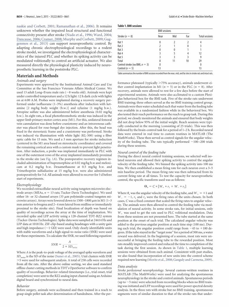
Table 1. BMI sessions
Stroke (n � 8)
BMI sessions
Total sessionsNear Mid Far
Rat 1 0 2 2 4Rat 2 2 1 0 3Rat 3 0 0 1 1Rat 4 3 5 4 12Rat 5 3 3 3 9Total 8 11 10 29Control stroke (no BMI, n � 3) 0 0Control (n � 5) 13 13
Table summarizes the number of BMI sessions recorded from the near, mid, and far sites in stroke rats and intact rats.
8654 • J. Neurosci., June 3, 2015 • 35(22):8653– 8661 Gulati et al. • Stroke Brain–Machine Interface

went BMI training. We used the Chronux toolbox to calculate bothpower spectrum and spike-field coherence (SFC; Mitra and Bokil, 2008).SFC measures phase synchronization between the LFP and spike times asa function of frequency. The magnitude of SFC is a function of frequencyand takes values between 0 and 1. We segmented the spontaneous peri-ods into 15s segments and then averaged the coherency measures acrosssegments. Spontaneous segments lasted 3–10 min preceding or after di-rect neural control. Because the SFC can vary due to the number of spikesthat are used to calculate it (Zeitler et al., 2006), we equaled the numberof spikes in the spontaneous segments by randomly selecting a subsampleof spikes from the group with greater number of spikes (Mitchell et al.,2009; Rutishauser et al., 2010). For the multitaper analyses we used atime-bandwidth (TW) product of 4 with 7 tapers. To compare coher-ences across groups, a z-score was calculated using the programs avail-able in the Chronux toolkit. Coherence between activity in two regionswas calculated and defined as follows:
Cxy ��Rxy�
��Rxx� ��Ryy�
Where Rxx and Ryy are the power spectra and Rxy is the cross-spectrum.Spectral analyses were calculated in segmented spontaneous epochs andaveraged across these epochs across animals. Mean coherences were cal-culated in the delta (�, 0.3– 4 Hz), theta (�, 6 –10 Hz), alpha (�, 8 –15 Hz),beta (�, 18 –25 Hz), and gamma (, 30 – 60 Hz) frequency band ranges.Significance testing on coherence magnitudes was performed using t testsfor comparisons in stroke and control animals in different frequencyranges. The power spectrum of the LFP channels used in the coherencecalculation, as well as for overall power change in spontaneous segments,was also determined using the multitaper method.
Sessions and changes in performance. A total of 42 training sessions(recorded from the 10 rats) were used for our analyses. Thirteen BMIsessions were recorded in five control animals and 29 BMI sessions wererecorded in five stroke animals. Table 1 summarizes the session details.Sessions in stroke animals were recorded when the upper limb was stillsubstantially impaired (i.e., poor performance on the reach to grasptask). Changes in task performance were compared between and acrosssessions. Specifically, we compared the performance change betweenearly and late trials by calculating the mean and SE of the time to com-pletion during the last 30 trials and the first 30 trials in a session. Thepercentage improvement was calculated from these values. We also as-sessed the proportion of unsuccessful trials in these early and late trialsegments. We used a paired t test to assess statistical significance andone-way ANOVA to see improvement changes between groups. In selectsessions, we videotaped the rat during BMI training blocks. Also consis-tent with multiple reports, we did not observe movements that system-atically predicted feeding tube movements (Ganguly et al., 2011; Koraleket al., 2012). Specifically, we analyzed whether limb movements mea-sured using the video recording (i.e., markers manually assigned to thehead, torso, and each limb using image processing software) covariedwith movements of the feeding tube. Across multiple sessions, we did notfind evidence for significant covariation (data not shown). This is likelydue to the fact that non-movement-related random weights were as-signed to the decoder units.
Ensemble activation analyses. We used activation strength analysis(Peyrache et al., 2009; Lopes-dos-Santos et al., 2013; Gulati et al., 2014) toestimate the similarity of neural activity patterns in late trials and earlytrials using the “instantaneous activation strength matrix.” This analysisexamines the evolution of stereotyped ensemble activity with learning.For this, we used both the “direct” neural activity (i.e., units causally likedto BMI control by the transform) and the “indirect” activity (i.e., activitythat was recorded but not used for BMI control (Ganguly et al., 2011).Therefore, an increase in activation would indicate that there is coinci-dent activation of neural activity and the formation of cell assemblies. Weconsistently observed that the transition to a plateau performance levelsheralded the transition to increased activation. For this analysis, we firstcomputed a pairwise neural activity correlation matrix (spike trains werebinned, �tbin � 50 ms). The eigenvector with the largest eigenvalue inthis correlation matrix served as the learning-related cell assembly. Spe-
cifically, the obtained spike counts for each cell [si (t), i � 1:n, t � 1:B,where n is the number of cells and B is the number of time bins in theepoch] were z-scored, obtaining the Q matrix as follows:
Q �si�t � si �
si
where � si � is the mean and is si is the SD. The pairwise cell activitycorrelation matrix can be presented in following equation:
C �1
BQQt
We thus obtained spike count matrices, QBMI, and the correlation matri-ces CBMI. Peyrache et al. (2009) further proposed the use of the Marcen-ko–Pastur distribution as the null hypothesis for the existence of cellassemblies. It was demonstrated that eigenvalues of the correlation ma-trix of a normal random matrix R with statistically independent rowsfollows a probability distribution described as follows:
p�� �q
2� 2
���max � �� �min
�
Where �minmax � 2�1 � �1/q 2 and 2 is the variance of the elements of
the random matrix R, which is 1 here (due to z scoring), and q � Rcolumns/Rrows � 1. Under the null hypothesis of an uncorrelated matrix, thecorrelations between spike trains are determined only by random fluctu-ations and the eigenvalues of template awake matrix must lie between�min and �max. Eigenvalues greater than �max are therefore a sign ofnonrandom correlations in the matrix and, for this reason, we refer tothese principal components as signal. A reactivation time series mea-sured the instantaneous match of this cell assembly to the ongoing activ-ity (Peyrache et al., 2009). The output of this analysis are principlecomponents (PCs), consisting of an array of weights assigned to each unitin the identified ensemble, and the eigenvalue, a numerical value thatrepresents the extent of total variance that is captured by a given PC (PCswith the largest eigenvalues capture the most variance) and also an in-stantaneous activation strength of these signal components. Whether acalculated PC represents a significant temporally correlated pattern ofactivity is determined by �max, the highest eigenvalue that arises out of anequivalently sized random matrix based on the Marchenko–Pastur.Therefore, PCs with eigenvalues greater than �max were considered “sig-nal components,” whereas those below �max were considered to havearisen from chance interactions.
We started by estimating the signal components (i.e., ensemble pat-terns of activity) linked to successful learning. For quantification of theinstantaneous activation, we isolated the reactivations of identified cellassembly in early and late trials. We specifically examined the first 4 safter the “GO” cue was given. We also averaged the respective values forthe first 30 trials (i.e., early) and the last 30 trials (i.e., late). For compar-ison across stroke and control learning sessions, we compared the peakactivation during early and late trials.
Statistical analysisWe performed one-way ANOVA with multiple comparisons whereversignificance assessment was required in more than three groups. We alsoused a paired t test for comparisons between early and late trials features(e.g., time to reward and proportion of unsuccessful trials in and earlyand late trials, etc.). We also used linear regression to evaluate trendsbetween reach success and improvement/rate of learning in BMIsessions.
ResultsMicrowire probes were inserted into the PLC of five rats imme-diately after a photothrombotic stroke of the upper-limb M1. Theefficacy of stroke was determined by histological examination(Fig. 1A) and the ability to perform a single-pellet reach and grasptask (Foroud and Whishaw, 2006; Wong et al., 2015). In a subsetof animals, we used ex vivo microcomputerized tomography to
Gulati et al. • Stroke Brain–Machine Interface J. Neurosci., June 3, 2015 • 35(22):8653– 8661 • 8655

measure the infarct volume (7.067 � 0.052, mm 3) and to deter-mine the “near” electrode distance from the stroke site (580.00 �137.61 �m; Fig. 1A). We also implanted five uninjured rats as acontrol group.
Network dysfunction in the stroke PLCWe first compared the recorded neural activity across the array;that is, on “near,” “mid,” and “far” electrodes (Fig. 1A,B). In thefirst week after stroke, there was a significant reduction in thenumber of neurons recorded on near versus far channels (one-way ANOVA, p � 104), further indicating that the implantedarray was in the PLC. After 2 weeks, the distribution was similaracross the array and was not significantly different from M1 re-cordings obtained from five intact animals (one-way ANOVA,p � 0.05).
Next, we assessed whether spontaneous firing rates were al-tered in the PLC. We grouped neurons based on the measured
width of the recorded spike; past literature suggests that suchmeasurements can distinguish putative fast spiking interneuronsand pyramidal neurons (Vinck et al., 2013; Courtin et al., 2014).Interestingly, we found that neurons with broader action poten-tials had significantly higher spontaneous firing rate in the strokegroup (compared with the control group, t test, p � 0.05, n � 64in control group and n � 33 in stroke group for broad-width cellsand n � 12 in control group and n � 16 in stroke group fornarrow-width cells; Fig. 1C). The control group spontaneous fir-ing rates are comparable to what has been observed for sponta-neous firing in other rodent motor cortical studies (Isomura etal., 2009). We also looked at SFC magnitudes (SFCmag; Fig. 2A)for these units and found that broad-width spikes had signifi-cantly elevated SFCmag in the � (0.3– 4 Hz) and � (6 –10 Hz) bandscompared with controls (t test, p � 0.005; Fig. 2B). They were notsignificantly different for the other higher-frequency bands (i.e.,�, 8 –15 Hz; �, 18 –25 Hz; and , 30 – 60 Hz). For narrow-width
Figure 1. Spontaneous spiking activity and field oscillations in the PLC. A, Schematic of electrophysiological monitoring from the PLC. Histology above shows example sagittal view of stroke siteand tract from a near electrode. B, Comparison of the change in recording yield over time (week 1 vs week 2.5) on electrodes as a function of distance from the stroke site (near, mid, and far electrodesare indicated at top). Error bars indicate SEM. *p � 0.01. C, Difference in the spiking activity for broad-width (left gray column) and narrow-width units (blue right column). *p � 0.05.
Figure 2. A, Schematic depicting SFC. Top row shows out-of-phase unit spikes and LFP indicating a lower SFC and bottom row shows phase-synchronized spikes and LFP resulting in a higher SFC.B, Comparison of differences in the SFCmag in control and stroke animals. C, Power spectral density of LFPs in control and stroke animals. *p � 0.05; **p � 0.005.
8656 • J. Neurosci., June 3, 2015 • 35(22):8653– 8661 Gulati et al. • Stroke Brain–Machine Interface

units, none of the five frequencies showed a significant difference(data not shown, p � 0.05 for all five frequency bands). More-over, the power spectral density of the LFPs from stroke animalsshowed significantly elevated power in the slowest-frequencybands compared with controls (t test, p � 0.05; Fig. 2C). Theseresults are consistent with the notion that stroke leads to a dys-functional network state marked by slow oscillations (Schiene etal., 1996; Carmichael and Chesselet, 2002).
Direct control of the PLC spiking activityWe next quantified whether spiking activity recorded from theelectrodes in the PLC could be volitionally modulated in a task-dependent manner (Fig. 3). Specifically, spiking activity wastransformed via a linear decoder into the angular velocity of amechanical actuator that could also deliver water (see Materialsand Methods and Fig. 3A,B). The decoder weights were heldconstant during the session to rely exclusively on neural learning.Each trial started with the simultaneous delivery of an auditorytone and the opening of a door to allow access to the tube. At thestart of each trial, the angular position of the tube was set to 0°(P1). If the angular position of the tube was held for � 300 ms atposition P2 (90°), a defined amount of water was delivered (i.e.,successful trial). A trial was stopped if this was not achievedwithin 15 s (i.e., unsuccessful trial). At the end of a trial, the doorwas closed and the actuator was returned to position P1. After atypical 1–2 h practice session, animals showed improvements intask performance with a steady reduction in the time to trialcompletion and a decrease in the number of unsuccessful trials.Figure 3B shows an example of the learning curve when usingnear spiking activity of two neurons that were respectively as-signed a positive and a negative weight. There were also bidirec-tional changes in the task related activation of the positive and the
negative weight neurons (Fig. 3C). In 29 such training sessions infive rats, there was consistent evidence of practice-related im-provements in task performance (control group n � 13 sessionsand stroke group n � 29 sessions, paired t tests, p � 0.05 for eachcomparison; Fig. 3D). Interestingly, the net drop in time to taskcompletion was not significantly different for the stroke and thecontrol group.
We also investigated whether there were differences in perfor-mance characteristics for neurons recorded from near, mid, andfar sites. For the 29 BMI training sessions described above, eightwere from near sites, 11 from mid and 10 from far sites (Table 1).When near, mid, and far sites were compared separately, the timeto task completion reduced markedly for activity recorded oneach sites in a manner similar to intact animals (near: 11.58 �0.53 to 6.17 � 0.44 s; mid: 12.67 � 0.45 to 6.84 � 0.37 s, far:12.65 � 0.47 to 7.02 � 0.39 s, control: 11.92 � 0.41 to 6.38 �0.34 s, all paired t tests, p � 0.05; Fig. 4A). Interestingly, therewere also no differences compared with intact animals learningthe neuroprosthetic control (one-way ANOVA, p � 0.05). Theproportion of unsuccessful trials dropped significantly for eachgroup comparing early and late trials (23.6 � 7.5 to 6.1 � 3.2% atnear sites, t test, t6 � 4.12, p � 0.005; 52.1 � 6.4 to 10.1 � 2.7% atmid sites, t test, t7 � 3.79, p � 105; 55.7 � 6.7 to 5.7 � 2.9%at far sites, t test, t9 � 7.7, p � 104; and 24.4 � 5.9% to 5.5 �2.1% in control rats, t test, t12 � 7.09, p � 104; Fig. 4A). Therewere also no significant differences in modulation depth (MD�)with BMI learning (102.5 � 22.5%, 111.8 � 18.5%, 130.2 �19.3%, and 120.3 � 18.2% change for 17, 25, 23 units at near,mid, and far sites in stroke rats and 26 units in control rats,respectively, one-way ANOVA, F(3,87) � 0.33, p � 0.80; Fig. 4B).Even when MD� was segregated for broad-width and narrow-width units, there were no significant differences. Further, the
Figure 3. Direct brain control from the PLC. A, Schematic of the setup used to assess direct neural control of the PLC. B, Change in time to task completion with practice using volitional control ofPLC neurons. Solid line represents mean time using a moving window of 20 trials. W� and W, respectively, indicate positive and negative weights. C, Change in task-related activation for twoneurons. Shown at top are the respective mean firing rates during early and late trials. Shaded region is the SEM. At the bottom are single-trial rasters from corresponding early and late periods. D,Comparison of the change in time to task completion for control and stroke animals. *p � 0.01.
Gulati et al. • Stroke Brain–Machine Interface J. Neurosci., June 3, 2015 • 35(22):8653– 8661 • 8657
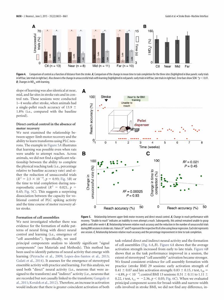
slope of learning was also identical at near,mid, and far sites in stroke rats and in con-trol rats. These sessions were conducted1– 4 weeks after stroke, when animals hada single-pellet reach accuracy of 13.9 �1.8% (i.e., compared with the baselineperiod).
Direct cortical control in the absence ofmotor recoveryWe next examined the relationship be-tween upper-limb motor recovery and theability to learn transforms using PLC neu-rons. The example in Figure 5A illustratesthat learning was possible even when ratswere unable to attempt reaches. Acrossanimals, we did not find a significant rela-tionship between the ability to completethe physical reaching task (i.e., percentagerelative to baseline accuracy rate) and ei-ther the reduction of unsuccessful trials(R 2 � 2.5 � 104, p � 0.93; Fig. 5B) orthe time to trial completion during neu-roprosthetic control (R 2 � 0.021, p �0.45; Fig. 5C). This suggests a surprisingdissociation between the capacity for vo-litional control of PLC spiking activityand the time course of motor recovery af-ter stroke.
Formation of cell assembliesWe next investigated whether there wasevidence for the formation of stable pat-terns of neural firing with direct neuralcontrol and learning (i.e., emergence of“cell assemblies”). Specifically, we usedprincipal components analysis to identify significant “signalcomponents” (see Materials and Methods). This method hasbeen used to identify patterns of neural activity that emerge withlearning (Peyrache et al., 2009; Lopes-dos-Santos et al., 2013;Gulati et al., 2014). It assesses for the emergence of stereotypedensemble activity with practice and learning. For this analysis, weused both “direct” neural activity (i.e., neurons that were as-signed to the transform) and “indirect” activity (i.e., neurons thatare recorded but not causally linked to the transform; Ganguly etal., 2011; Koralek et al., 2012). Therefore, an increase in activationwould indicate that there is greater coincident activation of both
task-related direct and indirect neural activity and the formationof cell assemblies (Fig. 6A,B). Figure 6A shows that the averageactivation strength increased from early to late trials. Figure 6Bshows that as the task performance improved in a session, theextent of stereotyped “cell assembly” activation became stronger.We found consistent evidence for cell assembly formation withpractice (stroke BMI 29 sessions: early activation strength of0.41 � 0.07 and late activation strength: 0.83 � 0.15, t test, t28 �4.89, p � 104; control BMI 13 sessions: 0.51 � 0.11 to 1.11 �0.22, t test, t12 � 2.36, p � 0.05; Fig. 6C). When we evaluatedprinicipal component scores for broad-width and narrow-widthcells involved in stroke BMI, we did not find any difference, in-
Figure 4. Comparison of control as a function of distance from the stroke. A, Comparison of the change in mean time to task completion for the three sites (highlighted in blue panels: early trialsin left bar, late trials in right bar). Also shown is the change in unsuccessful trials with learning (highlighted in red panels: early trials in left bar, late trials in right bar). Error bars show SEM. *p � 0.01.B, Changes in MD� with learning.
Figure 5. Relationship between upper-limb motor recovery and direct neural control. A, Change in reach performance withrecovery. “Unable to reach” indicates an inability to even attempt a reach. Subsequently, this animal remained unable to grasppellets until after week 4. B, Relationship between relative reach accuracy and the reduction in the number of unsuccessful trialsduring BMI sessions in stroke rats. Values (R 2 and P) represent the respective fit of a line using linear regression. Each dot representsone session. C, Relationship between relative reach accuracy and the percentage improvement in time to task completion.
8658 • J. Neurosci., June 3, 2015 • 35(22):8653– 8661 Gulati et al. • Stroke Brain–Machine Interface

dicating that both cell types participated in a similar way in as-sembly formation (PC scores for narrow-width cells: 0.021 �0.019%, broad-width cells: 0.016 � 0.029%; t test, t63 � 0.75, p �0.46). Therefore, cortical networks in the PLC appear to be capa-ble of forming and stabilizing new large-scale functional associa-tions among emergent ensembles.
Persistent changes in spike-field couplingWe next evaluated whether prolonged direct neural control per-sistently modified the spike-field relationship in the PLC. Inter-estingly, we found that, during spontaneously recorded activityafter the first successful training session, the SFCmag for directneurons that underwent direct neural control reduced signifi-cantly (p � 0.05, n � 10 for � and � frequency range; Fig. 7A,B).Furthermore, we found that the spontaneous firing rates of directunits reduced significantly (p � 0.05; Fig. 7C). When we checkedfor firing rates in three additional rats that underwent stroke butno BMI training, the firing rates did not change (n � 72, p � 0.05;Fig. 7C). The firing rates were equalized before SFCmag calcula-tion (see Materials and Methods). In addition, the power in thesefrequency bands appeared to reduce slightly, but this was notstatistically significant. Therefore, the spike-field relationship forneurons that underwent direct neural control was persistentlymodified after the training period.
DiscussionOur results indicate that spiking activity from neurons in the PLCcan be used to achieve neuroprosthetic control. There was nodifference relative to distance from the stroke site or comparedwith intact animals. Interestingly, the observed abnormal slowingof the LFP and its associated modulation of neural firing did notappear to impede the network-level physiological plasticity nec-essary to achieve neuroprosthetic control. This further indicates
that the microstructural and physiological reorganization afterstroke is sufficient to permit direct cortical control and theformation of functional cell assemblies. Moreover, we alsofound a surprising dissociation between the capacity for BMIcontrol from the PLC and the process of motor recovery.
Our findings have direct implications for the development ofintracortical BMIs compatible with cortical injury. In our exper-iments, rodents were required to modulate PLC neural activityvolitionally to control an artificial actuator; volitional modula-tion of neural activity is at the core of neuroprosthetic control(Fetz, 1969; Ganguly and Carmena, 2009; Ganguly et al., 2011).We also found that the injury triggered slow oscillations (i.e., inthe � and � frequency range; Van Huffelen et al., 1984; Laaksonenet al., 2013) did not necessarily preclude learning neuroprostheticcontrol. Moreover, the elevated firing rates of the putative pyra-midal cells (i.e., broader action potential widths) also did notappear to impede learning. Although a general hyperexcitabilitypoststroke has been observed using multiunit recordings (Schiene etal., 1996; Hagemann et al., 1998; Carmichael and Chesselet,2002), we specifically found that such hyperexcitability may besignificantly more pronounced for regular-spiking pyramidalcells. It is possible that the altered local and long-range con-nectivity (Dancause, 2006), the changes in receptor profiles(Schiene et al., 1996), and/or the changes in extracellular neu-rotransmitter content (Clarkson et al., 2010) affects the twoclasses of neurons differentially. Remarkably, despite the ab-normal oscillations and the elevated baseline firing rates, wefound clear evidence for robust volitional control of the peri-lesional neural activity. Moreover, such control was also pos-sible even when physical upper limb movements were severelyimpaired. This suggests that the PLC in subjects with perma-nent deficits may be capable of supporting neuroprosthetic
Figure 6. Emergence of functional cell assemblies in the PLC. A, Shown are the mean activation profiles for early versus late trials from a representative session. Shaded region is the SEM. B, Soliddark blue line is the moving average mean using 20 trials of the time to task completion for the session used in A. The solid red line is a continuous trial-by-trial measure of the activation strength.The dotted line in dark red is the moving average mean of the activation strength using 20 trials. Early and late portions of the trials are highlighted in same colors as A. C, Comparison of the respectivechanges in activation for intact animals BMI sessions and BMI sessions in stroke animals. Error bars indicate SEM. *p � 0.01; **p � 10 4.
Figure 7. Persistent change in cortical state after direct neural control. A, Example of a persistent reduction of SFCmag for a direct unit after a learning session. B, Averaged SFCmag for direct unitsbefore and after first training (i.e., in the �- and �-frequency bands). C, Averaged firing rate for direct units before and after direct neural control in stroke animal with BMI training and no BMItraining. *p � 0.05.
Gulati et al. • Stroke Brain–Machine Interface J. Neurosci., June 3, 2015 • 35(22):8653– 8661 • 8659

control. Together, our results indicate that, despite the dis-rupted connectivity after stroke, the PLC may serve as an ef-fective target for intracortical neural interfaces specificallydesigned for stroke patients.
We also found that direct neural control modified neural ac-tivity patterns outside of the training period (i.e., changes inspontaneous activity after training compared with that beforetraining). Specifically, we found that the firing rates of the con-ditioned direct neurons became significantly reduced and ap-proached that of normal animals. Moreover, these neurons weresignificantly less likely to be modulated by the abnormal low-frequency oscillations evident in the PLC. These findings suggestthat direct neural control can trigger longer-lasting plasticitythrough modulation of the functional connectivity. It may beconsistent with our recent finding of “offline” processing duringslow-wave sleep after neuroprosthetic learning (Gulati et al.,2014). The observed persistent changes in neural firing may alsobe closely related to the growing body of work suggesting that“neurofeedback” can be used to treat a range of neurological andpsychiatric disorders (Strehl et al., 2006; Birbaumer et al., 2009;Ramos-Murguialday et al., 2013). During neurofeedback, nonin-vasive scalp EEG recordings are used to assess brain states and toprovide feedback. Interestingly, a recent study found that voli-tional modulation of the �-rhythm using neurofeedback couldpromote recovery after stroke (Birbaumer and Cohen, 2007;Ramos-Murguialday et al., 2013). It is difficult to compare ourinvasive recordings from a single cortical area with scalp EEGrecordings directly; moreover, the exact cortical and subcorticalgenerators of the �-rhythm remain unclear. However, it suggeststhe possibility that feedback-dependent �-rhythm modulationmay exert its effects, at least in part, by modifying the activity ofthe PLC.
Interestingly, the notable dissociation between the temporalcourse and the extent of recovery and the ability for direct neuralcontrol suggests that the functional state and the potential forplasticity of the PLC may not be sufficient to support motorrecovery. There is increasing evidence that volitional modulationof neural activity requires both top-down and local processes(Fetz, 2007; Halder et al., 2011). Moreover, it requires the recruit-ment of NMDA-R-dependent plasticity (Koralek et al., 2012).Together, the evidence suggests that existing cortical and subcor-tical mechanisms of plasticity are recruited during the process oflearning direct neural control. In this context, the ability toachieve successfully direct neural control of the PLC suggests thatmechanisms of plasticity are present and capable of being re-cruited. It further suggests that such mechanisms are not neces-sarily sufficient to promote motor recovery. This may beconsistent with the finding in clinical stroke studies that cortico-spinal projection integrity is essential for recovery of motor func-tion (Stinear et al., 2012; Ganguly et al., 2013). In other words,whereas the PLC is capable of expressing plasticity, the lack of asubstrate to transmit the information ultimately prevents motorrecovery. Moreover, our finding of robust direct neural control ofthe PLC may have implications for emerging therapeutics; forexample, cell-based therapies aim to augment the plasticity andthe recovery potential of cortical areas after stroke (Bliss et al.,2007).
In summary, our results indicate that direct neural controland learning was possible from the immediate PLC. There wassurprisingly no difference relative to distance from the stroke siteor compared with intact animals. More broadly, our results sug-gest that PLC may be a viable substrate for intracortical neuralinterfaces in patients with severe disability after stroke.
ReferencesArduin PJ, Fregnac Y, Shulz DE, Ego-Stengel V (2013) “Master” neurons
induced by operant conditioning in rat motor cortex during a brain–machine interface task. J Neurosci 33:8308 – 8320. CrossRef Medline
Biernaskie J, Corbett D (2001) Enriched rehabilitative training promotesimproved forelimb motor function and enhanced dendritic growth afterfocal ischemic injury. J Neurosci 21:5272–5280. Medline
Birbaumer N, Cohen LG (2007) Brain-computer interfaces: communica-tion and restoration of movement in paralysis. J Physiol 579:621– 636.CrossRef Medline
Birbaumer N, Ramos Murguialday A, Weber C, Montoya P (2009) Neuro-feedback and brain-computer interface clinical applications. Int Rev Neu-robiol 86:107–117. CrossRef Medline
Bliss T, Guzman R, Daadi M, Steinberg GK (2007) Cell transplantationtherapy for stroke. Stroke 38:817– 826. CrossRef Medline
Brinkman J, Kuypers HG (1973) Cerebral control of contralateral and ipsi-lateral arm, hand and finger movements in the split-brain rhesus monkey.Brain 96:653– 674. CrossRef Medline
Brown CE, Boyd JD, Murphy TH (2010) Longitudinal in vivo imagingreveals balanced and branch-specific remodeling of mature cortical pyra-midal dendritic arbors after stroke. J Cereb Blood Flow Metab 30:783–791. CrossRef Medline
Bundy DT, Wronkiewicz M, Sharma M, Moran DW, Corbetta M, LeuthardtEC (2012) Using ipsilateral motor signals in the unaffected cerebralhemisphere as a signal platform for brain-computer interfaces in hemi-plegic stroke survivors. J Neural Eng 9:036011. CrossRef Medline
Carmena JM, Lebedev MA, Crist RE, O’Doherty JE, Santucci DM, DimitrovDF, Patil PG, Henriquez CS, Nicolelis MA (2003) Learning to control abrain–machine interface for reaching and grasping by primates. PLoS Biol1:E42. Medline
Carmichael ST, Chesselet MF (2002) Synchronous neuronal activity is asignal for axonal sprouting after cortical lesions in the adult. J Neurosci22:6062– 6070. Medline
Chapin JK, Moxon KA, Markowitz RS, Nicolelis MA (1999) Real-time con-trol of a robot arm using simultaneously recorded neurons in the motorcortex. Nat Neurosci 2:664 – 670. CrossRef Medline
Clarkson AN, Huang BS, Macisaac SE, Mody I, Carmichael ST (2010) Re-ducing excessive GABA-mediated tonic inhibition promotes functionalrecovery after stroke. Nature 468:305–309. CrossRef Medline
Collinger JL, Wodlinger B, Downey JE, Wang W, Tyler-Kabara EC, Weber DJ,McMorland AJ, Velliste M, Boninger ML, Schwartz AB (2013) High-performance neuroprosthetic control by an individual with tetraplegia.Lancet 381:557–564. CrossRef Medline
Courtin J, Chaudun F, Rozeske RR, Karalis N, Gonzalez-Campo C, Wurtz H,Abdi A, Baufreton J, Bienvenu TC, Herry C (2014) Prefrontal parvalbu-min interneurons shape neuronal activity to drive fear expression. Nature505:92–96. Medline
Cramer SC (2008) Repairing the human brain after stroke: I. Mechanisms ofspontaneous recovery. Ann Neurol 63:272–287. CrossRef Medline
Daly JJ, Wolpaw JR (2008) Brain-computer interfaces in neurological reha-bilitation. Lancet Neurol 7:1032–1043. CrossRef Medline
Dancause N (2006) Vicarious function of remote cortex following stroke:recent evidence from human and animal studies. Neuroscientist 12:489 –499. CrossRef Medline
Fetz EE (1969) Operant conditioning of cortical unit activity. Science 163:955–958. CrossRef Medline
Fetz EE (2007) Volitional control of neural activity: implications for brain-computer interfaces. J Physiol 579:571–579. CrossRef Medline
Foroud A, Whishaw IQ (2006) Changes in the kinematic structure and non-kinematic features of movements during skilled reaching after stroke: aLaban Movement Analysis in two case studies. J Neurosci Methods 158:137–149. CrossRef Medline
Ganguly K, Carmena JM (2009) Emergence of a stable cortical map for neu-roprosthetic control. Plos Biol 7:e1000153. CrossRef Medline
Ganguly K, Secundo L, Ranade G, Orsborn A, Chang EF, Dimitrov DF, WallisJD, Barbaro NM, Knight RT, Carmena JM (2009) Cortical representa-tion of ipsilateral arm movements in monkey and man. J Neurosci 29:12948 –12956. CrossRef Medline
Ganguly K, Dimitrov DF, Wallis JD, Carmena JM (2011) Reversible large-scale modification of cortical networks during neuroprosthetic control.Nat Neurosci 14:662– 667. CrossRef Medline
8660 • J. Neurosci., June 3, 2015 • 35(22):8653– 8661 Gulati et al. • Stroke Brain–Machine Interface

Ganguly K, Byl NN, Abrams GM (2013) Neurorehabilitation: Motor recov-ery after stroke as an example. Ann Neurol 74:373–381. CrossRef Medline
Guggenmos DJ, Azin M, Barbay S, Mahnken JD, Dunham C, Mohseni P,Nudo RJ (2013) Restoration of function after brain damage using a neu-ral prosthesis. Proc Natl Acad Sci U S A 110:21177–21182. CrossRefMedline
Gulati T, Ramanathan DS, Wong CC, Ganguly K (2014) Reactivation ofemergent task-related ensembles during slow-wave sleep after neuropros-thetic learning. Nat Neurosci 17:1107–1113. CrossRef Medline
Hagemann G, Redecker C, Neumann-Haefelin T, Freund HJ, Witte OW(1998) Increased long-term potentiation in the surround of experimen-tally induced focal cortical infarction. Ann Neurol 44:255–258. CrossRefMedline
Halder S, Agorastos D, Veit R, Hammer EM, Lee S, Varkuti B, Bogdan M,Rosenstiel W, Birbaumer N, Kubler A (2011) Neural mechanisms ofbrain-computer interface control. Neuroimage 55:1779 –1790. CrossRefMedline
Hochberg LR, Serruya MD, Friehs GM, Mukand JA, Saleh M, Caplan AH,Branner A, Chen D, Penn RD, Donoghue JP (2006) Neuronal ensemblecontrol of prosthetic devices by a human with tetraplegia. Nature 442:164 –171. CrossRef Medline
Hochberg LR, Bacher D, Jarosiewicz B, Masse NY, Simeral JD, Vogel J, Had-dadin S, Liu J, Cash SS, van der Smagt P, Donoghue JP (2012) Reach andgrasp by people with tetraplegia using a neurally controlled robotic arm.Nature 485:372–375. CrossRef Medline
Hummel FC, Cohen LG (2006) Non-invasive brain stimulation: a newstrategy to improve neurorehabilitation after stroke? Lancet Neurol5:708 –712. CrossRef Medline
Isomura Y, Harukuni R, Takekawa T, Aizawa H, Fukai T (2009) Microcir-cuitry coordination of cortical motor information in self-initiation ofvoluntary movements. Nat Neurosci 12:1586 –1593. CrossRef Medline
Jarosiewicz B, Chase SM, Fraser GW, Velliste M, Kass RE, Schwartz AB(2008) Functional network reorganization during learning in a brain-computer interface paradigm. Proc Natl Acad Sci U S A 105:19486 –19491. CrossRef Medline
Koralek AC, Long JD, Costa RM, Carmena JM (2010) Corticostriatal dy-namics during learning and performance of a neuroprosthetic task. ConfProc IEEE Eng Med Biol Soc 2010:2682–2685. Medline
Koralek AC, Jin X, Long JD 2nd, Costa RM, Carmena JM (2012) Corticos-triatal plasticity is necessary for learning intentional neuroprostheticskills. Nature 483:331–335. CrossRef Medline
Laaksonen K, Helle L, Parkkonen L, Kirveskari E, Makela JP, Mustanoja S,Tatlisumak T, Kaste M, Forss N (2013) Alterations in spontaneous brainoscillations during stroke recovery. PLoS One 8:e61146. CrossRefMedline
Langhorne P, Bernhardt J, Kwakkel G (2011) Stroke rehabilitation. Lancet377:1693–1702. CrossRef Medline
Lopes-dos-Santos V, Ribeiro S, Tort AB (2013) Detecting cell assemblies inlarge neuronal populations. J Neurosci Methods 220:149 –166. CrossRefMedline
Mitchell JF, Sundberg KA, Reynolds JH (2009) Spatial attention decorre-lates intrinsic activity fluctuations in macaque area V4. Neuron 63:879 –888. CrossRef Medline
Mitra P, Bokil H (2008) Observed brain dynamics. New York: OUP.Moritz CT, Perlmutter SI, Fetz EE (2008) Direct control of paralysed mus-
cles by cortical neurons. Nature 456:639 – 642. CrossRef MedlineMurphy TH, Corbett D (2009) Plasticity during stroke recovery: from syn-
apse to behaviour. Nat Rev Neurosci 10:861– 872. CrossRef MedlineMusallam S, Corneil BD, Greger B, Scherberger H, Andersen RA (2004)
Cognitive control signals for neural prosthetics. Science 305:258 –262.CrossRef Medline
Norrving B, Kissela B (2013) The global burden of stroke and need for acontinuum of care. Neurology 80:S5–12. CrossRef Medline
Nudo RJ, Milliken GW (1996) Reorganization of movement representa-tions in primary motor cortex following focal ischemic infarcts in adultsquirrel monkeys. J Neurophysiol 75:2144 –2149. Medline
Nudo RJ, Wise BM, SiFuentes F, Milliken GW (1996) Neural substrates forthe effects of rehabilitative training on motor recovery after ischemicinfarct. Science 272:1791–1794. CrossRef Medline
Peyrache A, Khamassi M, Benchenane K, Wiener SI, Battaglia FP (2009)Replay of rule-learning related neural patterns in the prefrontal cortexduring sleep. Nat Neurosci 12:919 –926. CrossRef Medline
Ramanathan D, Conner JM, Tuszynski MH (2006) A form of motor corticalplasticity that correlates with recovery of function after brain injury. ProcNatl Acad Sci U S A 103:11370 –11375. CrossRef Medline
Ramos-Murguialday A, Broetz D, Rea M, Laer L, Yilmaz O, Brasil FL, LiberatiG, Curado MR, Garcia-Cossio E, Vyziotis A, Cho W, Agostini M, SoaresE, Soekadar S, Caria A, Cohen LG, Birbaumer N (2013) Brain–machineinterface in chronic stroke rehabilitation: a controlled study. Ann Neurol74:100 –108. CrossRef Medline
Rutishauser U, Ross IB, Mamelak AN, Schuman EM (2010) Human mem-ory strength is predicted by theta-frequency phase-locking of single neu-rons. Nature 464:903–907. CrossRef Medline
Santhanam G, Ryu SI, Yu BM, Afshar A, Shenoy KV (2006) A high-performance brain-computer interface. Nature 442:195–198. CrossRefMedline
Schiene K, Bruehl C, Zilles K, Qu M, Hagemann G, Kraemer M, Witte OW(1996) Neuronal hyperexcitability and reduction of GABAA-receptorexpression in the surround of cerebral photothrombosis. J Cereb BloodFlow Metab 16:906 –914. Medline
Serruya MD, Hatsopoulos NG, Paninski L, Fellows MR, Donoghue JP (2002)Instant neural control of a movement signal. Nature 416:141–142.CrossRef Medline
Stinear CM, Barber PA, Petoe M, Anwar S, Byblow WD (2012) The PREPalgorithm predicts potential for upper limb recovery after stroke. Brain135:2527–2535. CrossRef Medline
Strehl U, Leins U, Goth G, Klinger C, Hinterberger T, Birbaumer N (2006)Self-regulation of slow cortical potentials: a new treatment for childrenwith attention-deficit/hyperactivity disorder. Pediatrics 118:e1530 –1540.CrossRef Medline
Suner S, Fellows MR, Vargas-Irwin C, Nakata GK, Donoghue JP (2005) Re-liability of signals from a chronically implanted, silicon-based electrodearray in non-human primate primary motor cortex. IEEE Trans NeuralSyst Rehabil Eng 13:524 –541. CrossRef Medline
Taylor DM, Tillery SI, Schwartz AB (2002) Direct cortical control of 3Dneuroprosthetic devices. Science 296:1829 –1832. CrossRef Medline
Van Huffelen AC, Poortvliet DC, Van der Wulp CJ (1984) Quantitativeelectroencephalography in cerebral ischemia: detection of abnormalitiesin “normal” EEGs. Prog Brain Res 62:3–28. CrossRef Medline
Velliste M, Perel S, Spalding MC, Whitford AS, Schwartz AB (2008) Corticalcontrol of a prosthetic arm for self-feeding. Nature 453:1098 –1101.CrossRef Medline
Vinck M, Womelsdorf T, Buffalo EA, Desimone R, Fries P (2013) Atten-tional modulation of cell-class-specific gamma-band synchronization inawake monkey area v4. Neuron 80:1077–1089. CrossRef Medline
Ward NS (2004) Functional reorganization of the cerebral motor systemafter stroke. Curr Opin Neurol 17:725–730. CrossRef Medline
Wong CC, Ramanathan DS, Gulati T, Won SJ, Ganguly K (2015) An auto-mated behavioral box to assess forelimb function in rats. J Neurosci Meth-ods 246:30 –37. CrossRef Medline
Zeitler M, Fries P, Gielen S (2006) Assessing neuronal coherence withsingle-unit, multi-unit, and local field potentials. Neural Comput 18:2256 –2281. CrossRef Medline
Gulati et al. • Stroke Brain–Machine Interface J. Neurosci., June 3, 2015 • 35(22):8653– 8661 • 8661
View publication statsView publication stats